Embed Size (px)
Citation preview
1
Bi / CNS 150
Lecture 24
Monday November 26, 2012
Two neurodegenerative diseases
Henry Lester
Kandel, Chapters 43 & 58
2
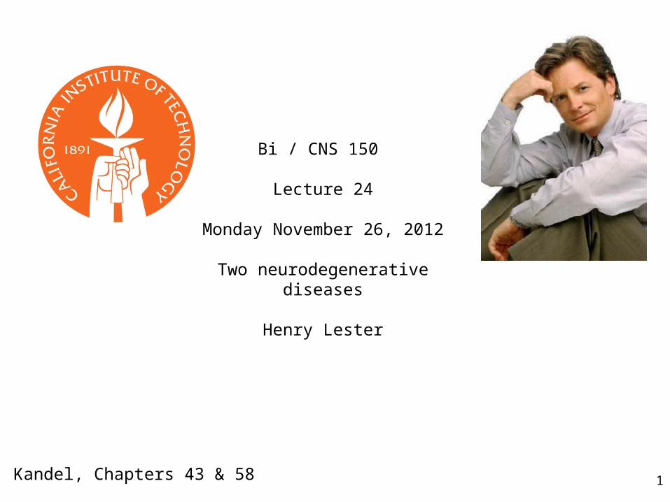
https://class.coursera.org/drugsandbrain-2012-001/class/index
https://www.coursera.org/#course/drugsandbrain
Inspect the discussion forums:https://class.coursera.org/drugsandbrain-2012-001/forum/index
Proposal for Bi/CNS 150 extra credit:a.Enroll for the course. b.If you wish to continue after the first 2 weeks, inform Henry Lester.c.Pass the course. Should be quite easy.d.Contribute to the forums with technical and scientific explanations.e.Contribute to the Caltech Bi/CNS 150 students forum.Result: at least 1/3 grade (i. e., B+ to A-). Retroactive, will not destroy the curve.
After 12/1: Inspect the “Course Page”:
Inspect the “Landing Page”:
Inspect miniLecture 5
3
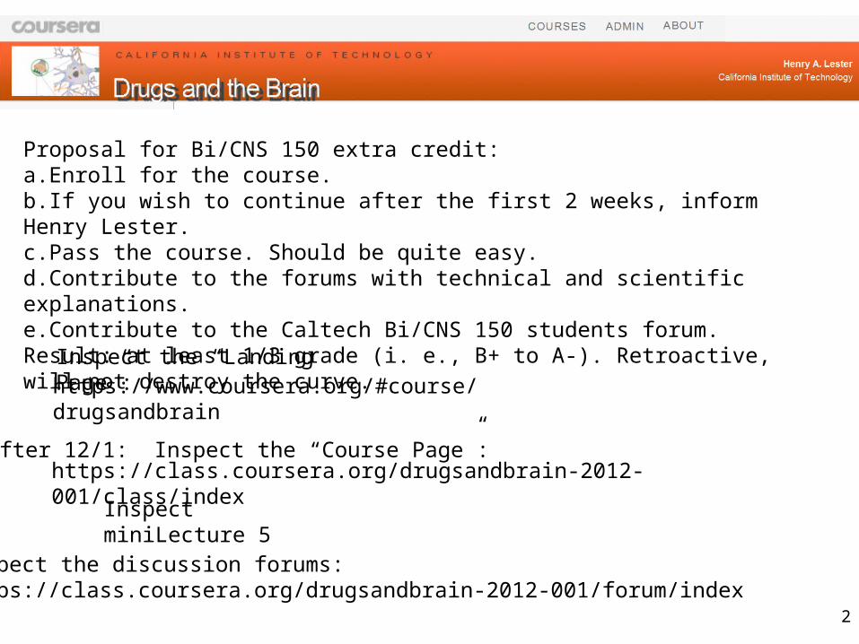
James Parkinson, apothecary surgeon1817, An Essay on the Shaking Palsy, described "paralysis agitans", from observations of 6 individuals during his daily walks in London
Parkinson’s disease (tremor at rest 3-5 Hz, “pill-rolling”, slow movements, particularly when starting; short, rapid steps)
but most Parkinson patients are either medicated or stimulated
Parkinson’s disease
1. Clinical description
2. Genetics
3. Pathophysiology
4. Biomarkers and animal models
5. Heterozygote advantage: none known
6. Therapeutic approaches
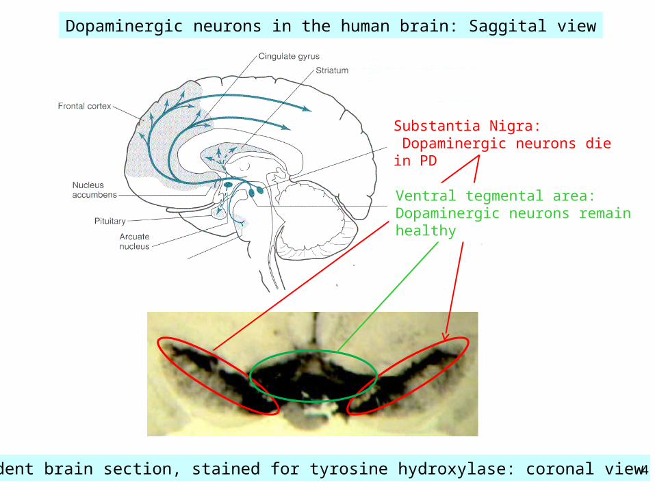
Rodent brain section, stained for tyrosine hydroxylase: coronal view 4
Dopaminergic neurons in the human brain: Saggital view
Substantia Nigra: Dopaminergic neurons die in PD
Ventral tegmental area: Dopaminergic neurons remain healthy
5
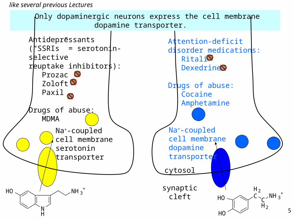
Only dopaminergic neurons express the cell membrane dopamine transporter.
Antidepressants (“SSRIs” = serotonin-selectivereuptake inhibitors): Prozac Zoloft Paxil
Drugs of abuse: MDMA
Attention-deficit disorder medications: Ritalin Dexedrine
Drugs of abuse: Cocaine Amphetamine
like several previous Lectures
Na+-coupledcell membrane serotonintransporter
Na+-coupledcell membrane dopamine transporter
NH
HO NH3+
HO
HO
H2C
CH2
NH3+
cytosol
synapticcleft
6
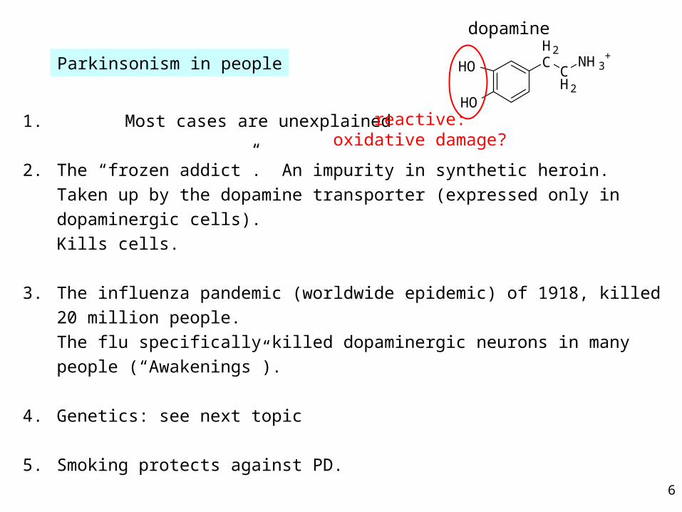
1. Most cases are unexplained
2. The “frozen addict”. An impurity in synthetic heroin.
Taken up by the dopamine transporter (expressed only in dopaminergic cells).
Kills cells.
3. The influenza pandemic (worldwide epidemic) of 1918, killed 20 million people.
The flu specifically killed dopaminergic neurons in many people (“Awakenings”).
4. Genetics: see next topic
5. Smoking protects against PD.
HO
HO
H2C
CH2
NH3+
dopamine
reactive:oxidative damage?
Parkinsonism in people
7
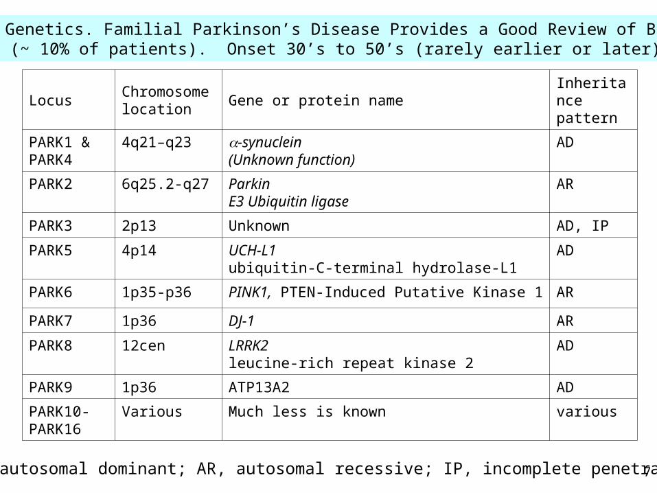
3. Genetics. Familial Parkinson’s Disease Provides a Good Review of Bi 8/9(~ 10% of patients). Onset 30’s to 50’s (rarely earlier or later)
LocusChromosome location
Gene or protein nameInheritance pattern
PARK1 & PARK4
4q21–q23 -synuclein(Unknown function)
AD
PARK2 6q25.2-q27 ParkinE3 Ubiquitin ligase
AR
PARK3 2p13 Unknown AD, IP
PARK5 4p14 UCH-L1ubiquitin-C-terminal hydrolase-L1
AD
PARK6 1p35-p36 PINK1, PTEN-Induced Putative Kinase 1 AR
PARK7 1p36 DJ-1 AR
PARK8 12cen LRRK2leucine-rich repeat kinase 2
AD
PARK9 1p36 ATP13A2 AD
PARK10-PARK16
Various Much less is known various
AD, autosomal dominant; AR, autosomal recessive; IP, incomplete penetrance
8
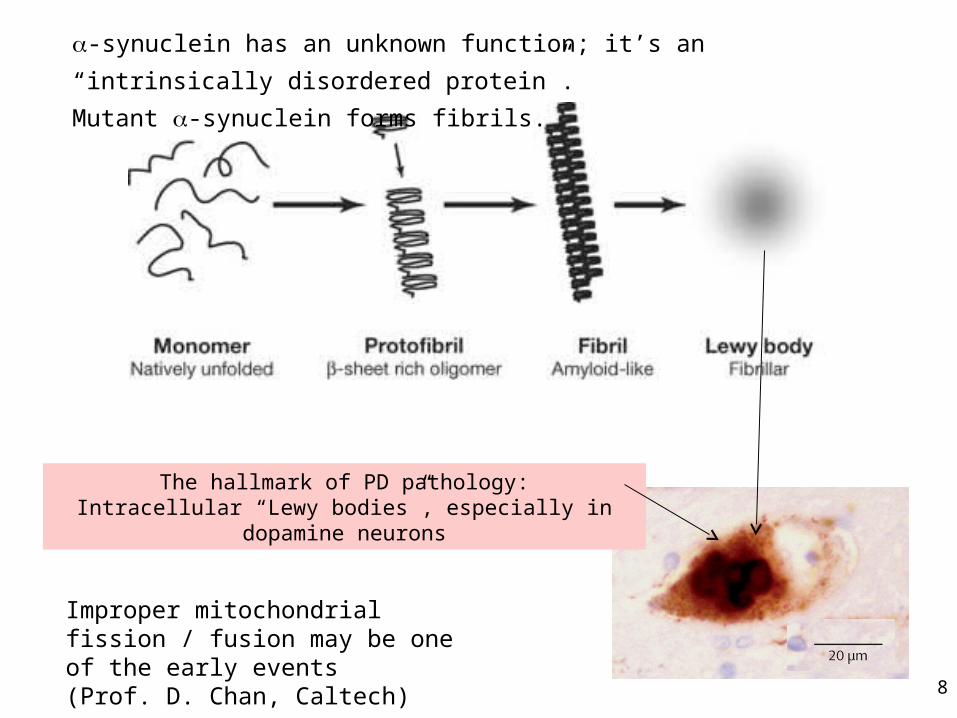
-synuclein has an unknown function; it’s an “intrinsically disordered protein”.
Mutant -synuclein forms fibrils.
Improper mitochondrial fission / fusion may be one of the early events(Prof. D. Chan, Caltech)
The hallmark of PD pathology:Intracellular “Lewy bodies”, especially in dopamine neurons
9
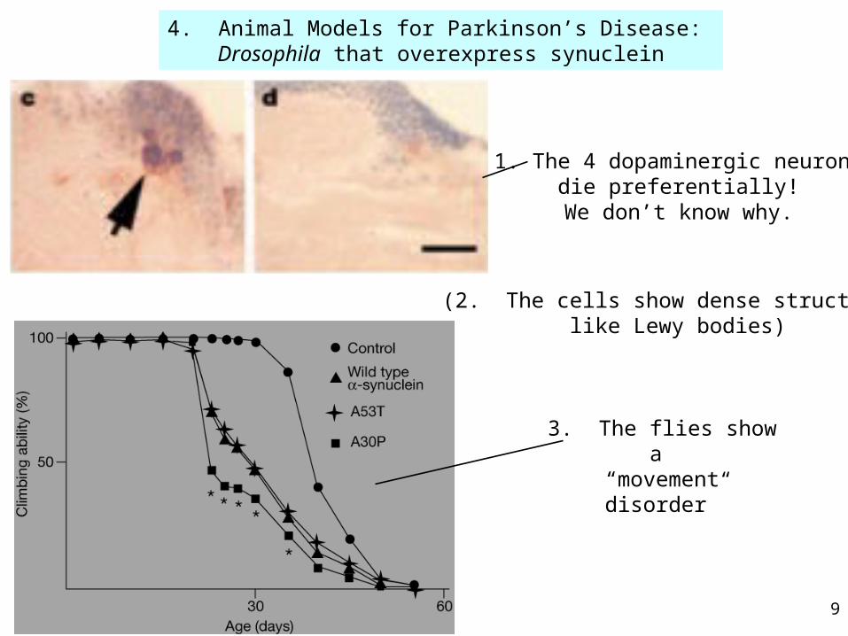
4. Animal Models for Parkinson’s Disease: Drosophila that overexpress synuclein
1. The 4 dopaminergic neurons die preferentially!
We don’t know why.
3. The flies show a “movement disorder”
(2. The cells show dense structures like Lewy bodies)
10
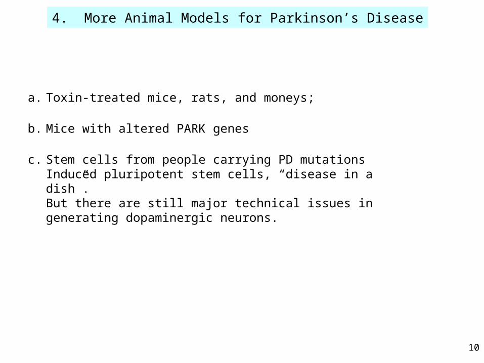
4. More Animal Models for Parkinson’s Disease
a. Toxin-treated mice, rats, and moneys;
b. Mice with altered PARK genes
c. Stem cells from people carrying PD mutationsInduced pluripotent stem cells, “disease in a dish”. But there are still major technical issues in generating dopaminergic neurons.

ACh
ACh
GPi
DirectPathway
GPe
Thalamus
Cortex
Excitation
Inhibition
(Regardless of color)
INs
ACh GABA Glu
INs
Transmitters
dorsalstriatum
?STN +SNr
=SNc
?
PPTg
MSN D1RMSN D2R
Indirect pathway
INs
DA
Tremor arises in a malfunctioning feedback loop: substantia nigra, striatum, and other structures in basal ganglia.
Implanted stimulating electrodes retune this loop.
Deep brain stimulation for Parkinson’s Disease like previous lectures
Axons passing through
12
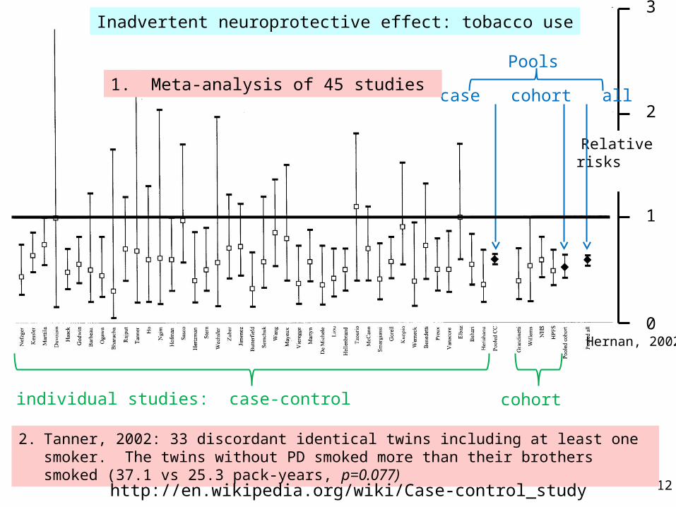
Inadvertent neuroprotective effect: tobacco use3
2
1
0
Relative risks
individual studies: case-control
Hernan, 2002
cohort
case cohort all
2. Tanner, 2002: 33 discordant identical twins including at least one smoker. The twins without PD smoked more than their brothers smoked (37.1 vs 25.3 pack-years, p=0.077)
1. Meta-analysis of 45 studies
http://en.wikipedia.org/wiki/Case-control_study
Pools
13(Lester research group)
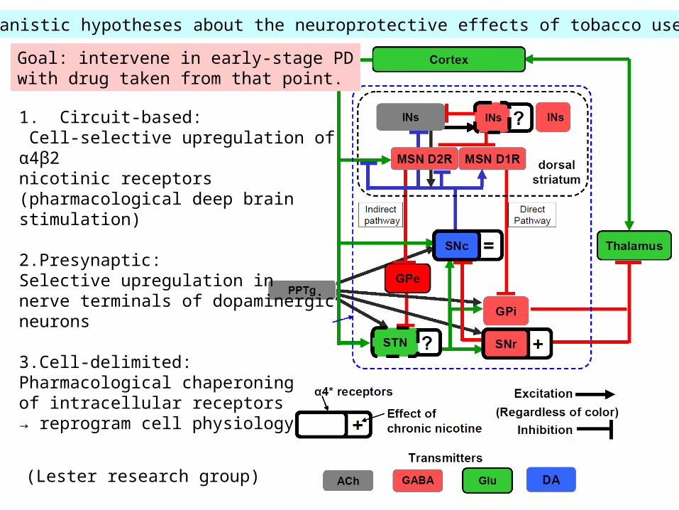
1. Circuit-based: Cell-selective upregulation of α4β2nicotinic receptors (pharmacological deep brain stimulation)
2.Presynaptic:Selective upregulation in nerve terminals of dopaminergic neurons
3.Cell-delimited:Pharmacological chaperoning of intracellular receptors→ reprogram cell physiology
Mechanistic hypotheses about the neuroprotective effects of tobacco use in PD
Goal: intervene in early-stage PDwith drug taken from that point.
14
Transplantation of stem cells induced to release dopamine
No reproducible success to date.
Alzheimer’s disease
1. Clinical description
2. Genetics
3. Pathophysiology
4. Biomarkers and animal models
5. Heterozygote advantage: none known
6. Therapeutic approaches
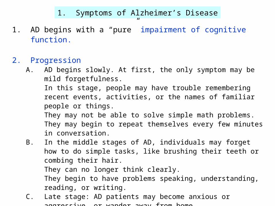
1. Symptoms of Alzheimer’s Disease
1. AD begins with a “pure” impairment of cognitive function.
2. ProgressionA. AD begins slowly. At first, the only symptom may be mild forgetfulness.
In this stage, people may have trouble remembering recent events, activities, or the names of familiar people or things. They may not be able to solve simple math problems. They may begin to repeat themselves every few minutes in conversation.
B. In the middle stages of AD, individuals may forget how to do simple tasks, like brushing their teeth or combing their hair. They can no longer think clearly. They begin to have problems speaking, understanding, reading, or writing.
C. Late stage: AD patients may become anxious or aggressive, or wander away from home. Eventually, patients need total care.
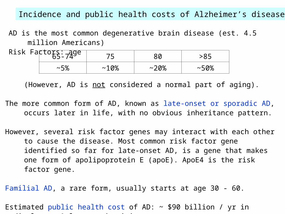
Incidence and public health costs of Alzheimer’s disease
AD is the most common degenerative brain disease (est. 4.5 million Americans) Risk Factors: age
(However, AD is not considered a normal part of aging). The more common form of AD, known as late-onset or sporadic AD, occurs later in life,
with no obvious inheritance pattern. However, several risk factor genes may interact with each other to cause the disease.
Most common risk factor gene identified so far for late-onset AD, is a gene that makes one form of apolipoprotein E (apoE). ApoE4 is the risk factor gene.
Familial AD, a rare form, usually starts at age 30 - 60.
Estimated public health cost of AD: ~ $90 billion / yr in medical care & lost productivity.
65-74 75 80 >85
~5% ~10% ~20% ~50%
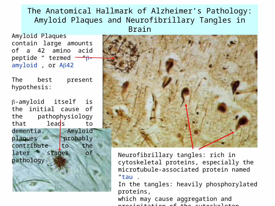
The Anatomical Hallmark of Alzheimer’s Pathology:Amyloid Plaques and Neurofibrillary Tangles in Brain
Amyloid Plaquescontain large amounts of a 42 amino acid peptide termed “-amyloid”, or A42
The best present hypothesis:
-amyloid itself is the initial cause of the pathophysiology that leads to dementia. Amyloid plaques probably contribute to the later stages of pathology
Neurofibrillary tangles: rich in cytoskeletal proteins, especially the microtubule-associated protein named “tau”. In the tangles: heavily phosphorylated proteins, which may cause aggregation and precipitation of the cytoskeleton,
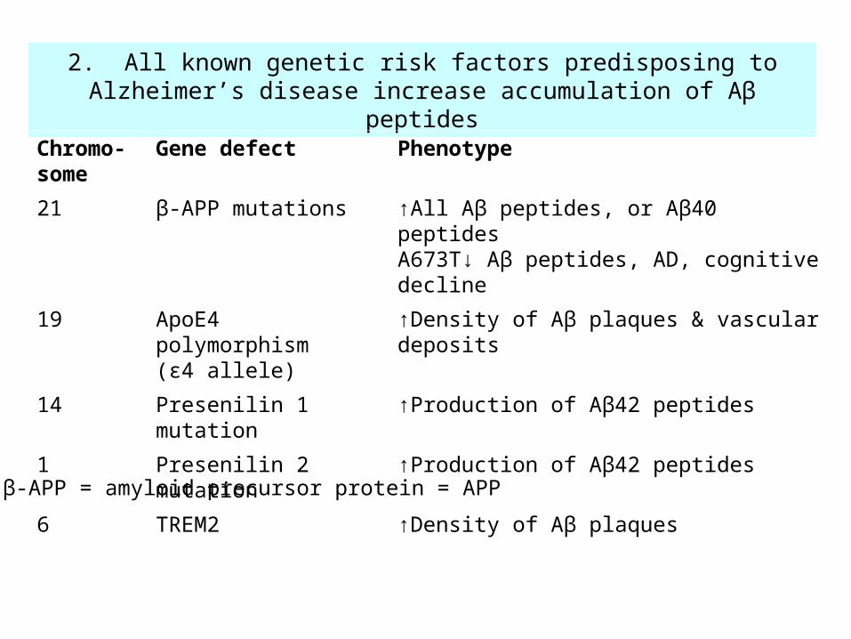
2. All known genetic risk factors predisposing to Alzheimer’s disease increase accumulation of Aβ peptides
Chromo-some
Gene defect Phenotype
21 β-APP mutations ↑All Aβ peptides, or Aβ40 peptidesA673T↓ Aβ peptides, AD, cognitive decline
19 ApoE4 polymorphism(ε4 allele)
↑Density of Aβ plaques & vascular deposits
14 Presenilin 1 mutation ↑Production of Aβ42 peptides
1 Presenilin 2 mutation ↑Production of Aβ42 peptides
6 TREM2 ↑Density of Aβ plaques
β-APP = amyloid precursor protein = APP
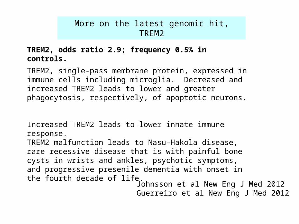
More on the latest genomic hit, TREM2
TREM2, odds ratio 2.9; frequency 0.5% in controls.
TREM2, single-pass membrane protein, expressed in immune cells including microglia. Decreased and increased TREM2 leads to lower and greater phagocytosis, respectively, of apoptotic neurons.
Increased TREM2 leads to lower innate immune response. TREM2 malfunction leads to Nasu–Hakola disease, rare recessive disease that is with painful bone cysts in wrists and ankles, psychotic symptoms, and progressive presenile dementia with onset in the fourth decade of life.
Johnsson et al New Eng J Med 2012Guerreiro et al New Eng J Med 2012
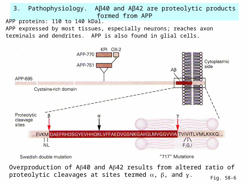
3. Pathophysiology. Aβ40 and Aβ42 are proteolytic products formed from APP
Overproduction of A40 and A42 results from altered ratio of proteolytic cleavages at sites termed ,, and.
Fig. 58-6
APP proteins: 110 to 140 kDal. APP expressed by most tissues, especially neurons; reaches axon terminals and dendrites. APP is also found in glial cells.
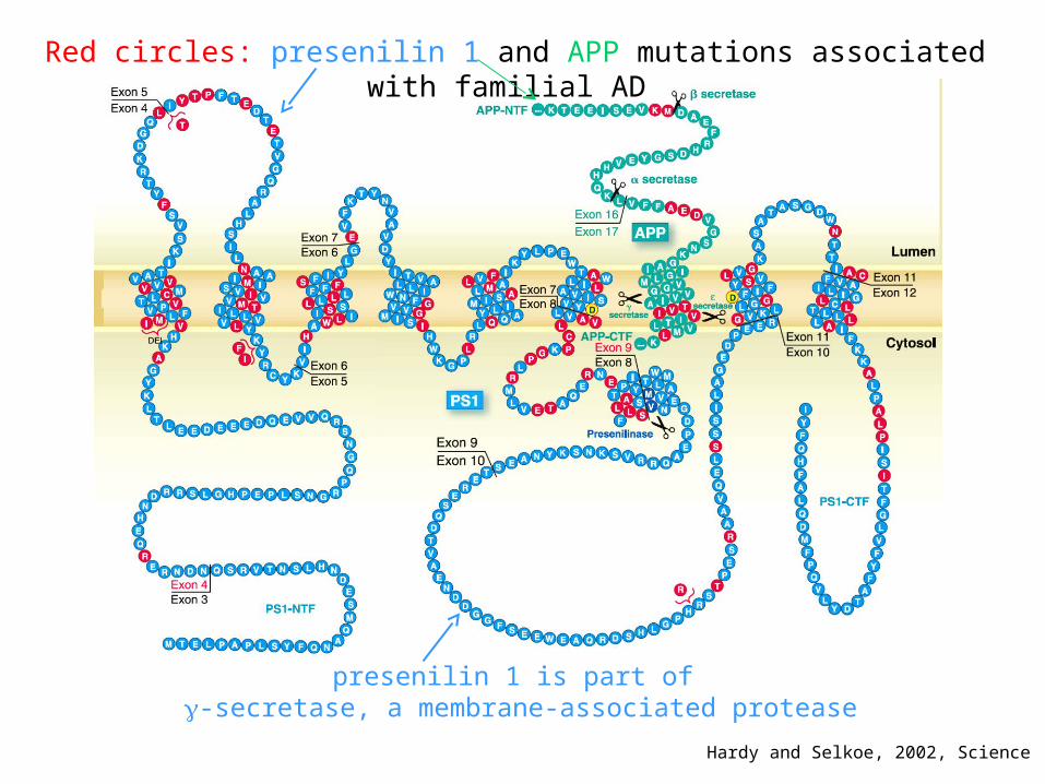
Red circles: presenilin 1 and APP mutations associated with familial AD
Hardy and Selkoe, 2002, Science
presenilin 1 is part of -secretase, a membrane-associated protease
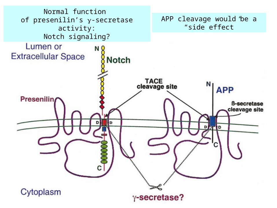
Normal function of presenilin’s -secretase activity:
Notch signaling?APP cleavage would be a
“side effect”
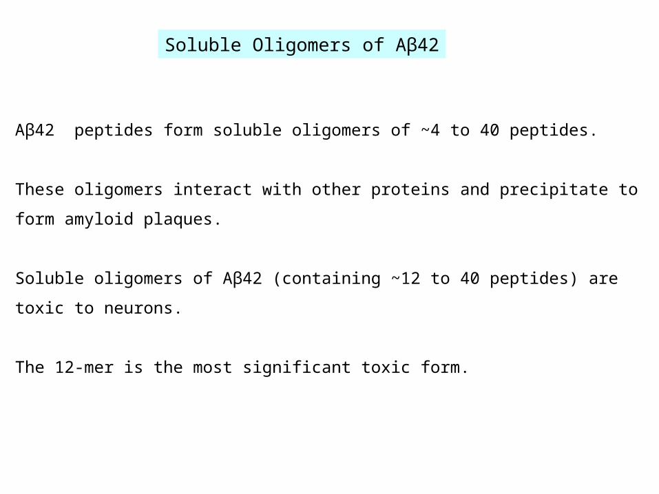
Aβ42 peptides form soluble oligomers of ~4 to 40 peptides.
These oligomers interact with other proteins and precipitate to form amyloid plaques.
Soluble oligomers of Aβ42 (containing ~12 to 40 peptides) are toxic to neurons.
The 12-mer is the most significant toxic form.
Soluble Oligomers of Aβ42
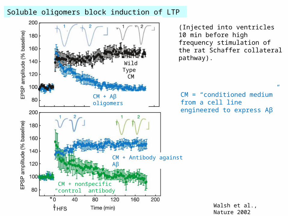
(Injected into ventricles 10 min before high frequency stimulation of the rat Schaffer collateral pathway).
Walsh et al., Nature 2002
CM = “conditioned medium” from a cell line engineered to express Aβ
Soluble oligomers block induction of LTP
Wild Type CM
CM + Aβ oligomers
CM + nonspecific“control” antibody
CM + Antibody against Aβ
From Selkoe, 2002, Science
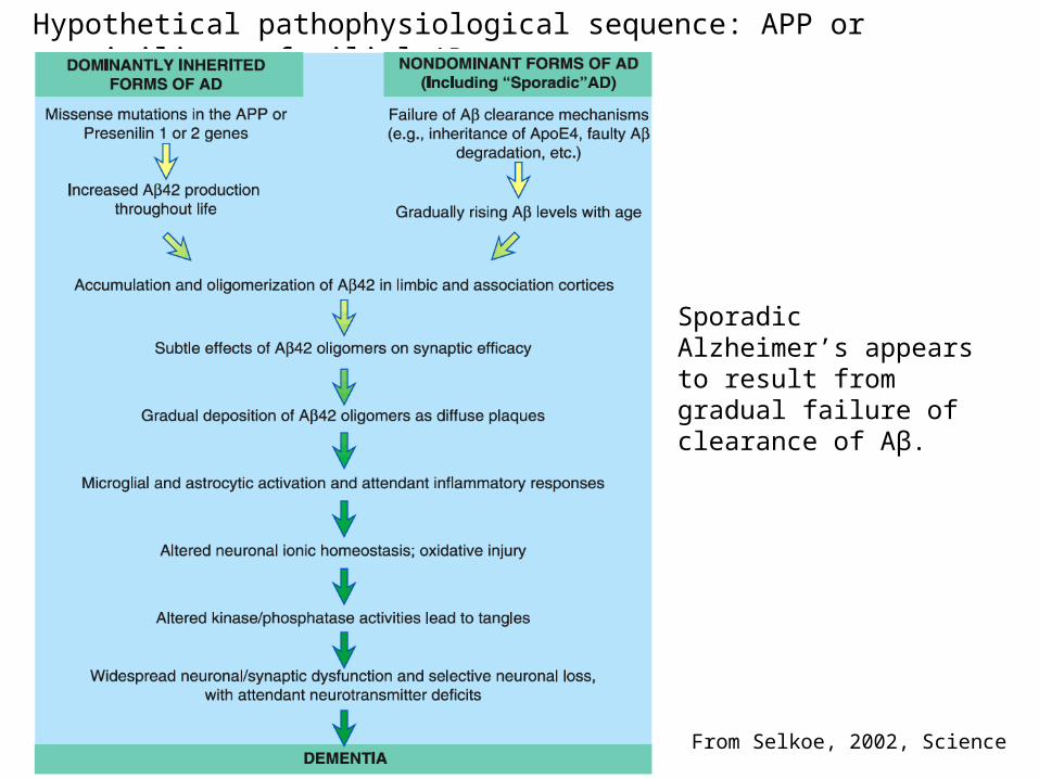
Hypothetical pathophysiological sequence: APP or presinilin → familial AD
Sporadic Alzheimer’s appears to result from gradual failure of clearance of Aβ.
27
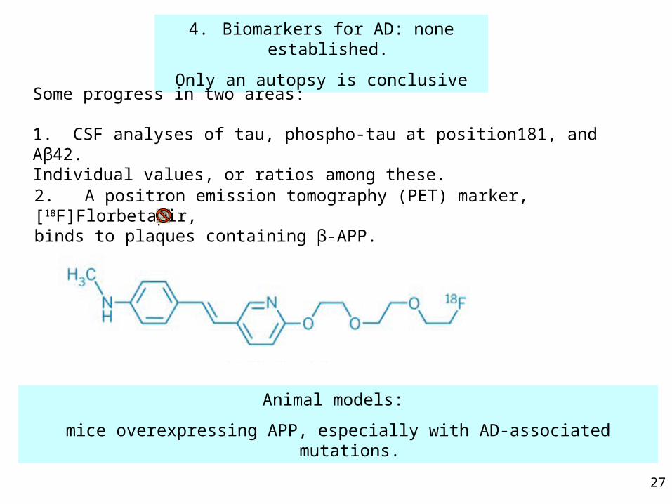
4. Biomarkers for AD: none established.
Only an autopsy is conclusive
Some progress in two areas:
1. CSF analyses of tau, phospho-tau at position181, and Aβ42.Individual values, or ratios among these.
Animal models:
mice overexpressing APP, especially with AD-associated mutations.
2. A positron emission tomography (PET) marker, [18F]Florbetapir, binds to plaques containing β-APP.
28
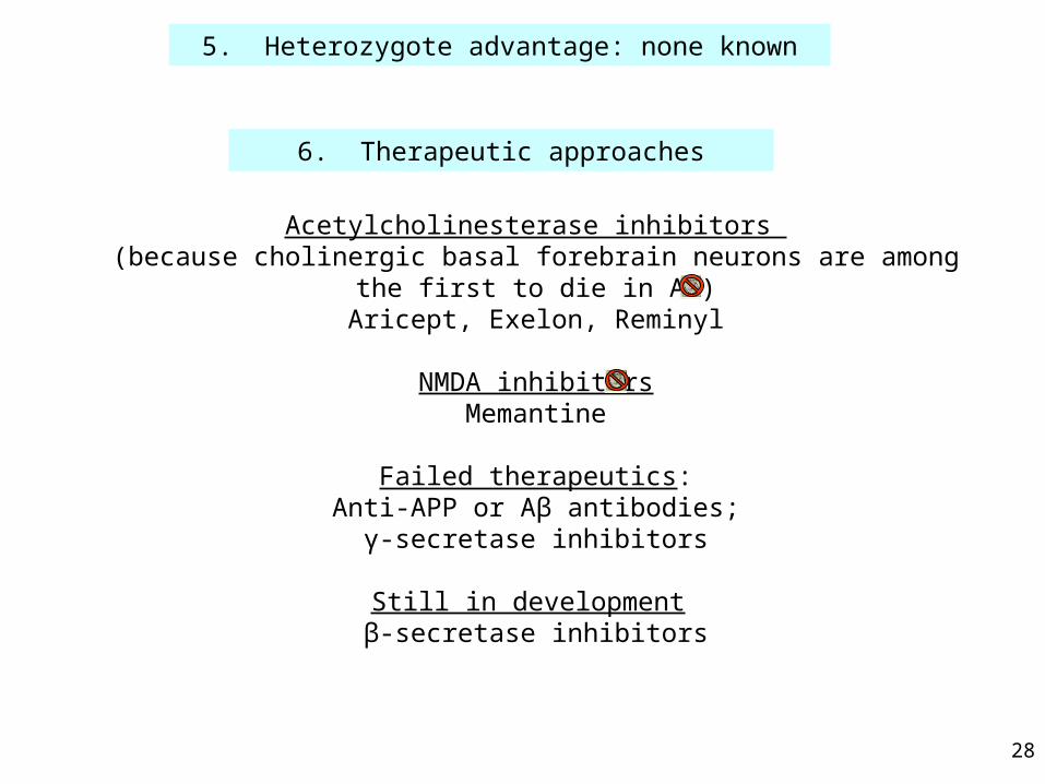
5. Heterozygote advantage: none known
Acetylcholinesterase inhibitors (because cholinergic basal forebrain neurons are among the first to die in AD)
Aricept, Exelon, Reminyl
NMDA inhibitorsMemantine
Failed therapeutics:Anti-APP or Aβ antibodies;
γ-secretase inhibitors
Still in development β-secretase inhibitors
6. Therapeutic approaches
29
End of lecture
Henry Lester’s “office” hours, Monday this week 1:15-2 Red Door
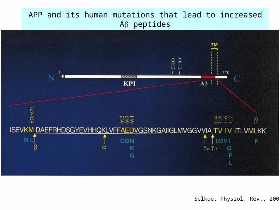
Selkoe, Physiol. Rev., 2001
APP and its human mutations that lead to increased A peptides
QuickTime™ and aTIFF (Uncompressed) decompressor
are needed to see this picture.
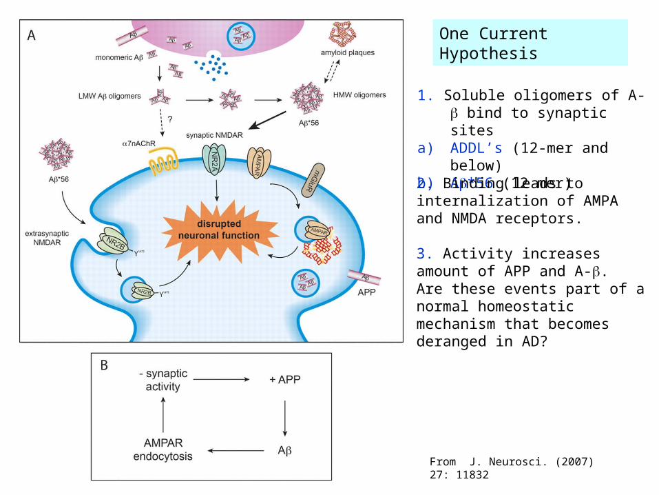
1. Soluble oligomers of A- bind to synaptic sites
a) ADDL’s (12-mer and below)b) A*56 (12 mer)
2. Binding leads to internalization of AMPA and NMDA receptors.
3. Activity increases amount of APP and A-. Are these events part of a normal homeostatic mechanism that becomes deranged in AD?
One Current Hypothesis
From J. Neurosci. (2007) 27: 11832