Embed Size (px)
DESCRIPTION
TJPRC JOURNALS
Citation preview

www.tjprc.org [email protected]
LAMINATE VENEERS A VIABLE ALTERNATIVE TO FULL
COVERAGE CROWNS – A CASE REPORT
KAMATCHI. K 1, KRISHNA MEERA. N 2 & KRISHNA RAJ. R 3 1,2Department of Prosthodontics, Madha Dental College and Hospital, Tamil Nadu, India
3Department of Prosthodontics, Rajah Muthiah Dental College and Hospital, Tamil Nadu, India
ABSTRACT
Everyday there is growing number of patients even seeking esthetic enhancement of already healthy teeth.
Porcelain laminate veneers are used frequently for achieving esthetics in natural dentition. These veneer only need
minimal tooth preparation compared to the extensive tooth preparation in case of full coverage all ceramic and metal
ceramic restorations.This article discusses about the indications, advantages, various clinical steps, disadvantages and
contraindication of the porcelain laminate veneers along with a case report.
KEYWORDS: Wrap Around Preparation, Spot Etch And Spot Bonding, Silane Coupling Agent, Dual Cure Composite
Resin
INTRODUCTION
The early work of Buonocore on acid etching along with resin composite bonding and dental ceramic
etching and bonding by Simonsen and Calamia has made the conservative tooth preparation and restoring with
porcelain laminate veneers possible1. Laminate veneers are indicated in areas such as to close mild diastemas, to
correct tooth forms and position, discolourations, incisal abrasions and to replace old extensive restorations2,3.
Even though porcelain has a long history as one of the biocompatible and only esthetic alternative as full crowns,
using it as a labial veneer along with the concept of acid etching and bonding was first documented in the year
1975 by Rochette 4.
Porcelain as laminate veneer offers advantages like inherent colour control, improving the periodontal
health by reducing plaque accumulation due to its highly glazed surface, provide greater resistance to abrasion and
wear, its resistance to fluid sorption, finally its excellent esthetics5,6. The ultimate success of laminate veneers lies
in proper tooth preparation, impression making, accurate shade selection and finally implementing proper bonding
technique. This case report will be a useful guide for all clinical procedures in fabrication of porcelain laminate
veneers.
Case Report
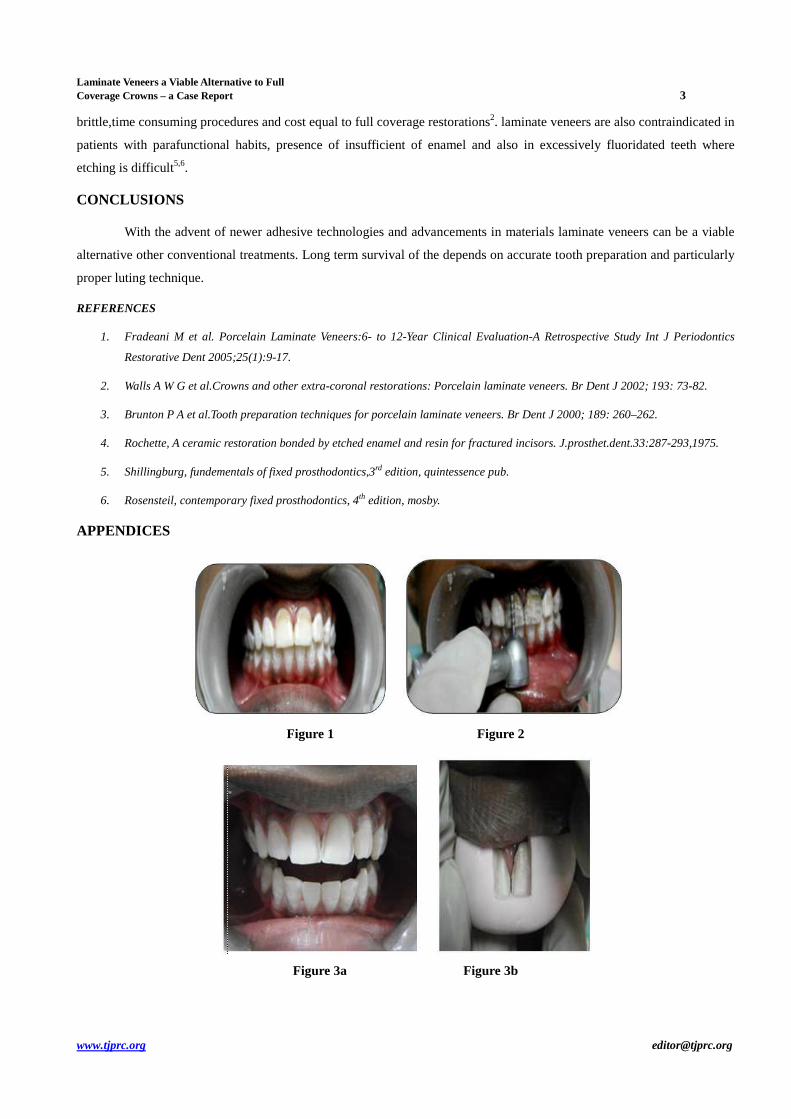
A male patient aged 21yrs reported to the department of prosthodontics complained of discoloration in his
front teeth. On examination it was observed that the central incisors of the patient had grade 2 discoloration due to
enamel flourosis (Figure 1). A more conservative mode of treatment of laminate veneers was suggested that
involved minimal tooth preparation and a more esthetic alternative.
Original A
rticle International Journal of Dental Research & Development (IJDRD) ISSN(P): 2250-2386; ISSN(E): 2321-0117 Vol. 5, Issue 4, Dec 2015, 1-4 © TJPRC Pvt. Ltd

2 Kamatchi. K, Krishna Meera. N & Krishna Raj. R
Impact Factor (JCC): 1.9876 Index Copernicus Value(ICV): 3.0
The diagnostic impressions were taken in irreversible hydrocolloid and poured in type 2 gypsum product. Putty
index for assessing the amount of tooth reduction and for provisional restorations were made. Initially depth orientation
grooves were made using 0.5 mm three tier self limiting depth gauge bur(Figure 2) after which axial tooth reduction is
done using parallel sided torpedo bur, the tip of the bur automatically prepares the chamfer finish margins when preparing
the axial portion of the tooth. Proximally the tooth preparation is extended into the embrasure space and ends just before
the contact area to hide the restoration margin. Finish margins are kept supra gingivally in the cervical region is done
(Figure 3a). All the line angles and point angles of the preparation is rounded using soft flex disc. Putty index was placed
and verified for assessing the amount of tooth preparation (Figure 3b)
Before making the final impression, one sided fine diamond abrasive strip is used to shape the contact areas. This
reshaping the contact area allows proper recording of the impression inter proximally without affecting the integrity and
stability of the arch. Tissue retraction is done using retraction cord placed inside the sulcus for about 1minute and the final
impression is made using addition silicone impression material using multiple mix technique. Shade is recorded as A1.
Provisional acrylic veneers are fabricated and cemented to the prepared tooth using spot etch and spot bonding technique
for the easy retrieval of the temporary in later stages. Lithium disilicate crystals has been used for the pressed ceramic for
the fabrication of laminates and thickness of the laminates were also evaluated( Figure 4a &4b)
Before bonding Porcelain laminate veneers adaptation of the veneers margins, relationship of one laminate to
other and the to the adjacent teeth and the esthetics of the restoration is checked using the try in paste of the dual cure resin
cement. Bonding of the porcelain laminate veneers is done with good moisture control technique. Prepared tooth surface is
acid etched using 37% Ortho phosphoric acid for 30 seconds followed by rinsing with water and air drying. Frosty white
appearance of the etched surface indicates adequate etching of the tooth surface. Bonding agent is applied to the etched
surface and allowed it to dry. Inner surface of the veneers are etched using 9% Hydroflouric acid and rinsed with water. A
silane coupling agent is applied to the etched veneer surface and allowed it to dry. Finally dual polymerizing luting resin
cement is used to bond the veneer to the tooth. Unset excess resin cement is removed using metallic instrument cervically
and using the dental floss interproximally. Once the excess cements are removed, visible light is used to polymerize the
resin composite cement for 30 seconds from all sides (Figure 5). Final stage is the checking the sulcus and other areas for
the excess cement and remove it and checking for the contact points and finishing those areas using soft flex discs and 10
mm particle size diamond polishing paste. The patient was esthetically satisfied with the appearance and tooth form of the
laminates(Figure 6). Review was done that revealed a satisfactory outcome
DISCUSSIONS
Tooth preparations for laminate veneers should always be limited only to enamel and most certainly the finish
margins to ensure adequate seal. It is recommended that at least 50% of preparation should be in enamel to enhance the
bonding of the veneer to the underlying tooth. However reduced bond strength along with pulpal hyperemia due the
exposure of dental tubules to acid etchant and the bonding material itself are the consequence of bringing the tooth
preparation to the dentin level.. Study conducted by Fradeani M et al on laminate veneers after 12 years of clinical service
provides a 94.4 of clinical survival rate and most of the failure is attributed only due to improper bonding techniques1.
Study conducted by Leempoel et al on laminate veneers reported survival rate of 99% after 5 years and 95% after 11
years1.Even though porcelain laminate veneers are viable alternative to direct composite resin veneering and full coverage
restorations they do have some disadvantages like repairing is difficult once it is luted, technique sensitive, margins are
Laminate Veneers a Viable Alternative to Full Coverage Crowns – a Case Report 3
www.tjprc.org [email protected]
brittle,time consuming procedures and cost equal to full coverage restorations2. laminate veneers are also contraindicated in
patients with parafunctional habits, presence of insufficient of enamel and also in excessively fluoridated teeth where
etching is difficult5,6.
CONCLUSIONS
With the advent of newer adhesive technologies and advancements in materials laminate veneers can be a viable
alternative other conventional treatments. Long term survival of the depends on accurate tooth preparation and particularly
proper luting technique.
REFERENCES
1. Fradeani M et al. Porcelain Laminate Veneers:6- to 12-Year Clinical Evaluation-A Retrospective Study Int J Periodontics
Restorative Dent 2005;25(1):9-17.
2. Walls A W G et al.Crowns and other extra-coronal restorations: Porcelain laminate veneers. Br Dent J 2002; 193: 73-82.
3. Brunton P A et al.Tooth preparation techniques for porcelain laminate veneers. Br Dent J 2000; 189: 260–262.
4. Rochette, A ceramic restoration bonded by etched enamel and resin for fractured incisors. J.prosthet.dent.33:287-293,1975.
5. Shillingburg, fundementals of fixed prosthodontics,3rd edition, quintessence pub.
6. Rosensteil, contemporary fixed prosthodontics, 4th edition, mosby.
APPENDICES
Figure 1 Figure 2
Figure 3a Figure 3b

4 Kamatchi. K, Krishna Meera. N & Krishna Raj. R
Impact Factor (JCC): 1.9876 Index Copernicus Value(ICV): 3.0
Figure 4a Figure 4b
Figure 4 Figure 5
FIGURE LEGENDS
Figure 1: Preoperative
Figure 2: Preparation with self limiting bur
Figure 3a: Finished incisal preparation
Figure 3b: Verification with putty index
Figure 4a &4b: Pressed ceramic and thickness of laminates checked
Figure 5: Curing of the dual cure resin cement done
Figure 6: Post treatment