Embed Size (px)
Citation preview
1
History of Melanosome Research Jan Borovansk ý
1.1 Introduction
Melanosomes were fi rst proposed as specifi c organelles, unique to pigment cells, in a preliminary publication that appeared on 30 July 1960 [1] . An announcement had been made at the 21st Annual Meeting of the Society for Investigative Der-matology, at Miami Beach, Florida, USA on 13 June 1960 [2] and the news, that the chemical composition and enzyme activities in melanosomes and mitochon-dria are completely different, was considered to be of such signifi cance that it appeared in a newspaper report (Figure 1.1 ). Similar data, with an emphasis on terminology, were published in 1963 [3] .
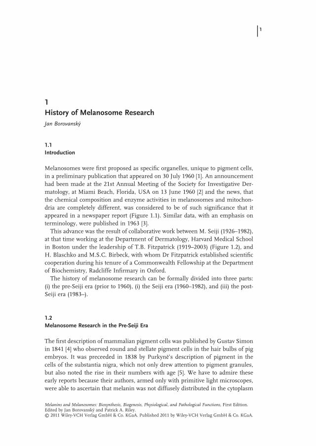
This advance was the result of collaborative work between M. Seiji (1926 – 1982), at that time working at the Department of Dermatology, Harvard Medical School in Boston under the leadership of T.B. Fitzpatrick (1919 – 2003) (Figure 1.2 ), and H. Blaschko and M.S.C. Birbeck, with whom Dr Fitzpatrick established scientifi c cooperation during his tenure of a Commonwealth Fellowship at the Department of Biochemistry, Radcliffe Infi rmary in Oxford.
The history of melanosome research can be formally divided into three parts: (i) the pre - Seiji era (prior to 1960), (i) the Seiji era (1960 – 1982), and (iii) the post - Seiji era (1983 – ).
1.2 Melanosome Research in the Pre - S eiji Era
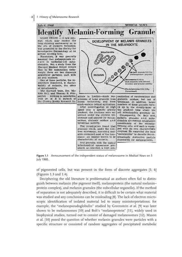
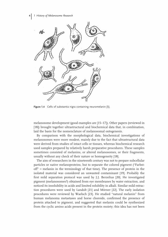
The fi rst description of mammalian pigment cells was published by Gustav Simon in 1841 [4] who observed round and stellate pigment cells in the hair bulbs of pig embryos. It was preceeded in 1838 by Purkyn ĕ ’ s description of pigment in the cells of the substantia nigra, which not only drew attention to pigment granules, but also noted the rise in their numbers with age [5] . We have to admire these early reports because their authors, armed only with primitive light microscopes, were able to ascertain that melanin was not diffusely distributed in the cytoplasm
Melanins and Melanosomes: Biosynthesis, Biogenesis, Physiological, and Pathological Functions, First Edition.Edited by Jan Borovanský and Patrick A. Riley.© 2011 Wiley-VCH Verlag GmbH & Co. KGaA. Published 2011 by Wiley-VCH Verlag GmbH & Co. KGaA.
1
2 1 History of Melanosome Research
of pigmented cells, but was present in the form of discrete aggregates [5, 6] (Figures 1.3 and 1.4 ).
Deciphering the old literature is problematical as authors often fail to distin-guish between melanin (the pigment itself), melanoprotein (the natural melanin – protein complex), and melanin granules (the subcellular organelle). If the method of separation is not adequately described, it is diffi cult to be certain what material was studied and any conclusions can be misleading [8] . The lack of electron micro-scopic identifi cation of isolated material led to many misinterpretations; for example, the “ melanopseudoglobulin ” studied by Greenstein et al. [9] was later shown to be melanosomes [10] and Bolt ’ s “ melanoprotein ” [11] , widely used in biophysical studies, turned out to consist of damaged melanosomes [12] . Mason et al . [10] posed the question of whether melanin granules were particles with a specifi c structure or consisted of random aggregates of precipitated metabolic
Figure 1.1 Announcement of the independent status of melanosome in Medical News on 5 July 1960.
1.2 Melanosome Research in the Pre-Seiji Era 3
products. The introduction of electron microscopy was able to resolve this matter and Laxer et al . [13] were able to discern an inner ultrastructure in isolated melano-somes. The fi rst clear pictures were obtained only in 1956 [14] .
An avalanche of papers in subsequent years brought with it enormous amounts of information on the ultrastructure of melanosomes and its changes during
Figure 1.2 Professor Makoto Seiji (left) and Professor Thomas B. Fitzpatrick (right) in 1972.
Figure 1.3 “ Chromatophore ” from donkey conjuctiva [7] .
4 1 History of Melanosome Research
melanosome development (good examples are [15 – 17]) . Other papers (reviewed in [18] ) brought together ultrastructural and biochemical data that, in combination, laid the basis for the nomenclature of melanosomal ontogenesis.
By comparison with the morphological data, biochemical investigations of melanosomes were more modest, mainly due to the fact that ultrastructural data were derived from studies of intact cells or tissues, whereas biochemical research used samples prepared by relatively harsh preparative procedures. These samples sometimes consisted of melanins, or altered melanosomes, or their fragments, usually without any check of their nature or homogeneity [18] .
The aim of researchers in the nineteenth century was not to prepare subcellular particles or native melanoproteins, but to separate the colored pigment ( “ Farbst-off ” = melanin in the terminology of that time). The presence of protein in the isolated material was considered an unwanted contaminant [19] . Probably the fi rst mild separation protocol was used by J.J. Berzelius [20] . He investigated pigment (melanosomes?) obtained from eye membranes by water extraction, and noticed its insolubility in acids and limited solubility in alkali. Similar mild extrac-tion procedures were used by Landolt [21] and M ö rner [22] . The early isolation procedures were reviewed by Waelsch [23] . He studied “ natural melanin ” from human melanoma metastases and horse choroids, confi rmed the presence of protein attached to pigment, and suggested that melanin could be synthesized from the cyclic amino acids present in the protein moiety; this idea has not been
Figure 1.4 Cells of substantia nigra containing neuromelanin [5] .
1.3 Melanosome Research in the Seiji Era 5
abandoned till now. Herrmann and Boss [24] demonstrated dopa oxidase activity in the fraction of melanin granules from ciliary bodies of cattle eyes, but, as their samples were contaminated with mitochondria, they demonstrated the presence of mitochondrial enzyme markers as well. In 1949, du Buy et al . concluded that melanosomes are modifi ed mitochondria typical of pigment cells [25] . It is interesting that du Buy [26] and other authors [27] did not abandon the mito-chondrial theory of melanosome origin even in 1963 (i.e., 2 years after the for-mulation of Seiji ’ s melanosomal concept) and even published their papers in the same volume in which Seiji et al . published detailed confi rmation of their model [28] .
It is interesting that history has disregarded the contribution of Stein [29] who, several years before the work of Seiji et al. , using a separation procedure of his own, isolated melanin granules from ox choroids and analyzed their content not only of melanin, but also lipids, carbohydrates, RNA, and metals (including the pioneer fi nding of a high level of zinc), and concluded that the chemical composi-tion of melanin granules is completely different from mitochondria.
The ability of melanin in melanin granules, isolated from Harding - Passey melanoma and from the ink sac of Loligo opalescens , to act as a cation exchanger [30] , and the demonstration of free radical activity in melanin - containing tissues [31] also rank among the observations of the pre - Seiji era.
1.3 Melanosome Research in the S eiji Era
1.3.1 Terminology of Melanosomes
The demonstration of melanosomes as unique pigment cell organelles possessing developmental stages prompted the introduction of a system of terminology that refl ected the characteristics of the various states. Until 1961 the common term for all varieties of these organelles was melanin (or pigment) granule [1, 2] . The fi rst system of nomenclature [2] described three stages in the ontogenesis of melanosomes:
i) Premelanosomes: spherical organelles. ii) Melanosomes: organelles with an internal structure and tyrosinase activity.
iii) Melanin granules: melanoprotein polymer.
A second terminological system was proposed [3, 26] consisting of three devel-opmental stages plus a fi nal product. Thus:
• Stage I (fi rst stage): biosynthesis of protein. • Stage II (intermediate stage): biosynthesis of organelle. • Stage III (late phase): biosynthesis of melanin. • Final product: melanin granule.
6 1 History of Melanosome Research
These nomenclature systems introduced a certain degree of confusion, particu-larly as the term melanin granule had been used to describe pigment granules at any developmental stage. In an attempt to establish a consensus, Fitzpatrick et al . [32, 33] circulated a postal questionnaire seeking opinions about the adequacy of the terms in common use in pigment cell research and, with the approval of the participants of the Sixth International Pigment Cell Conference in 1965 in Sofi a, Bulgaria, recommended the use of two terms:
• Melanosome : a discrete melanin - containing organelle in which melanization is complete as indicated by its almost uniform density by electron microscopy and the absence of demonstrable tyrosinase activity.
• Premelanosome : a term applied to all the stages in melanosome biogenesis that precede the fully developed state. Within the restrictions of this general defi nition, the premelanosomal stage might, at the discretion of the investiga-tor, be subdivided into early, intermediate, and late phases.
The nomenclature in general use today does not adhere to any of the three systems outlined above, but is essentially a system proposed by Toda et al . [34 – 36] refl ecting the earlier descriptions of Birbeck [37, 38] which employs the uniform term “ melanosome ” with a numerical indication (I – IV) of the degree its ontoge-netic development.
However, in practice, chaos prevails. While the system of Toda et al . is widely – if somewhat erratically – used, some European authors refer, often incorrectly, to the stages proposed in the second system of nomenclature [3, 26] and some American authors tend to cite nomenclature introduced in their previous papers or those of their friends.
1.3.2 Ultrastructural and Histochemical Studies
The concept of subcellular biosynthesis and localization of melanins and melano-proteins in melanosomes was further confi rmed by (i) autoradiographic evidence with [ 3 H]dopa and [2 - 14 C]dopa [39 – 43] ., (ii) incorporation of [2 - 14 C]dopa and moni-toring radioactivity in subcellular fractions [44, 45] ., and (c) isolation of melano-somes and analysis of their chemical composition [46, 47] .
Electron microscopy enabled the defi nition of the basic morphometric data of isolated melanosomes (i.e., their size, shape, and ultrastructural appearance). The most extensive data were published by Hach et al . [48, 49] . For discussion concerning the ultrastructural appearances of melanosomes, see Section 12.3 in Chapter 12 .
Various pathological states may be manifested by changes in melanosome mor-phology. Mishima et al . [50] considered that melanosome polymorphism, such as changes in size, shape, ultrastructural matrix, the manner of melanin deposition, and the degree of melanosome maturation, as a criterion of molecular pathology that could fi nd practical use in the differential diagnosis of various pigmentary disorders.
1.3 Melanosome Research in the Seiji Era 7
In melanoma cells, various irregularities in the architecture of melanosomes are common. Deposition of melanin can be uneven, leading to a bizarre appearance of melanosomes [51 – 57] ; the presence of melanosomes of all stages of develop-ment is typical [51, 52] . Melanoma melanosomes also often exhibit defects of their limiting membranes that may lead to leakage of toxic melanin precursors into the cytosol. The pathological consequences of this failure of containment of melano-genic intermediates are discussed in Section 12.4.2 . Extracellular deposition of melanin on fi brils resembling melanosomal matrix fi brils has also been observed in melanoma cells [58, 59] .
Early ideas on melanosomal biogenesis were summarized in several studies [52, 60, 61] .
Fitzpatrick and Breathnach defi ned a functional unit in human epidermis named the “ epidermal melanin unit. ” This was viewed as a symbiotic relationship between melanocytes and keratinocytes in which each melanocyte supplies approximately 36 keratinocytes with melanosomes [62] . The mechanism of melanosome transport and transfer to keratinocytes was outlined in Mottaz and Zelickson [63] . Szab ó et al . [64] demonstrated racial differences in the fate of melanosomes in human epidermis.
1.3.3 Biochemical Studies
A prerequisite for classical biochemical studies is the ability to isolate native and pure melanosomes [65] . To this end 17 isolation protocols suggested for the isola-tion of melanosomes between 1940 and 1973 were critically reproduced [18, 65] , and it was concluded that the best samples could be obtained using the procedures described by Stein [29] , Doezema [66] , and Haberman and Menon [67] . Isolation of melanosomes from keratinous material turned out to be more diffi cult because the need to release melanosomes from hair required chemical means of tissue disintegration, which always engenders a search for a compromise between suf-fi cient tissue disintegration and minimizing the extent of melanosome modifi ca-tion by the isolation procedure [61, 68] . Ten methods for isolating melanosomes from hair were critically reviewed and half of them, which from the assessment of the isolation conditions seemed to be promising, were reproduced [68] . The best results were obtained with the isolation protocol of Borovansk ý and Hach [69] .
The availability of melanosome samples of adequate quality [18] opened the gate to the subsequent establishment of their basic chemical composition. Melanin and protein moieties were shown to be dominant constituents of melanosomes [46, 47, 61, 70 – 72] . Isolated melanosomes were reported to contain 5 – 10% of carbohy-drates [29] . Tyrosinase, as a glycoprotein, also brings into melanosomes sialic acids containing N - acetylneuraminic acid [73] . In addition to gangliosides mentioned in Section 12.2 , many other lipid constituents in melanosomes have been reported including cholesterol and free fatty acids [74] , and phospholipids [3, 75] . The level of total lipids was found to vary between 1 – 5% [29] and 5 – 11% [76] . Jimbow et al . [77] made a complete qualitative and quantitative analysis of lipids and their

8 1 History of Melanosome Research
fractions in isolated Harding - Passey and B16 melanoma melanosomes, and found quantitative differences between them (Figure 1.5 ). The demonstration of the absence of DNA in isolated melanosomes was clear evidence of the difference between melanosomes and mitochondria [78] . Melanosomes are abundant in various metals as described in detail in the Section 12.6 . The description of the development of analytical methods including classical chemical techniques such as titration, spectrophotometry, electrochemical and isotope methods, neutron activation analysis, mass spectrometry, and inductively coupled plasma tech-niques, together with cell biological methods such as histochemistry, autometal-lography, autoradiography, and microanalytical techniques (using electron, proton, laser, X - ray, and ion beams), and their use in melanosome analyses is comprehen-sively covered in a review [79] .
Nowadays it sounds incredible, but in the 1960s there were doubts as to whether melanosomes merely represented an association of tyrosinase with melanin or whether the organelles contained other proteins as suggested by the electron microscopic appearance. The matter was investigated by electrophoretic studies of isolated melanosomes treated with detergents. Of the many studies summarized in [61] , only four used samples of melanosomes that had been checked for purity by electron microscopy [66, 80 – 82] and these publications unambiguously dem-onstrated that melanosomes contain several proteins, some of them of brown color and rapid anionic mobility on electrophoresis suggesting their melanoprotein nature [80 – 82] . The presence of more proteins in melanosomes was later con-fi rmed by comparison of the amino acid composition of tyrosinase with that of melanosome hydrolysates [11] . Matrix proteins were characterized by means of sodium dodecyl sulfate – polyacrylamide gel electrophoresis in melanosomes iso-lated from Harding - Passey and B16 melanomas and treated with Brij - 35 and
Figure 1.5 Professor Kowichi Jimbow, a pioneer in melanosome research, in 1981.
1.4 Melanosome Research in the Post-Seiji Era 9
guanidine hydrochloride. A simultaneous ultrastructural study revealed that treat-ment of melanosomes with guanidine hydrochloride induced partial degradation detectable by electron microscopy [83] .
A strong stream of research represented studies aimed at demonstrating the presence of enzymes in melanosomes. Naturally, special attention was paid to the melanosomal marker enzyme – tyrosinase (EC1.14.18.1) [1 – 3, 84 – 89] .
Among the common constituents of melanosomes are acid phosphatase (EC 3.1.3.2.) [90 – 95] and other lysosomal hydrolases, such as β - galactosidase (EC 3.2.1.23) [75] , β - glucuronidase (EC 3.2.1.31) [74, 96] , β - N - acetyl glucosaminidase (EC 3.2.1.30) [74] , cathepsin D (EC 3.4.23.5) [74] , and arylsulfatase (EC 3.1.6.1) [97] . The presence of acid phosphatase and other acid hydrolases used to be explained by adhesion of lysosomal enzymes because during isolation melanosomes are contaminated with phagosomes [90, 96] and autophagosomes [94, 97] . However, as removal of superfi cially bound proteins by detergents [96] or enzymes [74] did not remove the activity of lysosomal enzymes, they seemed to be integral constitu-ents of melanosomes.
Tyrosine - 2 - oxoglutarate amino transferase (EC 2.6.1.5) and tryptophan - 2,3 - dioxygenase (EC1.13.11.11) were demonstrated to be a constant constituent of melanosomes from guinea pig skin [98] . The presence of ATPase (EC 3.6.1.3) is not surprising [45] . γ - Glutamyltransferase (EC 2.3.2.2) was demonstrated in melano-somes and premelanosomes of B16 melanoma cells [99] . γ - Glutamyltransferase is thought to have a role both in melanogenesis and in cellular protection against oxi-dative stress.
Progress in melanosome research was quite rapid. Ten years after the recogni-tion of the melanosome as a unique subcellular particle of pigment cells, the basic biological processes associated with pigmentation were shown to be related to: (i) formation of melanosomes in melanocytes, (ii) melanization of melanosomes in melanocytes, (iii) transfer of melanosomes into keratinocytes, and (iv) transport of melanosomes by keratinocytes, with or without degradation, in lysosome - like organelles [100] . These four processes were partially characterized, and biochemi-cal knowledge of melanosomes reached a level enabling consideration of their function and the possibilities of exploiting these functions in clinical practice [101 – 105] . A well - balanced review on the melanosome and melanogenesis, describ-ing the situation at the beginning of the twenty - fi rst century, was written by Toles-son [106] . The advent of the techniques of molecular biology has still further accelerated the growth of our knowledge of melanosomes.
1.4 Melanosome Research in the Post - S eiji Era
Professor Makoto Seiji died in 1982. In recognition of his key role and fundamental achievements in melanosome research the Seiji Memorial Lectureship was established in his memory by the International Federation of Pigment Cell Socie-ties to be given every third year at the International Pigment Cell Conferences.
10 1 History of Melanosome Research
Symbolically, the fi rst Seiji Memorial Lecture was given by Professor T.B. Fitz-patrick at the International Pigment Cell Conference in Giessen in 1983. In parallel with the “ epidermal melanin unit ” a “ follicular melanin unit ” was introduced [107] .
Modern analytical techniques of high sensitivity make heavy demands on the purity of the melanosomal fractions studied. Hence, the problem of isolation has resurfaced. For the isolation of melanosomes from keratinized structures enzy-matic tissue disintegration has been introduced by Arnaud and Bor é [108] . However, they used preliminary treatment of hair either with dimethylsulfoxide at 120 ° C or treatment of hair under refl ux with an aqueous solution of lithium bromide. Such methods are in absolute contradiction to principles of denaturation - free separation. Isolation methods strongly predetermine the quality of the samples obtained, such as surface area - to - mass ratio as demonstrated by Liu and Simon [109] . In 2000, Prota et al. developed an isolation procedure based only on enzyme digestion [110] . Melanosomes of various stages could be separately isolated by inserting into the protocol a free - fl ow electrophoresis step [111] . Percoll gradients were also introduced into melanosome isolation [112] .
Tyrosine - induced increase of melanin was shown to infl uence melanosomal size and shape, especially of those originating from the light skin types [113] .
The list of lysosomal hydrolases was extended by the detection of cathepsin B (EC 3.4.22.1) and L (EC 3.4.22.15) [114] , and α - mannosidase (EC 3.2.1.24) [99] . After the discovery of the acidic pH of melanosomes [115] , the presence both of acid hydrolases and lysosome - associated membrane proteins 1, 2, and 3, and evidence of phagocytotic ability, melanosomes were designated as specialized lyso-somes [116, 117] and their existence as specifi c organelles was endangered. The ranking of melanosomes among lysosome - related organelles [118] was the only appropriate solution, which readily explains the common participation of lyso-somes and melanosomes in some pigmentary disorders such as Chediak – Higashi syndrome and He ř mansk ý – Pudl á k syndrome.
The mechanism of melanosome disintegration and degradation has been studied for a long time [119] . There are many histochemical reports of the presence of acid hydrolases, and particularly acid phosphatase, in melanosome complexes and these have been interpreted as implying their presumptive role in melanosome degrada-tion (reviewed in [119, 120] ). However, the reaction specifi city of acid phosphatase consists of hydrolyzing phosphate esters and there have been no reports to suggest that phosphates play any part in maintaining the aggregation pattern of melanin or melanosome architecture. Acid hydrolases may have a role the degradation of the protein moiety of the melanosome, but melanin seems to be susceptible mainly to redox reactions [119 – 122] (see also Sections 12.4.1 and 12.4.2 ).
Immunological techniques have helped a great deal in understanding melano-some structure and biogenesis. Monoclonal antibodies prepared by immunization with melanosomal proteins [123] , but especially antibodies prepared by Hearing against synthetic peptides corresponding to the C - termini of melanosomal pro-teins, have proved to be invaluable tools in melanosome research [124] . In the post - Seiji era the contribution of molecular biological techniques has been enor-mous and is refl ected in Chapters 2 , 9 , 10 , and 11 . The group of Professor John
1.5 Other Historical Aspects 11
Simon has recently introduced new sophisticated biophysical and chemical tech-niques into melanosome research (e.g., [109, 125] ).
The combined consensus of the current knowledge of melanins and melano-somes that has emerged from the many investigations briefl y alluded to above constitutes the material contained within the chapters written by the leading authorities in the fi eld that illuminate this book.
1.5 Other Historical Aspects
The author has been engaged in pigment research since 1968 and this chapter refl ects his subjective preferences for the articles taking into account melanosomes as subcellular organelles. Hundreds of articles (and their authors) dealing with the investigation of processes, control factors, and molecular characteristics of melano-cytes, which have no direct relation to melanosomes as functional units, can be found in other reviews. The description of pigment cell research along a time axis was monitored in a unique way by Nordlund et al. [126] and there are also articles with a geographical emphasis on pigment cell research [127, 128] .
The history of melanocyte research, mentioning the fi rst description by Sangio-vanni in 1819 [129] of a pigment cell as a “ chromatophore ” in the squid, was summarized by Westerhof [130] and repeated by Falabella [131] . Brief historical remarks can be found in [132, 133] . Melanoproteins were fi rst defi ned in 1910 [134] , and studied again by Serra [135] and reviewed in [6, 136] . Since the formula-tion of an exact defi nition of specifi c melanosomal proteins the general term “ melanoprotein ” has been fading. However, the terminology reappears on occa-sion in descriptions of the manner of melanin attachment to proteins such as Pmel - 17. Of course, the history of melanin and the development of knowledge in the fi eld is much longer than the time since it was given its name by Berzelius in 1840 [20] , and is covered in considerable depth in the books by Nicolaus [137] and Prota [138] , and in several reviews (e.g., [139] ). In his book, Nicolaus [137] divides the development of melanin chemistry into three periods. (i) The period of frustra-tion, which started with the studies of Dressler and Pribram in 1856 and termi-nated with Raper ’ s fundamental work in the 1930s. (ii) The period of uncertainty 1930 – ?. In 1968, Nicolaus predicted that ever - increasing interest would soon lead to entry into the third period – (iii) the period of elucidation. It is undoubtedly to this era that the articles of this book belong.
The ever - increasing interest in the investigation of melanosomes can be illus-trated by data from the ISI Web of Knowledge (Table 1.1 ).
Until 1960 the term melanosome on the ISI Web of Knowledge did not exist and a slow increase took place up to the period 1981 – 1985. The decrease in the subsequent period can be explained by the failure to use the term melanosome among the “ key words ” as investigators concentrated more on the molecular level. A further complication is that the term melanosome has two meanings: (i) a sub-cellular particle of pigment cells as described in this book and (ii) a dark region
12 1 History of Melanosome Research
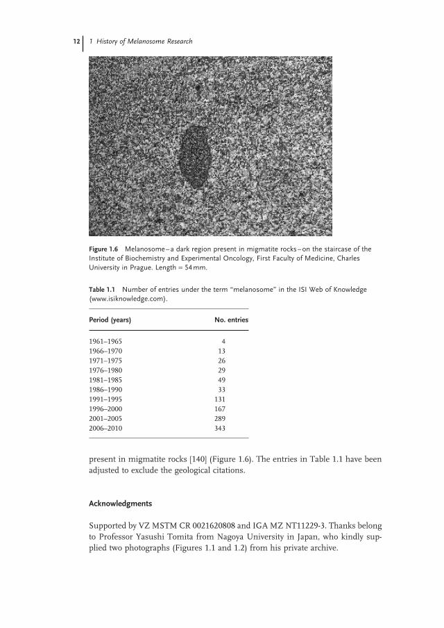
present in migmatite rocks [140] (Figure 1.6 ). The entries in Table 1.1 have been adjusted to exclude the geological citations.
Acknowledgments
Supported by VZ MSTM CR 0021620808 and IGA MZ NT11229 - 3. Thanks belong to Professor Yasushi Tomita from Nagoya University in Japan, who kindly sup-plied two photographs (Figures 1.1 and 1.2 ) from his private archive.
Figure 1.6 Melanosome – a dark region present in migmatite rocks – on the staircase of the Institute of Biochemistry and Experimental Oncology, First Faculty of Medicine, Charles University in Prague. Length = 54 mm.
Table 1.1 Number of entries under the term “ melanosome ” in the ISI Web of Knowledge ( www.isiknowledge.com ).
Period (years) No. entries
1961 – 1965 4 1966 – 1970 13 1971 – 1975 26 1976 – 1980 29 1981 – 1985 49 1986 – 1990 33 1991 – 1995 131 1996 – 2000 167 2001 – 2005 289 2006 – 2010 343
References 13
References
1 Baker , R.V. , Birbeck , M.S. , Blaschko , H. , Fitzpatrick , B. , and Seiji , M. ( 1960 ) Melanin granules and mitochondria . Nature , 187 , 392 – 394 .
2 Seiji , M. , Fitzpatrick , T.B. , and Birbeck , M.S.C. ( 1961 ) The melanosome: a distinctive subcellular particle of mammalian melanocytes and the site of melanogenesis . J. Invest. Dermatol. , 36 , 243 – 252 .
3 Seji , M. , Fitzpatrick , T.B. , Simpson , R.T. , and Birbeck , M.S.C. ( 1963 ) Chemical composition and terminology of specialized organelles (melanosomes and melanin granules) in mammalian melanocytes . Nature , 197 , 1082 – 1084 .
4 Simon , G. ( 1841 ) Z ü r Entwickelungsgeschichte der Haare . Joh. Muller ’ s Arch. Anat. , 367 .
5 Purkyn ĕ , J.E. ( 1838 ) Bericht ü ber die Versammlung deutscher Naturforscher und Aerzte in Prag im September 1837 (eds K. Sternberg and J.V. Krombholz ), Prague , pp. 174 – 180 .
6 Sorby , H.C. ( 1878 ) On the colouring matters found in human hair . J. Anthropol. Inst. , 8 , 1 – 24 .
7 Kromayer , E. ( 1893 ) Oberhautpigment der S ä ugethiere . Arch. Mikrosk. Anat. , 42 , 1 – 15 .
8 Ducho ň , J. , Fitzpatrick , T.B. , and Seiji , M. ( 1968 ) Melanin 1968: some defi nitions and problems , in The 1967 – 68 Year Book of Dermatology (eds A.W. Kopf and R. Andrade ), Year Book Medical , Chicago, IL , pp. 6 – 33 .
9 Greenstein , J.P. , Turner , J.C. , and Jenrette , W.V. ( 1940 ) Chemical studies on the components of normal and neoplastic tissues. IV. The melanin - containing melanopseudoglobulin of the malignant melanoma of mice . J. Nat. Cancer Inst. , 1 , 377 – 385 .
10 Mason , H.S. , Kahler , E. , Mac Cardle , R.C. , and Dalton , A.J. ( 1947 ) Chemistry of melanin. IV. Electron micrography of natural melanins . Proc. Soc. Exp. Biol. Med. , 66 , 421 – 431 .
11 Bolt , A.G. ( 1967 ) Interactions between human melanoprotein and chlorpromazine derivatives. I. Isolation
and purifi cation of human melanoprotein from hair and melanoma tissue . Life Sci. , 6 , 1277 – 1283 .
12 Borovansk ý , J. and Hach , P. ( 1973 ) Comments on Bolt ’ s tumour melanoprotein . Neoplasma , 20 , 325 – 329 .
13 Laxer , G. , Sikorski , J. , Whewell , C.S. , and Woods , H.J. ( 1954 ) The electron microscopy of melanin granules isolated from pigmented mammalian fi bres . Biochim. Biophys. Acta , 15 , 174 – 185 .
14 Birbeck , M.S.C. , Mercer , E.H. , and Barnicot , N.A. ( 1956 ) The structure and formation of pigment granules in human hair . Exp. Cell Res. , 10 , 505 – 514 .
15 Wellings , S.R. and Siegel , J. ( 1959 ) Role of Golgi apparatus in the formation of melanin granules in human malignant melanoma . J. Natl. Cancer Inst. , 3 , 131 – 140 .
16 Drochmans , P. ( 1960 ) Electron microscope studies of epidermal melanocytes, and the fi ne structure of melanin granules . J. Biophys. Biochem. Cytol. , 8 , 165 – 180 .
17 Drochmans , P. ( 1960 ) Study by the electron microscope of the mechanism of melanin pigmentation . Arch. Belg. Dermatol. Syphiligr. , 16 , 155 – 163 .
18 Borovansk ý , J. ( 1975 ) Isolation of melanosomes and an attempt to quantify melanin content in tissues . PhD thesis, Faculty of General Medicine, Charles University Prague.
19 Sieber , N. ( 1886 ) Ueber die Pigmente der Chorioidea und der Haare . Arch. f. Exp. Pathol. , 20 , 362 – 367 .
20 Berzelius , J.J. ( 1840 ) Lehrbuch der Chemie (aus der Schwedischen Handschrift des Verfassers ü bersetzt von F. Woehler). Dritte ungearbeitete und vermehrte Original Aufl age, Dresden & Leipzig: in der Arnoldischen Buchhandlung, vol. 9 , 22 – 24 .
21 Landolt , H. ( 1899 ) Ueber das Melanin der Augenh ä ute . Hoppe Seylers Z. Physiol. Chem. , 28 , 192 – 211 .
22 M ö rner , K.A.H. ( 1887 ) Zur Kenntnis von der Farbstoffen der melanotischen Geschw ü lste . Z. Physiol. Chem. , 11 , 66 – 141 .

14 1 History of Melanosome Research
23 Waelsch , H. ( 1932 ) Zur Kenntnis der nat ü rlichen Melanine . Hoppe Seylers Z. Physiol. Chem. , 213 , 35 – 57 .
24 Hermann , H. and Boss , M.B. ( 1945 ) Dopa oxidase activity in extracts from ciliary body and in isolated pigment granules . J. Cell. Comp. Physiol. , 26 , 131 – 138 .
25 Du Buy , H.G. , Woods , M.W. , Burk , D. , and Lackey , M.D. ( 1949 ) Enzymatic activities of isolated amelanotic and melanotic granules of mouse melanomas and suggested relationship with mitochondria . J. Am. Cancer Inst. , 9 , 325 – 336 .
26 Du Buy , H.G. , Showacre , J.L. , and Hesselbach , M.L. ( 1963 ) Enzymic and other similarities of melanoma granules and mitochondria . Ann. NY Acad. Sci. , 100 , 569 – 583 .
27 Woods , M. , Burk , D. , and Hunter , J. ( 1963 ) The ontogenic status of melanin granules . Ann. NY Acad. Sci. , 100 , 534 – 539 .
28 Seiji , M. , Shimao , K. , Birbeck , M.S.C. , and Fitzpatrick , T.B. ( 1963 ) Subcellular localization of melanin biosynthesis . Ann. NY Acad. Sci. , 100 , 497 – 533 .
29 Stein , W.D. ( 1955 ) Chemical composition of the melanin granule and its relation to the mitochondrion . Nature , 175 , 256 – 257 .
30 White , L.P. ( 1956 ) Melanin: a naturally occurring cation exchange material . Nature , 182 , 1427 – 1428 .
31 Commoner , J.B. , Townsend , J. , and Pake , G.E. ( 1954 ) Free radicals in biological materials . Nature , 174 , 689 – 691 .
32 Fitzpatrick , T.B. , Quevedo , W.C. , Jr , Levene , A.L. , Mc Govern , V.J. , Mishima , Y. , and Oettle , A.G. ( 1966 ) Terminology of vertebrate melanin - containing cells . Science , 152 , 88 – 89 .
33 Fitzpatrick , T.B. , Quevedo , W.C. , Levene , A.L. , Mc Govern , V.J. , Mishima , Y. , and Oettle , A.G. ( 1966 ) Terminology of vertebrate melanin - containing cells, their precursors and related cells. A report of the nomenclature committee of the 6th International Pigment Cell Conference , in Structure and Control of the Melanocyte (eds G. Della Porta and O. Muhlbock ), Springer , New York , pp. 1 – 5 .
34 Toda , K. , Hori , Y. , and Fitzpatrick , T.B. ( 1968 ) Isolation of the intermediate “ vesicles ” during ontogeny of melanosomes in embryonic chick retinal pigment epithelium . Fed. Proc. , 27 , 722 .
35 Toda , K. and Fitzpatrick , T.B. ( 1971 ) The origin of melanosomes , in Biology of Normal and Abnormal Melanocytes (eds T. Kawamura , T.B. Fitpatrick , and M. Seiji ), University Park Press , Baltimore, MD , pp. 265 – 278 .
36 Fitzpatrick , T.B. , Hori , Y. , Toda , K. , Kinebuchi , S. , and Szab ó , G. ( 1971 ) The mechanism of normal human melanin pigmentation and of some pigmentary disorders , in Biology of Normal and Abnormal Melanocytes (eds T. Kawamura , T.B. Fitpatrick , and M. Seiji ), University Park Press , Baltimore, MD , pp. 369 – 401 .
37 Birbeck , M.S.C. ( 1962 ) Electron microscopy of melanocytes . Br. Med. Bull. , 18 , 220 – 222 .
38 Birbeck , M.S.C. ( 1963 ) Electron microscopy of melanocytes: the fi ne structure of hair bulb premelanosomes . Ann. NY Acad. Sci. , 100 , 540 – 548 .
39 Brumbaugh , J.A. and Froiland , T.G. ( 1973 ) DOPA and cysteine incorporation into premelanosomes: effects of cycloheximide and gene substitution . J. Invest. Dermatol. , 60 , 172 – 178 .
40 Hempel , K. and Deimel , M. ( 1963 ) Untersuchungen zur gezielten Strahlentherapie des Melanoms und des chromaffi nen Systems durch selektive 3 H - Inkorporation nach Gabe von 3 H markierten DOPA . Strahlentherapie , 121 , 22 – 45 .
41 Hempel , K. ( 1966 ) Investigation on the structure of melanin in malignant melanoma with 3 H and C 14 - DOPA labeled at different positions , in Structure and Control of the Melanocyte (eds G. Della Porta and O. Muhlbock ), Springer, New York , pp. 162 – 175 .
42 Nakai , T. and Shubik , P. ( 1964 ) Electronmicroscopic radioautoraphy: the melanosome as a site of melanogenesis in neoplastic melanocytes . J. Invest. Dermatol. , 43 , 267 – 269 .
43 Zelickson , A.S. , Hirsch , H.M. , and Hartmann , J.F. ( 1964 ) Melanogenesis – an autoradiographic
References 15
study at the ultrastructural level . J. Invest. Dermatol. , 43 , 327 - 332 .
44 Seiji , M. and Iwashita , S. ( 1963 ) On the site of melanin formation in melanocytes . J. Biochem. , 54 , 465 – 467 .
45 Seiji , M. and Iwashita , S. ( 1965 ) Intracellular organisation of tyrosinase and site of melanin formation in melanocyte . J. Invest. Dermatol. , 45 , 305 – 314 .
46 Borovansk ý , J. and Ducho ň , J. ( 1974 ) Chemical composition of hair melanosomes . Dermatologica , 149 , 116 – 120 .
47 Hach , P. , Borovansk ý , J. and Ducho ň , J. ( 1973 ) Melanosomes of horse benign melanoma . Folia Morphol. (Prague) , 21 , 275 – 277 .
48 Hach , P. and Borovansk ý , J. ( 1972 ) Ultrastructure of melanosomes of different origin . Folia Morphol. (Prague) , 20 , 82 – 84 .
49 Hach , P. , Ducho ň , J. , and Borovansk ý , J. ( 1977 ) Ultrastructural and biochemical characteristics of isolated melanosomes . Folia Morphol. (Prague) , 25 , 407 – 410 .
50 Mishima , Y. ( 1965 ) Macromolecular changes in pigmentary disorders . Arch. Dermatol. , 91 , 519 – 557 .
51 C é sarini , J.P. ( 1971 ) Recent advances in the ultrastructure of malignant melanoma . Rev. Eur. É tud. Clin. Biol. , 16 , 316 – 322 .
52 Foa , C. and Aubert , C.H. ( 1977 ) Cellular localization of tyrosinase in human malignant melanoma . J. Invest. Dermatol. , 68 , 369 – 378 .
53 Hirone , T. , Nagai , T. , Matsubara , T. , and Fukushiro , R. ( 1971 ) Human malignant melanoma of the skin and their preexisting conditions , in Biology of Normal and Abnormal Melanocytes (eds T. Kawamura , T.B. Fitpatrick , and M. Seiji ), University Park Press , Baltimore, MD , pp. 329 – 348 .
54 Hunter , J.A.A. , Paterson , W.D. , and Fairley , D.J. ( 1978 ) Human malignant melanoma. Melanosomal polymorphism and the ultrastructural DOPA reaction . Br. J. Dermatol. , 98 , 381 – 390 .
55 Hunter , J.A.A. , Zaynoun , W.D. , Paterson , W.D. , Bleehen , S.S. , Mackie , R. , and Cochran , A.J. ( 1978 ) Cellular fi ne structure in the invasive nodules of
different histogenetic types of malignant melanoma . Br. J. Dermatol. , 98 , 255 – 272 .
56 Moyer , F.H. ( 1963 ) Genetic effects on melanosome fi ne structure and ontogeny in normal and malignant cells . Ann. NY Acad. Sci. , 100 , 584 – 606 .
57 Szekeres , L. ( 1975 ) Fine structure and X - ray microanalysis of melanosomes in pigmented nevi and melanoma . Arch. Derm. Forsch. , 252 , 297 – 304 .
58 Klingmuller , G. and Schmoeckel , C. ( 1971 ) Frei im Cytoplasma liegende Melanin - synthesierende Membranaordnungen beim malignem Melanom . Arch. Derm. Forsch. , 241 , 115 – 121 .
59 McGovern , V.J. and Lane Brown , M.M. ( 1969 ) The Nature of Melanoma , Thomas, Springfi eld, IL .
60 Fitzpatrick , T.B. ( 1971 ) The biology of pigmentation . Birth Defects Orig. Artic. Ser. , 7 , 5 – 12 .
61 Borovansk ý , J. ( 1978 ) Biochemical parameters of melanosomes and pigment tissues . Habilitation thesis. Charles University, Prague.
62 Fitzpatrick , T.B. and Breathnach , A.S. ( 1963 ) Das epidermale melanin einheit - system . Dermatol. Wochenschr. , 147 , 481 – 489 .
63 Mottaz , J.H. and Zelickson , A.S. ( 1967 ) Melanin transfer: a possible phagocytic process . J. Invest. Dermatol. , 49 , 605 – 610 .
64 Szab ó , G. , Gerald , A.B. , Pathak , M.A. , and Fitzpatrick , T.B. ( 1969 ) Racial differences in the fate of melanosomes in human epidermis . Nature , 222 , 1081 – 1082 .
65 Borovansk ý , J. , Hach , P. , Vedralov á , E. , and Ducho ň , J. ( 1978 ) Strategy of melanosome isolation , in XXIst Colloqiuum Scientifi cum Facultatis Medicae Universitatis Carolinae et XIXth Morphological Congress Symposia (ed. E. Klika ), Univerzita Karlova , Prague , pp. 613 – 618 .
66 Doezema , P. ( 1973 ) Proteins from melanosomes of mouse and chick pigment cells . J. Cell. Physiol. , 89 , 201 – 208 .
67 Habermann , H.F. and Menon , I.A. ( 1973 ) A modifi ed method for the isolation of melanosomes from B - 16
16 1 History of Melanosome Research
melanoma . J. Invest. Dermatol. , 60 , 67 – 72 .
68 Borovansk ý , J. and Hach , P. ( 1986 ) Isolation of melanosomes from keratinous structures: current state of the art . Arch. Dermatol. Res. , 279 , 54 – 58 .
69 Borovansk ý , J. and Hach , P. ( 1972 ) Isolation of melanosomes from keratinous material – a new method . Dermatologica , 145 , 37 – 41 .
70 Ducho ň , J. , Borovansk ý , J. , and Hach , P. ( 1973 ) Chemical composition of ten kinds of various melanosomes , in Mechanisms in Pigmentation (eds V.J. McGovern and P. Russell ), Karger , Basel , pp. 165 – 170 .
71 Ito , S. and Jimbow , K. ( 1983 ) Quantitative analysis of eumelanin and pheomelanin in hair and melanomas . J. Invest. Dermatol. , 80 , 268 – 272 .
72 Jimbow , K. , Miyake , Y. , Homma , K. , Yasuda , K. , Izumi , Y. , Tsutsumi , A. , and Ito , S. ( 1984 ) Characterization of melanogenesis and morphogenesis of melanosomes by physicochemical properties of melanin and melanosomes in malignant melanoma . Cancer Res. , 44 , 1128 – 1134 .
73 Miyazaki , K. and Ohtaki , N. ( 1975 ) Tyrosinase as a glycoprotein . Arch. Dermatol. Forsch. , 252 , 211 – 216 .
74 Siakotos , A.N. , Patel , V. , and Cantaboni , A. ( 1973 ) The isolation and chemical composition of premelanosomes and melanosomes: human and mouse melanomas . Biochem. Med. , 7 , 14 – 24 .
75 Seiji , M. ( 1966 ) Subcellular particles and melanin formation in melanocytes , in Advances in the Biology of Skin VIII: The Pigmentary System (eds W. Montagna and F. Hu ), Pergamon Press , Oxford , pp. 189 – 222 .
76 Vedralov á , E. and Ducho ň , J. ( 1977 ) Lipid constituents in melanosomes of tumour origin . Sborn í k l é k. , 79 , 335 – 339 .
77 Jimbow , M. , Kanoh , H. , and Jimbow , K. ( 1982 ) Characterization of biochemical properties of melanosomes for structural and functional differentiation: analysis of the compositions of lipids and proteins in melanosomes and their subfractions . J. Invest. Dermatol. , 79 , 97 – 102 .
78 Vedralov á , E. , Duchon , J. , and Hach , P. ( 1987 ) RNA and DNA in melanosomes of hamster melanoma . Pigment Cell Res. , 1 , 76 – 80 .
79 Borovansk ý , J. ( 1997 ) Detection of metals in tissues, cells and subcellular particles . Sborn í k L é k. , 98 , 77 – 97 .
80 Borovansk ý , J. , Ducho ň , J. , Proch á zkov á , B. , and Hach , P. ( 1972 ) [An attempt to disintegrate melanosomes into protein subunits] . Č as. L é k. Č es. , 111 , 218 – 220 .
81 Bratosin , S. ( 1973 ) Disassembly of melanosomes in detergents . J. Invest. Dermatol. , 60 , 224 – 230 .
82 Hearing , V.J. and Lutzner , M.A. ( 1973 ) Mammalian melanosomal proteins: characterization by polyacrylamide gel electrophoresis . Yale J. Biol. Med. , 46 , 553 – 559 .
83 Jimbow , K. , Jimbow , M. , and Chiba , M. ( 1982 ) Characterization of structural properties for morphological differentiation of melanosomes: II. Electron microscopic and SDS – PAGE comparison of melanosomal matrix proteins in B16 and Harding - Passey melanomas . J. Invest. Dermatol. , 78 , 76 – 81 .
84 Seiji , M. and Fitzpatrick , T.B. ( 1961 ) The reciprocal relationship between melanization and tyrosinase activity in melanosomes (melanin granules) . J. Biochem. , 49 , 700 – 706 .
85 Menon , I.A. and Haberman , H.F. ( 1970 ) Activation of tyrosinase in microsomes and melanosomes from B - 16 and Harding - Passey mouse melanoma . Arch. Biochem. Biophys. , 137 , 231 – 242 .
86 Miyazaki , K. and Seiji , M. ( 1971 ) Tyrosinase isolated from mouse melanoma melanosomes . J. Invest. Dermatol. , 57 , 81 – 86 .
87 Hearing , V.J. ( 1973 ) Tyrosinase activity in subcellular fractions of black and albino mice . Nat. New Biol. , 245 , 81 – 83 .
88 Mufson , R.A. ( 1975 ) The tyrosinase activity of melanosomes from the Harding - Passey melanoma: the absence of a peroxidase component in vitro . Arch. Biochem. Biophys. , 167 , 338 – 343 .
89 Blagoeva , P.M. ( 1977 ) Solubilizing effect of Triton X100 on melanosome tyrosinase in hamster pigmented melanoma . Neoplasma , 24 , 291 – 294 .
References 17
90 Kikuchi , A. ( 1968 ) Acid phosphatase activity in melanosomes of melanocytes . Bull. Tokyo Med. Dent. Univ. , 15 , 279 – 294 .
91 Novikoff , A.B. , Albala , A. , and Biempica , L. ( 1968 ) Ultrastructural and cytochemical observations on B - 16 and Harding - Passey mouse melanomas. The origin of premelanosomes and compound melanosomes . J. Histochem. Cytochem. , 16 , 299 – 319 .
92 Olson , R.L. , Nordquist , J. , and Everett , M.A. ( 1970 ) The role of epidermal lysosomes in melanin physiology . Br. J. Dermatol. , 83 , 189 – 199 .
93 Seiji , M. and Kikuchi , A. ( 1969 ) Acid phosphatase activity in melanosomes . J. Invest. Dermatol. , 52 , 212 – 216 .
94 Wolff , K. and Schreiner , E. ( 1971 ) Melanosomal acid phosphatase . Arch. Dermatol. Forsch. , 241 , 255 – 272 .
95 Wolff , K. and H ö nigsmann , H. ( 1972 ) Are melanosome complexes lysosomes? J. Invest. Dermatol. , 59 , 170 – 176 .
96 Mufson , R.A. ( 1974 ) The subcellular distribution of lysosomal hydrolases in the Harding - Passey melanoma . J. Cell. Physiol. , 83 , 75 – 84 .
97 Ohtaki , N. ( 1970 ) Melanosome and lysosome: I. Lysosomal activity in relation to growth of melanoma . Bull. Tokyo Med. Dent. Univ. , 17 , 89 – 102 .
98 Kurbanov , C. , Abaskina , L.I. , Karimova , L.S. , and Krivorotova , L.S. ( 1977 ) Investigation of tyrosine - α - ketoglutarate transaminase and tryptophane pyrrolase activities in subcellular fractions of skin of guinea pigs . Izv. Akad Nauk Turkmenskoj SSR, Ser. Biol. , 72 – 76 .
99 Borovansk ý , J. and Hach , P. ( 1999 ) Disparate behaviour of two melanosomal enzymes α - mannosidase and γ - glutamyltransferase . Cell. Mol. Biol., 45 , 1047 – 1052 .
100 Fitzpatrick , T.B. and Quevedo , W.C. , Jr ( 1971 ) Biological processes underlying melanin pigmentation and pigmentary disorders , in Modern Trends in Dermatology 4 (ed. P. Borrie ), Butterworths , London , pp. 122 – 149 .
101 Hill , H.Z. ( 1992 ) The function of melanin or six blind people examine an elephant . Bioassays , 14 , 49 – 56 .
102 Hu , D.N. , Simon , J.D. , and Sarna , T. ( 2008 ) Role of ocular melanin in ophthalmic physiology and pathology . Photochem Photobiol. , 84 , 639 – 644 .
103 Borovansk ý , J. ( 1993 ) Properties of melanosomes and their exploitation in the diagnosis and treatment of melanoma . Pigment Cell Res. , 3 , 181 – 186 .
104 Riley , P.A. ( 1991 ) Melanogenesis: a realistic target for antimelanoma therapy . Eur. J. Cancer , 27 , 1172 – 1177 .
105 Hearing , V.J. ( 2005 ) Biogenesis of pigment granules: a sensitive way to regulate melanocyte function . J. Dermatol. Sci. , 37 , 3 – 14 .
106 Tolleson , W.H. ( 2005 ) Human melanocyte biology, toxicology, and pathology . J. Environ. Sci. Health C , 23 , 105 – 161 .
107 Ortonne , J.P. and Prota , G. ( 1993 ) Hair melanins and hair color. Ultrastructure and biochemical aspects . J. Invest. Dermatol. , 101 , 82s – 89s .
108 Arnaud , J.C. and Bor é , P. ( 1981 ) Isolation of melanin pigments from human hair . J. Soc. Cosmet. Chem. , 32 , 137 – 152 .
109 Liu , Y. and Simon , J.D. ( 2003 ) Isolation and biophysical studies of natural eumelanins: application of imaging technologies and ultrafast spectroscopy . Pigment Cell Res. , 16 , 606 – 618 .
110 Novellino , L. , Napolitano , A. , and Prota , G. ( 2000 ) Isolation and characterization of mammalian eumelanins from hair and irides . Biochim. Biophys. Acta , 1475 , 295 – 306 .
111 Kushimoto , T. , Basrur , V. , Valencia , J. , Matsunaga , J. , Vieira , W.D. , Ferrans , V.J. , Muller , J. , Appella , E. , and Hearing , V.J. ( 2001 ) A model for melanosome biogenesis based on the purifi cation and analysis of early melanosomes . Proc. Natl. Acad. Sci. USA , 98 , 10698 – 10703 .
112 Orlow , S.J. , Boissy , R.E. , Moran , D.J. , and Pifko - Hirst , S. ( 1993 ) Subcellular distribution of tyrosinase and tyrosinase - related protein - 1: implications for melanosomal biogenesis . J. Invest. Dermatol. , 100 , 55 – 64 .
113 Van Nieuwpoort , F. , Smit , N.P.M. , Kolb , R. , van der Meulen , H. , Koerten , H. , and Pavel , S. ( 2004 ) Tyrosine - induced
18 1 History of Melanosome Research
melanogenesis show differences in morphologic and melanogenic preference of melanosomes from light and dark skin types . J. Invest. Dermatol. , 122 , 1251 – 1255 .
114 Diment , S. , Eidelman , M. , Rodriguez , G.M. , and Orlow , S.J. ( 1995 ) Lysosomal hydrolases are present in melanosomes and are elevated in melanizing cells . J. Biol. Chem. , 270 , 4213 – 4215 .
115 Bhatnagar , V. , Anjaiah , S. , Puri , N. , Darshanam , B.N. , and Ramaiah , A. ( 1993 ) pH of melanosomes of B16 murine melanoma is acidic: its physiological importance in the regulation of melanin biosynthesis . Arch. Biochem. Biophys. , 307 , 183 – 192 .
116 Mishima , Y. ( 1994 ) Molecular and biological control of melanogenesis through tyrosinase genes and intrinsic and extrinsic regulatory factors . Pigment Cell Res. , 7 , 376 – 387 .
117 Orlow , S.J. ( 1995 ) Melanosomes are specialized members of the lysosomal lineage of organelles . J. Invest. Dermatol. , 105 , 3 – 7 .
118 Dell ’ Angelica , E.C. , Mullins , C. , Caplan , S. , and Bonifacino , J.S. ( 2000 ) Lysosome - related organelles . FASEB J. , 14 , 1265 – 1278 .
119 Borovansk ý , J. , Hach , P. , Smetana , K. , Jr , Elleder , M. , and Matou š - Malbohan , I. ( 1999 ) Attempts to induce melanosome degradation in vivo . Folia Biol. (Praha) , 45 , 47 – 52 .
120 Borovansk ý , J. and Elleder , M. ( 2003 ) Melanosome degradation: fact or fi ction . Pigment Cell Res. , 16 , 280 – 286 .
121 Kayatz , P. , Thumann , G. , Luther , T.T. , Jordan , J.F. , Bartz - Schmidt , K.U. , Esser , P.J. , and Schraermeyer , U. ( 2001 ) Oxidation causes melanin fl uorescence . Invest. Ophthalmol. Vis. Sci. , 42 , 241 – 246 .
122 Sarna , T. , Burke , J.M. , Korytowski , W. , R ó zanowska , M. , Skumatz , C.M. , Zareba , A. , and Zareba , M. ( 2003 ) Loss of melanin from human RPE with aging: possible role of melanin photooxidation . Exp. Eye Res. , 76 , 89 – 98 .
123 Jimbow , K. , Yamana , K. , Akutsu , Y. , and Maeda , K. ( 1988 ) Nature and biosynthesis of structural matrix protein in melanosomes: melanosomal
structural protein as differentiation antigen for neoplastc melanocytes , in Advances in Pigment Cell Research (ed. R. Alan ), Liss , New York , pp. 169 – 182 .
124 Anonymous ( 2009 ) Antibodies specifi c to melanocyte - specifi c proteins available from the Hearing laboratory . Pigment Cell Melanoma Res. , 22 , 651 .
125 Simon , J.D. , Hong , L. , and Peles , D.N. ( 2008 ) Insights into melanosomes and melanin from some interesting spatial and temporal properties . J. Phys. Chem. B , 112 , 13201 – 13217 .
126 Nordlund , J.J. , Abdel - Malek , Z. , Biossy , R.E. , and Rheins , L.A. ( 1989 ) Pigment cell biology: an historical review . J. Invest. Dermatol. , 4 , 53S – 60S .
127 Aquaron , R. ( 1999 ) Tradition of basic and applied pigment cell research in Marseille . Cell. Mol. Biol. , 45 , 877 – 892 .
128 Ducho ň , J. ( 1999 ) Tradition of pigment cell research at Charles University in Prague . Cell Mol. Biol. , 45 , 893 – 898 .
129 Sangiovanni , G. ( 1819 ) Descrizione di un particolare sistema di organi cromoforo espansivo - dermoideo e dei fenomeni che esso produce, scoperto nei molluschi cefaloso . G. Enciclopedico Napoli , 9 , 1 – 13 .
130 Westerhof , W. ( 2006 ) The discovery of the human melanocyte . Pigment Cell Res. , 19 , 183 – 193 .
131 Falabella , R. ( 2009 ) Vitiligo and the melanocyte reservoir . Indian J. Dermatol. , 54 , 313 – 318 .
132 Kenney , J.A. , Jr ( 1961 ) Melanin pigmentation . J. Nat. Med. Assoc. , 53 , 447 – 454 .
133 Schallreuter , K.U. ( 2007 ) Advances in melanocyte basic science research . Dermatol. Clin. , 25 , 283 – 291 .
134 Gortner , R.A. ( 1910 ) Studies on melanin. I. Methods of isolation. The effect of alkali on melanin . J. Biol. Chem. , 8 , 341 – 364 .
135 Serra , A.J. ( 1946 ) Constitution of hair melanins . Nature , 157 , 771 .
136 Borovansk ý , J. and Ducho ň , J. ( 1971 ) Melanoproteins (in Czech) . Chem. Listy , 65 , 500 – 528 .
137 Nicolaus , R.A. ( 1968 ) Melanins , Hermann , Paris .

References 19
138 Prota , G. ( 1992 ) Melanins and Melanogenesis , Academic Press , San Diego, CA .
139 Swan , G.A. ( 1974 ) Structure, chemistry, and biosynthesis of the melanins . Prog. Chem. Org. Nat. Prod. , 31 , 522 – 582 .
140 Harris , N.B.W. , Caddick , M. , Kosler , J. , Goswami , S. , Vance , D. , and Tindle , A.G. ( 2004 ) The pressure – temperature – time path of migmatites from the Sikkim Himalaya . J. Metamorphic Geol. , 22 , 249 – 264 .


![FlexibleRadio:AFrameworkforOptimizedMultimodal ... · larly, looking “down” at the platform/circuit level [5], we see intense activity on flexible and malleable platforms and](https://img.pdfslide.net/doc/110x75/5e14a0fddc76a662540cf8e7/flexibleradioaframeworkforoptimizedmultimodal-larly-looking-aoedowna-at.jpg)


![1 History of Melanosome Research · somes and analysis of their chemical composition [46, 47] . Electron microscopy enabled the defi nition of the basic morphometric data of isolated](https://img.pdfslide.net/doc/110x75/5edca82bad6a402d66676a8d/1-history-of-melanosome-research-somes-and-analysis-of-their-chemical-composition.jpg)