Embed Size (px)
Citation preview
Path: K:/ASP-SOFTNANO-07-0502/Application/ASP-SOFTNANO-07-0502-008.3dDate: 3rd November 2007 Time: 14:30 User ID: tamilmanir BlackLiningEnabled
ISBN: 978-1-58883-xxx-xCopyright � 2008 by American Scientific PublishersAll rights of reproduction in any form reserved.
Soft NanomaterialsEdited by Hari Singh Nalwa
Volume XY: Pages (1–28)
CHAPTER 8
Polypeptide Multilayer Films andMicrocapsules
Bingyun LiDepartment of Orthopedics, School of Medicine and WVNano Initiative, West Virginia University,Morgantown, WV 26506, USA
CONTENTS
1. Introduction . . . . . . . . . . . . . . . . . . . . . . . . . . . . . . . . . . . . . . . . . . . 1
2. Polypeptide and Polypeptide Synthesis . . . . . . . . . . . . . . . . . . . . . . . . 3
2.1. Polypeptide—An Innovative Biomaterial . . . . . . . . . . . . . . . . . . 3
2.2. Chemical Synthesis of Polypeptides . . . . . . . . . . . . . . . . . . . . . . 4
3. Techniques for Fabrication of Polypeptide Multilayers . . . . . . . . . . . . . 5
3.1. Langmuir-Blodgett (LB) Technique . . . . . . . . . . . . . . . . . . . . . . 5
3.2. Electrostatic Layer-by-Layer (LBL) Self-Assembly . . . . . . . . . . . 6
4. Techniques for Monitoring and Characterizing Polypeptide Multilayer
Formation . . . . . . . . . . . . . . . . . . . . . . . . . . . . . . . . . . . . . . . . . . . . . 7
4.1. Quartz Crystal Microbalance (QCM) . . . . . . . . . . . . . . . . . . . . . 7
4.2. UV-Vis . . . . . . . . . . . . . . . . . . . . . . . . . . . . . . . . . . . . . . . . . . . 7
4.3. Atomic Force Microscopy (AFM) . . . . . . . . . . . . . . . . . . . . . . . 8
4.4. Fluorescence Microscopy . . . . . . . . . . . . . . . . . . . . . . . . . . . . . 8
4.5. Ellipsometry . . . . . . . . . . . . . . . . . . . . . . . . . . . . . . . . . . . . . . . 8
4.6. Fourier Transform Infrared Spectroscopy (FTIR) . . . . . . . . . . . . 9
4.7. Circular Dichroism (CD) Spectroscopy . . . . . . . . . . . . . . . . . . . 9
5. Polypeptide Multilayer Films and Microcapsules . . . . . . . . . . . . . . . . . 9
5.1. Polypeptide Multilayer Films Prepared Using LB Technique . . . . 9
5.2. LBL Multilayers of Poly(L-lysine)/Poly(L-glutamic acid) . . . . . . . 10
5.3. LBL Multilayers of Designed Polypeptides . . . . . . . . . . . . . . . . . 14
5.4. Multilayers Consisting of One Polypeptide and One
Non-Polypeptide Component . . . . . . . . . . . . . . . . . . . . . . . . . . . . . . . 22
5.5. Polypeptide Microcapsules . . . . . . . . . . . . . . . . . . . . . . . . . . . . 22
6. Applications of Polypeptide Multilayers . . . . . . . . . . . . . . . . . . . . . . . 23
6.1. Chiral Separation . . . . . . . . . . . . . . . . . . . . . . . . . . . . . . . . . . . 23
6.2. Anti-Inflammatory Coatings . . . . . . . . . . . . . . . . . . . . . . . . . . . 24
6.3. Polypeptide Multilayers as Electrochemical Interfaces
for Ion and Electron Transport . . . . . . . . . . . . . . . . . . . . . . . . . . . . . . 25
6.4. Polypeptide Multilayers in Nanofiltration for Ion Separation . . . . 25
7. Conclusions and Future Prospects . . . . . . . . . . . . . . . . . . . . . . . . . . . . 25
References . . . . . . . . . . . . . . . . . . . . . . . . . . . . . . . . . . . . . . . . . . . . 26
Page Number: 1
Path: K:/ASP-SOFTNANO-07-0502/Application/ASP-SOFTNANO-07-0502-008.3dDate: 3rd November 2007 Time: 14:30 User ID: tamilmanir BlackLiningEnabled
1. INTRODUCTIONIn recent years, polypeptide multilayers, including multilayer films and microcapsules, haveattracted considerable attention due mainly to the close relationship of polypeptides to nativebiological molecules and the ordered secondary structures (a-helix and b-sheet) of polypep-tides and polypeptide multilayers. For instance, the interaction of oppositely charged 32-merpolypeptides leads to multilayer films presenting a predominantly b-sheet content [1, 2] andpoly(L-lysine)/poly(L-glutamic acid), i.e., PLL/PLGA films, a high a-helix content [3]. Theordered secondary structures are useful in creating self-organized functional surfaces [4, 5],and their biological nature makes polypeptides useful for numerous applications requiringbiocompatible interfaces [6]. Moreover, by designing suitable primary (and secondary) struc-tures, polypeptides and polypeptide multilayers can be endowed with specific biofunctional-ities. The number of possible, and, indeed, realizable polypeptide sequences is effectivelyunlimited; this provides flexibility in polypeptide design and the polypeptide multilayer designprocess.
Polypeptide multilayers are the subject of fundamental structural studies [7–9] for biomedicaland pharmaceutical applications [10–13] and for biomimetic surface modifications [1–3]. Differ-ent approaches have been taken in polypeptide multilayer formation. Two well-known techni-ques are Langmuir-Blodgett (LB) deposition and electrostatic layer-by-layer self-assembly(LBL). The latter, first proposed by Iler [14] and extended by Decher [15], provides a simple,versatile, environmentally efficient method of preparing polypeptide multilayers on the nanome-ter scale [1, 16].
Polypeptides and proteins are natural polymers. Proteins including cytochrome c, lysozyme,histone f3, myoglobin, hemoglobin, pepsin, albumin, immunoglobulin G, peroxidase, glucoseoxidase, and catalase have been used for LBL of multilayers. The applications of proteins forLBL and LB multilayers have been reviewed [17–19] and will not be discussed here. Polypep-tides are generally less complex than proteins, which may present a number of difficulties formultilayer formation. Direct assembly of oppositely charged protein molecules has been foundto be difficult; the preparation of glucose oxidase/lysozyme composite layers has failed [17]. Thisis probably because electrostatic attraction cannot be maximized with globular proteins in theLBL process.
This chapter provides an overview of polypeptide multilayer films and microcapsules with par-ticular emphasis on multilayers made of designed polypeptides. Multilayers made of commer-cially available polypeptides such as PLL and PLGA are also discussed in detail. The chemicalmethods for polypeptide synthesis are described, and the approaches used to form polypeptidemultilayer films and microcapsules are outlined. The applications of widely used physical techni-ques for monitoring polypeptide multilayer formation are briefly reviewed. The applications ofpolypeptide multilayers are discussed and some examples given.
2. POLYPEPTIDE AND POLYPEPTIDE SYNTHESIS
2.1. Polypeptide—An Innovative Biomaterial
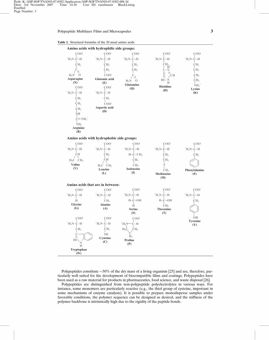
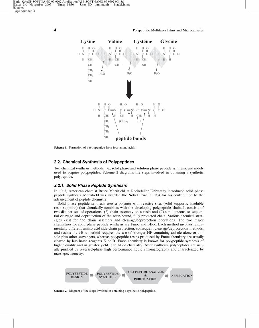
There are four major types of biological macromolecules; polypeptides are one type. Polypep-tides are natural or synthetic linear polymers based on approximately 20 amino acid monomers[Table 1]. They contain an amino group (�NH2) and a carboxyl group (�COOH) attached to acentral carbon atom. However, each amino acid has a different side group attached to the cen-tral carbon that lends unique character to that amino acid. The side group can be hydrophobic,hydrophilic, positively charged, or negatively charged at neutral pH [20]. The amino acids arelinked covalently by peptide bonds. Scheme 1 shows how four amino acids linked by peptidebonds form a tetrapeptide.
There are four levels of structures found in polypeptides: primary, secondary, tertiary, andquaternary structures. Depending on the sequential order of the amino acids (primary structureof the polypeptide), the polypeptide assumes different structures along the polymer chain (sec-ondary structure of the polypeptide). The two main secondary structures are a-helix and b-sheet,which are observed in polypeptide solutions [1, 21] and polypeptide multilayers [1–3, 9, 22–24].The tertiary structure of the polypeptide reflects how secondary structures organize relative toeach other to form overall globular, fibrous, or random polypeptide structure. Quaternary struc-ture occurs when whole polypeptides interact with each other to provide a unique structure orbiological activity.
Page Number: 2
2 Polypeptide Multilayer Films and Microcapsules
Path: K:/ASP-SOFTNANO-07-0502/Application/ASP-SOFTNANO-07-0502-008.3dDate: 3rd November 2007 Time: 14:30 User ID: tamilmanir BlackLiningEnabled
Polypeptides constitute �50% of the dry mass of a living organism [25] and are, therefore, par-ticularly well suited for the development of biocompatible films and coatings. Polypeptides havebeen used as a raw material for products in pharmaceutics, food science, and waste disposal [26].
Polypeptides are distinguished from non-polypeptide polyelectrolytes in various ways. Forinstance, some monomers are particularly reactive (e.g., the thiol group of cysteine, important insome mechanisms of enzyme catalysis). It is possible to prepare monodisperse samples underfavorable conditions, the polymer sequence can be designed as desired, and the stiffness of thepolymer backbone is intrinsically high due to the rigidity of the peptide bonds.
Table 1. Structural formulas of the 20 usual amino acids.
Page Number: 3
Polypeptide Multilayer Films and Microcapsules 3
Path: K:/ASP-SOFTNANO-07-0502/Application/ASP-SOFTNANO-07-0502-008.3dDate: 3rd November 2007 Time: 14:30 User ID: tamilmanir BlackLiningEnabled
2.2. Chemical Synthesis of Polypeptides
Two chemical synthesis methods, i.e., solid phase and solution phase peptide synthesis, are widelyused to acquire polypeptides. Scheme 2 diagrams the steps involved in obtaining a syntheticpolypeptide.
2.2.1. Solid Phase Peptide SynthesisIn 1963, American chemist Bruce Merrifield at Rockefeller University introduced solid phasepeptide synthesis. Merrifield was awarded the Nobel Prize in 1984 for his contribution to theadvancement of peptide chemistry.
Solid phase peptide synthesis uses a polymer with reactive sites (solid supports, insolubleresin supports) that chemically combines with the developing polypeptide chain. It consists oftwo distinct sets of operations: (1) chain assembly on a resin and (2) simultaneous or sequen-tial cleavage and deprotection of the resin-bound, fully protected chain. Various chemical strat-egies exist for the chain assembly and cleavage/deprotection operations. The two majorchemistries for solid phase peptide synthesis are Fmoc and t-Boc. Each method involves funda-mentally different amino acid side-chain protection, consequent cleavage/deprotection methods,and resins; the t-Boc method requires the use of stronger HF containing anisole alone or ani-sole plus other scavengers, whereas polypeptide resins produced by Fmoc chemistry are usuallycleaved by less harsh reagents K or R. Fmoc chemistry is known for polypeptide synthesis ofhigher quality and in greater yield than t-Boc chemistry. After synthesis, polypeptides are usu-ally purified by reversed-phase high performance liquid chromatography and characterized bymass spectrometry.
Scheme 1. Formation of a tetrapeptide from four amino acids.
Scheme 2. Diagram of the steps involved in obtaining a synthetic polypeptide.
Page Number: 4
4 Polypeptide Multilayer Films and Microcapsules
Path: K:/ASP-SOFTNANO-07-0502/Application/ASP-SOFTNANO-07-0502-008.3dDate: 3rd November 2007 Time: 14:30 User ID: tamilmanir BlackLiningEnabled
2.2.2. Solution Phase Peptide SynthesisSolution phase peptide synthesis is not used as widely as the solid phase method. Solution phasepeptide synthesis may be applicable for polypeptides that are longer than 100 amino acids or forlarge-scale synthesis of well-known polypeptides. PLL and PLGA, commercially available withdifferent molecular weights, are usually made from solution phase peptide synthesis and the prod-ucts are polydisperse. Solution phase peptide synthesis is usually labor-, time-, and skill-intensivelargely due to the unpredictable solubility characteristics of intermediates.
Different from the aforementioned chemical synthesis methods, recombinant synthesis (ormicrobial or biotic synthesis) is based on gene synthesis technology. It has been used in recentyears for the production of proteins or polypeptides with high molecular weights.
3. TECHNIQUES FOR FABRICATION OF POLYPEPTIDEMULTILAYERS
Chemists have used a variety of techniques, e.g., thermal evaporation, sputtering, electrodeposi-tion, molecular beam epitaxy, adsorption from solution, LB technique, and LBL, to fabricate mul-tilayer films and coatings on solid substrates. Among these, the LB and LBL techniques are thetwo most well known for preparing polypeptide multilayers.
3.1. Langmuir-Blodgett (LB) Technique
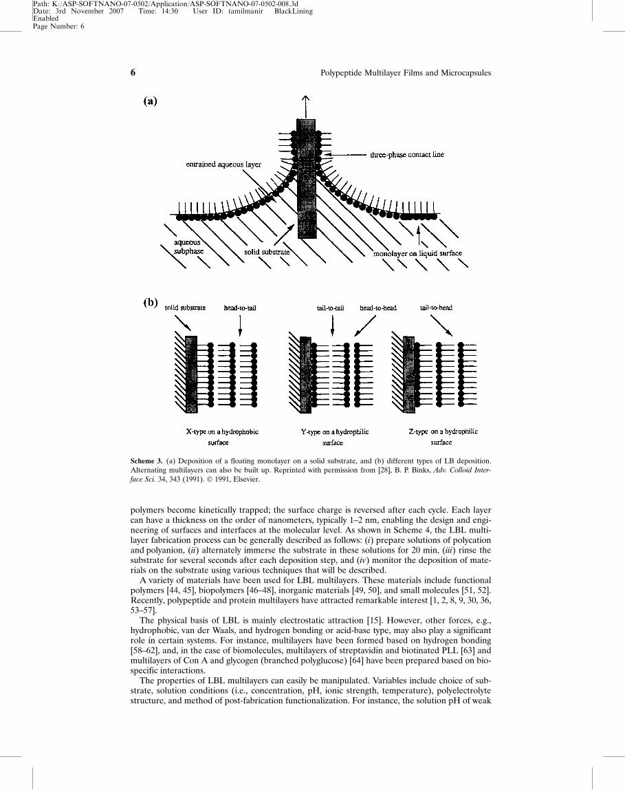
The term LB comes from the names of a research scientist and his assistant, Irving Langmuir andKatherine Blodgett, who discovered unique properties of thin films in the early 1900s. Langmuir’soriginal work involved the transfer of monolayers from liquid to solid substrates. He was awardedthe Nobel Prize for his systematic studies on monolayers. Several years later, Blodgett expandedon Langmuir’s research to deposit multilayer films on solid substrates [27]. After the pioneeringwork done by Langmuir and Blodgett, it took almost half a century before scientists worldwidestarted to realize the potential of this unique technique.
Scheme 3 displays the LB deposition process [28]. One molecular layer is formed by dippinga solid substrate into water containing a polymer, i.e., subphase, that forms a single layer ofmolecular chains on the surface [Scheme 3a]. This layer is then transferred from the water tothe substrate. An ordered, multilayer film can be created by repeating the dipping process[Scheme 3b]. There are a variety of dipping schemes resulting in predominantly three types ofLB multilayer films. The most common is the Y-type multilayer, which is produced when themonolayer deposits to the solid substrate in both up and down directions. When the monolayerdeposits only in one direction (down or up), the multilayer structure is called either X-type orZ-type [Scheme 3b].
Many factors may influence the film-forming characteristics:
¥ subphase composition and temperature,¥ pH of subphase,¥ purity of subphase,¥ addition of ions to stabilize films,¥ nature of the spread film, and¥ type and nature of the solid substrate.
The main advantages of LB over other methods are its ability to offer an enhanced degree oforder and packing density and its ability to form oriented monolayers of molecules whose activitymay exceed that of those immobilized by other methods. However, this technique has difficultiesin assemblies on substrates with irregular shape, and it needs a complicated setup to form alter-nating multilayers.
3.2. Electrostatic Layer-by-Layer (LBL) Self-Assembly
LBL is the most promising method for preparation of multilayer nanofilms of controlled thick-ness and molecular architecture. This method was pioneered by Iler [14] and extended as a newultrathin film preparation by Decher et al. in the early 1990s [15].
LBL has been used to make polyelectrolyte multilayer films, coatings, and microcapsules fromoppositely charged polymers [1, 2, 15, 16, 29–43]. The procedure involves the repetitive sequen-tial dipping of a substrate in solutions of oppositely charged polyelectrolytes. On adsorption, the
Page Number: 5
Polypeptide Multilayer Films and Microcapsules 5
Path: K:/ASP-SOFTNANO-07-0502/Application/ASP-SOFTNANO-07-0502-008.3dDate: 3rd November 2007 Time: 14:30 User ID: tamilmanir BlackLiningEnabled
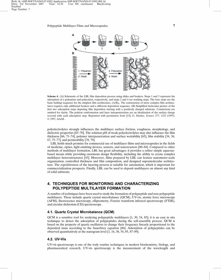
polymers become kinetically trapped; the surface charge is reversed after each cycle. Each layercan have a thickness on the order of nanometers, typically 1–2 nm, enabling the design and engi-neering of surfaces and interfaces at the molecular level. As shown in Scheme 4, the LBL multi-layer fabrication process can be generally described as follows: (i) prepare solutions of polycationand polyanion, (ii) alternately immerse the substrate in these solutions for 20 min, (iii) rinse thesubstrate for several seconds after each deposition step, and (iv) monitor the deposition of mate-rials on the substrate using various techniques that will be described.
A variety of materials have been used for LBL multilayers. These materials include functionalpolymers [44, 45], biopolymers [46–48], inorganic materials [49, 50], and small molecules [51, 52].Recently, polypeptide and protein multilayers have attracted remarkable interest [1, 2, 8, 9, 30, 36,53–57].
The physical basis of LBL is mainly electrostatic attraction [15]. However, other forces, e.g.,hydrophobic, van der Waals, and hydrogen bonding or acid-base type, may also play a significantrole in certain systems. For instance, multilayers have been formed based on hydrogen bonding[58–62], and, in the case of biomolecules, multilayers of streptavidin and biotinated PLL [63] andmultilayers of Con A and glycogen (branched polyglucose) [64] have been prepared based on bio-specific interactions.
The properties of LBL multilayers can easily be manipulated. Variables include choice of sub-strate, solution conditions (i.e., concentration, pH, ionic strength, temperature), polyelectrolytestructure, and method of post-fabrication functionalization. For instance, the solution pH of weak
Scheme 3. (a) Deposition of a floating monolayer on a solid substrate, and (b) different types of LB deposition.Alternating multilayers can also be built up. Reprinted with permission from [28], B. P. Binks, Adv. Colloid Inter-
face Sci. 34, 343 (1991). � 1991, Elsevier.
Page Number: 6
6 Polypeptide Multilayer Films and Microcapsules
Path: K:/ASP-SOFTNANO-07-0502/Application/ASP-SOFTNANO-07-0502-008.3dDate: 3rd November 2007 Time: 14:30 User ID: tamilmanir BlackLiningEnabled
polyelectrolytes strongly influences the multilayer surface friction, roughness, morphology, anddielectric properties [65–70]. The solution pH of weak polyelectrolytes may also influence the filmthickness [66, 71–74], polymer interpenetration and surface wettability [65], film stability [34, 36,67, 75–77], and permeability [78, 79].
LBL holds much promise for commercial use of multilayer films and microcapsules in the fieldsof medicine, optics, light-emitting devices, sensors, and nanoreactors [80–84]. Compared to othermethods of multilayer formation, LBL has great advantages: it provides a rather simple aqueous-based means while providing enormous design flexibility, including the ability to create complexmultilayer heterostructures [43]. Moreover, films prepared by LBL can feature nanometer-scaleorganization, controlled thickness and film composition, and designed supramolecular architec-ture. The repetitiveness of the layering process is suitable for automation, which is important forcommercialization prospects. Finally, LBL can be used to deposit multilayers on almost any kindof solid substrate.
4. TECHNIQUES FOR MONITORING AND CHARACTERIZINGPOLYPEPTIDE MULTILAYER FORMATION
A number of techniques have been used to study the formation of polypeptide and non-polypeptidemultilayers. These include quartz crystal microbalance (QCM), UV-vis, atomic force microscopy(AFM), fluorescence microscopy, ellipsometry, Fourier transform infrared spectroscopy (FTIR),and circular dichroism (CD) spectroscopy.
4.1. Quartz Crystal Microbalance (QCM)
QCM is a sensitive tool for analyzing polypeptide multilayers [1, 30, 54, 85]; it is an easy in situtechnique to detect the adsorption of polypeptides during the self-assembly process. QCM isbased on the property of quartz oscillators to change their frequency linearly proportional to thedeposited mass according to the Sauerbrey equation [86]. Adsorption of polypeptides can beobserved quantitatively at the nanogram level [1, 16, 30, 54, 85, 87–89].
4.2. UV-Vis
UV-vis spectroscopy is one of the truly routine techniques in modern biochemistry, biology, andpharmaceutical research. UV-vis spectroscopy is the measurement of the wavelength and
Scheme 4. (A) Schematic of the LBL film deposition process using slides and beakers. Steps 1 and 3 represent theadsorption of a polyanion and polycation, respectively, and steps 2 and 4 are washing steps. The four steps are thebasic buildup sequence for the simplest film architecture, (A/B)n. The construction of more complex film architec-tures requires only additional beakers and a different deposition sequence. (B) Simplified molecular picture of thefirst two adsorption steps depicting film deposition starting with a positively charged substrate. Counterions areomitted for clarity. The polyion conformation and layer interpenetration are an idealization of the surface chargereversal with each adsorption step. Reprinted with permission from [15], G. Decher, Science 277, 1232 (1997).� 1997, AAAS.
Page Number: 7
Polypeptide Multilayer Films and Microcapsules 7
Path: K:/ASP-SOFTNANO-07-0502/Application/ASP-SOFTNANO-07-0502-008.3dDate: 3rd November 2007 Time: 14:30 User ID: tamilmanir BlackLiningEnabled
intensity of absorption of near-ultraviolet and visible light by a sample. An absorption spectrumshows a number of absorption bands corresponding to structural groups within the molecule.Any species with an extended system of alternating double and single bonds absorb UV light,and anything with color absorbs visible light, making UV-vis spectroscopy applicable to a widerange of samples. Because of its wide availability, UV-vis has been widely used for monitoringthe formation of polypeptide multilayers [2, 16, 51]. Note that UV-vis spectra have broad fea-tures that are of limited use for sample identification but are very useful for quantitative mea-surements as the absorbance of a sample increases with its concentration.
4.3. Atomic Force Microscopy (AFM)
AFM is one of the most versatile types of scanning probe microscopes. It is an imaging techniquethat, in some cases, can resolve atomic lattice in the real space. To use this system, a cantilever tipis brought in contact with the surface of a solid sample. As the tip nears the sample surface, somerepulsive force between the tip and the surface appears. This force from the surface applied onthe cantilever tip bends the cantilever upward. A laser beam is reflected over the cantilever, form-ing a spot that is reflected on a split photodetector. The photodetector monitors the bending ofthe cantilever and can be used to calculate the force. Keeping the force constant while scanningthe tip across the surface, the vertical movement of the tip follows the surface profile, which isthen recorded as the surface topography. AFM has been used to investigate the surface morphol-ogy of polypeptide multilayers [2, 16, 36] and protein film structure [90–92].
4.4. Fluorescence Microscopy
Fluorescence is the property of some atoms and molecules to absorb light at a particular wave-length and to subsequently emit light of longer wavelength after a brief interval. A certain type ofdye attaches a polypeptide molecule; fluorescence reveals the adsorption of this polypeptide mol-ecule. Using this technique, fluorescent-labeled polypeptide microcapsules have been visualized[93].
4.5. Ellipsometry
Ellipsometry is also a sensitive optical method for measuring properties such as the thickness andrefractive index of thin films at interfaces. This method has been used for nearly one hundredyears to derive information about surfaces. It makes use of the fact that the polarization state oflight may change when the light beam is reflected from a surface. If the surface is covered by apolypeptide thin multilayer, the entire optical system of multilayers and substrates influences thechange in polarization. It is therefore possible to deduce information about the polypeptide multi-layer properties including thickness. Using ellipsometry, the thickness and refractive index ofmultilayers made of designed polypeptides [16] and the buildup and stability of PLL/PLGA mul-tilayers [56, 85] have been studied.
4.6. Fourier Transform Infrared Spectroscopy (FTIR)
FTIR is perhaps the most powerful tool for identifying types of chemical bonds (functionalgroups). This technique measures the absorption of various infrared light wavelengths by thematerial of interest. Because chemical bonds absorb infrared energy at specific wavelengths, thebasic structure of compounds can be determined by the spectral locations of their infrared absorp-tions. For most common materials, the spectrum of an unknown can be identified by comparisonto a library of known compounds. FTIR has been used to study the secondary structure of PLL/PLGA multilayers [9, 12, 55, 94]. FTIR can also be used for some quantitative analyses becausethe strength of the absorption is proportional to concentration. FTIR has been used to monitorthe assembly process of polypeptide multilayers [1]; the absorption intensity of a certain charac-teristic peak, such as Amide I, increases with adsorption steps.
4.7. Circular Dichroism (CD) Spectroscopy
CD spectroscopy is another technique that has been used to characterize the secondary structureof polypeptides [1, 21, 95, 96] and polypeptide multilayers [1, 30, 36, 51]. CD measures the differ-ences in the absorption of left-handed polarized light versus right-handed polarized light thatarise due to structural asymmetry. The absence of regular structure results in zero CD intensity,while an ordered structure results in a spectrum that can contain both positive and negative
Page Number: 8
8 Polypeptide Multilayer Films and Microcapsules
Path: K:/ASP-SOFTNANO-07-0502/Application/ASP-SOFTNANO-07-0502-008.3dDate: 3rd November 2007 Time: 14:30 User ID: tamilmanir BlackLiningEnabled
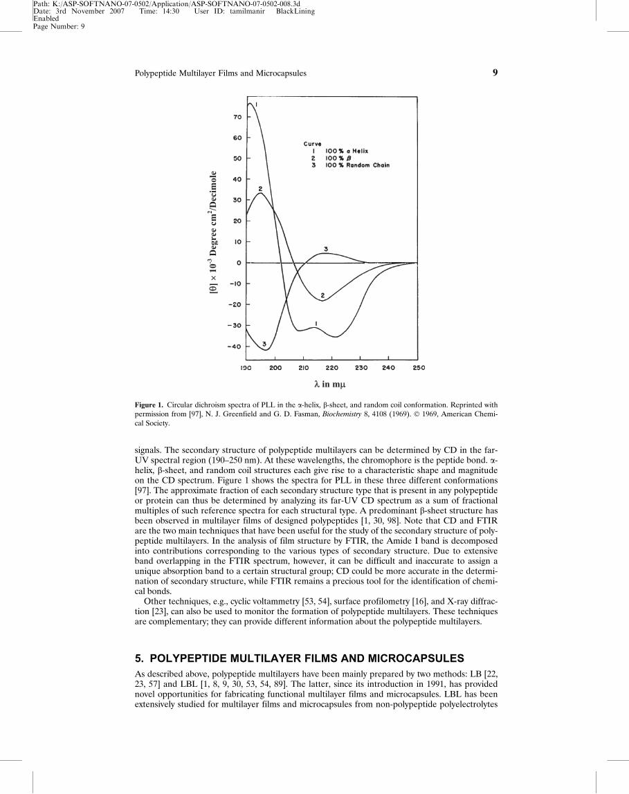
signals. The secondary structure of polypeptide multilayers can be determined by CD in the far-UV spectral region (190–250 nm). At these wavelengths, the chromophore is the peptide bond. a-helix, b-sheet, and random coil structures each give rise to a characteristic shape and magnitudeon the CD spectrum. Figure 1 shows the spectra for PLL in these three different conformations[97]. The approximate fraction of each secondary structure type that is present in any polypeptideor protein can thus be determined by analyzing its far-UV CD spectrum as a sum of fractionalmultiples of such reference spectra for each structural type. A predominant b-sheet structure hasbeen observed in multilayer films of designed polypeptides [1, 30, 98]. Note that CD and FTIRare the two main techniques that have been useful for the study of the secondary structure of poly-peptide multilayers. In the analysis of film structure by FTIR, the Amide I band is decomposedinto contributions corresponding to the various types of secondary structure. Due to extensiveband overlapping in the FTIR spectrum, however, it can be difficult and inaccurate to assign aunique absorption band to a certain structural group; CD could be more accurate in the determi-nation of secondary structure, while FTIR remains a precious tool for the identification of chemi-cal bonds.
Other techniques, e.g., cyclic voltammetry [53, 54], surface profilometry [16], and X-ray diffrac-tion [23], can also be used to monitor the formation of polypeptide multilayers. These techniquesare complementary; they can provide different information about the polypeptide multilayers.
5. POLYPEPTIDE MULTILAYER FILMS AND MICROCAPSULESAs described above, polypeptide multilayers have been mainly prepared by two methods: LB [22,23, 57] and LBL [1, 8, 9, 30, 53, 54, 89]. The latter, since its introduction in 1991, has providednovel opportunities for fabricating functional multilayer films and microcapsules. LBL has beenextensively studied for multilayer films and microcapsules from non-polypeptide polyelectrolytes
Figure 1. Circular dichroism spectra of PLL in the a-helix, b-sheet, and random coil conformation. Reprinted withpermission from [97], N. J. Greenfield and G. D. Fasman, Biochemistry 8, 4108 (1969). � 1969, American Chemi-cal Society.
Page Number: 9
Polypeptide Multilayer Films and Microcapsules 9
Path: K:/ASP-SOFTNANO-07-0502/Application/ASP-SOFTNANO-07-0502-008.3dDate: 3rd November 2007 Time: 14:30 User ID: tamilmanir BlackLiningEnabled
[82, 99]. It has been getting more and more attention in a variety of biological macromoleculessuch as polypeptides [1, 9, 30] and nucleic acids [100–102]. What has been learned about multi-layers from non-polypeptide polyelectrolytes, including weak ones, may not be suitable to predictproperties, including biological properties, of polypeptide multilayers.
5.1. Polypeptide Multilayer Films Prepared Using LB Technique
Most polypeptides can be adjusted to be anionically or cationically charged by simply adjustingthe solution pH to be higher or lower than the isoelectric point. This property has been previ-ously applied in LB technique to build a composite lipid/enzyme monolayer structure at the air-water interface [103]. The monolayers can be transferred onto a solid planar substrate to buildmultilayer assemblies.
Poly(c-methyl-L-glutamate) multilayers have been prepared using the LB technique, and a-helical structure has been observed [22]. Highly ordered polypeptide multilayer films have alsobeen fabricated from chloroform solutions of PLGA derivatives [23], which are deposited ontosolid substrates (e.g., glass slides) by the vertical dipping method as Z-type films with almost unityratio. FTIR, X-ray diffraction, and CD measurements show that these polypeptide multilayershave a-helix structure. Functional organic molecules such as phthalocyaninato metals have beendeposited onto the ordered LB multilayer films, and the in-plane electrical conductivity of themultilayer thin films can be as high as 10�4 S cm�1 after doping with I2.
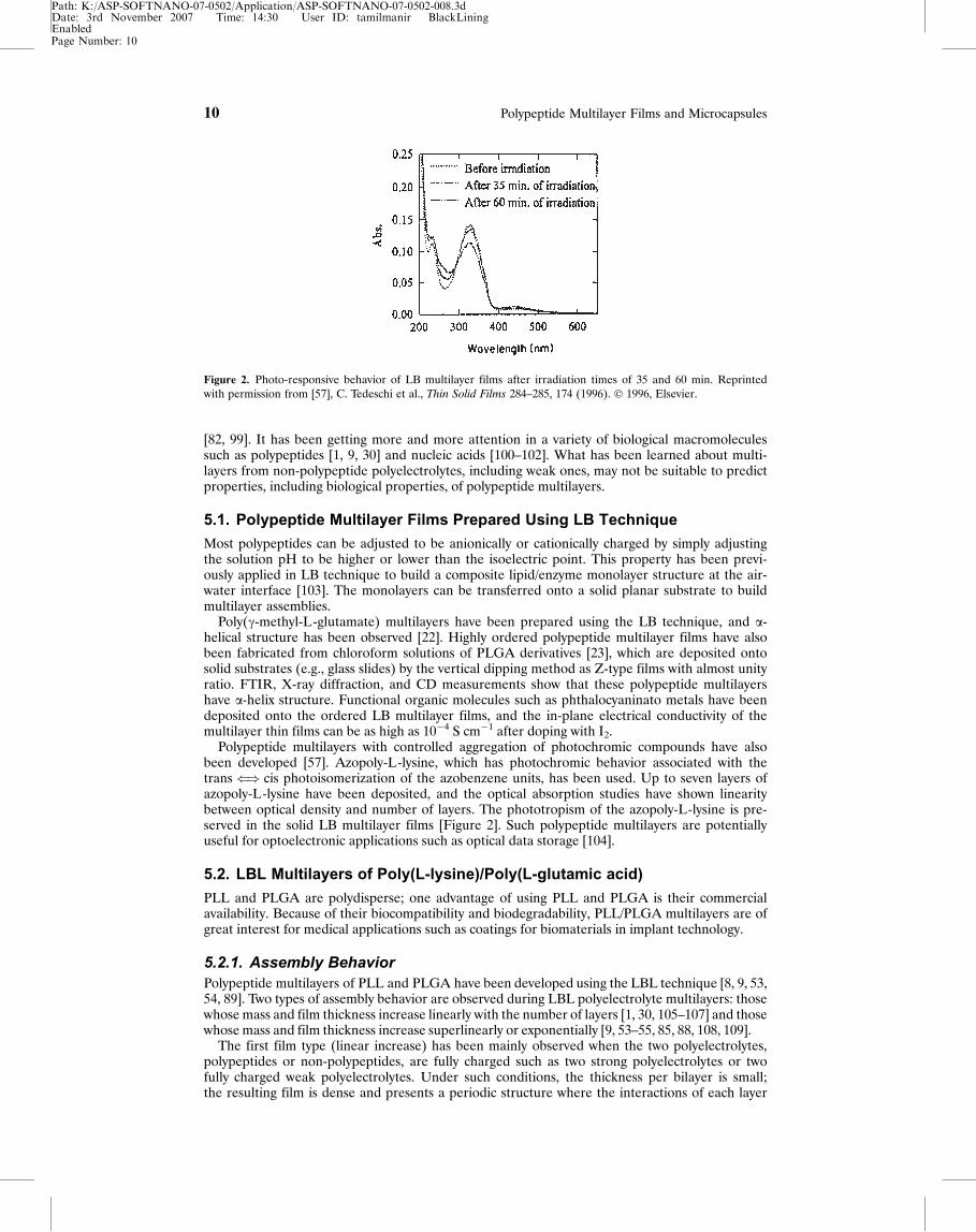
Polypeptide multilayers with controlled aggregation of photochromic compounds have alsobeen developed [57]. Azopoly-L-lysine, which has photochromic behavior associated with thetrans() cis photoisomerization of the azobenzene units, has been used. Up to seven layers ofazopoly-L-lysine have been deposited, and the optical absorption studies have shown linearitybetween optical density and number of layers. The phototropism of the azopoly-L-lysine is pre-served in the solid LB multilayer films [Figure 2]. Such polypeptide multilayers are potentiallyuseful for optoelectronic applications such as optical data storage [104].
5.2. LBL Multilayers of Poly(L-lysine)/Poly(L-glutamic acid)
PLL and PLGA are polydisperse; one advantage of using PLL and PLGA is their commercialavailability. Because of their biocompatibility and biodegradability, PLL/PLGA multilayers are ofgreat interest for medical applications such as coatings for biomaterials in implant technology.
5.2.1. Assembly BehaviorPolypeptide multilayers of PLL and PLGA have been developed using the LBL technique [8, 9, 53,54, 89]. Two types of assembly behavior are observed during LBL polyelectrolyte multilayers: thosewhose mass and film thickness increase linearly with the number of layers [1, 30, 105–107] and thosewhose mass and film thickness increase superlinearly or exponentially [9, 53–55, 85, 88, 108, 109].
The first film type (linear increase) has been mainly observed when the two polyelectrolytes,polypeptides or non-polypeptides, are fully charged such as two strong polyelectrolytes or twofully charged weak polyelectrolytes. Under such conditions, the thickness per bilayer is small;the resulting film is dense and presents a periodic structure where the interactions of each layer
Figure 2. Photo-responsive behavior of LB multilayer films after irradiation times of 35 and 60 min. Reprintedwith permission from [57], C. Tedeschi et al., Thin Solid Films 284–285, 174 (1996). � 1996, Elsevier.
Page Number: 10
10 Polypeptide Multilayer Films and Microcapsules
Path: K:/ASP-SOFTNANO-07-0502/Application/ASP-SOFTNANO-07-0502-008.3dDate: 3rd November 2007 Time: 14:30 User ID: tamilmanir BlackLiningEnabled
are constrained to its neighbors [15]. Therefore, the polyelectrolytes from the solution interactonly with the outer part of the multilayer and do not interact with polyelectrolyte layers moredeeply embedded in the film, and, therefore, the film grows linearly [1, 30, 105–107].
Multilayers made of at least one weak polyelectrolyte may grow superlinearly or exponentially.Hoffmannov�a et al. have observed that the mass of PLL/PLGA deposited increases superlinearlywith increasing adsorption cycles, and much more PLL than PLGA is deposited in each cycle [54].These findings are consistent with the observation by Cheng and Corn, who have found that PLLlayers are thicker on average than PLGA layers [53]. Similar mass increment behavior has beenobserved by Halthur and Elofsson [85]. This exponential growth is believed to be due to the diffu-sion in and out through the whole film at each adsorption cycle of at least one of the two polyelec-trolyte components [109].
The film density of PLL/PLGA multilayers has been studied during the assembly process [85].The measurements of the refractive index and the water content in the multilayers have shownthat the density of PLL/PLGA multilayers increases with increasing adsorption cycles. Thisincrease in film density is proposed as a result of polymers diffusing into and attaching insidethe underlying layers.
The mass deposition of polypeptides may depend on the properties of the buffer present in thepolypeptide solutions [54]. At pH 8.5, the PLL/PLGA multilayers grown in phosphate buffersshow a mass twice that of the corresponding films grown in carbonate buffer. However, it shouldbe noted that in these experiments [54], the concentration for the two buffers are very different(0.005 M for carbonate buffer and 0.02 or 0.1 M for phosphate buffer). This changes the ionicstrength, and, therefore, could have a big influence on the assembly behavior.
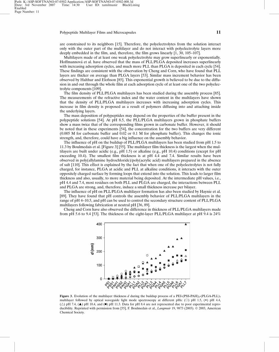
The influence of pH on the buildup of PLL/PLGA multilayers has been studied from pH 1.5 to11.3 by Boulmedais et al. [Figure 3] [55]. The multilayer film thickness is the largest when the mul-tilayers are built under acidic (e.g., pH 1.5) or alkaline (e.g., pH 10.4) conditions (except for pHexceeding 10.4). The smallest film thickness is at pH 4.4 and 7.4. Similar results have beenobserved in poly(allylamine hydrochloride)/poly(acrylic acid) multilayers prepared in the absenceof salt [110]. This effect is explained by the fact that when one of the polyelectrolytes is not fullycharged, for instance, PLGA at acidic and PLL at alkaline conditions, it interacts with the outeroppositely charged surface by forming loops that extend into the solution. This leads to larger filmthickness and also, usually, to more material being deposited. At the intermediate pH values, i.e.,pH 4.4 and 7.4, most residues on both PLL and PLGA are charged, the interactions between PLLand PLGA are strong, and, therefore, induce a small thickness increase per bilayer.
The influence of pH on PLL/PLGA multilayer formation has also been studied by Haynie et al.[89]. They have found that pH controls the assembly behavior of PLL/PLGA multilayers in therange of pH 4–10.5, and pH can be used to control the secondary structure content of PLL/PLGAmultilayers following fabrication at neutral pH [36, 89].
Cheng and Corn have also observed the difference in thickness of PLL/PLGA multilayers madefrom pH 5.6 to 9.4 [53]. The thickness of the eight-layer PLL/PLGA multilayer at pH 9.4 is 24%
Figure 3. Evolution of the multilayer thickness d during the buildup process of a PEI-(PSS-PAH)2-(PLGA-PLL)7
multilayer followed by optical waveguide light mode spectroscopy at different pHs: (s) pH 1.5, (¥) pH 4.4,(n) pH 7.4, (m) pH 10.4, and (n) pH 11.3. Data for pH 8.4 are not represented due to poor experimental repro-ducibility. Reprinted with permission from [55], F. Boulmedais et al., Langmuir 19, 9873 (2003). � 2003, AmericanChemical Society.
Page Number: 11
Polypeptide Multilayer Films and Microcapsules 11
Path: K:/ASP-SOFTNANO-07-0502/Application/ASP-SOFTNANO-07-0502-008.3dDate: 3rd November 2007 Time: 14:30 User ID: tamilmanir BlackLiningEnabled
greater than the multilayer fabricated from pH 8.0, and the eight-layer film created at pH 5.4 is10% thinner than at pH 8.0. Also, for all pH solutions used, the thickness of a PLL layer is greaterthan that of a PLGA layer. Because the operating pH values (5.6, 8.0, and 9.4) are within the pKasrange of PLGA (4.9) and PLL (10.5), the vast majority of the residues on both PLL and PLGAare charged. It seems that the variations in the film thickness with solution pH in this range shouldnot be so large. Therefore, it is believed that the change in film thickness at these pH ranges is notsimply related to the solution pH; it may also be related to the intrinsic structure, e.g., hydropho-bicity, of the PLL polycation relative to the PLGA polyanion.
Note that most PLL/PLGA multilayers have been fabricated with pre-layers of non-polypeptidepolyelectrolytes [8, 9, 55, 85]. The assembly behavior with and without pre-layers has been dis-cussed by Halthur and Elofsson [85]; they have shown that the buildup of polypeptide multilayersis sensitive to the pre-layers. The use of pre-layers of non-polypeptide polyelectrolytes such as poly-ethylimine (PEI) induces a difference in the thickness increments for PLL (40–55 A/layer) andPLGA (25–35 A/layer) in the first several layers, whereas this difference diminishes when the influ-ence of PEI pre-layers is omitted.
5.2.2. Film StabilityThe stability of polypeptide multilayers is important not only in terms of construction and storage,but also for understanding how the polypeptide multilayer reacts when in contact with variousenvironments. The stability of polypeptide multilayers could depend on pH, temperature, or sol-vent. At neutral pH, the vast majority of the residues on both PLL and PLGA are charged, andthe cooperative electrostatic interaction between PLL and PLGA is strong and can prevent disso-ciation of PLL/PLGA multilayers. However, under certain physical or chemical conditions, a poly-peptide multilayer could show an undesirable tendency to disintegrate [36].
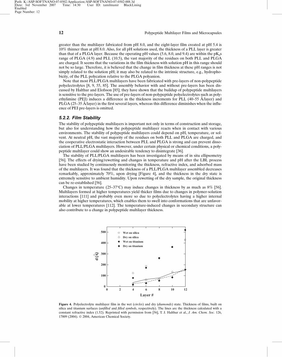
The stability of PLL/PLGA multilayers has been investigated by means of in situ ellipsometry[56]. The effects of drying/rewetting and changes in temperature and pH after the LBL processhave been studied by continuously monitoring the thickness, refractive index, and adsorbed massof the multilayers. It was found that the thickness of a PLL/PLGA multilayer assembled decreasesremarkably, approximately 70%, upon drying [Figure 4], and the thickness in the dry state isextremely sensitive to ambient humidity. Upon rewetting of the dry sample, the original thicknesscan be re-established [56].
Changes in temperature (25–37�C) may induce changes in thickness by as much as 8% [56].Multilayers formed at higher temperatures yield thicker films due to changes in polymer-solutioninteractions [111] and probably even more so due to polyelectrolytes having a higher internalmobility at higher temperatures, which enables them to swell into conformations that are unfavor-able at lower temperatures [112]. The temperature-induced changes in secondary structure canalso contribute to a change in polypeptide multilayer thickness.
Figure 4. Polyelectrolyte multilayer film in the wet (circles) and dry (diamonds) state. Thickness of films, built onsilica and titanium surfaces (unfilled and filled symbols, respectively). The lines are the thickness calculated with aconstant refractive index (1.52). Reprinted with permission from [56], T. J. Halthur et al., J. Am. Chem. Soc. 126,17009 (2004). � 2004, American Chemical Society.
Page Number: 12
12 Polypeptide Multilayer Films and Microcapsules
Path: K:/ASP-SOFTNANO-07-0502/Application/ASP-SOFTNANO-07-0502-008.3dDate: 3rd November 2007 Time: 14:30 User ID: tamilmanir BlackLiningEnabled
A more obvious change in thickness has been observed by changing the solution pH. Thechange in pH (3.9–10.1) enables the PLL/PLGA multilayers to undergo a nonreversible swelling/deswelling behavior, and a change of up to 10–20% has been reported [56]. A similar phenom-enon has been observed in PLL/hyaluronic acid multilayers, where the multilayers swell one toeight times when they are immersed in solutions of different pH [70].
Several research groups [36, 55] have reported the dissociation of PLL/PLGA multilayersrelated to pH jumps. Boulmedais et al. have observed that PLL/PLGA multilayers undergo apartial dissociation on contact with a solution of pH 1.5, and complete dissolution has beenobserved upon immersion into a solution of pH 13.0 [55]. Zhi and Haynie have reported thatPLL/PLGA multilayers dissociate about 8% at pH 2.0 and more than 30% at pH 12.0 [36]. Theinfluence of pH on polypeptide multilayer stability is probably governed by two main factors: theloss and gain of charge density and changes in secondary structure. Both might lead to structuralchanges in the polypeptide multilayer. At an extreme pH, i.e., strong acid or base, either PLGAor PLL becomes uncharged or less charged, and the other component molecules become fullycharged and repel themselves. This repulsion leads to multilayer dissociation.
5.2.3. Secondary Structure and Structural StabilityPolypeptides are known to interact not only through electrostatic interactions but also throughhydrogen bonding. Their chemical nature allows them to form secondary structures such as a-helices and b-sheets. At the same time, polyelectrolyte multilayers are known to preserve thesecondary structure of proteins [10], and the helical structure of DNA [101] or PLL [113, 114].Cooper et al. have reported that the interactions between PLL and Congo Red dye lead to a-helical structures [51], and the interactions of PLL and PLGA result in a mixture of a-helices andb-sheets [24]. Boulmedais et al. have shown that it is possible to develop polypeptide multilayerswith significant secondary structure content such as a-helices and b-sheets [9]. They have alsoshown that the precursor layers, i.e., two bilayers of poly(allylamine hydrochloride)/poly(styrenesulfonate) PAH/PSS, only influence the secondary structure of PLL/PLGA multilayers in the firstfew bilayers. The b-sheet content increases at the expense of random coil with an increasing num-ber of bilayers; except this is not the case for pH 1.5, where no b-sheet contribution has beenobserved. After several bilayers, the secondary structures of the PLL/PLGA multilayers becomeindependent of the number of bilayers [55]. All of these have shown that local order can be gen-erated in polypeptide multilayers by specific interactions between polycations and polyanions.
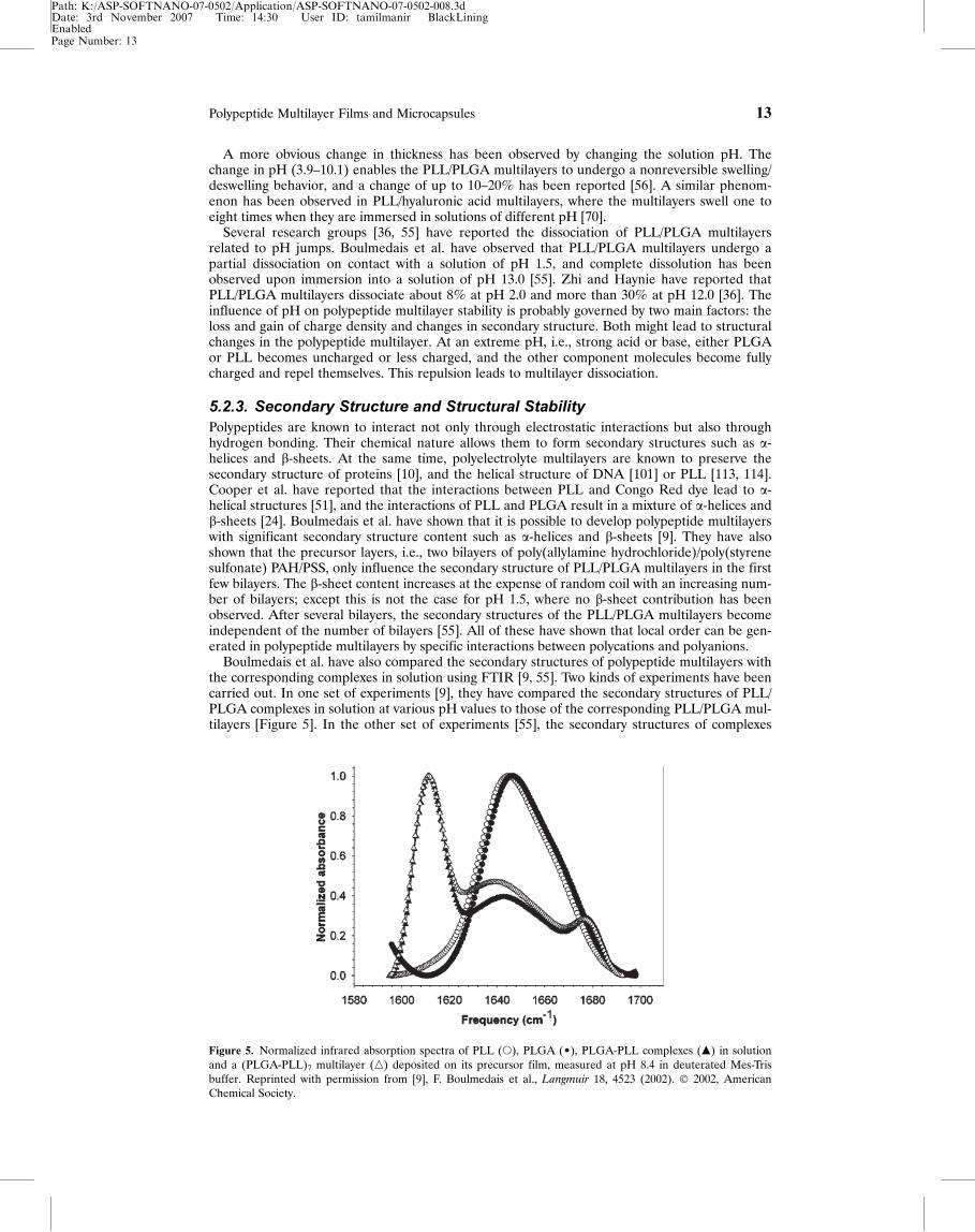
Boulmedais et al. have also compared the secondary structures of polypeptide multilayers withthe corresponding complexes in solution using FTIR [9, 55]. Two kinds of experiments have beencarried out. In one set of experiments [9], they have compared the secondary structures of PLL/PLGA complexes in solution at various pH values to those of the corresponding PLL/PLGA mul-tilayers [Figure 5]. In the other set of experiments [55], the secondary structures of complexes
Figure 5. Normalized infrared absorption spectra of PLL (s), PLGA (¥), PLGA-PLL complexes (m) in solutionand a (PLGA-PLL)7 multilayer (n) deposited on its precursor film, measured at pH 8.4 in deuterated Mes-Trisbuffer. Reprinted with permission from [9], F. Boulmedais et al., Langmuir 18, 4523 (2002). � 2002, AmericanChemical Society.
Page Number: 13
Polypeptide Multilayer Films and Microcapsules 13
Path: K:/ASP-SOFTNANO-07-0502/Application/ASP-SOFTNANO-07-0502-008.3dDate: 3rd November 2007 Time: 14:30 User ID: tamilmanir BlackLiningEnabled
and multilayers have been studied for various polyanion/polycation systems at a given pH. Forinstance, poly(L-aspartic acid)/PLL and PLGA/poly(L-ornithine) systems have been studied atpH 7.4; poly(L-aspartic acid) and poly(L-ornithine) differ from PLGA and PLL, respectively, bythe reduction of the side chains by one methylene group. Moreover, PLL has been replaced withpoly(D-lysine), which has different chirality. In all the systems studied, the secondary structuresof polypeptide multilayers always closely resemble those of the corresponding complexes in solu-tion [55, 115].
The stability in the local order structure of PLL/PLGA multilayers toward external stressessuch as pH jumps and temperature rise has been studied [36, 55]. It has been found that the sec-ondary structures of PLL/PLGA multilayers appear very stable against pH jumps in the range ofpH 4–10.5; b-sheets stabilize the polypeptide multilayers. However, the sudden exposure of aPLL/PLGA multilayer formed at pH 7.4 to a solution at pH 1.5 or 13.5 leads to a strong reductionof its b-sheet content together with a partial or total dissociation of the polypeptide multilayer. Asimilar change in secondary structure on pH shift has been observed by Zhi and Haynie based onCD experiments. A substantial conformational change, from b-sheet to predominantly a-helicalstructure, has been observed on contact of PLL/PLGA multilayers with strongly acidic (pH � 2.5)or strongly basic (pH � 12.0) solutions [36].
The secondary structural response of PLL/PLGA multilayers to a temperature rise (up to89�C) depends on the rate of temperature increase [55]. A slow temperature increase rate inducesa reversible decrease of the b-sheet content at the expense of a-helices. When the multilayer isheated rapidly, however, the b-sheet content increases, and a further increase is observed duringthe ensuing cooling process. Boulmedais et al. have proposed that the interactions between theamino groups of PLL and the carboxyl groups of PLGA favor a high temperature and lead to theformation of amide links [116]. The formation of such links between chains, even if their numberis small, tends to stabilize the structure of the multilayer film. On the other hand, an increase intemperature tends to destabilize the film’s secondary structures. Such a destabilization takesplace, however, only when the chains can move freely with respect to each other. When the multi-layer is heated slowly, the destabilization process happens before the formation of amide links. Bycontrast, when the multilayer is heated rapidly, some amide links may form before the b-sheetsare destabilized. The relative freedom of movement of the chains gained by the temperatureincrease only allows the extension of the b-sheet domains, but it forbids their destruction (whichwould require larger movements). In the ensuing cooling process, b-sheets become even more sta-ble, thus leading to a further increase of their content.
The above results have suggested that polypeptide multilayers may present significant second-ary structures (a-helix and b-sheet), and their structural stability may change in response to exter-nal stresses.
5.3. LBL Multilayers of Designed Polypeptides
The extreme flexibility in sequence design as well as the inclusion of specific biofunctional sequen-ces enables great potential for the application and formation of multilayers from designedpolypeptides.
In comparison with the large number of reports on the formation and uses of non-polypeptidepolyelectrolyte multilayers, very few studies have been undertaken for the construction of multi-layers from designed polypeptides [1, 2, 16, 30, 93, 98]. Strong polyions such as PSS, poly(vinylsulfate) (PVS), poly(diallyldimethylammonium chloride) (PDDA), and PEI with a molecularweight (MW) between 50,000 and 100,000 have been used widely for the formation of polyelec-trolyte multilayer films and microcapsules. These polyions contain hundreds of ionized groups inthe range of pH 3–9. Similarly, PLL and PLGA contain hundreds of ionized groups at neutralpH. The influence of the polymerization degree on the formation of stable interpolyelectrolytebulk complexes has been analyzed by Kabanov and Zezin [117, 118]. They have found that suchcomplexes are stable when a polyion contains more than 20 charged groups in the sequence.However, complexes of our designed polypeptides [1, 2, 16, 30, 93, 98], which have approximately16 charged groups, have also been found to be stable as described below. In our designed poly-peptides, hydrogen bonding and hydrophobicity may also play an important role in the complexformation.
5.3.1. Polypeptide DesignIn general, polypeptide properties such as hydrophobicity, linear charge density, propensity toform secondary structure, and ability to form chemical cross-links can be varied by the designof the polypeptide sequence. Other polypeptide properties, including degree of dispersity and
Page Number: 14
14 Polypeptide Multilayer Films and Microcapsules
Path: K:/ASP-SOFTNANO-07-0502/Application/ASP-SOFTNANO-07-0502-008.3dDate: 3rd November 2007 Time: 14:30 User ID: tamilmanir BlackLiningEnabled
chemical modification of chain termini or side chains, can be controlled by selecting themethod of synthesis or protocol. Due to recent advancements in methods of peptide synthesis,the number of possible, and, indeed, realizable polypeptide sequences is effectively unlimited.
Polypeptides have been designed for LBL of multilayers based on a highly interdisciplinaryapproach [105]. One main concern is that the electrostatic properties of the polypeptides must becompatible with the basic principle of LBL. A series of oppositely charged 32-mer and 48-merpolypeptides has been designed and tested extensively for LBL. Examples are given in Table 2,where K, E, V, G, C, D, Q, S, R, N, and Y represent, respectively, the amino acids lysine, glutamicacid, valine, glycine, cysteine, aspartic acid, glutamine, serine, arginine, asparagines, and tyrosine.Y is included for determination of the concentration of polypeptides in solution by absorbance at274 nm. Cysteine is included in some of the polypeptides to show the ability to form chemicalcross-linking. The net charge per unit length of these polypeptides is �0.5 at neutral pH, enablingself-assembly based on electrostatic attraction.
A polypeptide sequence could be designed on various principles, for instance, based on humangenome information [105]. A highly interdisciplinary approach aimed at minimizing the immunoge-nicity of polypeptide multilayers has been developed. This approach is to base the amino-acidsequences on solvent-exposed regions in the folded states of proteins from the same organism. Theapproach may become more specifically tailored for intravenous applications by requiring anemployed sequence to correspond to a known blood protein. In Table 2, the P4, N4, P8, and N8sequences are designed using structural motifs that have been identified in the human genome [105].
Compared to proteins, polypeptides of low MW such as our designed 32-mers have advantagesin purification and control over secondary structure before and after multilayer assembly. More-over, the designed polypeptides allow exceptional control over assembly, and physical propertiesof the resulting multilayers can be tuned on the nanometer scale [1, 16]. On oxidation of theformed multilayers of cysteine-containing polypeptides, cysteine side chains reversibly form chem-ical cross-links between polymer chains [1, 2, 30, 93, 98]. By controlling the number, sequence,and type of polymer layers incorporated, it is possible to create a wide variety of chemically andstructurally diverse polypeptide multilayers.
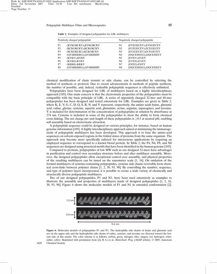
Two of our designed polypeptides, P1 and N1, have been used extensively as examples toillustrate the assembly and properties of multilayers made of designed polypeptides [1, 2, 16,30, 93, 98]. Figure 6 shows the molecular models of P1 and N1 in extended conformation [2].
Figure 6. Molecular models of polypeptides P1 and N1. The hydrophilic side chains of lysine and glutamic acidare on the upper side and the hydrophobic side chains of valine, cysteine, and tyrosine are directed toward the bot-tom side of this model. The color scheme is as follows: carbon, green; nitrogen, blue; oxygen, red; hydrogen, white;sulfur, yellow. Reprinted with permission from [2], B. Li et al., Biotechnol. Prog. (ASAP article). � 2005, AmericanChemical Society.AQ3
Table 2. Examples of designed polypeptides for LBL multilayers.
Positively charged polypeptide Negatively charged polypeptide
P1 (KVKGKCKV)3KVKGKCKY N1 (EVEGECEV)3EVEGECEYP2 (KCKGKCKV)3KCKGKCKY N2 (ECEGECEV)3ECEGECEYP3 (KCKGKCKC)3KCKGKCKY N3 (ECEGECEC)3ECEGECEYP4 (QVSRRRRG)3QVSRRRRY N4 (DQCEDEEG)3DQCEDEEYP5 (KVKV)7KVKY N5 (EVEV)7EVEYP6 (KVKS)7KVKY N6 (EVEN)7EVEYP7 (KKKK)7KKKY N7 (EEEE)7EEEYP8 (QVSRRRRG)5QVSRRRRY N8 (DQCEDEEG)5DQCEDEEY
Page Number: 15
Polypeptide Multilayer Films and Microcapsules 15
Path: K:/ASP-SOFTNANO-07-0502/Application/ASP-SOFTNANO-07-0502-008.3dDate: 3rd November 2007 Time: 14:30 User ID: tamilmanir BlackLiningEnabled
All hydrophobic side chains point in the same direction, all hydrophilic ones in the oppositedirection. Both polypeptides have a contour length of �9.4 nm and a thickness of �1.0 nm.These two polypeptides are oppositely charged at pH 7.4; P1 is positive, whereas N1 is nega-tive. The absolute charge density is �0.5 electronic charge per residue at pH 7.4. These twopolypeptides bind to each other in a multilayer by electrostatic attraction. Other types ofinteraction, however, may also contribute to the multilayer assembly and secondary structureformation.
5.3.2. Property Control at Nanometer Scale of Multilayers Made ofDesigned Polypeptides
A variety of ways have been used to control the properties of LBL multilayers made of designedpolypeptides.
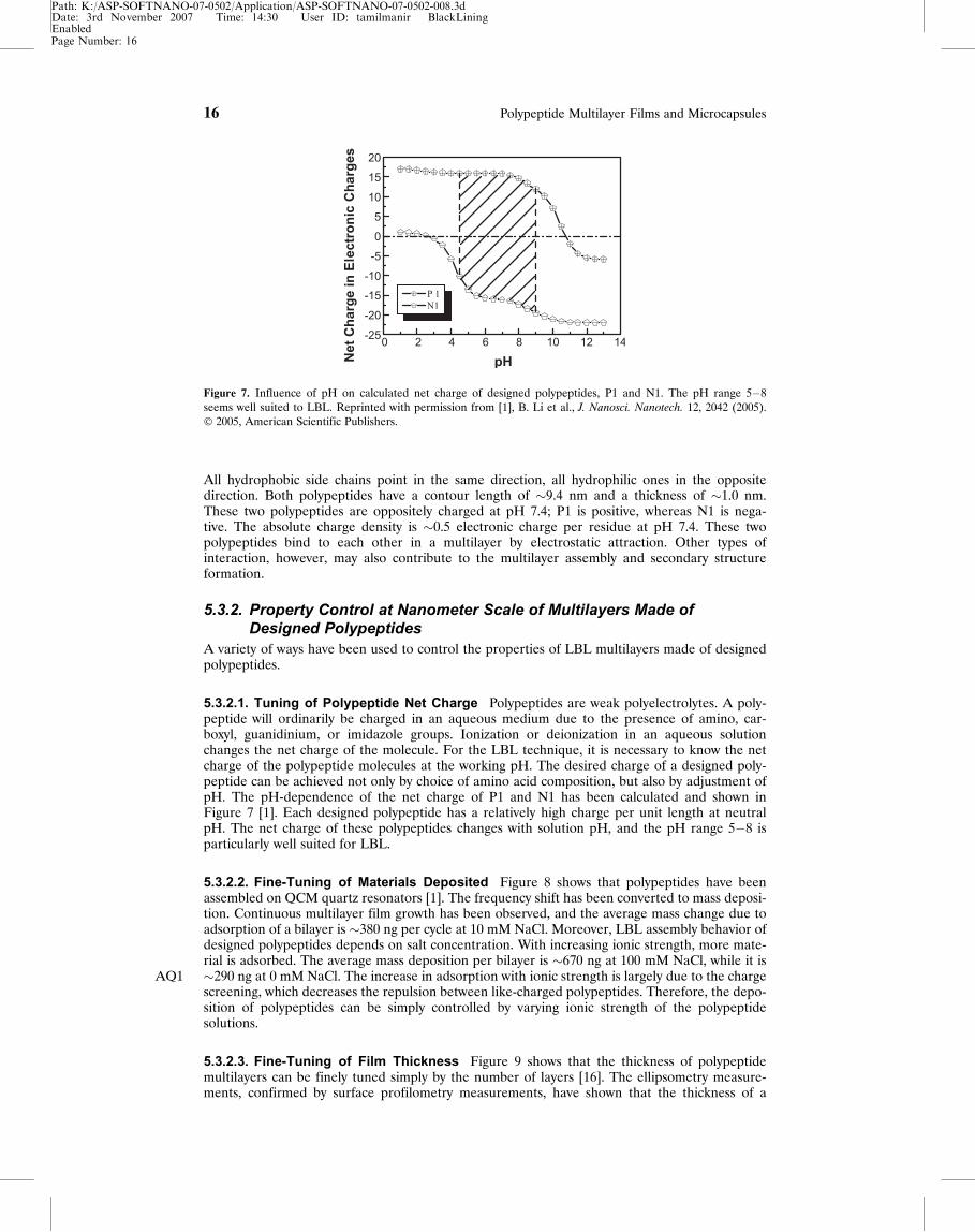
5.3.2.1. Tuning of Polypeptide Net Charge Polypeptides are weak polyelectrolytes. A poly-peptide will ordinarily be charged in an aqueous medium due to the presence of amino, car-boxyl, guanidinium, or imidazole groups. Ionization or deionization in an aqueous solutionchanges the net charge of the molecule. For the LBL technique, it is necessary to know the netcharge of the polypeptide molecules at the working pH. The desired charge of a designed poly-peptide can be achieved not only by choice of amino acid composition, but also by adjustment ofpH. The pH-dependence of the net charge of P1 and N1 has been calculated and shown inFigure 7 [1]. Each designed polypeptide has a relatively high charge per unit length at neutralpH. The net charge of these polypeptides changes with solution pH, and the pH range 5�8 isparticularly well suited for LBL.
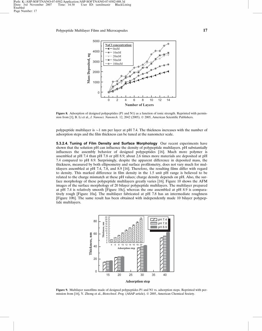
5.3.2.2. Fine-Tuning of Materials Deposited Figure 8 shows that polypeptides have beenassembled on QCM quartz resonators [1]. The frequency shift has been converted to mass deposi-tion. Continuous multilayer film growth has been observed, and the average mass change due toadsorption of a bilayer is �380 ng per cycle at 10 mM NaCl. Moreover, LBL assembly behavior ofdesigned polypeptides depends on salt concentration. With increasing ionic strength, more mate-rial is adsorbed. The average mass deposition per bilayer is �670 ng at 100 mM NaCl, while it is�290 ng atAQ1 0 mM NaCl. The increase in adsorption with ionic strength is largely due to the chargescreening, which decreases the repulsion between like-charged polypeptides. Therefore, the depo-sition of polypeptides can be simply controlled by varying ionic strength of the polypeptidesolutions.
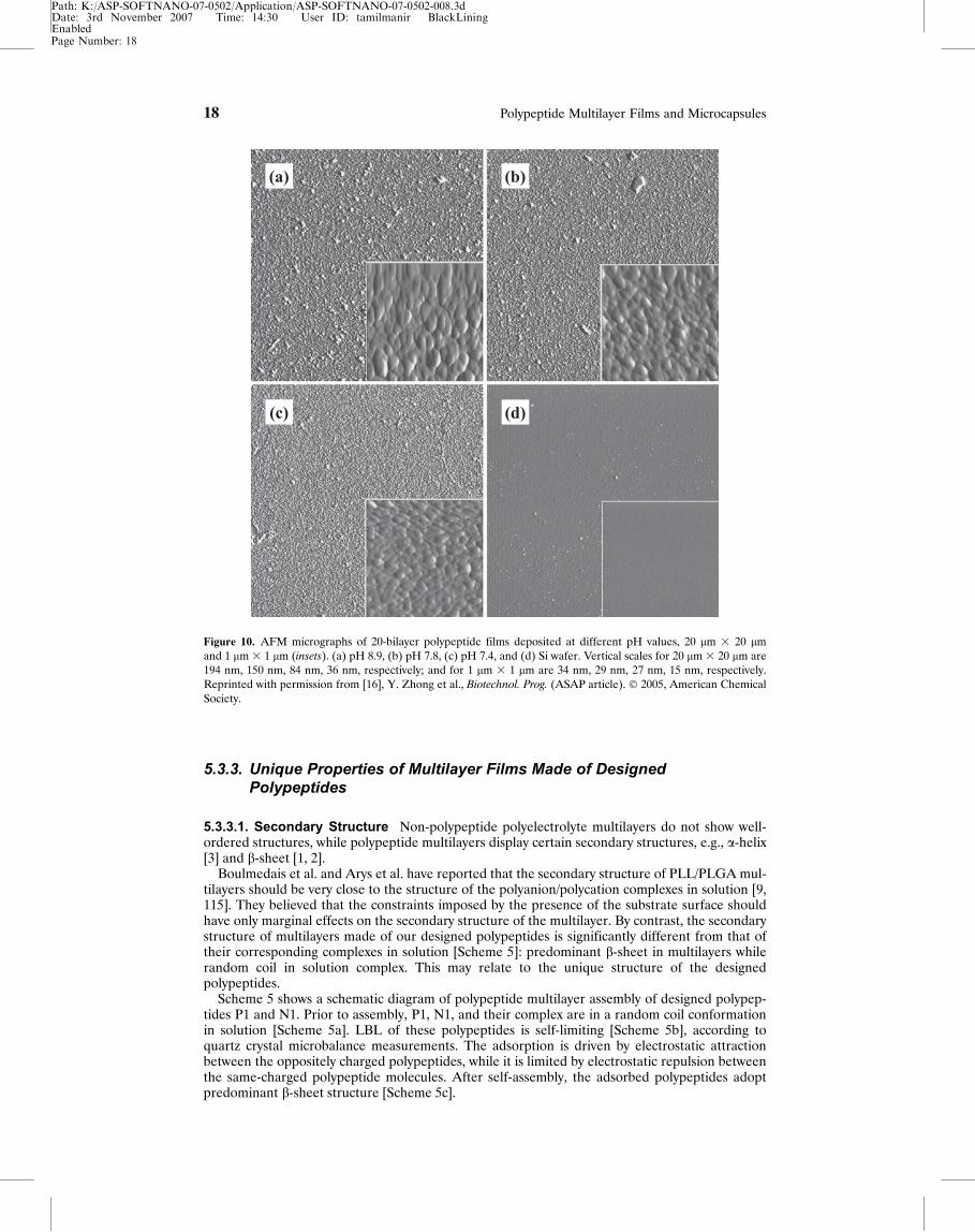
5.3.2.3. Fine-Tuning of Film Thickness Figure 9 shows that the thickness of polypeptidemultilayers can be finely tuned simply by the number of layers [16]. The ellipsometry measure-ments, confirmed by surface profilometry measurements, have shown that the thickness of a
Figure 7. Influence of pH on calculated net charge of designed polypeptides, P1 and N1. The pH range 5�8seems well suited to LBL. Reprinted with permission from [1], B. Li et al., J. Nanosci. Nanotech. 12, 2042 (2005).� 2005, American Scientific Publishers.
Page Number: 16
16 Polypeptide Multilayer Films and Microcapsules
Path: K:/ASP-SOFTNANO-07-0502/Application/ASP-SOFTNANO-07-0502-008.3dDate: 3rd November 2007 Time: 14:30 User ID: tamilmanir BlackLiningEnabled
polypeptide multilayer is �1 nm per layer at pH 7.4. The thickness increases with the number ofadsorption steps and the film thickness can be tuned at the nanometer scale.
5.3.2.4. Tuning of Film Density and Surface Morphology Our recent experiments haveshown that the solution pH can influence the density of polypeptide multilayers. pH substantiallyinfluences the assembly behavior of designed polypeptides [16]. Much more polymer isassembled at pH 7.4 than pH 7.8 or pH 8.9; about 2.6 times more materials are deposited at pH7.4 compared to pH 8.9. Surprisingly, despite the apparent difference in deposited mass, thethickness, measured by both ellipsometry and surface profilometry, does not vary much for mul-tilayers assembled at pH 7.4, 7.8, and 8.9 [16]. Therefore, the resulting films differ with regardto density. This marked difference in film density in the 1.5 unit pH range is believed to berelated to the charge mismatch at these pH values; charge density depends on pH. Also, the sur-face morphology of these polypeptide multilayers greatly varies [16]. Figure 10 shows the AFMimages of the surface morphology of 20 bilayer polypeptide multilayers. The multilayer preparedat pH 7.4 is relatively smooth [Figure 10c], whereas the one assembled at pH 8.9 is compara-tively rough [Figure 10a]. The multilayer fabricated at pH 7.8 has an intermediate roughness[Figure 10b]. The same result has been obtained with independently made 10 bilayer polypep-tide multilayers.
Figure 9. Multilayer nanofilms made of designed polypeptides P1 and N1 vs. adsorption steps. Reprinted with per-mission from [16], Y. Zhong et al., Biotechnol. Prog. (ASAP article). � 2005, American Chemical Society.
Figure 8. Adsorption of designed polypeptides (P1 and N1) as a function of ionic strength. Reprinted with permis-sion from [1], B. Li et al., J. Nanosci. Nanotech. 12, 2042 (2005). � 2005, American Scientific Publishers.
Page Number: 17
Polypeptide Multilayer Films and Microcapsules 17
Path: K:/ASP-SOFTNANO-07-0502/Application/ASP-SOFTNANO-07-0502-008.3dDate: 3rd November 2007 Time: 14:30 User ID: tamilmanir BlackLiningEnabled
5.3.3. Unique Properties of Multilayer Films Made of DesignedPolypeptides
5.3.3.1. Secondary Structure Non-polypeptide polyelectrolyte multilayers do not show well-ordered structures, while polypeptide multilayers display certain secondary structures, e.g., a-helix[3] and b-sheet [1, 2].
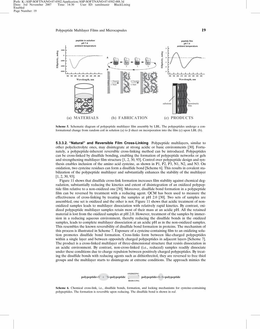
Boulmedais et al. and Arys et al. have reported that the secondary structure of PLL/PLGA mul-tilayers should be very close to the structure of the polyanion/polycation complexes in solution [9,115]. They believed that the constraints imposed by the presence of the substrate surface shouldhave only marginal effects on the secondary structure of the multilayer. By contrast, the secondarystructure of multilayers made of our designed polypeptides is significantly different from that oftheir corresponding complexes in solution [Scheme 5]: predominant b-sheet in multilayers whilerandom coil in solution complex. This may relate to the unique structure of the designedpolypeptides.
Scheme 5 shows a schematic diagram of polypeptide multilayer assembly of designed polypep-tides P1 and N1. Prior to assembly, P1, N1, and their complex are in a random coil conformationin solution [Scheme 5a]. LBL of these polypeptides is self-limiting [Scheme 5b], according toquartz crystal microbalance measurements. The adsorption is driven by electrostatic attractionbetween the oppositely charged polypeptides, while it is limited by electrostatic repulsion betweenthe same-charged polypeptide molecules. After self-assembly, the adsorbed polypeptides adoptpredominant b-sheet structure [Scheme 5c].
Figure 10. AFM micrographs of 20-bilayer polypeptide films deposited at different pH values, 20 lm 3 20 lmand 1 lm 3 1 lm (insets). (a) pH 8.9, (b) pH 7.8, (c) pH 7.4, and (d) Si wafer. Vertical scales for 20 lm 3 20 lm are194 nm, 150 nm, 84 nm, 36 nm, respectively; and for 1 lm 3 1 lm are 34 nm, 29 nm, 27 nm, 15 nm, respectively.Reprinted with permission from [16], Y. Zhong et al., Biotechnol. Prog. (ASAP article). � 2005, American ChemicalSociety.
Page Number: 18
18 Polypeptide Multilayer Films and Microcapsules
Path: K:/ASP-SOFTNANO-07-0502/Application/ASP-SOFTNANO-07-0502-008.3dDate: 3rd November 2007 Time: 14:30 User ID: tamilmanir BlackLiningEnabled
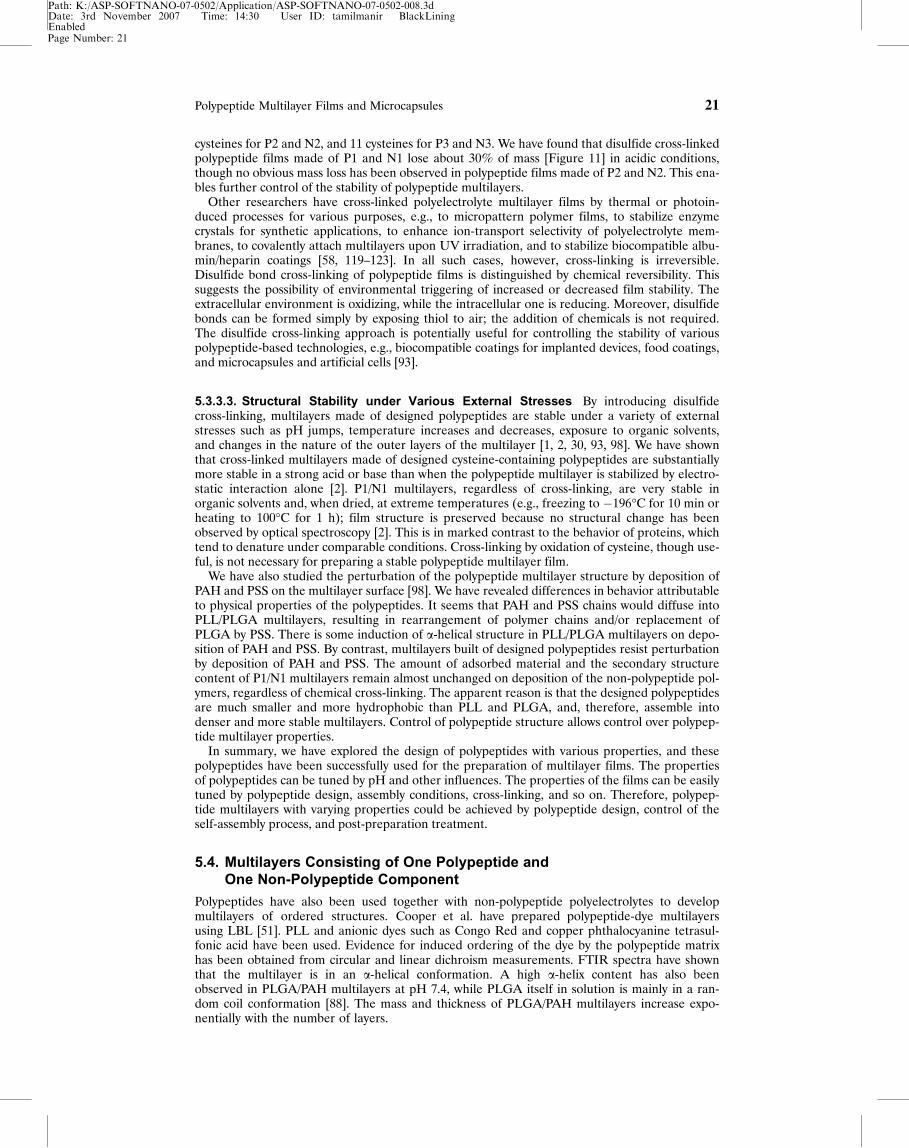
5.3.3.2. �Natural� and Reversible Film Cross-Linking Polypeptide multilayers, similar toother polyelectrolyte ones, may disintegrate at strong acidic or basic environments [30]. Fortu-nately, a polypeptide-inherent reversible cross-linking method can be introduced. Polypeptidescan be cross-linked by disulfide bonding, enabling the formation of polypeptide networks or gelsand strengthening multilayer film structure [1, 2, 30, 93]. Control over polypeptide design and syn-thesis enables inclusion of the amino acid cysteine, as shown in P1, P2, P3, N1, N2, and N3. Onoxidation, two cysteine residues can form a disulfide bond [Scheme 6]. This results in covalent sta-bilization of the polypeptide multilayer and substantially enhances the stability of the multilayer[1, 2, 30, 93].
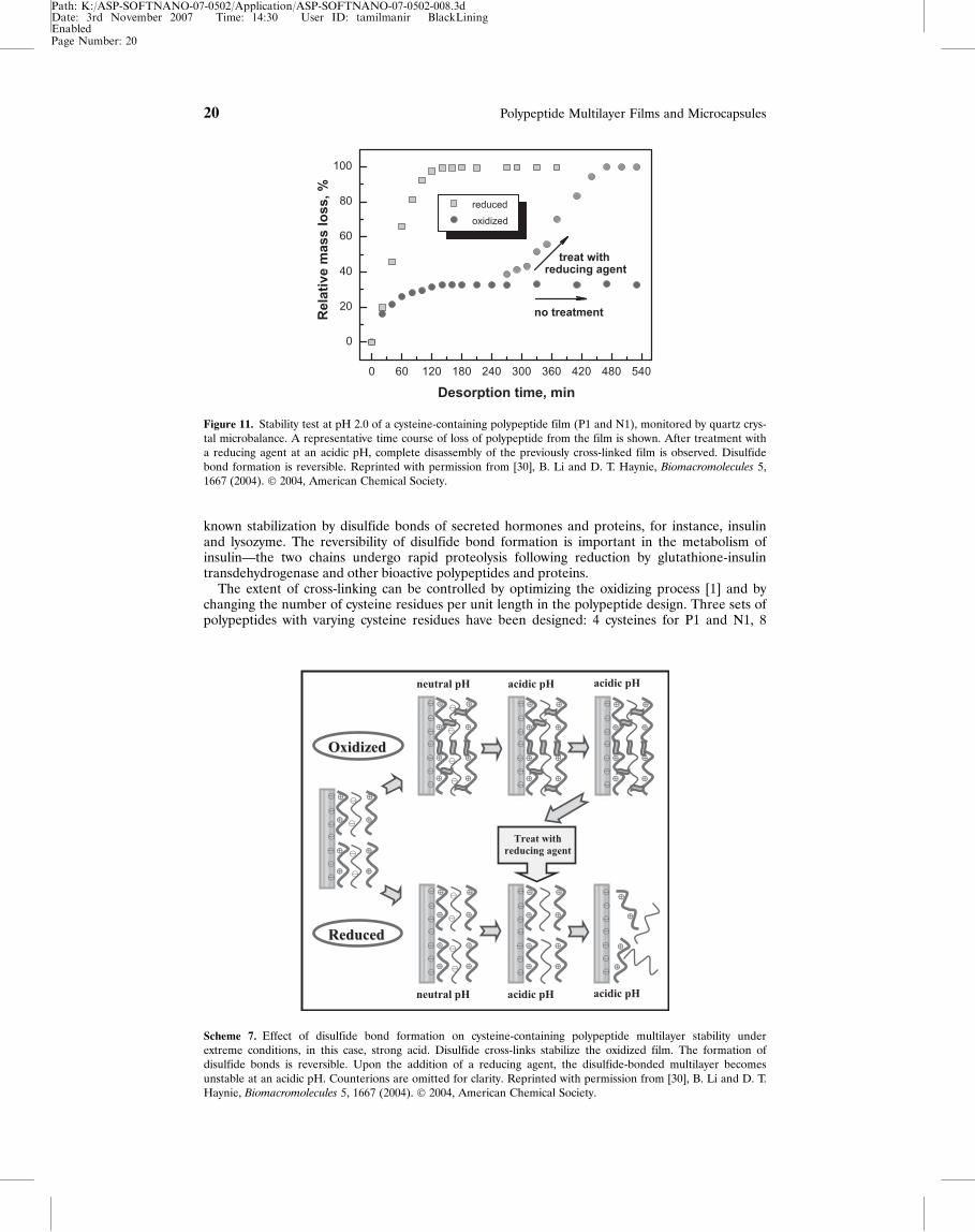
Figure 11 shows that disulfide cross-link formation increases film stability against chemical deg-radation, substantially reducing the kinetics and extent of disintegration of an oxidized polypep-tide film relative to a non-oxidized one [30]. Moreover, disulfide bond formation in a polypeptidefilm can be reversed by treatment with a reducing agent. QCM has been used to measure theeffectiveness of cross-linking by treating the samples at pH 2.0 [30]. Two sets of samples areassembled, one set is oxidized and the other is not. Figure 11 shows that acidic treatment of non-oxidized samples leads to multilayer dissociation with relatively rapid kinetics. By contrast, oxi-dized polypeptide multilayer samples retain most of their mass at an acidic pH. All the retainedmaterial is lost from the oxidized samples at pH 2.0. However, treatment of the samples by immer-sion in a reducing aqueous environment, thereby reducing the disulfide bonds in the oxidizedsamples, leads to complete multilayer dissociation at an acidic pH as in the non-oxidized samples.This resembles the known reversibility of disulfide bond formation in proteins. The mechanism ofthis process is illustrated in Scheme 7. Exposure of a cysteine-containing film to an oxidizing solu-tion promotes disulfide bond formation. Cross-links form between like-charged polypeptideswithin a single layer and between oppositely charged polypeptides in adjacent layers [Scheme 7].The product is a cross-linked multilayer of three-dimensional structure that resists dissociation inan acidic environment. By contrast, non-cross-linked (i.e., reduced) samples readily dissociateunder these conditions due to charge repulsion between positively charged polypeptides. By treat-ing the disulfide bonds with reducing agents such as dithiothreitol, they are reversed to free thiolgroups and the multilayer starts to disintegrate at extreme conditions. The approach mimics the
Scheme 5. Schematic diagram of polypeptide multilayer film assembly by LBL. The polypeptides undergo a con-formational change from random coil in solution (a) to b sheet on incorporation into the film (c) upon LBL (b).
Scheme 6. Chemical cross-link, i.e., disulfide bonds, formation, and locking mechanisms for cysteine-containingpolypeptides. The formation is reversible upon reducing. The disulfide bond is shown in red.
Page Number: 19
Polypeptide Multilayer Films and Microcapsules 19
Path: K:/ASP-SOFTNANO-07-0502/Application/ASP-SOFTNANO-07-0502-008.3dDate: 3rd November 2007 Time: 14:30 User ID: tamilmanir BlackLiningEnabled
known stabilization by disulfide bonds of secreted hormones and proteins, for instance, insulinand lysozyme. The reversibility of disulfide bond formation is important in the metabolism ofinsulin—the two chains undergo rapid proteolysis following reduction by glutathione-insulintransdehydrogenase and other bioactive polypeptides and proteins.
The extent of cross-linking can be controlled by optimizing the oxidizing process [1] and bychanging the number of cysteine residues per unit length in the polypeptide design. Three sets ofpolypeptides with varying cysteine residues have been designed: 4 cysteines for P1 and N1, 8
Figure 11. Stability test at pH 2.0 of a cysteine-containing polypeptide film (P1 and N1), monitored by quartz crys-tal microbalance. A representative time course of loss of polypeptide from the film is shown. After treatment witha reducing agent at an acidic pH, complete disassembly of the previously cross-linked film is observed. Disulfidebond formation is reversible. Reprinted with permission from [30], B. Li and D. T. Haynie, Biomacromolecules 5,1667 (2004). � 2004, American Chemical Society.
Scheme 7. Effect of disulfide bond formation on cysteine-containing polypeptide multilayer stability underextreme conditions, in this case, strong acid. Disulfide cross-links stabilize the oxidized film. The formation ofdisulfide bonds is reversible. Upon the addition of a reducing agent, the disulfide-bonded multilayer becomesunstable at an acidic pH. Counterions are omitted for clarity. Reprinted with permission from [30], B. Li and D. T.Haynie, Biomacromolecules 5, 1667 (2004). � 2004, American Chemical Society.
Page Number: 20
20 Polypeptide Multilayer Films and Microcapsules
Path: K:/ASP-SOFTNANO-07-0502/Application/ASP-SOFTNANO-07-0502-008.3dDate: 3rd November 2007 Time: 14:30 User ID: tamilmanir BlackLiningEnabled
cysteines for P2 and N2, and 11 cysteines for P3 and N3. We have found that disulfide cross-linkedpolypeptide films made of P1 and N1 lose about 30% of mass [Figure 11] in acidic conditions,though no obvious mass loss has been observed in polypeptide films made of P2 and N2. This ena-bles further control of the stability of polypeptide multilayers.
Other researchers have cross-linked polyelectrolyte multilayer films by thermal or photoin-duced processes for various purposes, e.g., to micropattern polymer films, to stabilize enzymecrystals for synthetic applications, to enhance ion-transport selectivity of polyelectrolyte mem-branes, to covalently attach multilayers upon UV irradiation, and to stabilize biocompatible albu-min/heparin coatings [58, 119–123]. In all such cases, however, cross-linking is irreversible.Disulfide bond cross-linking of polypeptide films is distinguished by chemical reversibility. Thissuggests the possibility of environmental triggering of increased or decreased film stability. Theextracellular environment is oxidizing, while the intracellular one is reducing. Moreover, disulfidebonds can be formed simply by exposing thiol to air; the addition of chemicals is not required.The disulfide cross-linking approach is potentially useful for controlling the stability of variouspolypeptide-based technologies, e.g., biocompatible coatings for implanted devices, food coatings,and microcapsules and artificial cells [93].
5.3.3.3. Structural Stability under Various External Stresses By introducing disulfidecross-linking, multilayers made of designed polypeptides are stable under a variety of externalstresses such as pH jumps, temperature increases and decreases, exposure to organic solvents,and changes in the nature of the outer layers of the multilayer [1, 2, 30, 93, 98]. We have shownthat cross-linked multilayers made of designed cysteine-containing polypeptides are substantiallymore stable in a strong acid or base than when the polypeptide multilayer is stabilized by electro-static interaction alone [2]. P1/N1 multilayers, regardless of cross-linking, are very stable inorganic solvents and, when dried, at extreme temperatures (e.g., freezing to �196�C for 10 min orheating to 100�C for 1 h); film structure is preserved because no structural change has beenobserved by optical spectroscopy [2]. This is in marked contrast to the behavior of proteins, whichtend to denature under comparable conditions. Cross-linking by oxidation of cysteine, though use-ful, is not necessary for preparing a stable polypeptide multilayer film.
We have also studied the perturbation of the polypeptide multilayer structure by deposition ofPAH and PSS on the multilayer surface [98]. We have revealed differences in behavior attributableto physical properties of the polypeptides. It seems that PAH and PSS chains would diffuse intoPLL/PLGA multilayers, resulting in rearrangement of polymer chains and/or replacement ofPLGA by PSS. There is some induction of a-helical structure in PLL/PLGA multilayers on depo-sition of PAH and PSS. By contrast, multilayers built of designed polypeptides resist perturbationby deposition of PAH and PSS. The amount of adsorbed material and the secondary structurecontent of P1/N1 multilayers remain almost unchanged on deposition of the non-polypeptide pol-ymers, regardless of chemical cross-linking. The apparent reason is that the designed polypeptidesare much smaller and more hydrophobic than PLL and PLGA, and, therefore, assemble intodenser and more stable multilayers. Control of polypeptide structure allows control over polypep-tide multilayer properties.
In summary, we have explored the design of polypeptides with various properties, and thesepolypeptides have been successfully used for the preparation of multilayer films. The propertiesof polypeptides can be tuned by pH and other influences. The properties of the films can be easilytuned by polypeptide design, assembly conditions, cross-linking, and so on. Therefore, polypep-tide multilayers with varying properties could be achieved by polypeptide design, control of theself-assembly process, and post-preparation treatment.
5.4. Multilayers Consisting of One Polypeptide andOne Non-Polypeptide Component
Polypeptides have also been used together with non-polypeptide polyelectrolytes to developmultilayers of ordered structures. Cooper et al. have prepared polypeptide-dye multilayersusing LBL [51]. PLL and anionic dyes such as Congo Red and copper phthalocyanine tetrasul-fonic acid have been used. Evidence for induced ordering of the dye by the polypeptide matrixhas been obtained from circular and linear dichroism measurements. FTIR spectra have shownthat the multilayer is in an a-helical conformation. A high a-helix content has also beenobserved in PLGA/PAH multilayers at pH 7.4, while PLGA itself in solution is mainly in a ran-dom coil conformation [88]. The mass and thickness of PLGA/PAH multilayers increase expo-nentially with the number of layers.
Page Number: 21
Polypeptide Multilayer Films and Microcapsules 21
Path: K:/ASP-SOFTNANO-07-0502/Application/ASP-SOFTNANO-07-0502-008.3dDate: 3rd November 2007 Time: 14:30 User ID: tamilmanir BlackLiningEnabled
Muller et al. have developed PLGA/PDDA and PLL/PVS multilayers [96]. An increase of orderhas been observed with decreasing polyelectrolyte concentration in the PLGA/PDDA multilayerand with an increasing number of layers in the PLL/PVS multilayer.
5.5. Polypeptide Microcapsules
Polymer microcapsules are an important class of functional materials widely used in encapsu-lation, separation, and biological applications [124]. Microcapsules of polypeptides are of particu-lar interest because of their intrinsic compatibility with biomolecules, high biodegradability, andcertain functions that can be endowed by designing suitable primary (and secondary) structures.In spite of these promising features, there have been only a few studies on self-assembly ofpolypeptide microcapsules [93, 125–127]. Polypeptide microcapsules have been prepared byMorikawa et al. [125] from simple polypeptides, e.g., poly(c-benzyl L-glutamate), with well-defined structures. Kidchob et al. have developed polypeptide microcapsules that showpH-responsive release of encapsulated dextran [126]. Wang et al. have prepared and studied poly-peptide microcapsules as drug carriers [127].
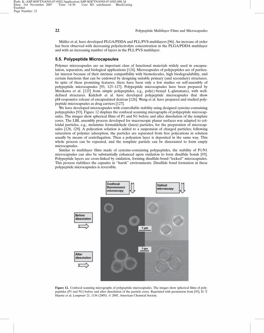
We have developed microcapsules with controllable stability using designed cysteine-containingpolypeptides [93]. Figure 12 displays the confocal scanning micrographs of polypeptide microcap-sules. The images show spherical films of P1 and N1 before and after dissolution of the templatecores. The LBL assembly process developed for macroscopic planar surfaces was adapted to col-loidal particles, e.g., melamine formaldehyde (latex) particles, for the preparation of microcap-sules [128, 129]. A polycation solution is added to a suspension of charged particles; followingsaturation of polymer adsorption, the particles are separated from free polycations in solutionusually by means of centrifugation. Then a polyanion layer is deposited in the same way. Thiswhole process can be repeated, and the template particle can be dissociated to form emptymicrocapsules.
Similar to multilayer films made of cysteine-containing polypeptides, the stability of P1/N1microcapsules can also be substantially enhanced upon oxidation to form disulfide bonds [93].Polypeptide layers are cross-linked by oxidation, forming disulfide-bond-‘‘locked’’ microcapsules.This process stabilizes the capsules in ‘‘harsh’’ environments. Disulfide bond formation in thesepolypeptide microcapsules is reversible.
Figure 12. Confocal scanning micrographs of polypeptide microcapsules. The images show spherical films of poly-peptides (P1 and N1) before and after dissolution of the particle cores. Reprinted with permission from [93], D. T.Haynie et al. Langmuir 21, 1136 (2005). � 2005, American Chemical Society.
Page Number: 22
22 Polypeptide Multilayer Films and Microcapsules
Path: K:/ASP-SOFTNANO-07-0502/Application/ASP-SOFTNANO-07-0502-008.3dDate: 3rd November 2007 Time: 14:30 User ID: tamilmanir BlackLiningEnabled
6. APPLICATIONS OF POLYPEPTIDE MULTILAYERSIn general, polyelectrolyte multilayers can be used as biosensors, nanoreactors, drug delivery sys-tems, and so on [17]. The biocompatibility, biofunctionality, and immunogenicity of polypeptideswill be more favorable for biomedical applications of LBL structures than those made from non-polypeptide polyelectrolytes, for instance, PAH and PSS, particularly if the sequences are basedon genomic information [105]. Applications of polypeptide multilayers are very promising in avariety of areas including membranes for ion separation, drug delivery, artificial cells, cardiovascu-lar applications, multifunctional molecular coatings, biosensors, nanoreactors, liquid/liquid elec-trochemical interfaces, and so on. Following are a few examples.
6.1. Chiral Separation
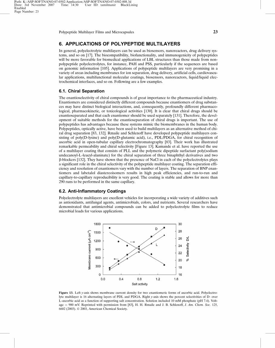
The enantioselectivity of chiral compounds is of great importance to the pharmaceutical industry.Enantiomers are considered distinctly different compounds because enantiomers of drug substan-ces may have distinct biological interactions, and, consequently, profoundly different pharmaco-logical, pharmacokinetic, or toxicological activities [130]. It is clear that chiral drugs should beenantioseparated and that each enantiomer should be used separately [131]. Therefore, the devel-opment of suitable methods for the enantioseparation of chiral drugs is important. The use ofpolypeptides has advantages because these systems mimic the biomembranes in the human body.Polypeptides, optically active, have been used to build multilayers as an alternative method of chi-ral drug separation [83, 132]. Rmaile and Schlenoff have developed polypeptide multilayers con-sisting of poly(D-lysine) and poly(D-glutamic acid), i.e., PDL/PDGA, for chiral recognition ofascorbic acid in open-tubular capillary electrochromatography [83]. Their work has illustratedremarkable permeability and chiral selectivity [Figure 13]. Kamande et al. have reported the useof a multilayer coating that consists of PLL and the polymeric dipeptide surfactant poly(sodiumundecanoyl-L-leucyl-alaninate) for the chiral separation of three binaphthyl derivatives and twob-blockers [132]. They have shown that the presence of NaCl in each of the polyelectrolytes playsa significant role in the chiral selectivity of the polypeptide multilayer coating. The separation effi-ciency and resolution of enantiomers vary with the number of layers. The separation of BNP enan-tiomers and labetalol diastereoisomers results in high peak efficiencies, and run-to-run andcapillary-to-capillary reproducibility is very good. The coating is stable and allows for more than290 runs to be performed in the same capillary.
6.2. Anti-Inflammatory Coatings
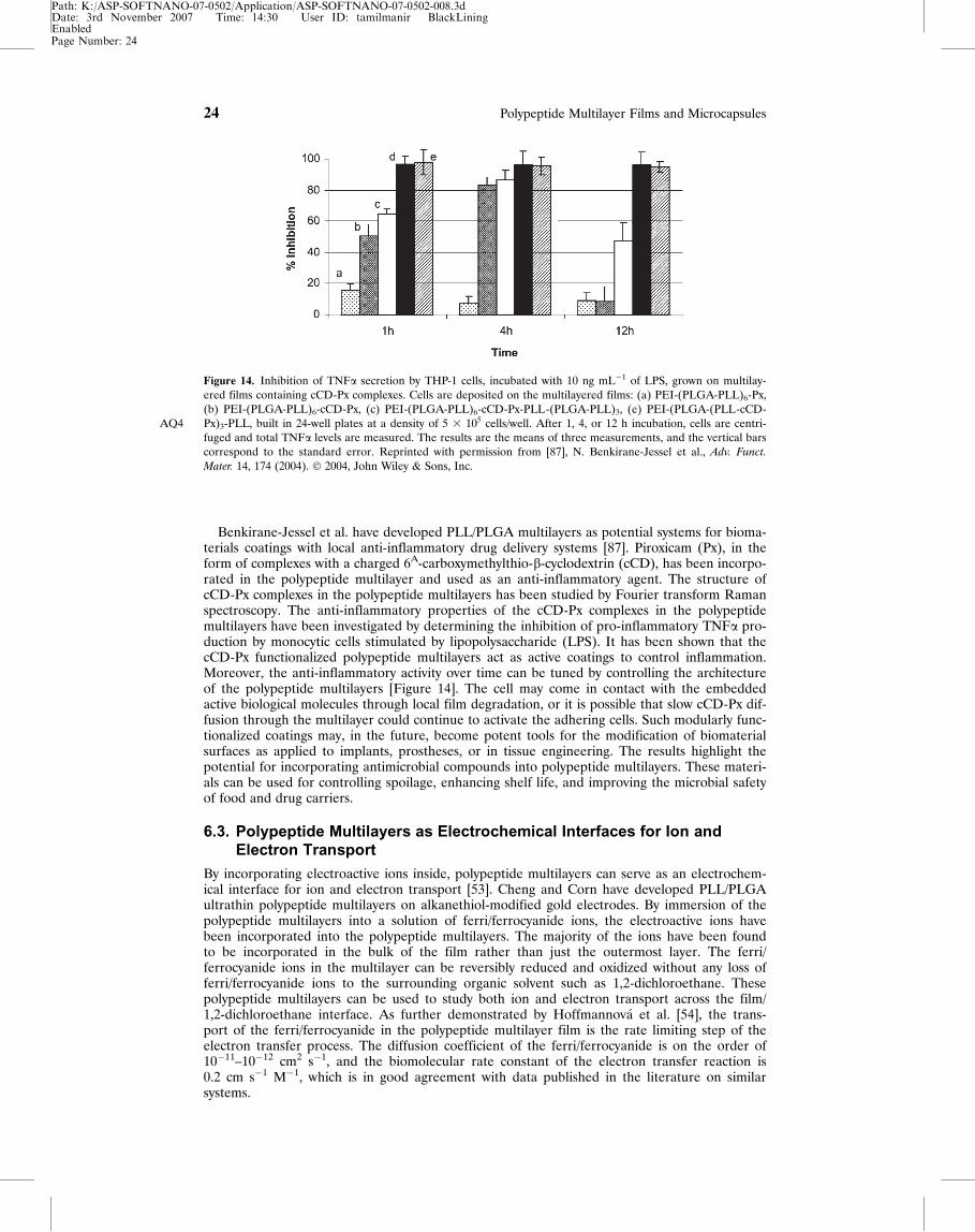
Polyelectrolyte multilayers are excellent vehicles for incorporating a wide variety of additives suchas antioxidants, antifungal agents, antimicrobials, colors, and nutrients. Several researchers havedemonstrated that antimicrobial compounds can be added to polyelectrolyte films to reducemicrobial loads for various applications.
Figure 13. Left y-axis shows membrane current density for two enantiomeric forms of ascorbic acid. Polyelectro-lyte multilayer is 16 alternating layers of PDL and PDGA. Right y-axis shows the percent selectivities of D- overL-ascorbic acid as a function of supporting salt concentration. Solution included 10 mM phosphate (pH 7.4). Volt-age ¼ 900 mV. Reprinted with permission from [83], H. H. Rmaile and J. B. Schlenoff, J. Am. Chem. Soc. 125,6602 (2003). � 2003, American Chemical Society.
Page Number: 23
Polypeptide Multilayer Films and Microcapsules 23
Path: K:/ASP-SOFTNANO-07-0502/Application/ASP-SOFTNANO-07-0502-008.3dDate: 3rd November 2007 Time: 14:30 User ID: tamilmanir BlackLiningEnabled
Benkirane-Jessel et al. have developed PLL/PLGA multilayers as potential systems for bioma-terials coatings with local anti-inflammatory drug delivery systems [87]. Piroxicam (Px), in theform of complexes with a charged 6A-carboxymethylthio-b-cyclodextrin (cCD), has been incorpo-rated in the polypeptide multilayer and used as an anti-inflammatory agent. The structure ofcCD-Px complexes in the polypeptide multilayers has been studied by Fourier transform Ramanspectroscopy. The anti-inflammatory properties of the cCD-Px complexes in the polypeptidemultilayers have been investigated by determining the inhibition of pro-inflammatory TNFa pro-duction by monocytic cells stimulated by lipopolysaccharide (LPS). It has been shown that thecCD-Px functionalized polypeptide multilayers act as active coatings to control inflammation.Moreover, the anti-inflammatory activity over time can be tuned by controlling the architectureof the polypeptide multilayers [Figure 14]. The cell may come in contact with the embeddedactive biological molecules through local film degradation, or it is possible that slow cCD-Px dif-fusion through the multilayer could continue to activate the adhering cells. Such modularly func-tionalized coatings may, in the future, become potent tools for the modification of biomaterialsurfaces as applied to implants, prostheses, or in tissue engineering. The results highlight thepotential for incorporating antimicrobial compounds into polypeptide multilayers. These materi-als can be used for controlling spoilage, enhancing shelf life, and improving the microbial safetyof food and drug carriers.
6.3. Polypeptide Multilayers as Electrochemical Interfaces for Ion andElectron Transport
By incorporating electroactive ions inside, polypeptide multilayers can serve as an electrochem-ical interface for ion and electron transport [53]. Cheng and Corn have developed PLL/PLGAultrathin polypeptide multilayers on alkanethiol-modified gold electrodes. By immersion of thepolypeptide multilayers into a solution of ferri/ferrocyanide ions, the electroactive ions havebeen incorporated into the polypeptide multilayers. The majority of the ions have been foundto be incorporated in the bulk of the film rather than just the outermost layer. The ferri/ferrocyanide ions in the multilayer can be reversibly reduced and oxidized without any loss offerri/ferrocyanide ions to the surrounding organic solvent such as 1,2-dichloroethane. Thesepolypeptide multilayers can be used to study both ion and electron transport across the film/1,2-dichloroethane interface. As further demonstrated by Hoffmannov�a et al. [54], the trans-port of the ferri/ferrocyanide in the polypeptide multilayer film is the rate limiting step of theelectron transfer process. The diffusion coefficient of the ferri/ferrocyanide is on the order of10�11–10�12 cm2 s�1, and the biomolecular rate constant of the electron transfer reaction is0.2 cm s�1 M�1, which is in good agreement with data published in the literature on similarsystems.
Figure 14. Inhibition of TNFa secretion by THP-1 cells, incubated with 10 ng mL�1 of LPS, grown on multilay-ered films containing cCD-Px complexes. Cells are deposited on the multilayered films: (a) PEI-(PLGA-PLL)6-Px,(b) PEI-(PLGA-PLL)6-cCD-Px, (c) PEI-(PLGA-PLL)6-cCD-Px-PLL-(PLGA-PLL)3, (e) PEI-(PLGA-(PLL-cCD-Px)3-PLL, built in 24-well plates at a density of 5 3 105 cells/wellAQ4 . After 1, 4, or 12 h incubation, cells are centri-fuged and total TNFa levels are measured. The results are the means of three measurements, and the vertical barscorrespond to the standard error. Reprinted with permission from [87], N. Benkirane-Jessel et al., Adv. Funct.
Mater. 14, 174 (2004). � 2004, John Wiley & Sons, Inc.
Page Number: 24
24 Polypeptide Multilayer Films and Microcapsules
Path: K:/ASP-SOFTNANO-07-0502/Application/ASP-SOFTNANO-07-0502-008.3dDate: 3rd November 2007 Time: 14:30 User ID: tamilmanir BlackLiningEnabled
6.4. Polypeptide Multilayers in Nanofiltration for Ion Separation
Polypeptide multilayers have been deposited in microporous membranes for ion separation[133]. The deposition of PLL/PLGA multilayers within confined pore geometries in microporousmembranes leads to an enhanced volume density of ionizable groups in the membrane phase.This increase in the effective charge density allows for Donnan or charge-based exclusion ofionic species (especially divalent co-ions) using microporous materials characterized by perme-ability values that exceed conventional membrane processes. The properties of the resultingmaterials have been found to depend highly on the ionic strength of the carrier solvent used forthe multilayer deposition. Compared to assemblies in the absence of salt, the multilayer mem-branes assembled at high salt concentration display lower pure water permeability and enhancedoverall adsorption. This is because in the deposition process, the number of ionic site interac-tions per adsorbed chain is smaller at high salt concentration than at low salt concentration.Thus, larger segments of the adsorbing polymer extend into the pore cross-section at high saltconcentration and lead to greater solvent resistance. In addition, an excess of these charged con-stituents away from the pore assembly facilitates ion exclusion in open membrane materials.Multilayer assemblies prepared at high ionic strength, e.g., 0.5 M NaCl solution, provide excel-lent separation of divalent co-ions, even in the ultradilute regime. This work and other recentwork on non-polypeptide polyelectrolytes [119, 134–138] have shown great promise in nanofiltra-tion and reverse osmosis applications.
As aforementioned, great efforts have been made to explore the applications of polypeptidemultilayers over the past years. Multilayers made of designed polypeptides could have a broaderrange of application because polypeptide design may enable a great degree of control over bioac-tive properties in defined and ‘‘natural’’ ways. Certain functional polypeptide sequences, forinstance, sequences from extracellular and matricellular proteins that contain the Arg-Gly-Asp(RGD) cell adhesive sequence, could be incorporated into the designed polypeptide sequence.This RGD motif is responsible for integrinAQ2 -mediated cell adhesion and has been found in fibro-nectin and many other extracellular matrix and matricellular proteins. The RGD motif has beenincluded in streptavidin widely utilized proteins, and its incorporation has facilitated cell adhesion[139]. This RGD sequence could be inserted into our designed polypeptide sequences to makethese polypeptide multilayers possess a cell-adhesion property. Also, sequences having antimicro-bial properties could be easily included when designing polypeptide sequences.
7. CONCLUSIONS AND FUTURE PROSPECTSThis chapter has described the development and fabrication as well as the applications of polypep-tide multilayer films and microcapsules. Polypeptide multilayers are prepared by two main meth-ods, i.e., LBL and LB.
Increasing activity in the field of polypeptide multilayer films and microcapsules is due to sev-eral factors. First, the application of the technique, i.e., LBL, allows one to easily deposit filmsand microcapsules containing polypeptides and/or other biomacromolecules. LBL also allows oneto deposit multilayers and to form complex structures with controlled assembly behavior and mul-tilayer properties. Second, polypeptides can now be chemically synthesized or recombinantlymade in cells or plants with little difficulty due to the advancement in the past decades of chemicalsynthesis methods of biopolymers and biotechnology. Third, there is flexibility and diversity indesigning polypeptides; polypeptides with specific biofunctionalities and structures can beobtained, and polypeptide design offers the possibility of a reversible ‘‘polypeptide-inherent’’cross-linking method. Therefore, the future of polypeptide multilayers, especially those ofdesigned polypeptides, looks extremely promising as we continue to find novel applications forthese advanced materials. We have explored the design of 32-mer polypeptides for LBL of multi-layer films and microcapsules. New developments may include the design of polypeptides with spe-cific biofunctionalities such as including an RGD sequence for ‘‘bio-active’’ coatings that can beused to promote the adhesion of cells.
ACKNOWLEDGMENTSThe support from the Department of Orthopedics, the School of Medicine, WVNano, and WVUResearch Corporation, at West Virginia University is gratefully acknowledged. I would thankSuzanne Smith for proofreading the manuscript and assisting with the figures.
Page Number: 25
Polypeptide Multilayer Films and Microcapsules 25
Path: K:/ASP-SOFTNANO-07-0502/Application/ASP-SOFTNANO-07-0502-008.3dDate: 3rd November 2007 Time: 14:30 User ID: tamilmanir BlackLiningEnabled
REFERENCES1. B. Li, D. T. Haynie, N. Palath, and D. Janisch, J. Nanosci. Nanotech. 12, 2042 (2005).2. B. Li, J. Rozas, and D. T. Haynie, Biotechnol. Prog. (In press).AQ33. M. Muller, T. Reihs, B. Kebler, H. J. Adler, and K. Lunkwitz, Macromol. Symp. 211, 217 (2004).4. T. Jaworek, D. Neher, G. Wegner, R. H. Wieringa, and A. J. Schouten, Science 279, 57 (1998).5. L. Hartmann, T. Kratzmuller, H. G. Braun, and F. Kremer, Macromol. Rapid Commun. 21, 814 (2000).6. T. Strother, W. Cai, X. Zhao, R. J. Hamers, and L. M. Smith, J. Am. Chem. Soc. 122, 1205 (2000).7. C. Picart, P. Lavalle, P. Hubert, F. J. G. Cuisinier, G. Decher, P. Schaaf, and J. C. Voegel, Langmuir 17, 7414
(2001).8. P. Lavalle, C. Gergely, F. J. G. Cuisinier, G. Decher, P. Schaaf, and J. C. Voegel, Macromolecules 35, 4458
(2002).9. F. Boulmedais, P. Schwinte, C. Gergely, J. C. Voegel, and P. Schaaf, Langmuir 18, 4523 (2002).
10. P. Schwinte, J. C. Voegel, C. Picart, Y. Haikel, P. Schaaf, and B. Szalontai, J. Phys. Chem. B 105, 11906(2001).
11. J. Chluba, J. C. Voegel, G. Decher, P. Erbacher, P. Schaaf, and J. Ogier, Biomacromolecules 2, 800 (2001).12. P. Schwinte, V. Ball, B. Szalontai, Y. Haikel, J. C. Voegel, and P. Schaaf, Biomacromolecules 3, 1135 (2002).13. L. Richert, Ph. Lavalle, D. Vautier, B. Senger, J. F. Stoltz, P. Schaaf, J. C. Voegel, and C. Picart, Biomacromo-
lecules 3, 1170 (2002).14. R. K. Iler, J. Colloid Interface Sci. 21, 569 (1966).15. G. Decher, Science 277, 1232 (1997).16. Y. Zhong, B. Li, and D. T. Haynie, Biotechnol. Prog. (In press).17. Y. Lvov, in Protein Architecture, Interfacial Molecular Assembly and Immobilization Biotechnology, Y. Lvov
and H. M€ohwald, Eds., Marcel Dekker, New York (2000), p. 125.18. J. F. Rusling, in Protein Architecture: Interfacial Molecular Assembly and Immobilization Biotechnology, Y. Lvov
and H. M€ohwald, Eds., Marcel Dekker, New York (2000), p. 337.19. V. Erokhin, in Protein Architecture: Interfacial Molecular Assembly and Immobilization Biotechnology, Y. Lvov
and H. M€ohwald, Eds., Marcel Dekker, New York (2000), p. 99.20. J. C. Cheftel, J. L. Cuq, and D. Lorient, in Food Chemistry, O. R. Fennema, Ed., Marcel Dekker, New York
(1985), p. 245.21. I. D. Campbell and R. A. Dwek, Biological Spectroscopy, Benjamin/Cummings, CA (1984).22. C. S. Winter and R. H. Tredgold, Thin Solid Films 123, L1 (1985).23. M. Rikukawa, K. Sanui, and N. Ogata, Synth. Met. 84, 393 (1997).24. T. M. Cooper, A. L. Campbell, C. Noffsinger, J. Gunther-Greer, R. L. Crane, and W. Adams, Mater. Res. Soc.
Symp. Proc. 351, 239 (1994).25. T. E. Creighton, Proteins: Structures and Molecular Properties. 2nd ed., Freeman, New York (1993), p. 287.26. A. Gennadios, Protein-based Films and Coatings, CRC Press, Boca Raton, FL (2002), p. 1.27. K. B. Blodgett, J. Am. Chem. Soc. 57, 1007 (1935).28. B. P. Binks, Adv. Colloid Interface Sci. 34, 343 (1991).29. O. N. Oliveira, Jr., J. A. He, V. Zucolotto, S. Balasubramanian, L. Li, H. S. Nalwa, J. Kumar, and S. K. Tripa-
thy, in Handbook of Polyelectrolytes and Their Applications, S. K. Tripathy, J. Kumar, and H. S. Nalwa, Eds.,American Scientific Publishers, Stevenson Ranch, CA (2002), Vol. 1, p. 1.
30. B. Li and D. T. Haynie, Biomacromolecules 5, 1667 (2004).31. S. A. Sukhishvili and S. Granick, J. Am. Chem. Soc. 122, 9550 (2002).32. C. Schuler and F. Caruso, Biomacromolecules 2, 921 (2001).33. A. J. Chung and M. F. Rubner, Langmuir 18, 1176 (2002).34. J. A. Hiller and M. F. Rubner, Macromolecules 36, 4078 (2003).35. K. C. Wood, J. Q. Boedicker, D. M. Lynn, and P. T. Hammond, Langmuir 21, 1603 (2005).36. Z. L. Zhi and D. T. Haynie, Macromolecules 37, 8668 (2004).37. G. B. Sukhorukov, M. Brumen, E. Donath, and H. M€ohwald, J. Phys. Chem. B 103, 6434 (1999).38. A. A. Antipov, G. B. Sukhorukov, E. Donath, and H. M€ohwald, J. Phys. Chem. B 105, 2281 (2001).39. M. K. Park, S. Deng, and R. C. Advincula, Langmuir 21, 5272 (2005).40. O. P. Tiourina, A. A. Antipov, G. B. Sukhorukov, N. L. Larionova, Y. Lvov, and H. M€ohwald, Macromol.
Biosci. 1, 209 (2001).41. A. J. Khopade and F. Caruso, Biomacromolecules 3, 1154 (2002).42. R. V. Klitzing and H. M€ohwald, Macromolecules 29, 6901 (1996).43. A. Wu, D. Yoo, J. K. Lee, and M. F. Rubner, J. Am. Chem. Soc. 121, 4883 (1999).44. A. C. Fou, O. Onitsuka, M. Ferreira, M. F. Rubner, and B. R. Hsieh, J. Appl. Phys. 79, 7501 (1996).45. W. Kong, X. Zhang, M. Gao, H. Zhou, W. Li, and J. Shen, Macromol. Rapid Commun. 15, 405 (1994).46. Y. Lvov, H. Haas, G. Decher, H. M€ohwald, A. Michailov, B. Mtchedlishvily, E. Morgunova, and B. Vain-
shtain, Langmuir 10, 4232 (1994).47. Y. Lvov, K. Ariga, and T. Kunitake, J. Am. Chem. Soc. 117, 6117 (1995).48. F. Caruso, D. N. Furlong, K. Ariga, I. Ichinose, and T. Kunitake, Langmuir 14, 4559 (1998).49. A. Kotov, I. D�ek�any, and J. H. Fendler, J. Phys. Chem. 99, 13065 (1995).50. Y. Lvov, K. Ariga, I. Ichinose, and T. Kunitake, Langmuir 12, 3038 (1996).51. T. M. Cooper, A. L. Campbell, and R. L. Crane, Langmuir 11, 2713 (1995).52. K. Ariga, Y. Lvov, and T. Kunitake, J. Am. Chem. Soc. 119, 2224 (1997).
Page Number: 26
26 Polypeptide Multilayer Films and Microcapsules
Path: K:/ASP-SOFTNANO-07-0502/Application/ASP-SOFTNANO-07-0502-008.3dDate: 3rd November 2007 Time: 14:30 User ID: tamilmanir BlackLiningEnabled
53. Y. Cheng and R. M. Corn, J. Phys. Chem. B 103, 8726 (1999).54. H. Hoffmannov�a, D. Fermin, and P. Krtil, J. Electroanal. Chem. 562, 261 (2004).55. F. Boulmedais, M. Bozonnet, P. Schwinte, J. C. Voegel, and P. Schaaf, Langmuir 19, 9873 (2003).56. T. J. Halthur, P. M. Claesson, and U. M. Elofsson, J. Am. Chem. Soc. 126, 17009 (2004).57. C. Tedeschi, M. Polli, M. P. Fontana, and O. Pieroni, Thin Solid Films 284–285, 174 (1996).58. S. Y. Yang and M. F. Rubner, J. Am. Chem. Soc. 124, 2100 (2002).59. D. Lee, S. Y. Yang, R. E. Cohen, and M. F. Rubner, Polym. Mater. Sci. Eng. 90, 526 (2004).60. D. M. DeLongchamp and P. T. Hammond, Langmuir 20, 5403 (2004).61. V. Kozlovskaya, S. Ok, A. Sousa, M. Libera, and S. A. Sukhishvili, Macromolecules 36, 8590 (2003).62. H. Zhang, Z. Wang, Y. Zhang, and X. Zhang, Langmuir 20, 9366 (2004).63. J. D. Hong, K. Lowack, J. Schmitt, and G. Decher, Prog. Colloid Polym. Sci. 93, 98 (1993).64. M. Sano, Y. Lvov, and T. Kunitake, Annu. Rev. Mater. Sci. 26, 153 (1996).65. D. Yoo, S. S. Shiratori, and M. F. Rubner, Macromolecules 31, 4309 (1998).66. J. J. Harris and M. L. Bruening, Langmuir 16, 2006 (2000).67. J. D. Mendelsohn, C. J. Barrett, V. V. Chan, A. J. Pal, A. M. Mayes, and M. F. Rubner, Langmuir 16, 5017 (2000).68. A. Fery, B. Scholer, T. Cassagneau, and F. Caruso, Langmuir 17, 3779 (2001).69. J. Hiller, J. D. Mendelsohn, and M. F. Rubner, Nat. Mater. 1, 59 (2002).70. S. E. Burke and C. J. Barrett, Biomacromolecules 4, 1773 (2003).71. B. Y. Kim and M. L. Bruening, Langmuir 19, 94 (2003).72. O. Mermut and C. Barrett, J. Analyst 126, 1861 (2001).73. J. Q. Sun, T. Wu, F. Liu, Z. Q. Wang, X. Zhang, and J. C. Shen, Langmuir 16, 4620 (2000).74. B. Schoeler, E. Poptoschev, and F. Caruso, Macromolecules 36, 5258 (2003).75. N. G. Hoogeveen, M. A. C. Stuart, G. J. Fleer, and M. R. B€ohmer, Langmuir 12, 3675 (1996).76. E. Kharlampieva and S. A. Sukhishvili, Langmuir 19, 1235 (2003).77. S. T. Dubas and J. B. Schlenoff, Macromolecules 34, 3736 (2001).78. G. B. Sukhorukov, A. A. Antipov, A. Voigt, E. Donath, and H. M€ohwald, Macromol. Rapid Commun. 22, 44
(2001).79. H. H. Rmaile, T. R. Farhat, and J. B. Schlenoff, J. Phys. Chem. B 107, 14401 (2003).80. G. B. Sukhorukov, H. M€ohwald, G. Decher, and Y. Lvov, Thin Solid Films 284, 220 (1996).81. H. P. Zheng, I. Lee, M. F. Rubner, and P. T. Hammond, Adv. Mater. 14, 569 (2002).82. S. K. Tripathy, J. Kumar, and H. S. Nalwa, Eds., Handbook of Polyelectrolyte-based Thin Films for Electronic
and Photonic Applications, American Scientific Publishers, Stevenson Ranch, CA (2002), Vol. 1, p. 1.83. H. H. Rmaile and J. B. Schlenoff, J. Am. Chem. Soc. 125, 6602 (2003).84. M. J. McShane, J. Q. Brown, K. B. Guice, and Y. Lvov, J. Nanosci. Nanotech. 2, 411 (2002).85. T. J. Halthur and U. M. Elofsson, Langmuir 20, 1739 (2004).86. G. Sauerbrey, Z. Phyzik 155, 206 (1959).87. N. Benkirane-Jessel, P. Schwinte, P. Falvey, R. Darcy, Y. Haikel, P. Schaaf, J. C. Voegel, and J. Ogier, Adv.
Funct. Mater. 14, 174 (2004).88. F. Boulmedais, V. Ball, P. Schwinte, B. Frisch, P. Schaaf, and J. C. Voegel, Langmuir 19, 440 (2003).89. D. T. Haynie, S. Balkundi, N. Palath, K. Chakravarthula, and K. Dave, Langmuir 20, 4540 (2004).90. K. S. Birdi, D. T. Vu, L. Moesby, K. B. Anderson, and D. Kristensen, Surf. Coat. Technol. 67, 183 (1994).91. C. A. Nicolae, S. Cantin-Rivi�ere, A. El Abed, and P. Peretti, Langmuir 13, 5507 (1997).92. I. Fujiwara, M. Ohnishi, and J. Seto, Langmuir 8, 2219 (1992).93. D. T. Haynie, N. Palath, Y. Liu, B. Li, and N. Pargaonkar, Langmuir 21, 1136 (2005).94. M. Debreczeny, V. Ball, F. Boulmedais, B. Szalontai, J. C. Voegel, and P. Schaaf, J. Phys. Chem. B 107, 12734
(2003).95. S. Zhang and A. Rich, Proc. Natl. Acad. Sci. USA 94, 23 (1997).96. M. Muller, B. Kessler, and K. Lunkwitz, J. Phys. Chem. B 107, 8189 (2003).97. N. J. Greenfield and G. D. Fasman, Biochemistry 8, 4108 (1969).98. L. Zhang, B. Li, Z. L. Zhi, and D. T. Haynie, Langmuir 21, 5439 (2005).99. G. Decher and J. B. Schlenoff, Eds., Multilayer Thin Films: Sequential Assembly of Nanocomposite Materials,
Wiley-VCH, Weinheim (2002).100. G. B. Sukhorukov, M. M. Montrel, A. I. Petrov, L. I. Shabarchina, and B. I. Sukhorukov, Biosens. Bioelectron.
11, 913 (1996).101. M. M. Montrel, G. B. Sukhorukov, A. I. Petrov, L. I. Shabarchina, and B. I. Sukhorukov, Sens. Actuators, B
42, 225 (1997).102. X. L. Hou, L. X. Wu, L. Sun, H. Zhang, B. Yang, and J. C. Shen, Polym. Bull. 47, 445 (2002).103. Y. Okahata, T. Tsuruta, K. Ijiro and K. Ariga, Thin Solid Films 180, 65 (1989).104. M. Sawodny, A. Schmidt, C. Urban, H. Ringsdorf, and W. Knoll, Makromol. Chem. Macromol. Symp. 46,
217(1990).105. B. Zheng, D. T. Haynie, H. Zhong, K. Sabnis, V. Surpuriya, N. Pargaonkar, G. Sharma, and K. Vistakula,
J. Biomater. Sci. Polym. Ed. 16, 285 (2005).106. J. Ruths, F. Essler, G. Decher, and H. Riegler, Langmuir 16, 8871 (2000).107. R. Advincula, E. Aust, W. Meyer, and W. Knoll, Langmuir 12, 3536 (1996).108. P. Bertrand, A. Jonas, A. Laschewsky, and R. Legras, Macromol. Rapid Commun. 21, 319 (2000).109. C. Picart, J. Mutterer, L. Richert, Y. Luo, G. D. Prestwich, P. Schaaf, J. C. Voegel, and P. Lavalle, Proc. Natl.
Acad. Sci. USA 99, 12531 (2002).
Page Number: 27
Polypeptide Multilayer Films and Microcapsules 27
Path: K:/ASP-SOFTNANO-07-0502/Application/ASP-SOFTNANO-07-0502-008.3dDate: 3rd November 2007 Time: 14:30 User ID: tamilmanir BlackLiningEnabled
110. S. S. Shiratori and M. F. Rubner, Macromolecules 33, 4213 (2000).111. K. Buscher, K. Graf, H. Ahrens, and C. A. Helm, Langmuir 18, 3585 (2002).112. H. L. Tan, M. J. McMurdo, G. Pan, and P. G. van Patten, Langmuir 19, 9311 (2003).113. M. Muller, Biomacromolecules 2, 262 (2001).114. M. Muller, M. Brissova, T. Rieser, A. C. Powers, and K. Lunkwitz, Mat. Sci. Eng. C 8–9, 163 (1999).115. X. Arys, A. Laschewsky, and A. M. Jonas, Macromolecules 34, 3318 (2001).116. J. J. Harris, P. M. DeRose, and M. L. Bruening, J. Am. Chem. Soc. 121, 1978 (1999).117. V. Kabanov and A. Zezin, Pure Appl. Chem. 56, 343 (1984).118. V. Kabanov, Polym. Sci. 36, 143 (1994).119. J. L. Stair, J. J. Harris, and M. L. Bruening, Chem. Mater. 13, 2641 (2001).120. M. K. Park, C. J. Xia, R. C. Advincula, P. Schutz, and F. Caruso, Langmuir 17, 7670 (2001).121. E. Brynda and M. Houska, J. Colloid Interface Sci. 183, 18 (1996).122. W. Tong, C. Gao, and H. M€ohwald, Chem. Mater. 17, 4610 (2005).123. X. Zhang, J. Sun, and J. Shen, in Multilayer Thin Films: Sequential Assembly of Nanocomposite Materials,
G. Decher and J. B. Schlenoff, Eds., Wiley-VCH, Weinheim (2002), p. 301.124. H. Kawaguchi, Prog. Polym. Sci. 25, 1171 (2000).125. M. Morikawa, M. Yoshihara, T. Endo, and N. Kimizuka, Chem. Eur. J. 11, 1574 (2005).126. T. Kidchob, S. Kimura, and Y. Imanishi. J. Appl. Polym. Sci. 63, 453 (1997).127. S. B. Wang, F. H. Xu, H. S. He, and L. J. Weng, Macromol. Biosci. 5, 408 (2005).128. E. Donath, G. B. Sukhorukov, F. Caruso, S. Davis, and H. M€ohwald, Angew. Chem., Int. Ed. 37, 2201 (1998).129. Y. Lvov, A. A. Antipov, A. Mamedov, H. M€ohwald, and G. B. Sukhorukov, Nano. Lett. 1, 125 (2001).130. N. P. E. Vermeulen and J. M. te. Koppele, in Drug Stereochemistry: Analytical Methods and Pharmacology, 2nd
ed., W. I. Wainer, Ed., Marcel Dekker, New York (1993), p. 245.131. B. Li and D. T. Haynie, in Encyclopedia of Chem. Proc., Sungyu Lee, Ed., Taylor & Francis, New York
(2005), Vol. 1, p. 449.132. M. W. Kamande, X. Zhu, C. Kapnissi-Christodoulou, and I. M. Warner, Anal. Chem. 76, 6681 (2004).133. A. M. Hollman and D. Bhattacharyya, Langmuir 20, 5418 (2004).134. S. Berwald and J. Meier-Haack, in Handbook of Polyelectrolytes and Their Applications, S. K. Tripathy,
J. Kumar, and H. S. Nalwa, Eds., American Scientific Publishers, Stevenson Ranch, CA (2002), Vol. 1, p. 99.135. B. Tieke, in Handbook of Polyelectrolytes and Their Applications, S. K. Tripathy, J. Kumar, and H. S. Nalwa,
Eds., American Scientific Publishers, Stevenson Ranch, CA (2002), Vol. 3, p. 115.136. J. Dai, A. M. Balachandra, J. I. Lee, and M. L. Bruening, Macromolecules 35, 3164 (2002).137. W. Jin, A. Toutianoush, and B. Tieke, Langmuir 19, 2550 (2003).138. B. W. Stanton, J. J. Harris, M. D. Miller, and M. L. Bruening, Langmuir 19, 7038 (2003).139. T. C. McDevitt, K. E. Nelson, and P. S. Stayton, Biotechnol. Prog. 15, 391 (1999).
Page Number: 28
28 Polypeptide Multilayer Films and Microcapsules

Path: K:/ASP-SOFTNANO-07-0502/Application/ASP-SOFTNANO-07-0502-008.3dDate: 3rd November 2007 Time: 14:30 User ID: tamilmanir BlackLiningEnabled
Author Queries
AQ1: Correct that this is 0 mM NaCl?AQ2: Correct that this is integrin and not intergrin?AQ3: Please insert the appropriate updated publication information for any citations that were
from references 2 and 16 (those that were in press at the time of submission).AQ4: A description for bar (d) is missing from this legend.
Page Number: 29