Embed Size (px)
Citation preview
1
Phospholipid structure
Amphipathic molecule (phosphatidyl choline)hydrophobic part: fatty acidshydrophilic part: phosphate & choline
2
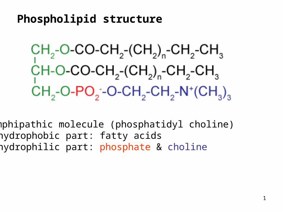
Membrane structure
3
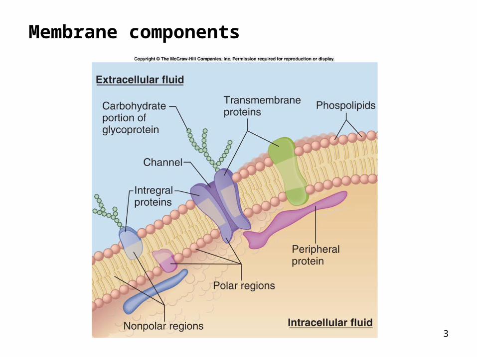
Membrane components
4
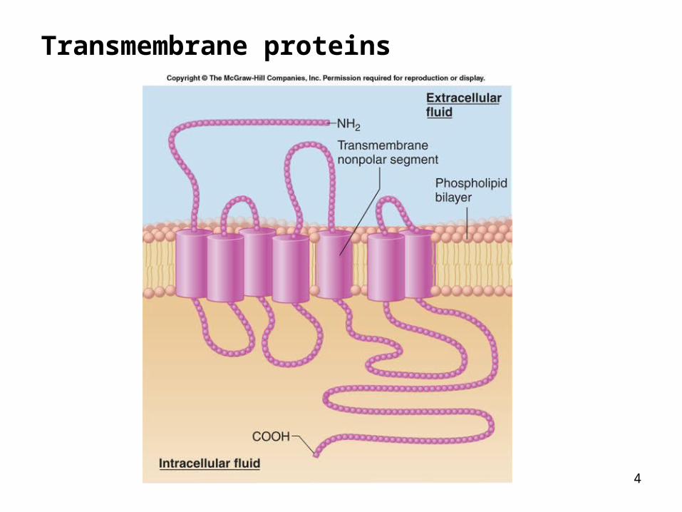
Transmembrane proteins
5
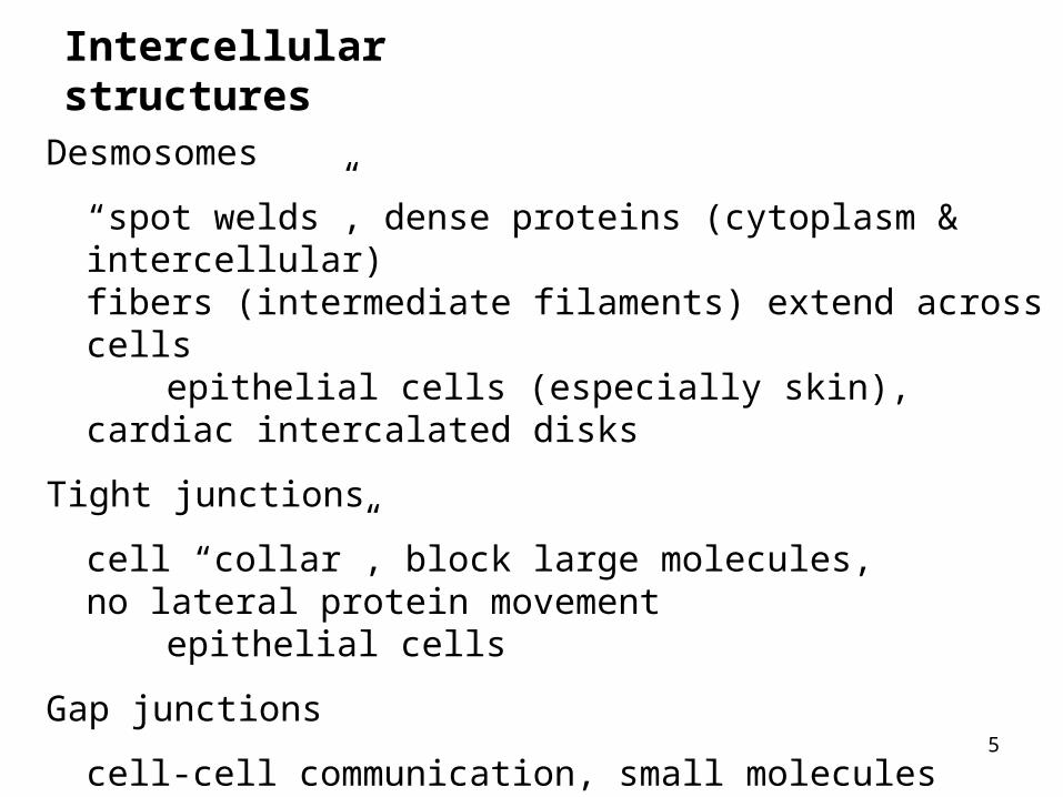
Intercellular structures
Desmosomes
“spot welds”, dense proteins (cytoplasm & intercellular) fibers (intermediate filaments) extend across cells
epithelial cells (especially skin), cardiac intercalated disks
Tight junctions
cell “collar”, block large molecules, no lateral protein movement
epithelial cells
Gap junctions
cell-cell communication, small molecules (<1000 MWt)cardiac intercalated disks, smooth muscle
6
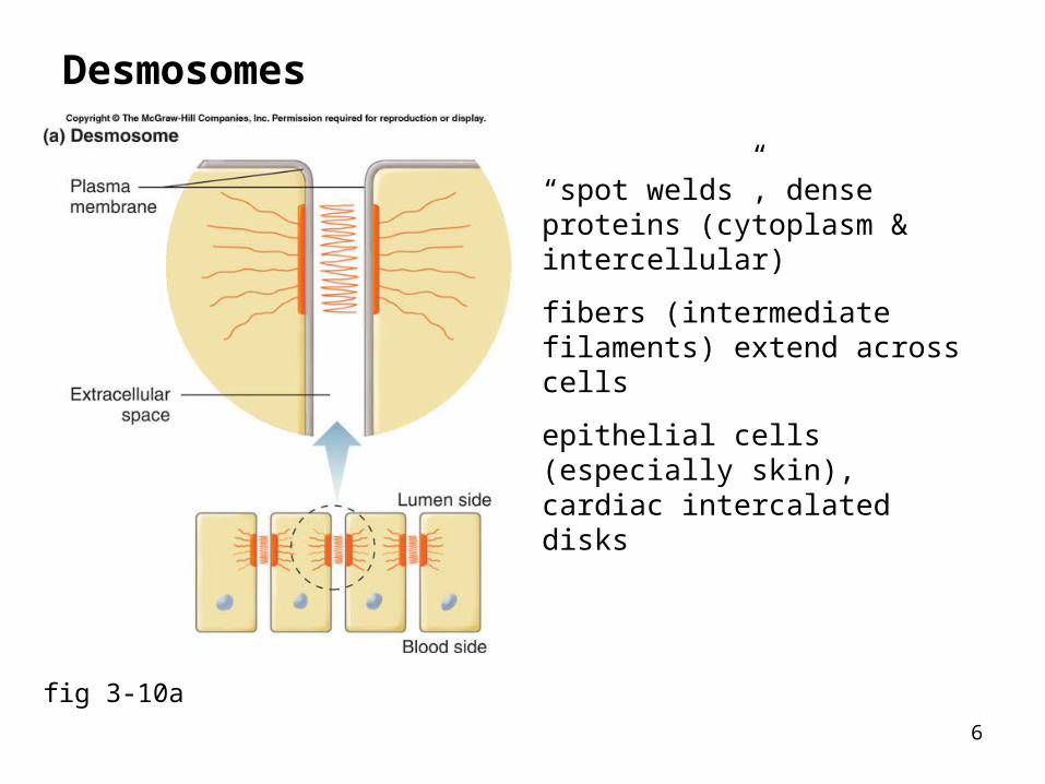
Desmosomes
fig 3-10a
“spot welds”, dense proteins (cytoplasm & intercellular)
fibers (intermediate filaments) extend across cells
epithelial cells (especially skin), cardiac intercalated disks
7
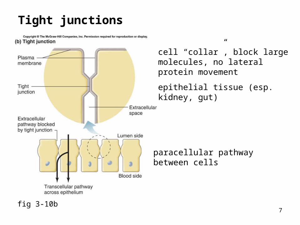
Tight junctions
fig 3-10b
cell “collar”, block large molecules, no lateral protein movement
epithelial tissue (esp. kidney, gut)
paracellular pathwaybetween cells
8
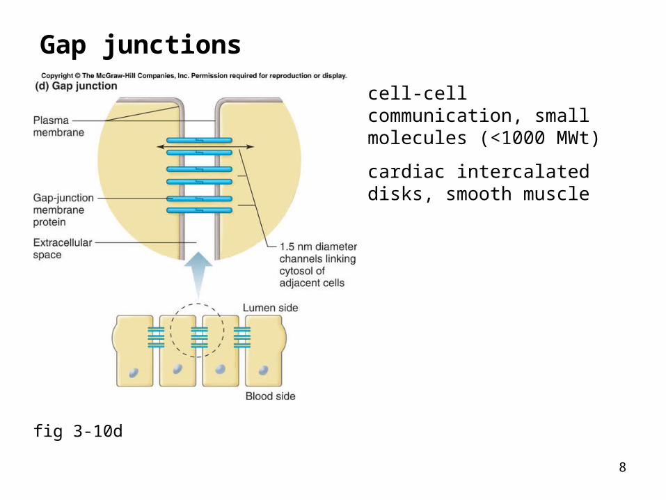
Gap junctions
fig 3-10d
cell-cell communication, small molecules (<1000 MWt)
cardiac intercalated disks, smooth muscle
9
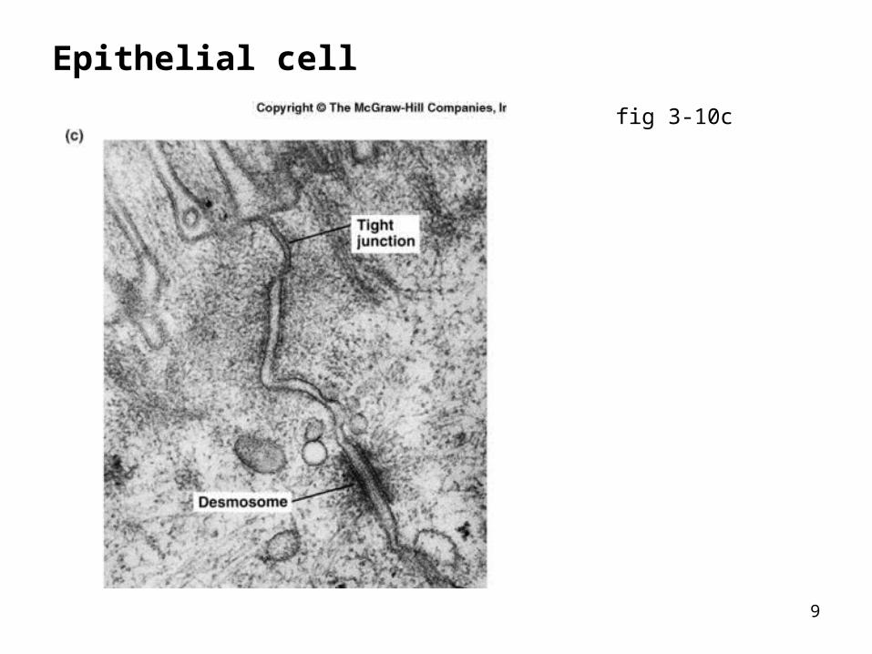
Epithelial cell
fig 3-10c
10
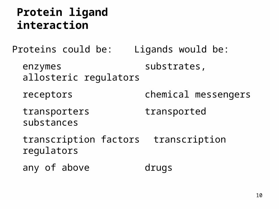
Protein ligand interaction
Proteins could be: Ligands would be:
enzymes substrates, allosteric regulators
receptors chemical messengers
transporters transported substances
transcription factors transcription regulators
any of above drugs

11
Protein-ligand binding properties
Specificity:binding depends on ligand size, shape, charge
Affinity:strength of binding: i.e. [ligand] at 50% binding
Saturation:there is a finite number of binding sites
Competition:structurally similar molecules can compete for binding
12
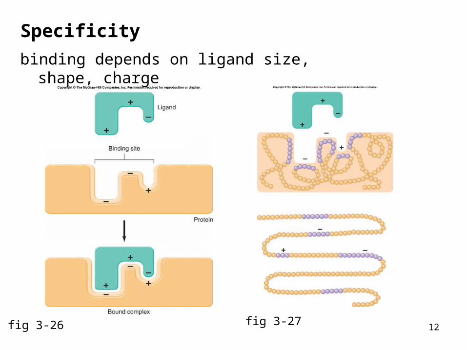
Specificity
binding depends on ligand size, shape, charge
fig 3-26 fig 3-27

13
Specificity
protein Y specificity
greater than
protein X specificity
fig 3-28
14
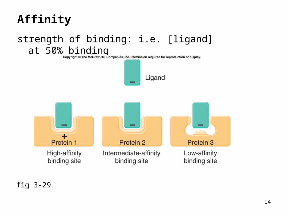
Affinity
strength of binding: i.e. [ligand] at 50% binding
fig 3-29
15
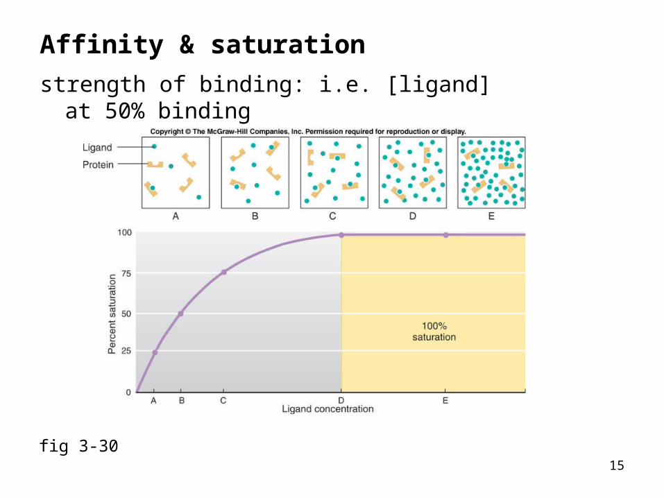
Affinity & saturation
strength of binding: i.e. [ligand] at 50% binding
fig 3-30
16
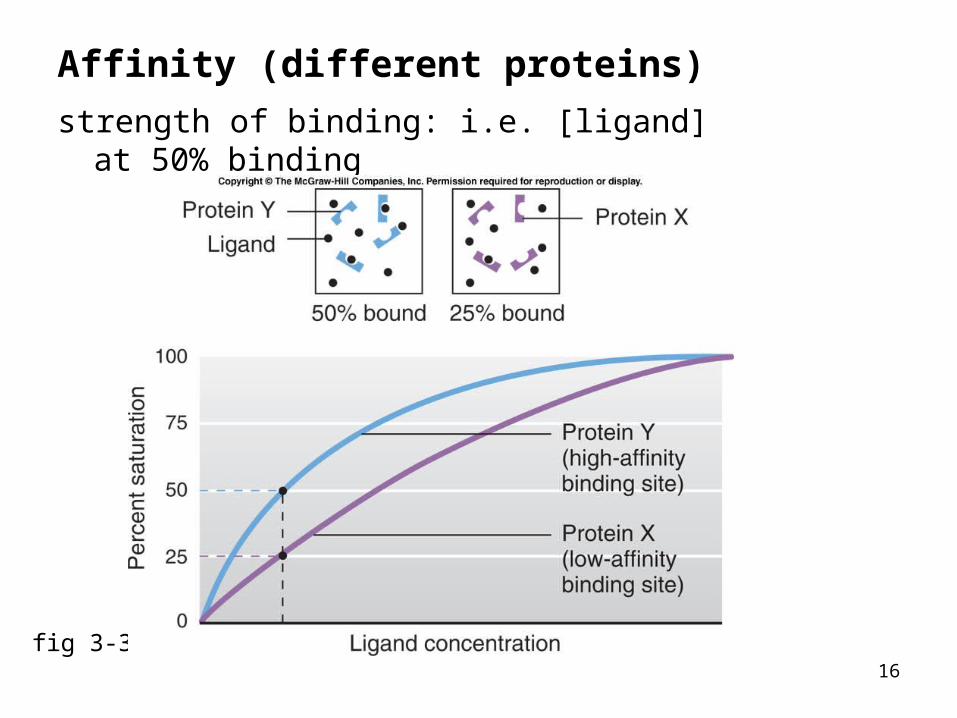
Affinity (different proteins)
strength of binding: i.e. [ligand] at 50% binding
fig 3-31
17
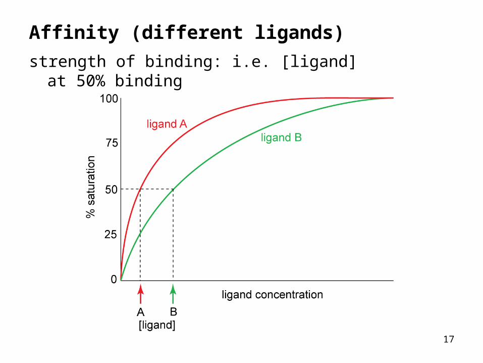
Affinity (different ligands)
strength of binding: i.e. [ligand] at 50% binding
18
Protein-ligand binding properties
Specificity:binding depends on ligand size, shape, charge
Affinity:strength of binding: i.e. [ligand] at 50% binding
Saturation:there is a finite number of binding sites
Competition:structurally similar molecules can compete for binding
and remember:the protein can be an enzyme, receptor, transporter, etc.
19
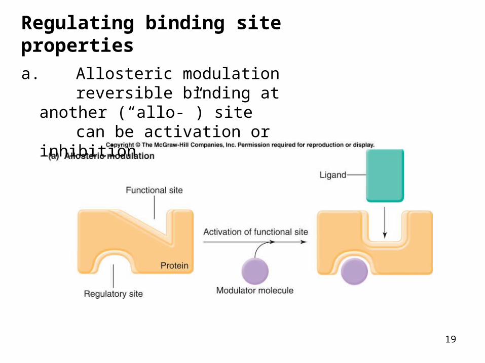
Regulating binding site properties
a. Allosteric modulationreversible binding at another (“allo-”)
sitecan be activation or inhibition
20
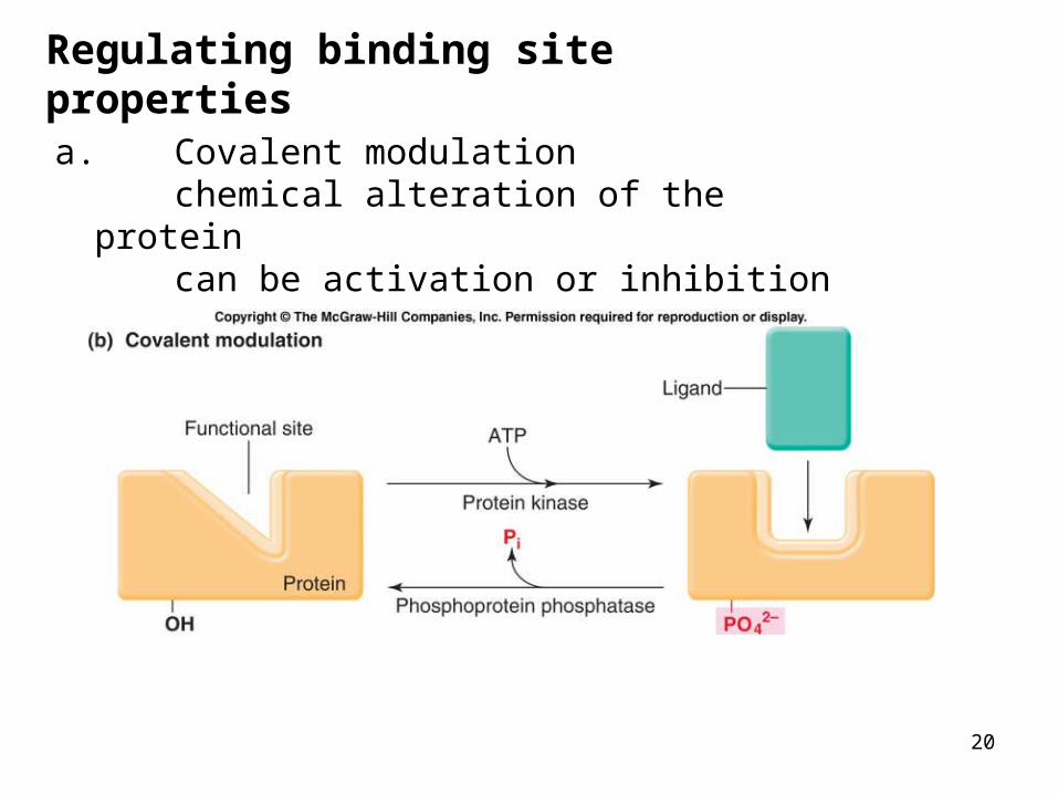
Regulating binding site properties
a. Covalent modulationchemical alteration of the proteincan be activation or inhibition
21
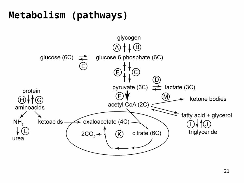
Metabolism (pathways)
22
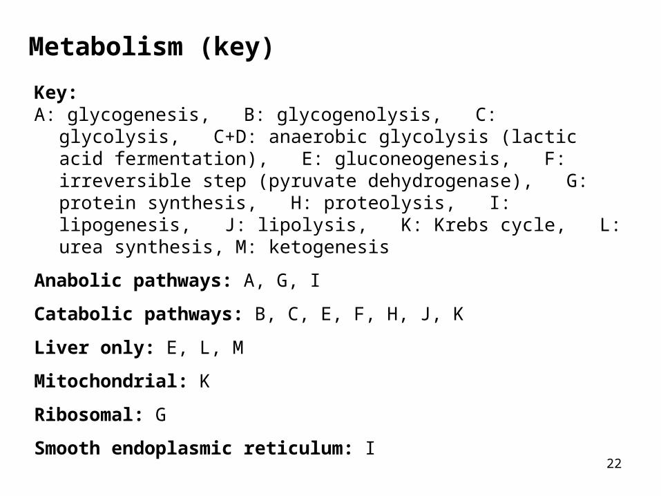
Metabolism (key)
Key:A: glycogenesis, B: glycogenolysis, C: glycolysis, C+D: anaerobic
glycolysis (lactic acid fermentation), E: gluconeogenesis, F: irreversible step (pyruvate dehydrogenase), G: protein synthesis, H: proteolysis, I: lipogenesis, J: lipolysis, K: Krebs cycle, L: urea synthesis, M: ketogenesis
Anabolic pathways: A, G, I
Catabolic pathways: B, C, E, F, H, J, K
Liver only: E, L, M
Mitochondrial: K
Ribosomal: G
Smooth endoplasmic reticulum: I
23
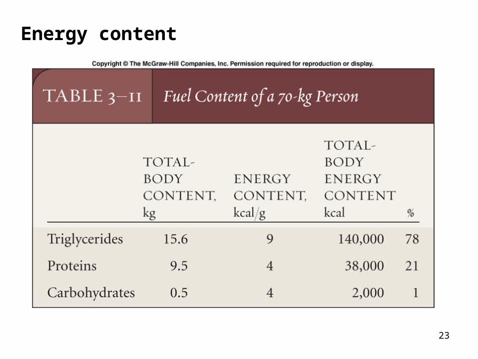
Energy content


![Ultrastructure and lipid composition of detergent-resistant ......sphingomyelin (SM) and dipalmitoyl phosphatidyl choline] were used in these model systems and the composition of these](https://img.pdfslide.net/doc/110x75/601030814f7f4358012f9b10/ultrastructure-and-lipid-composition-of-detergent-resistant-sphingomyelin.jpg)