Embed Size (px)
Citation preview

THE MAINTENANCE OF HUMAN NORMAL CELLS AND TUMORCELLS IN CONTINUOUS CULTURE
1. PRELIMINARY REPORT: CULTIVATION OF MESOBLASTIC TUMORS ANDNORMAL TISSUE AND NOTES ON METHODS OF CULTIVATION 1
GEORGE O. GEY AND MARGARET K. GEY
(From the Tissue Culture Lab oratory, Department oj Surgery, Johns Hopkins Hospital and University, and the Department oj Embryology, Carnegie Institution oj Washington)
In this communication we shall endeavor to present a description of someof the methods that we have used in establishing strains of human normal andtumor cells, and an account of some of our results. We believe that thevalue of this report lies not in the presentation of new and unique methodsfor maintaining tissue cultures under continuous cultivation, but in presentingevidence that very different methods and media can be used to obtain approximately the same end-results. This holds true, however, only when the highlyimportant factor of experience in the management of tissue cultures is usedto best advantage.
The opinion of many workers that human tissue cultures are difficult tomaintain is not justified. Just as much time and energy are involved in themaintenance of a rapidly growing rat sarcoma as in the maintenance of asimilar human tumor. In this laboratory, by the large lying-drop slidemethod, we have maintained the Walker rat sarcoma 338 for a period of overfour years and a half, and a human chondromyxosarcoma (Table IV) forapproximately the same length of time. Strains of cells from these tumorsare still under cultivation.
The advantage of knowing that many different types of human tumor cellscan be maintained under continuous cultivation for long periods of time maynot on first mention become apparent to many. When, however, one considers their use and their importance in collecting radiosensitivity data forspecific cell types, and in the study of their cytological, cultural, and metabolicactivities, the value of such knowledge is readily appreciated. The conceptthat pure cultures of human tumor cells can be used as sources of livingantigen for immunological and therapeutic purposes must not be overlooked.We are inclined to consider this aim as of greater significance than any of theothers.
One of the purposes of this paper is to offer definite evidence that a numberof different types of human tumor cells can be maintained in continuous culture, and to demonstrate the feasibility of maintaining large quantities ofpure malignant cells. We have already demonstrated by the use of the rollertube (Gey, 1) and from culture data (Table IV; E.Fl., R.Br., ].Da., et at.)that this can be done (Plate I). Dr. W. H. Lewis has adopted these methodsfor maintaining cultures of rat tumors and has reported good results (2, 3).
1 Supported by a grant from The International Cancer Research Foundation.45

46 GEORGE O. GEY AND MARGARET K. GEY
In developing technics for the management and handling of tissue cultureswe have found it necessary to devise certain instruments and apparatus whichare not in general use. More detailed reports covering the construction anduse of these specific aids will appear elsewhere.
TECHNIC
1. Transportation and Management of Fresh Tissue
The scarcity of human tumors that are richly cellular and sterile makesit highly important that one resort to the very best methods of preserving thetissue in order that time and effort be not wasted. Considering the generalrun of tumor specimens that are obtained from the operating room one isjustified in stating that fully 75 per cent of them are not suitable for tissueculture.
Although considerable emphasis has been put on the time factor as thereason for the failure of many primary explants to grow, we are inclined tobelieve that other factors are more important. On a number of occasionswe have obtained excellent cultures from tissue which had been stored forseveral days. On one occasion we were able to observe the emigration ofcells from fresh tissue stored for ten days in the ice-box. One reason whytissues do not grow after being stored is that they are contaminated. Excisedtissue fragments should, of course, be explanted as soon as possible. Ifstrict asepsis is carried out in obtaining a specimen, however, there is alwaysa good chance that, even though it is shipped by mail for a considerable distance, cultures can be obtained when it is received. The usual procedure inshipping fresh tissue is to put it in a sterile, air-tight bottle and moisten thetissue with a little of the patient's blood, which is allowed to clot around thespecimen. The use of saline is not to be encouraged, as prolonged washingin saline has a definite deleterious effect on fresh tissue.
2. Primary Explaniation and Manipulation of Cultures
The tissue culture worker is always desirous of getting specimens whichare composed almost wholly of healthy tumor cells. When such specimenscannot be obtained, he must resort to the explanting of a great number offragments, later, after a number of days of incubation, making a selection ofwhole cultures or portions of cultures which are composed chiefly of tumorcells. Rather infrequently he is faced with the problem of establishing a purestrain from one or several small fragments that appear promising. He must,of course, be able to recognize readily the difference between the tumor cellsand the normal cells. In the case of some primary tumors, when a great dealof reactive normal tissue is present, difficulty in recognizing the tumor cellsmay occur.
In the case of most primary tumors the tissue fragments when first explanted do not behave at all like the pure cultures which are established fromthem. Occasionally one is fortunate, and richly cellular, rapidly growingprimary tumors are obtained which in the primary explant behave like manyof the transplantable tumors of animals. Richly cellular primary explants

MAINTENANCE O F CELLS I N CONTINUOUS CULTURE 47
usually show massive emigration of tumor cells and normal cells from the stroma within the first twenty-four hours. Many of the primary tumor frag- ments which are not rich in healthy cells show a rather long lag period. In some instances several weeks are required before rapidly growing cultures are obtained.
In order to insure uniformity in size and selection of fragments for culture, we have found it best to cut up the tissue specimens with a pair of knives which are manipulated in a scissors-like fashion. For this purpose we find the Bard-Parker No. 11 knife to be quite suitable and inexpensive. The fragments selected are cut into 1.5 rnm. cubes, and groups of these are placed in small pools of balanced salt solution or culture fluid mixture, frequently with several changes to get rid of any traces of blood or plasma which might cause the fragments to stick together because of bits of newly formed clot. The fragments are then explanted in large lying-drop cultures or in roller tubes.
3. Primary Explantation to Large Lying-drop Slides
( a ) Selected fragments, usually 5 in number, are taken up in a tissue or plating pipette, and transferred to a small mixing tube (15 X 75 mm.). All excess fluid is then removed with the same pipette. Three drops of the pre- viously mixed fluid portion of the culture medium (for example: balanced salt solution 7 pts.; bovine embryo extract 2 pts.; placental cord serum 3 pts.) are added and the tube shaken momentarily in order to wet and thoroughly mix with the fragments.
(0) Two drops of plasma or plasma mixture (for example: human plasma 6 pts.; chicken plasma 2 pts.) are added and the tube is immediately shaken for a brief moment, during which time a tissue plating pipette is selected from the tube rack.
( G ) The freshly mixed composite medium and the fragments are rapidly taken up into the plating pipette and spread out on large cover glasses (40 X 45 mni.) to form an area about 28 mm. in diameter. A suitable plating and transfer table will be described in a subsequent paper.
( d ) A large depression slide of the Maximow type, spotted with small drops of vaseline, is immediately placed over the culture and the medium allowed to clot. When good visibility is desired, we prefer to use the drilled 2 X 3 inch culture slide chambers made of 3 p a r k 2
( c ) The cover slip is sealed with a sealing mixture composed of paraffine and vaseline (paraffine 6 ; vaseline 1).
( f ) Split rubber tubing skids are slipped over the ends of the culture slides to prevent contact of the seal with the bottom of the tray, and the culture is incubated with the cover slip down.
2 In a SO X 7 5 mm. ( 2 X 3 in.) slide of 1 to 2 mm. thickness a hole is drilled 35 mm. in diameter. With B suitable drilling apparatus this procedure is quite simple and rapid. T o this rlrilled slide is sealed a thinner slide ( 0 5 to 1.0 mm.) or cover glass, paraffine being used as a sealing medium. The cover glass with the culture or another thin slide completes the chamber. Details and preparatiori to be published later.

48 GEORGE 0. G E Y AND M A R G A R E T K. G E Y
4. Trans fe r and Suhrulture f rom Largr Lying-drop Slides
In the manipulation of cultures one must have considerable experience in judging the right time for transfer and for making subcultures. Frequently too generous subculturing causes the production of colonies which become unusually small, are composed of a good deal of hyaline contracted old clot depleted of cells, and which eventually peter out. I n Table IV will be found a few culture histories where this factor has played an important part in the total loss of the culture strain. Also, in transferring, one must learn to cut through the outermost peripheral area of the new growth which is still quite rich in cells. With most tissues it is safe to say that fully 20 per cent of the new growth (Figs. 9 and 9a) is lost in each transfer when the slide technic is used. For excising and subculturing the colonies we use a modified Bard- Parker No. 11 knife, which will be described elsewhere.
A most important factor in excising the tissue culture colonies is the avoidance of tags of clot or old medium. Usually no growth (Fig. 10) occurs in the area where the old clot is attached. More recently we have used the trypsin digestion procedure recommended by Vogelaar and Erlichman (4 ) for the digestion of old clots from the walls of roller tubes, and with promis- ing results. This procedure is not to be encouraged in the slide method, as proper care in excising the colonies is still the most reliable procedure in the hands of the experienced worker.
Procedures for transfer and subculture of slides may be briefly outlined as follows:
( 1) Immediately after removal of cultures from the incubator invert them in such a manner as to prevent a (‘ run-off ” of the liquefaction fluid which has formed. When a large liquefaction area is present and there is a large liquefaction droplet, it is always best to distribute the fluid by a roundabout rocking movement over the area covered by the culture medium, and to invert immediately. In this way no (‘ run-off ” will occur, even with a large drop.
( 2 ) With a small spatula lift up one corner of the hermetic seal; pull it off while holding the cover glass down with the spatula. When the sealing mixture is properly made up, and is still warm, it should come off easily. The seal of a slide that is cool must be chipped off, with the liability of cover slip breakage and of loss of culture.
( 3 ) Place the cover slip on a suitable microchirurgical tissue culture table or cutting block and cover.
( 4 ) Observe the colonies carefully and check and compare the gross observation of the extent of outgrowth with the microscopic notes previously made on the record form (Fig. 1).
( 5 ) Select a knife from the clean alcohol storage tube of the dry-heat rapid sterilizer and burnish and dry off the blade on a sterile cloth wiping pad held in a suitable dish. Excise the colony selected by a tip-to-base shearing maneuver of the knife blade. Make as many shearing cuts (Fig. 9 ) as will adequately remove the colony with a minimal loss of new growth. Be careful not to leave any tags (Fig. 10) of old medium.
( 6 ) \Irith the tip of the knife lift out the colony and place it in the cavity i Iktai ls to be published Inter.
MAINTENANCE O F CELLS I N CONTINUOUS CULTURE 49
of a depression dish' to which has been added previously the fluid portion of the medium to be used (example: 3 drops of the mixture, balanced salt solution 7 pts., bovine embryo extract 2 pts., placental cord serum 3 pts.).
( 7 ) Repeat the same excising procedure for other colonies on the cover glass. Cutting blocks which have a revolving vacuum-clutch plate ' make it possible to excise fragments without touching the culture side of the cover glass.
(8) Set up a group of clean cover glasses on a suitable plating table. For very thin cover slips a vacuum-clutch plate for holding the cover slip is best. Add a suitable quantity of plasma or plasma mixture (example: 2 drops of the plasma mixture, human plasma 6 pts.; chicken plasma 2 pts.) to the fluid and tissue colonies in the cavity of the depression dish.
and pull the fluid and colonies up into the constricted portion of the pipette several times to insure good mixing and immediately plate out to form a circular area about 28 mm. in diameter, a t the same time orienting the fragments. Withdraw sufficient medium so that the colonies are not completely immersed.
(10) When subculture is necessary, the excised colonies can either be bisected or the peripheral area of new outgrowth cut away from the central area. This can be done either in separate dishes with the aid of a microscope or binocular loupe or, as is the usual procedure, on the same cover slip imme- diately after the fragment is excised from the culture medium.
(11) For the transfer of cell colonies which rapidly liquefy most media, transfer can be effected by routinely pipetting off the floating cells and trans- ferring the clumps and free cells.
(9) Select a tissue or plating pipette
5. Primary Explantation into Rollcr Tubcs
The plain straight roller tube has been used by Lewis (3) and is satisfactory for most purposes. We have maintained many cultures in round Pyrex tubes, 16 X 150 mm. About the only objection to them is their circular form, which prohibits examination of uniformly flat fields. The hexagonal tube which has been described (1) offers distinct advantages with its flat sides permitting good visualization and with its somewhat constricted neck prevent- ing overflow and providing uniform distribution of fluid during incubation. These tubes have been perfected to such an extent that they can be made with walls of uniform thickness permitting observation under moderately high power.4
Inasmuch as a large number of fragments (30-50) can be explanted in one tube, it is highly important that this procedure be planned in such a way that there is a minimal loss of time. Otherwise, especially with fresh tissues which are rich in thromboplastic substances, premature clotting will occur long before this step is complete. T o avoid any such accident it would seem advisable to use plasma to which an additional amount of heparin has been added in order to delay clotting.
Roller tubes of different types have been used with good results.
4 The depression dish, vacuum-clutch plate, plating pipette and the molding apparatus for mak- ing the roller tubes will be described in a subsequent paper.

.5 0 GEORGE 0. GEY AND MARGARET K. GEY
When blood is taken for plasma into an oiled syringe, care must be taken that as little oil as possible be transferred when the plasma is taken off. Oil droplets which have been carried over by plasma interfere with the uniform wetting of the roller tube by the plasma. This precaution is highly important in order to insure a uniformly thin spread of medium that will adhere. The following procedure is one of several which have been routinely used with fairly good results.
In the preparation of these tubes, no cotton plugging is used, as this introduces particles of lint. Inverted tubes can be adequately and safely handled in clean sterilizing cans.
( h ) From a cold tube of plasma or plasma mixture (chicken plasma 2 pts.; human plasma 6 pts.) add 4 drops to the roller tube, and with the blunt side of the same pipette, or a special spreader, distribute the plasma through- out the tube. Complete and maintain the spread by gentle rolling movements.
(c ) With a curved-tip tissue or transfer pipette, select the number of fragments desired and concentrate them in the tip end of the pipette, a t the same time getting rid of as much of the pooling fluid as possible. Place these selected fragments in the upper portion of the tube where no plasma has been spread. If the tube is held in an almost horizontal position, the plasma and the fluid about the fragments will not run together. Remove all of the wash fluid about the fragments. With a platinum-iridium needle, or with the same pipette which was used in transferring the fragments, orient them as rapidly as possible, a t the same time allowing the excess of plasma to accumulate in the bottom of the tube.
( d ) With the tube upright add immediately 6 drops of a fluid mixture ‘( 7-2-3 ” ( i . ~ . balanced salt solution 7 pts.; bovine extract 2 pts.; placental cord serum 3 pts.) or whatever combination is desired. Shake gently for a very brief moment to allow admixture with the plasma which has collected at the bottom of the tube, and with a rolling and tipping movement gently dis- tribute the composite medium over and between the fragments, many of which will still remain in place. Quickly re-orient the fragments which have moved and, with the same transfer pipette used for explanting the fragments, remove all excess of the composite medium which drains as the tube is elevated, thus leaving the fragments fixed to the wall and held mostly by capillarity. In this way a very thin film of medium is obtained. A thicker layer of medium can be obtained by continuing to turn the tube with all of the medium in it until clotting occurs.
Continue to turn the tube until clotting is complete. Frequently it is only necessary to set the tube aside, slightly inclined so as to allow excess fluid to drain to the bottom or to a portion of the side wall where there are few or no fragments.
( f ) Add 1 2 to 14 drops of “ 7--2-3 ” fluid mixture or the expressed fluid from a clotted composite medium.
(g) Stopper with rubber stopper and label both tube and stopper. By it gentle rolling and tipping motion distribute the fluid over the clot and frag- ments. Sometimes it is quite difficult to get good wetting of a newly fornied clot. To insure good distribution of supernatant fluid, the same maneuver for wetting should be repeated sometime later, after incubation has begun.
( u ) Select from a sterilizing can a clean tube,
( c ) Place the tube on a suitable rack or grooved plate,

MAINTENANCE O F CELLS I N CONTINUOUS CULTURE 51
Rough nianipulation will unloose the clot quite readily. Incubate in a tube drum or rack which fits into a reiiable incubator. ,4 drum speed of 10 revo- lutions per hour has been found to be quite satisfactory. Suitable apparatus has been described by Gey ( 1 ) and Lewis ( 2 ) .
More recently we have been inclined to the view that shorter tubes are less cumbersome to use and give as good or better results. They require less than half the amount of medium specified in the above typical set-up. In describing a procedure of this sort it is most difficult to make specific recom- mendations. Most workers, in spite of recommendations, are inclined to develop their own methods which are adapted to their particular needs.
6 . Fluid Changes, Patching, Trans fcr, and Subculturing o j Roller Tubes
Fluid Chunges: During primary explantation the entire procedure must be so designed that cross contamination between tubes is limited to the tissue fragments from the same source. All subsequent procedures following pri- mary explantation should be so designed that there is no possible cross con- tamination between tubes. Even when microscopic examination reveals no evidence of contamination, each tube should be treated separately. After approximately four days of incubation the fluid mixture is removed and new fluid mixture added, either following a saline rinse or without it, as desired. Sometimes the fluid change is not the first procedure following explantation, as very frequently liquefaction areas form about some of the fragments within a few days. The use of a good deal of chicken plasma helps to prevent early liquefaction, but sometimes inhibits good early growth when used in high concentration.
Putching: Following removal of the fluid mixture, the tubes are usually patched with a half quantity of medium. This can be done in either of two ways: by patching with plasma added first to wet the old clot, followed by the addition of the fluid mixture, or by patching with the composite medium. In either case patching seems most effectual following careful rinsing with balanced salt solution. For some tissues patching and fluid changes can be carried out at specified intervals. For most tissues, however, the roller tube cultures should be treated in accordance with their requirements. When the fluid mixture is added to a patched tube, particular care must be taken to prevent the loosening of the clot. From the histories of a great number of roller tube cultures we have found it advisable to add fluid and to patch at alternate intervals of three to four days. Some cultures of freshly explanted tissue require daily patching a number of times for the establishment of good colonies.
Transjw and Subculture: The rapidity of liquefaction, the number of patches that have been made, the metabolic state and appearance of the colonies and cells determine the need for transfer or subculture. In Plate I is shown a group of cultures, Figs. 1-6, which were obtained from two tubes of the chondromyxosarconia strain, J.Ua., maintained 1490 days at the time the transfer was made and approximately 30 days after the last transfer. The procedure followed in making the transfer and subcultures in this case was to remove only the central portion of the culture that did not seem to be

52 GEORGE 0. GEY AND MARGARET I<. GEY
doing well, and to patch the remaining peripheral areas. In this instance the cultures were growing rather slowly in the medium and transfer was necessary. For other strains of cells entirely different results and treatment may have been necessary in a similar period. For some types of cells it is not unusual to find the entire tube (Fig. 11) filled with new colonies which have spread from the primary or transplanted secondary colonies. In the case of the Walker rat sarcoma 338, this kind of spread occurs in some media. With each type one must resort to a certain amount of experimentation in order to obtain the best results.
With the exception of a few cell types, most colonies of cells can be trans- ferred and subcultured, following the same general scheme used in primary explantation. The unusual precautions necessary to prevent premature clot- ting need not be followed in making transfers, as niost cell colonies once established have considerably less thromboplastic activity than fresh tissue.
7. Tho Significancc of Prrcisc Technics
One of the chief advantages of pure strains of cells maintained in con- tinuous culture is the uniformity in the behavior and activity of such cul- tured cells. When grown in media as nearly optimal as can be obtained and by suitable technics that can be consistently repeated, they constitute a con- stantly available source of uniform biologic material, becoming base-line con- trols for various experimental procedures. Such pure strains are as impor- tant to most of the experimental work done with tissue cultures as the pure strains of mice developed by the geneticist and used as his standard sources of biologic material. The results of work done in two different fields of scientific endeavor-that of Carrel in tissue culture and of Little in genetics, and of their followers-give added emphasis to the truth of these premises.
Although most of the procedures used in the management of tissue cultures are fairly complicated and must be carried out with a fair degree of precision of manipulation, and within fairly definite time limits, with suitable equipment each procedure can, nevertheless, be carried out wholly by one person.
The importance of experience, as the primary requisite for the maintenance rind establishment of successful cultures, cannot be over-estimated. In carry- ing out any tissue culture procedure, one must fully realize the multitude of influences which can injure the cells and alter their activity unfavorably. We have observed repeatedly that the best results seem to have been obtained by those who acquire a certain devotion to their work. The prudent and energetic worker is usually rewarded by a certain measure of good results which repay him fully for his efforts. Such an attitude must become con- tagious for the assistants, who should be imbued with the same standards of workmanship. We are greatly indebted to our assistants, Wm. Fitzwilliam and Thomas H. Stark, for their untiring efforts and devotion to their work over a period of years.

PLATE 1
ROLLER TUBE CKJLTURES OF HUMAN SARCOMA, STRAIN J.DA. MAINTAINED IN MEDIUM
(8 CHWKEN PLASMA) FOR 11 DAYS (SEE TABLE IV) 7-2-3-8 (3 ClrICKEN PLASMA; 5 HUMAN PLASMA) FOR 29 DAYS AND I N 7-2-3-8
1. Tube D-13, 1400 day old strain. In tube 29 days. 2 . Tube D-14, 1490 day old strain. In tube 29 days. 3. Tube D-15, 1501 day old strain. In tube 11 days. 4. Tube D-16, 1501 day old strain. I n tube 11 days.
areas of colonies of D--l3, without transfcr. following last transfer.
5. Tube 1)-17, 1501 day old strain. 6. Tube D-18, 1501 day old strain.
areas of colonies of D--14, without transfer. following last transfer.
This tube shows patched peripheral Note tendcncy for accumulation of old clot 40 days
In tube 20 days. In tube 29 days. This tube shows patched peripheral
Note tendency for accumulation of old clot 40 days
53

54
Na K C;1 Mg c1 (Serum) (Serum) (Serum) (Serum) (Plasma)
146 5.0 1.35 1.1 104 ~ ___ -~ ____ _ _ _ ~
GEORGE 0. GEY A N D M A R G A R E T K. G E Y
H C0:j PO4 SO, (I’lasnia) (Plasiiin) (Serurn)
2 7 1 .o 0.3 _____
MEDIA 1. Importance of Mcdiu of Known Composition
We are well aware that most of the media with which we have maintained cells in culture for long periods of time were possibly not optimal for growth. In Table IV are recorded a few of many variations in the medium which we have attempted to introduce in order to get good results. The reasons for the loss of some strains after they were established are clearly indicated.
Strict uniformity in the behavior of pure strains cannot be obtained so long as we must resort to varying biological sources for our supply of cell foodstuffs. A number of workers, among whom we are included, have at- tempted to prepare media which were erroneously designated ‘‘ artificial ” but on last analysis fell far short of being strictly artificial or synthetic. The results, though poor in comparison to results obtained by the usual proce- dures, were sufficiently encouraging (26) to justify the conclusion that a great (leal more work should be carried out in this field. At present, however, we must await the day when pure substances are made available in order to obtain strictly uniform results. At that stage we shall be able, to assume the r61e of the technocrat and design the automatic culture machine for the nianipula- tion, plating, and incubation of cultures. Until then we must remain content to use what is available, i.e., our own complicated products and those from animal sources.
2. Balanccd Salt Solution
In view of the important unphysiological properties of many of the so- called “ normal ” solutions which have been used in testing the responses of excised organs and tissues, an effort was made in connection with work done some time ago ( 5 , 6 ) to improve these solutions. An extensive review of the available literature revealed that even the most commonly used solutions of Ringer ( 7 ) , Locke (8), Hedon and Fleig (9) , Adler ( l o ) , and Tyrode ( 11 ) , as well as a host of others, were unphysiologic in many respects. Prom the literature on the analyses of blood, blood plasma, serum, lymph, and other body fluids, the various values of the chief inorganic ions (Table I) were
estimated for human plasma and serum only, as the results of analyses of these two were quite consistent. From these values a balanced physiologic solution was made up (Table 11).
The solution recommended must be used in equilibrium with a physiologic pressure of CO,. In many respects it is similar to the one described by van Dyke and Hastings ( 1 2 ) . We have used this solution with good results for cultures maintained in slides and flasks and to a limited extent in the roller

MAINTENANCE OF CELLS I N CONTINUOUS CULTURE 5.5
tube. However, one must have adequate facilities in order to use such a solution exclusively, as its proper use adds a considerable burden to routine cultural procedure with a large number of cultures. Several years ago we developed an adequate technic and designed suitable apparatus ' for main- taining cultures under controlled gas pressures. Up to the present the method has not been put to full use. Similar apparatus has been described by Earle (13) and Schade (14).
For work with slide cultures we use a somewhat similar solution to the one described, but in which the NaHCO, concentration is depleted to about one-tenth of its average millimolar concentration. This is done for solutions used in slide culture work, inasmuch as most of the procedures with slides are carried out under ordinary atmospheric conditions.
T.WI.E I1 1 Balanied Salt Solulion Recommended for 12quilibration with 5 Per Cent COz ~ - _. - - _.
Constituents
NaCl KCI NaHCO,,
'CaC12 (anhyd.)
' Na2HP04 X 2H20 MgClz X 6Hp0
KH2P04 hlgSOi X 7Hn0 d . Glucose-1.0 c.--weieh out
Concen- t ra tion
7.0 0.37 2.27 0.17 0.21 0.15 0.03 0.07
,esh.
.~ .- I
Stock Solution
Mol. coric.
5.0 1 .0 1 .0 0.1 1 .o 0.5 1 .0 0.1
~
gll
272.25 74.55 84.01 11.1
203.33 89.03
136.14 24.65
Quantities for H.S.S.
nil. per liter
19.2 5.0
27.0 13.5
1 .0 1.6 0.2 3.0
-_
Note: Add in the order given to freshly distilled water through which COZ has been passed. For slide cultures plated under ordinary atmospheric conditions use Sterilize by filtration.
1/10 the amount of NaHCOI and increase the NaCl to 8.0 g. per liter.
When changes in the concentration of any one ion are desired, these changes should be compensated for by changing the sodium chloride concen- tration. Such a change should have no deleterious influence, inasmuch as it has been shown repeatedly that considerable variations in the sodium chloride concentration have little or no effect on the vitality of living cells ( 1 5 ) . In Table I11 are presented the calcium and phosphorus values of a number of different combinations of the composite medium when high (50 per cent) and low ratios of the various ingredients are used. As will be noted, most of the media that we use are made up on a 20 parts basis. Single unit variations, therefore, represent changes of a magnitude of 5 per cent. The possibility that specific changes in the inorganic ion concentration of the entire medium would greatly improve it must not be lost sight of, especially since the whole question of the r81e of certain ions in tissue vitality and growth is still a con- troversial one.
I t is difficult to make specific recommendations when the proper facts are not available. A number of workers have studied the effects of varying the ionic environment of normal ( 2 7 ) and neoplastic tissue cultures in vitro. The
j A full dcicription of this apparatus and it5 ube will appear later.

5 6 GEORGE 0. GEY AND MARGARET K. GEY
12.0
evidence presented by Roffo ( l h ) , for example, that cultures of neoplastic tissue in z d r o are inhibited by concentrations of calcium salts which do not inhibit the growth of normal (embryonic) cultures may not hold true when such cultures are maintained as continztous cultures over long periods. Un- less cultures are maintained for long periods of time, the delayed effects on cells which many agents (radium, x-ray, et al) exhibit will not become ap- parent.
Tini,~: 111: Cumposition of Various Media
__ ~ _ _ 4.8 3.9 4.0
___
I't s . ~ ~~ ~ - _ _ _
ILilanced s,ilt solution 10 Hovine embryo extract 0 I'lacent,il serum 2 1 luman pl'lsIl1,l 8
('a 1 9.6
I' 1 4.0 7.0 ~ 4.3 5.1
__
I'tS.
7 2 3 83
11.3
4.1
Values for Single Constituents
I'ts. = Parts to make a total of 20. In this medium 2 p'irts of hunian and 1 part of chicken plasma were used.
Multiplied by 5 equals percentage composition.
3 . Tissuc Extract
Those who have attempted to maintain tissue d t u r e s in an actively growing condition for extended periods of time realize the great importance of a readily available source of tissue extract. For this reason extracts of chick embryos have been used a great deal. In an earlier publication ( 1 7 ) we reported that chick extract did not support the growth of human fibroblasts and epithelium indefinitely. Most of the cultures in which it was used could be maintained only about a month. The possibility that more suitable ex- tracts could be prepared from pig and bovine embryos obtained from a local abattoir," was considered. Pig extract was found to be definitely better than chick extract, allowing a much longer period of maintenance, but no cultures could be kept longer than three months. Chick and pig extract always pro- duced cells with an increasing accumulation of fat globules. Bovine embryo extract permitted the maintenance of cultures for much longer periods and in addition seemed to make the cultures grow more rapidly and to maintain in them much healthier looking cells more or less free of fat. In these earlier experiments we were able to maintain embryonic human fibroblasts in a very active state for a period of seven months (Fig. 9a). Further maintenance was interrupted 'through accidental loss. Cultures when maintained by the large (2s nim. spread) lying-drop method showed a very brief lag period and usually in two hours a good many cells had already migrated from the colony.
For the past eleven years we have used bovine extract almost exclusively as the chief source of nitrogenous foodstuff and growth-promoting agent for the
We a r e greatly indebted to The Wm. Schluderberg-T. J. Kurdlc Co. for their generous co- opcr,ition in m;ibing such material available to this laboratory.

MAINTENANCE O F CELLS IN CONTINUOUS CULTURE 57
maintenance of human cells. On a number of occasions we have attempted to stabilize the extract by desiccation and by freezing, with variable results. Fairly convincing evidence that desiccated extract is suitable for periods up to two months is found in the maintenance records (Table I V ) of strains A.Ha.I., L.Ti., D.Au., E.Ke.I., J.Ma., and L).Pr., which were kept from 31 to 54 days in nitro. These same cultures transferred to fresh mouse extract petered out in from 16 to 69 days in nitro. The use of mouse extract had to be resorted to, as we were trying to maintain a series of cultures in a summer laboratory."
From the above statements one might be led to believe that some form of tissue extract is an indispensable ingredient for human normal cells and tumor cells. As a matter of fact, a number of workers have reported the cultivation of different types of cells of several species in media containing 110 extract and in which serum was used. We also have had good results with some types of cells in media in which serum!' was the principal ingredient. For most tissues, however, the best results are obtained by the use of a com- posite medium containing both serum and extract as the chief ingredients. Krontowski (18) and others have reported good results with a composite medium. More recently Lewis ( 3 ) , using chiefly the composite medium, has obtained good results with rat tumors maintained in roller tubes. The addi- tion of other agents (various carbohydrates and salts and specific substances, as thyroxin, insulin (231 , and other products of cell metabolism) does not markedly improve the medium.
The following brief outline covers the most important steps in the prepara- tion of the extract.
1 . Careful selection of gravid uteri with intact embryos two or three months of age and with membranes unruptured.
2 . Removal of the embryo under strict aseptic conditions on the same day. ( u ) The uterus is palpated to determine the position of the embryo.
The distended amniotic sac containing the embryo is gently held close to the wall of the uterus and the whole amnion clamped off with a special clamp used for this purpose, somewhat similar to Payr's pyloric clamp. A suitable, divisible metal shield is then placed just beneath the isolated amnion and uterine segment to prevent tipping.
( b ) The uterine wall is sterilized twice with iodine and alcohol, each application bcing followed by free flaming with a Bunsen burner. 7 A number of preparations of freshly prepared bovine extract were desiccated by a procedure
essentially similar to one described by Flosdorf and Mudd ( 2 5 ) . Sonic of these preparations made from December 1930 to May 1931 were stored in the dark in sealed pyres tubcs. Storage in an atmosphere of nitrogen and in vnc'uo was tried. Experiments are being conducted at the present time on the activity of such preparations, most of which are now live years old.
We are greatly indebtcd to Ur. C. C. Little! who supplied us with suitable material a t the time this work was done.
Using stock cultures of Walker rat sarcoma 319, a small group of experiments was set up in a.hich hunian fetal serum and equine serum diluted 1 to 3 with balanced salt solution, werc used 3s the sole source of medium for the maintenance of these cells. In the medium containing diluted human fetal serum active colonies were maintaincd for several weeks and lost by contamination. In the medium containiiig diluted horse serum active colonies were kept growing for 125 days and lost in a n overhcated incubator. Maintenance in both series was by the roller tube method, the colonies jirowing on the plain glass wall.

58 GEORGE 0. GEY A N D MARGARET K. GEY
( r ) Hysterotomy is carried out in two stages in order to permit a sec- ond sterilization of the submucous layer with iodine and alcohol before presentation of the amniotic sac occurs. Sterilization of the amnion should not be quite as vigorous, as it is quite thin and easily ruptured. In order to prevent collapse of the incised uterine segment the edges of the uterine wall are clamped with a retaining clamp so as to hold them open and in place, and also help support the delicate and bulging amniotic sac.
( d ) A long-shanked scissors is then used to nick the amnion and while the amniotic fluid is flowing out of the hole thus made, a long forceps is inserted, the previously oriented head grasped, the embryo delivered, and the cord cut in successive maneuvers. The embryo is immediately placed in a suitable sterile dish and is ready for use. If these procedures are carried out correctly, the embryo will at no time touch anything except the edge of the amnion and will be continuously bathed by amniotic fluid during its removal. 3. Preparation of a fine embryo pulp with a suitable apparatus. 4. Dilution of pulp with an equal volume of balanced salt solution and
thorough mixing. 5, Separation of embryo juice by centrifugation twice, the first centrifu-
gation to get rid of the greater part of the fat and solid tissue, and the second to insure getting a cell free extract. In the second centrifugation we use a tightly packed sedimenting cotton filter inserted in the tube.
6 . Storage in an equilibrating jar under a physiologic pressure of carbon dioxide.
Bovine embryo extract so prepared can be used ten days or more with good results. Only occasionally do we use extract that is over a week old. As a precautionary measure, bouillon cultures are always made in order to detect contamination. To insure mairitaining clear media, all the ingredients are centrifuged imme- diately before using them.
‘The equilibrated extract (50 per cent), when ready for use, has a pH of 6.2, a calcium concentration of 10.5, and a phosphorus concentration of 12-14 (mg./100 c.c.). Just what effect a high phosphorus concentration has on cells in culture we are not prepared to say. As will be noted from the table of tissues cultured (Table IV) and the table of calcium and phosphorus values of the various media (Table H I ) , fairly high concentrations of extract, having a high concentration of phosphorus, have maintained tissue in a very active state for long periods of time. We have a good deal of evidence, however, especially in more recent work, that media containing a high concentration of extract do not give the best results. This seems to be especially true of freshly explanted tissues.
The percentage values for extract in the culture medium (Table IV) rec- ’ommended for a number of different tissues, range from 5 to 35 per cent (2.5 to 17.5 per cent of embryo derivative). More specific recommendations, however, based on the best average results for all types, show that extract concentrations of about 10 to 1.5 per cent are nearly optimal for most tissues under continuous cultivation. These values are somewhat lower than those recommended by most workers.
In this laboratory it is prepared several times a week.

MAINTENANCE O F CELLS I N CONTINUOUS CULTURE 59
4. Serum With the evidence presented by Carrel (19) and his coworkers that serum
from old individuals exerts a somewhat inhibitory effect on cells in culture, we were led some time ago to seek a source of serum which did not contain an inhibiting agent. Serum prepared from blood obtained from fresh human placentae was found to be the most suitable for this purpose. For the past twelve years we have used this placental (fetal) serum with very good results ( 17) . Theoretically it should not contain growth-inhibiting substances of the type discussed by Carrel, and we have a great deal of evidence that it contains growth-sustaining substances. The histories of a number of cultures show fairly long periods of maintenance in media containing fairly high concentra- tions of placental serum. The culture strain N.Du. (Table I V ) shows a maintenance of 125 days in a medium containing 35 per cent of placental serum. In data obtained from a number of other culture histories (to be published later) very definite evidence has been obtained that homologous and heterologous serum will support the growth of a number of different types of cells for long periods of time. Other workers have reported good results with other sera (20. 2 1).
The routine collection of placental serum requires the full cooperation of those attending the delivery of the patient from whom the placental blood is to be taken. As a rule, no difficulty is encountered, as the few requirements for obtaining sterile specimens of blood do not interfere with hospital routine. The following brief outline covers the essential steps.
1. When the cord is cut the placental cord clamp should not be removed so as to allow loss of blood from the placenta.
2. When the placenta is delivered it is set aside in a suitable pan and covered.
3. Within twenty minutes a large vein is properly exposed, swabbed with iodine and alcohol, and the blood in the distended cord milked back only once into the placenta by a single maneuver; the cord is then reclamped at the base.
4. A distended vein is punctured immediately and blood withdrawn slowly until the veins are well collapsed. No effort should be made to redistribute the blood in the placenta manually in order to get still more blood, as this introduces a possibility of contamination. Large needles of about 16 B & S gauge are best for this purpose in order to allow the passage of minute clots which are frequently encountered, especially in placentae that have been allowed to stand for a short time. On an average one should be able to get from 60 to 80 C.C. of blood.
Best results are obtained when it is allowed to stand for at least one hour, after which time the clot should be freed from the tube and the tube again set aside in an ice box to allow some syneresis to occur.
6. The blood is centrifuged at 3000 r.p.m. for at least fifteen minutes, the serum removed, and a second centrifugation done for five minutes to insure removal of all cells, Bouillon cultures are made as a check on possible bacterial contamination.
In this case only 5 per cent of extract was used.
5. Placental blood does not yield a great deal of serum.

00 GEORGE 0. GEY A N D MARGARET K. GEY
7. The serum is stored in an equilibrating jar under physiologic pressure of carbon dioxide.
5. Plasma
The old idea that the culture medium must supply a microscopic support- ing framework on which cells could grow, and that this framework constituted it tropismic stimulus to growth, is rapidly losing ground. In our culture his- tories of several different types of tumors both from human and animal sources, we can find records of fairly long continuous maintenance of cultures in media containing no plasma."' Tumors which produce colonies of compact cells with very little tendency to cell separation during active migration grow quite well in media containing no plasma. Cultures made from the sarcoma strains
&D. No. Set U p (date) TISSUE CUL
............
............ ................. .................. ...... Mo _._..
RD Saecimen No. Dr. Diag.
E y t - ........ mn nlLm ........... I.... tom. .. p k .......... p 4 ........... mpl ...........
...... CA ............ m i L ........ _....._ Pbnt.
. . . . . . . . . . . . .
FK. 1. KWORD F m b r
Il.Au., A.Ha.II., and H.Da. (Table I V ) , in spite of the fact that they were maintained in media containing plasma, always formed compact cell colonies which could be lifted out of a fluid pocket in the coagulated medium where little or no support was offered, except for a fine peripheral fringe of migrating cells. The cells in these colonies seemed to exhibit a natural cohesion result- ing in an expanding growth en massr. The rat sarcoma strain, Walker rat sarcoma 319, which has been maintained 1003 days, shows similar cultural characteristics, and grows well in media without plasma.
For the maintenance of many other types of tumors and of normal fibro- blasts, plasmatic media still oHer the best results from the practical stand- point. Tumor cell colonies which rapidly liquefy the medium, whether main- tained by the slide method or in roller tubes, also require plasma. Here it is used as a means of concentrating the small, free floating clumps of cells into
1 ' 1 'To be publ i ihcd

MAINTENANCE O F CELLS IN CONTINUOUS CULTURE 61
masses large enough to handle. Repeated patching of liquefied areas in these colonies with plasma seems to be the only method of choice in order to insure continued cultivation. In the sarcoma strain J.Da., which has been main- tained for 1583 days, two types of cell colonies were maintained, one of which rarely produced liquefaction in a composite medium containing human plasma, which medium is readily liquefied by most cells, while the other produced rapid liquefaction in media reinforced with added chicken plasma. Chicken plasma media resist to some extent digestion by the fibrinolytic ferments of most types of cells.
As regards the amount of plasma that should be used in the medium, our experience with a great number of tissues of different types has led us to believe that considerable variations can be made without affecting the growth a great deal. On the whole, we have had good results with media containing about 40 per cent of plasma. Media containing higher concentrations of human plasma usually liquefy more rapidly. Similar results have been re- ported by other workers. Dilution of the plasma alone is not sufficient to prevent liquefaction. The addition of avian plasma, as first suggested by Lambert and Hanes ( 2 2 ) , produces media of good stability. Cultures main- tained by the large lying-drop method seem to grow best in media containing about 15 per cent chicken plasma and 2 5 per cent homologous plasma.
Most work- ers are quite familiar with the necessary steps. In brief the following pro- cedure is the one that we have found satisfactory.
1. Withdrawal of blood from a fasting individual into an oiled syringe. Fasting plasma is necessary in order to avoid obtaining lipemic plasma, which produces a cloudy medium. I n this laboratory we have withdrawn from 30 to 50 C.C. of blood from 5 individuals repeatedly for a great many years without harmful effects. Collections are made usually once and sometimes twice a week, each individual thus being bled once every four to six weeks. Bleedings are made following an overnight fast and immediately after the morning meal.
2. Collection of blood in a paraffine-lined tube packed in ice to insure immediate chilling. To the tube there is previously added a sufficient amount of heparin solution to make the final concentration about 1 : 50,000 or less.
3. Centrifugation at 3000 r.p.m. for ten minutes in a centrifuge cup containing ice water.
4. Immediate removal of plasma from sedimented cells without admixture of cells.
5. Bouillon cultures made as a precautionary check. 6. Storage in an equilibrating chamber in the ice box under a physiologic
pressure of C02. Plasma obtained in this manner will remain suitable for use several weeks.
Occasionally a flocculent precipitate of denatured fibrinogen appears, but this does not affect the usefulness of the plasma. This plasma, when used with the other ingredients of the composite medium, forms a fairly firm clot in 20 to 40 seconds. The plating procedure used must be so planned that this time is ample for the thorough mixing of the medium and the plating of the colonies.
T o insure the best results, certain precautions must be taken.

( ' d l Strain
Spinrllr C e l l sar- coma iif nerve
-___- Ewing's tumor
Iswing's tiinior __
noinn) Axilla (mctnsta- sis from Im.ast carcinoma) Breast (carci- nomi)
.___-___
~
F r m u r
Knee
___ Feiiiiir, lower mil riglit
Sliouliler
Fcniur
FClllllt
-_-__ Femur. lower end riglit -__- Elhow _--__ Skull, mastoid prorrss liiimcrus. lower end Femur
Tihia , riglit
Claviclc
~ _ _
--___
________
W.M. 3 3
%',V. 11
W1.F. 47
(~.F. 5 1
W.F. 5 1
W.P. 63
W.F. 6.3 ____
\V.F. 27
___ W.V. 2 3
W.hl. 61
W . M . 40
____ C.1'. 38
W.P. 11
W.F. 11
\V.M. 13
W.M. 3 2
W . M . 17
(..IT. 39
W.F. 5.3
-~
-~_____-
~-
~
W.M. 2 1
_-_ W.1'. 3 2
W.F. 3 2
___.. W.M. 19
W.M. 5')
W.F. 11
W.M. 27
W.M. 15
W.M. 11
W.M. 46
___
____
1Sxtr:ic t "II. Srrum
V l X --I __ I I illllilll
I In) _
1 .i
sl. = Large sitting cultures on slides. r.t. = Roller tube cultures. B.S.S. = Balanced salt D.Bf. = Desiccated bovine eni-
P1.Cd. = Human placental (fetal) serum. solution (see Table 11) . br)m extract.
Bf. = Bovine embryo extract (50%). Mx = Mouse enlbryo extract (SOYo).

with a Few of the Tissues Cultured ______
Maintained in Culture
Date
Froni
I-ii-33 (1) 25-iv-35
_-_ . - 4-iv- 35 7-viii-.35
~,lJ 29 -iv--32
(12, 2-xi-31
4-iv- 3.5 (13) i-viii-.iS -~ ~~
10-i- 31
2S-ii--33
(16J 3CLiv-31
(17) 2 4 xii-30 __--
I-vii-34 (2L) 22-i-34
28-iv-31 (2.3)
(24) 24-ix-31
( L 5 J 15-x--30
.i&1-3 1 (16)
( L i ) 10-iii-33
(L*) 2wx-33 13-vi i i-34 23-x- 32
7--vi -35 9-v-.15
3 I-i--.ih 2X--ix--35 3-iv- 3 3
20-iii-33
9--v-.i I
~ _ _
_ _ _ . ~
~~
_ _ _ ~ 2 1-ix -.32 26-vii-3.3
12 viii-31 __--
30-i-32 _ _ ~
24-x- 35 9-ix-35
LO-vii-3 1
2 1-vii-33
1-x-3 1
27-ii 31
9-s .15 5-vi-35 3 1-i-36
____
~~
~~
~~
~~
_ _ _ ~ 11-vii-32
_____ 2CLvii-3 1
_______ 22--i- 35 15-iii-35
~~
25-vi-3 1
_ _ ~ 28-xi-31
~~
24-iii 31 __- 20-vii -J 1
2 -is- 33 _ _ ~
_ _ ~ 11-viii-34 31-x-34 25- ii 33
~~
Tntal Days in Vitro
sl. r.t.
856 14 870
25 1 51 302
369
324
137
385
155
40.5
1.59 30R 467
102
89
20.1 33
191
146
155
65
2 7 27 5 4
1583 129 15x3
11x
81
l(J5 52 247
5 8
65
160
17 1
176
-
295 74 360
125
Continuation of Cul- tures Interrupted by
Allowinr: colonies to xet too small with cell de- iiletion Still under cultivation 3
Use of Mx not support- ing growth Contamination
DisSontinuation of s tr1 I11
IJsr of Mx not s u n ~ o r t - .. in= erowtli .. .. IJse uf high extract incdirun and small colouies Alkiwing colonies to grt too small with cell de- nlction .Illowing colonies to get too small with cdl lie- plction Discoiltinuation of strain
.-
IJse of Mx not support- ing growth
Discontinuation of strain Allowing colonies to get ton small witli cell de- pletion IJse of Mx not support- ing growth Discontinuation of
.. ins growth Discontinuation of ,train Overlirated incubator
Still under cultivation 3
Discontinuation of strain
IJse of Mx not support- ing growth
Loss of rollrr tubes by: 1. non-supporting me-
2. contamination Use of unstable prepa- rations of D.Bf.
dium
Overlleatcd incubator. to 107.6' F. Discontinuation of straiu L'se n i Mx not suiifiurt- .. II~E growth Contamination
Contaniinatiun
Ibpid liquefaction of niedium witli ~irogres- 31ve loss of cells
Culture- rr)mposed Chiefly of
Pure strain o f fihrohlast:
Pure straiu of fibroblast:
Pure strain of large fibroblasts Pure strain of fibroblast!
Pure straiu of fibroblastr
Pure strain of I v g c fihroblasts Pure strain of l.rrge fibroblasts
Purestrainof fibroblasts
Pure strain of malig- nant fibroblasts (?)
Pure strainof fibroblastb
Pure strain of fibroblastb
Pure strain (if fibroblast6
Pure strain of nialig- nant fibroblasts (?)
Pure strain of sarcoma -rllr
Pure strain of sarcoma :ells Pure strain of sarcoma -ells Fibroblasts and sarcoina -rllq Pure strain of sarcoma :ells G m a l i g n a n t c e l l types 1. protochondroblasts 2. unrliff. Sarcoma cells Two ce!i types: 1. myxomatous fibro-
2 . malignant fibrohlasts blasts
Pure strain of sarcoura rells
Pure strain of sarcoma :ells
['ure strain of sarcoma :ells
E r e strain of sarcoma :ells Pure strain of ~iircufiia :ells l'urc strain of Sarcoma :ells Pure strain of sarcoina :e!ls Pure strain of sarcoma Y4ls Fibroblasts and tumor :ells with fibroblastic wrrgrowtli
Remarks
lox d ~ s in ~ I I ~ I I I . plasma
.- 47 da,s in D.Bf. 6') dxy \ in Mx
Large giant rrlls persist after 11X d.i.v. 3 1 days in D.Bi. 27 days in Mx G t . C persisted Early disappear- ance of giant rrlls -
59 ilays in D.Bf. X clay- in Mx
40 days in D.BI. 58 days in Mx 1.3 days in i iutol. plasnla ~ _ _ _
Oriz. r.t. strain lust in overheateii incubator
5 0 days in I).Bf. 4 days in Mx
54 (lays in D.Hf.
Hratetl cultnrr:s survived 18 days
1 These cnses selected from 332 cases explanted from 16-v-30 to 21-4-36. 2 Medium giving best average results italicized.
Total days in vitro to 31-1-36.

61 GEORGE 0. GEY AND hTARGARET K. GEY
The use of substitutes for plasma has never yielded very encouraging results. We have used fairly stable preparations of fibrinogen and only a few have given sufficiently encouraging results to warrant recommending them as a constant source for routine culture work.
TISSUE CULTURE HISTORIES AND RECORDS
In order to facilitate the orderly maintenance simultaneously of a number of cultures of different types and to record essential data a t suitable intervals, a standard record form should be used. Such forms save a great deal of time and effort in keeping records of continuous cultures. Fig. 1 shows a portion of the record form that we use for slide cultures. It is printed on sheets 9 % x 12 inches, which tit most standard file cases. Each sheet has
FIGS. 2 A N U 2a. E W I S ~ S TUMOR UP BONE: STAINED SECTION ( X 300) AND TISSUE CULTURE ( X 150) MADE FHOM AUJONINC AREA, SIIOWIXG NETWORK ARRAXGEMENT OF CELLS I N
I ~ I ~ ) L l F : l ~ A ( T l ( l N AREA 01” KIN(:-I .IKE C O L O N Y
1’. N. 50020. Slide Nn. 2 . Mcdium 7 1 3 7 . 21 days in vitro. Transfcrs 3. Krproduced from film No. 27.1 of living culturc.
sufficient space, front and back, for 2 7 separate and complete entries. Each entry can be used for a more or less full history of single colonies under ob- servation or for groups up to five colonies. The abbreviated captions are self-explanatory and the need for them is apparent to anyone familiar with this type of work. The use of this form for widely different experimental variations is, of course, limited. When such experimental procedures are introduced, more detailed observations are kept separately. As an aid in the planning and inanagement of each day’s work, a calendar of day notes show- ing a list of the cultures and the treatment that they are to receive is also kept. The record form is especially valuable for ready reference in going over tissue culture histories.
DISCUSSION OF RESULTS
In a great deal of the work that has been done on cultures of normal and malignant tissues, both human and animal, the period of cultivation was more
MAINTENANCE O F CELLS IN CONTINUOUS CULTURE 65
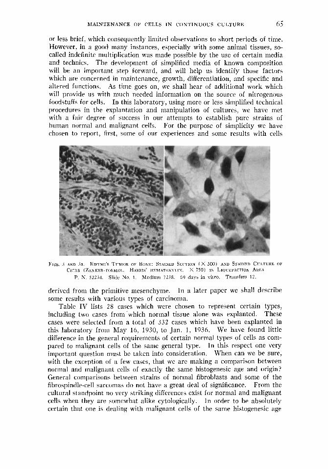
or less brief, which consequently limited observations to short periods of time. However, in a good many instances, especially with some animal tissues, so- called indefinite multiplication was made possible by the use of certain media and technics. The development of simplified media of known composition will be an important step forward, and will help us identify those factors which are concerned in maintenance, growth, differentiation, and specific and altered functions. As time goes on, we shall hear of additional work which will provide us with much needed information on the source of nitrogenous foodstuffs for cells. In this laboratory, using more or less simplified technical procedures in the explantation and manipulation of cultures, we have met with a fair degree of success in our attempts to establish pure strains of human normal and malignant cells. For the purpose of simplicity we have chosen to report, first, some of our experiences and some results with cells
FIGS. J ~ N D 3a. EWING'S TIJMOR OF BONE: STAINED SECTION ( X 300) AND STAISELI CULTURE 01' CELLS (ZENKER- L.'ORIMOL. HARRIS' IIEM.4TOXYI.IS. x 7 5 0 ) I N LIQUEFACTION AREA
P. N. 52234. Slidc No. 1 . Medium 7238. 60 days in vitro. Transfers 12.
derived from the primitive mesenchyme. In a later paper we shall describe some results with Lrarious types of carcinoma.
Table IV lists 28 cases which were chosen to represent certain types, including two cases from 'which normal tissue alone was explanted. These cases were selected from a total of 332 cases which have been explanted in this laboratory from May 16, 1930, to Jan. 1, 1936. We have found little difference in the general requirements of certain normal types of cells as com- pared to malignant cells of the same general type. In this respect one very important question must be taken into consideration. When can we be sure, with the exception of a few cases, that we are making a comparison between normal and malignant cells of exactly the same histogenesic age and origin? General comparisons between strains of normal fibroblasts and some of the fibrospindle-cell sarcomas do not have a great deal of significance. From the cultural standpoint no very striking differences exist for normal and malignant cells when they are somewhat alike cytologically. In order to be absolutely certain that one is dealing with malignant cells of the same histogenesic age

0 0 GEORGE 0. GEY AND MARGARET 1C. GEY
and type as a given type of normal cell, he must induce the normal cell to undergo an anaplastic change. Such a change must be definitely proved before specific comparisons can be made. In this laboratory we have tried repeatedly to effect such a change in strains of normal cells exposed continu-
FlCS. 4 AND 4a. FIHNOSYINDI.I'.-('EI.I. SARCOMA OF ~ R O B A I 3 I . E NERVE S l I E A T I i ORIGIN STAINED SECTION
( x .jo0) AND STAINED CIJLTZIRE FROM ADJOINING AREA (ZENKER-FORMOL. HARRIS' 1IEMATOXYI.IN. x . \oo), SHOWIN(; GENERAT. CHARACTER 01'' 0C;TcROWTII
P. N. 45010. Slide No. 13. Mcdium 3 7 3 7 . 20 days in vitro. Transfers 4.
Flc:s. 5 AND 5a. FlHROSPIND1,E-('El,L SARr0M.h: STAINED SECTlOi i ( x Boo) AXUD STAINED CULTUHE (~1~;NKliR-FORMOI. . HARKIS' III:MATOXYI.IN. x -300) FROM ADJOINING AREA SHOWIN(:
GI<NI;.RAI. CITARA('TI'R OF OUTCROWTI1
P. N. 44764. Slidc No. 4. Medium 3737. 20 days in vitro. Transfers 4.
ously to certain carcinogenic agents, but without success. The importance of a knowledge of reliable methods for the maintenance of pure strains of cells for indefinite periods is readily appreciated when such work is attempted.
On a number of occasions we have had an opportunity to explant both

MAINTENANCE OF CELLS I N CONTINUOUS CULTURE 67
normal tissue and tumor from the same patient and to maintain these tissues in autologous media. As compared to cells maintained in homologous media, those maintained in autologous media showed no noticeable difference in growth and appearance.
Fics. 6 ASD 6a. GIAXT-CELL TUMOR OF BONE: STAINED SECTION ( X 600) AND TISSUE CIJ1,- TURF. ( x 600) FROM .kDJOINING AREA, SHOWING LIVING MULTINUCLEATED T U M O R FIBROBLi%ST W H I C l r E X H l B l T E D ACTIVE PINOCYTOSIS I N T H E FILM (NO. 189) FROM WIIICH T I l E REPRODUCTION
WAS MADE.
similar tumor cells (see Table IV) up to 467 days. (H. PI. 5 ; Ch. PI. 2 ) .
Similar multinucleated tumor fibroblasts persisted throughout the period of cultivation of other Slide No. 402. Medium 3737 P. N. 45096.
18 days in vitro. Transfers 3 .
In Table IV are presented cultural data for a strain of normal fibroblasts, G.Ge., maintained for 108 days in media containing a large proportion of autologous serum and plasma. When transferred to homologous media these cultures did as well, if not better than in the autologous media. Some- what similar results were obtained with strain ES i . In strains V.As. I and 11, we were not able to observe any very great difference in cultural behavior between normal fibroblasts derived from quadriceps tendon and muscle, and malignant fibroblasts derived from a fibrospindle-cell sarcoma, when both tis- sues were obtained from the same patient, The question of identification of the sarcoma cells in such cultures, where there may be a possibility of confusion through contamination of the tumor cultures with fibroblasts and fibroblastic overgrowth, offers little difficulty to those who have made repeated observa- tions on the growth of sarcoma cells. A good many of the sarcoma cells that we have maintained were different from normal fibroblasts.
The question has often arisen whether or not tumor cells were grown and maintained from certain tumors. In Figs. 2 to 6 are shown portions of sarcoma colonies, in each case with massive emigration of all of the cells of which the tumors were composed. In each case direct comparison can be made between the section of the tumor and the portion of the culture, which in many cases show a striking similarity. For most types of sarcoma, when tissue is obtained from very cellular and healthy portions of tumor, the primary cultures and all subsequent cultures usually show a continued pro-

68 GEORGE 0. GEY A N D MARGARET K. GEY
liferation of the tumor cells with a certain minimal admixture of reactive normal cells. The early appearance of various ‘‘ wandering ” cells is com- monly observed. We have ob- served lymphocytes which have persisted for four months in fibroblast cul- tures. In some cases, where a good deal of normal supporting stroma is explanted with tumor cells, there is growth of both types and with the not uncommon result of fibroblastic overgrowth. The cultivation of sarcoma cells from cultures contaminated with fibroblasts usually offers no very great diffi- culty, but such contamination constitutes one of the chief difficulties in the establishment of carcinoma cultures. As noted above, we have come across very few tumor cells whose cultures could not be distinguished readily from cultures of normal cells. Only a few of the sarcomas that we have main-
Some of them persist for many months.
tained in culture have shown striking similarities to each other in the appear- ance of the cells and colonies and in cultural behavior.
In the general group of giant-cell tumors of bone (Table IV) the cultures of tumor fibroblasts which we maintained were more or less similar, and in all of these cases the large giant cells persisted for a considerable period of time. Multinucleated cells were present a t all times throughout the entire history of these cultures, but we were not able to observe the formation of new giant cells from them, containing large numbers (10-200 or more) of nuclei. With the exception of new giant-cell forms and the production of new multinucleated cells, these cultures behaved essentially like other fibro- blast cultures.
A study of the histories of the first 11 cases shown in Table IV has pro- vided us with evidence that there are fairly definite differences in different

MAINTENANCE O F CELLS I N CONTINUOUS CULTURE 69
strains of fibroblasts. Parker (24) has been able to identify certain so-called races which differ in growth rate and appearance. We also have noted con- siderable differences in the appearances of fibroblasts obtained from various sources. These differences have to do with cytological appearance and to a lesser degree with cultural behavior. We are inclined to the belief that the physiological state of the cells has a great deal to do with the variations in cytological appearance. These differences have also been noted by others. In some cases, (E.Ke. I and I1 and M.Sh.) strains of fibroblasts were estab- lished which were larger than other types (G.Ge., V.As. 11) and to some extent differed also in cytological appearance.”
I n the group of cases diagnosed osteogenic sarcoma and in the cases diagnosed spindle-cell sarcoma of probable nerve sheath origin, there are
certain similarities and certain striking differences in the behavior and ap- pearance of the sarcoma colonies. Although the diagnoses were different, the cell strains established from I).Au., H.T)a. and A.Ha. I and I1 were strikingly similar in cytological appearance and cultural behavior. The colonies in these cultures usually grew out as compact cell masses with only a thin peripheral fringe of emigrating cells. There was little tendency for the colonies to spread out in thin layers. The colonies were usually discoid and made up of niany cell layers whose peripheral margin was also thick and very cellular. Liquefaction of the medium seemed to be limited to the im- mediate vicinity of the colonies, which remained intact and could be lifted out of a small pool of fluid. This tendency to form compact cell colonies is somewhat similar to the behavior of colonies of the Walker rat sarcoma 319,
To be published

70 GEORGE 0. GEY AND MARGARET I<. CEY
which we have maintained for almost four years. In the group of cases diagnosed Ewing’s tumor, F.We., G.Em. and R.Br. (Table IV) , great simi- larity in cytological appearance and cultural behavior of the cell strains was
FIGS. 8 .ZND Xa. CIIONDI(OMYXOSARCOMA (SEF Frc. 7 ) X 3 7 5 . P. N. 46166.
Slitlc No. 14 ab. Medium 7238. 917 days in vitro. Transfers 206. Reproduced from film No. .w.
Fig. 8a shows a large m;ilignant protochondroblast from stained culture, with polymorphic nuclei. Tso- kited cells of this type are visible to the nnked eye. X 300. P. N. 46166. Slide No. 04. Medium 72.18. 329 days in vitro. Transfers 51.
Fig. 8 shows large tumor cells dividing abnormally in living culture.
Note enormous differencc in size as compared with other cells in the same culture.
found. The living tumor colonies (Fig. 10) are made up of small so-called reticulum cells which tend to produce in the colonies a (‘ reticular ” or sponge- Iike framework. These cells in no way resemble lymphocytes, nor do their

FIG. 8b. CHONDROMYXOSARCOMA (SEE Fic. 7 ) : PERSISTENCE OF TUMOR CELLS VARYING GREATLY I N SIZE AND WITH IXCREASIYG POLYMORPHISM OF NUCLEI O P THE LARGER CELLS AFTER
LOSG-(VSTINUEO CULTIVATION. x 300
P. N. 46166. Slide No. 14 abab. Medium 7238. 1241 days in vitro. Transfers 271.
FIG. 9. CIIOSDROMYXOSARCOMA (SEE FIG. 7 ) : STAINED CULTURE REMNANT or CELLS LEFT ON
COVER GLASS SHOWlNC WHERE SHEARING C U T S WERE M A D E AND INDICATED (ZENKER-FORMOT.. HARRIS' HEMATOXYLlN. x 50)
The portion A is included within the cut area and should have been removed with the excised The portions B and B' represent the
Slide No. 04 cgl. Medium colony which has left a more or less clear area at C. average amount of new growth usually lost in transfer. 7137. 119 days in vitro. Transfers 26.
P. N. 46166.
71

72 GEORGE 0. GEY A N D MARGARET I(. GEY
colonies resemble colonies of lymphocytes. When rounded up they are larger than lymphocytes, Whether or not liquefaction occurs, the colonies tend to form a network with ‘‘ spaces ” between small groups of cells and occasionally between but two cells. The formation of ring-like colonies may in some way be related to the perithelioinatous arrangement of these cells which is fre- quently seen in the original tumor. Furthermore, the cells do not resemble cndothelial cells, which are much larger.
F l C . 9tl. ‘rI5Kl<E-I)AY ~ l I I . I ’ I 7 K I : OF A SE R’I~~NTIIS-~I .T)~STRAIN OF HPMAN EMBRYONIC FIIIKIIHIASTS FROM TIIE S K I N : ~ I I O l 0 l ~ R A P I T O F I,IVIN(: CULTURE, M A Y 1 7 , 1926. x 50
The culture shows the gradual accumu1;ttion of indigestible cell by-products alid contracted clot These cut ;ireas, whose limits are shown a t a, b, and c, indi-
Coiideiiscd areas from previous At 11 ;ire shown 2 encysted acellular masses surrounded by epithelium which
left from the cut e&c of the colony. cate the numbcr of cuts and the angle at which thcy werr made. ruts arc also shown. still pcmists in this colony. P. N. 5271 (Col.). Slide No. S pc. Medium 3548.
Froin the chondromyxosarcoma cases, J.Da. and V.La. (Table I V ) . strains of cells were obtained which were very different from those of any other type of sarcoma that we have had under cultivation (Figs. 7-7c). From one of these cases, J.l>a., we have maintained cultures containing chiefly two types of cells : ( 1 ) large cells which we have designated protochondroblasts, differing greatly in size and cytological appearance, cultures of which liquefied ninny different types of media fairly rapidly; ( 2 ) undifferentiated sarcoma cells whose specific origin we have not ventured to suggest and which form colonies, rapidly liquefying most types of media. The strain of undiffer-

MAINTENANCE OF CELLS IN CONTINUOUS CULTURE 73
entiated sarcoma cells we have maintained over 1583 days to date. The strain of large sarcoma cells was maintained 1241 days.
Concerning the loss of strains when once established, a number of factors must be considered. All of them are equally important and should be guarded against. So-called accidental causes, such as contamination, over-heating of incubators, and faulty technic, are chiefly responsible for the loss of cultures.
FIG 10. LIVING CULTTIRE OF AN INDUCED (DIBENZANTHRACENE) RAT SARCOMA SHOWING AT A A FAIRLY DEXSE OUTGKOWTII AND AT B AN AREA WITH ATTACHED OLD CLOT OF FAIRLY SHARP
R. I. T. No. 5 . Slide No. 6a. Two-day culture. 49 days in vitro. OITTI.IKE, AS INDT(’ATED, AND WITH F E W CELLS PROLWERATTNG F R O M TITIS AREA. x 50
F I G . 11. WAl.KER RAT S A R ~ O M A No. 8.38; FIXED COLONIES IN HEXAGONAL ROLLER T U B E W H I ~ € I DI~~ELOPICD FROM A FINE SUSPEXSION OF SMALL CLUMPS AND SISGIX SARCOMA
CE1.l.S. NATTJRAT, SIZE
Multiple colonies of fair size developed in thirty-four days from barely visible explanted clumps. The entire medium is invaded by cells, most of which are single isolated cells. Zenker- formol. Not stained. No. S38R. In tube No. 7r for 34 days. Medium 72.38 (H. PI. 6 ; Ch. P1. 2 ) . 85 days in roller tubes. Maintained 1485 days in vitro.
One cannot over-emphasize the use of safety devices and precautionary pro- cedures as checks against many of these so-called accidents. Loss through the use of unsuitable or non-supporting media is but a part of the experi- mental evidence which is sought in obtaining maintenance data.
Speaking generally, we are more or less convinced that most types of human sarcoma can be maintained indefinitely in vitro. The possibility of Some variation in the cytological and cultural characteristics of tumor cells

74 GEORGE 0. GEY AND MARGARET K. GEY
maintained over long periods has been suggested. In the strain J.Da., which is the oldest human strain in this laboratory, some cytological changes occurred in a few of the cultures after long cultivation." We are not prepared to say that these changes were permanent or that they were temporary and due to variations in the medium which could not be controlled. Our observations on and experiences with the explanation of hundreds of human and animal tumors, and the maintenance by continuous culture of many of them, have led us to the conclusion that the tumor cells that we have cultured are mutant cells with rather specifically fixed properties which they manifest and retain for long periods of time and probably indefinitely. Though some may resemble sup- posedly related (histogenically) normal cells, we are impressed by a differ- ence in cultural behavior and cytological appearance when comparisons are attempted. Specific differences for all types are difficult to define.
SUMMARY
Evidence has been presented that : 1. Normal human fibroblasts can be maintained under continuous culti-
vation for long periods of time, and in media varying somewhat in composition. 2 . Several different types of human sarcoma cells can be maintained as
pure strains for long periods of time under continuous cultivation by fairly simple procedures.
3 . There are certain differences and certain similarities in the cytological and cultural behavior of pure strains of human sarcoma cells cultured from cases given the same histological diagnosis.
4. A good deal of variation in the composition of the culture medium can be made and still allow maintenance of some pure strains of cells for consider- able periods of time.
5. A composite medium containing balanced salt solution, bovine embryo extract, human placental (fetal) serum, and human plasma or human plasma with added chicken plasma, can maintain human normal and malignant tumor cells under continuous cultivation for long periods of time.
6. The same composite medium is as suitable for the maintenance of rat malignant cells as for human malignant cells, a number of tumor cells of both kinds having been maintained for varying periods up to over four and a half years.
7. The methods, media, technic, and apparatus herein referred to make possible the maintenance at the same time of some twenty to thirty different strains of cells by a small group of three or four workers. All of the pro- cedures involved can be carried out by one individual.
8. The possibility of maintaining, b y continuous culture, large quantities of human and animal malignant and normal cells for various experimental purposes is no more a matter for conjecture, but has actually been proved.
NOTE: We are greatly indebted to all of those who have so generously aided us in the wllcction of blood and tissue specimens. We are especially grateful to the late Dr. J. LV,
1 2 To bc published.

MAINTENANCE O F CELLS IN CONTINUOUS CULTURE 75
Williams who made possible the arrangements for collecting placental blood, and to the late Dr. J. C. Bloodgood for his unceasing efforts and generous cooperation in making material available. For the use of laboratory space and certain apparatus and for constant co- operation and invaluable help we are grateful to Doctors G. L. Streetcr and W. H. Lewis of the Carnegie Embryological Laboratory. Most of the fresh tumor tissues that we explanted have been made available through the generous cooperation of Dr. Dean Lewis and the house staff of this hospital. Acknowledgment is also made of a large supply of sectioned material prepared in the Surgical Pathological Laboratory under the supervision of Dr. C. F. Geschickter, to whom we are indebted for aid and cooperation. Some of our methods and technics were developed under other auspices than those mentioned. We are indeed grateful to Dr. J. L. Yates and others of the Columbia Hospital, Milwaukee, Wis., and to the Chemical Foundation, Inc., for their support of earlier work.
BIBLIOGRAPHY
1. GEY. G. 0.: An improved technic for massive tissue cultures, Am. J. Cancer 17: 752,
2. LEWIS, W. H.: Roller tube cultures, Anat. Rec. 58: 1934. 3. LEWIS, W. H.: Roller Tube Cultures, Contrib. to Embryology, No. 150, Carnegie Inst.
4. VOGELAAR, J. P. M., A N D ERLICHMAN, E.: Some remarks on the growth of human
5. LEWIS, W. H., A N D GEY, G. 0.: Clasmatocytes and tumor cells in cultures of mouse
6. CRON, R. S., AND GEY, G. 0.: The viability of the cast-off menstrual endometrium, Am.
7. RIKGER, S.: Further experiments regarding the influence of small quantities of lime,
8. LOCKE, F. S.: Die Wirkung der Metalle des Blutplasmas und verschiedener Zucker auf
9. HEDON, A N D FLEIG: Actions des serums artificiels et du serum sanguin sur le fonctionne-
10. ADLER, H. M.: Experiments toward a physiologically isotonic solution of salts, J. A. M.
1933. Supp. 55.
of Washington, July 1935.
fibroblasts in media containing copper, Am. J. Cancer 2 2 : 66, 1934.
sarcoma, Bull. Johns Hopkins Hosp. 34: 369, 1923.
J. Obst. & Gynec. 13: 645, 1927.
potassium. and other salts on muscular tissue, J. Physiol. 7 : 291, 1886.
des isolicrte Sangethierherz, Zentralbl. f . Physiol. 14: 670, 1901.
ment des organes isoles des mammifkres, Arch. int. de physiol. 3 : 95, 1905.
A. 51 : 752, 1908. 11. TYRODE, M. V.: The mode of action of some purgative salts. Arch. int. Pharmacol, 20:
205. 1910. 12. VAN DYKE, H. B. , AND HASTINGS, A. B.: Response of smooth muscle in different ionic
environments, Am. J. Physiol. 83: 563, 1928. 13. EARLE, W. R.: A technic for the adjustment of oxygen and carbon dioxide tensions and
hydrogen ion concentrations, in tissue cultures planted in Carrel flasks, Arch. f . exper. Zellforsch. 16: 116, 1934.
14. SCHADE, H.: Concerning a physiochemical method of carrying out tissue cultures in their own plasma without the additions hitherto necessary, Arch. f. exper. Zellforsch. 15: 121, 1934.
15. SHEAR, M. J.: The r81e of sodium. potassium, calcium, and magnesium in cancer: A review, Am. J. Cancer 18: 924, 1033.
16. ROFFO, A. H.: La acci6n de ciertos iones metilicos (potasio, calcio y magnesio) sobre el desarrollo de 10s tejidos normales y neo#isicos in vitro, Bol. Inst. med. exper. para el estud. y trat. del cancer 1 : 307, March-April 1925. And others (see review of Shear (15) . )
l i . GEY, G. 0.: Studies on the cultivation of human tissue outside the body, Wisconsin M. J. 28: 11, 1929.
IS. KRoNTowsKr, A. : Explanation und deren Ergebnisse fur die normale und pathologische Physiologie, Ergebn. d. Physiol. 26: 370, 1928.
19. CARREL, A,, A N D EBELING, A. H.: Antagonistic growth principles of serum and their relation to old age, J. Exper. Med. 38: 419, 1023.

76 GEORGE 0. G E Y AND MARGARET I<. GEY
20. ZAKKZEWSKT, Z.: Die Ziichtung von Gewehen in Serum niit 1)esonderer Betonung der Iieziehung zwischen Zcllwachstum und %elldifferenzierung, Arch. f . exper. Zellforsch. 15 : 113, 1034.
21. EARLE, W. K . : A study of the Walker rat mammary carcinoma 256 in vk7o and in vitro, Am. J . Cancer 24: 566, 1935.
22 . LAMBERT, K. A , . A N D HANES, I;. M.: The cultivation of tissue in plasma from alien species, J. Exper. Med. 14: 120, 1911.
23. GEY, G , O., A N D THALHIMER, W.: Observations on the effects of insulin introduced into thc nietiiurn of tissue cultures, J . A. M . A. 82: 1600, 1024.
24. PARKER, R. C. : The races that constitute the group of common fibroblasts. 111. lhf - ferences determined by origin of explant and age of donor, J. Exper. Metl. 58: 401, 1033.
2.5. FLOSDORF, E. W., A N D MLIDD, S.: Procedure and apparatus for the preservation in I ' lyophile " form of serum and other biological suhstitutcs, J. Immunol. 20: 380, 103.3.
26. BAKER, L. E., A N D CAKKEL, A.: Action on fihroblasts of the protein fraction of em- tiryonic tissue extract, J. Exper. Mcd. 44: 387, 1926. Effect of the amino acids and dialyzable constituents of embryonic tissue juice on the growth of fibroblasts, J. Exper. Med. 44: 307, 1926.
27. LEWIS, M. R., A N D LEWIS, W. H.: The cultivation of tissues from chick embryos in solutions of NuCI, CaCL, KCI, and NaHCO., Anat. Rec. 5 : 2 7 7 , 1011.