Embed Size (px)
Citation preview
10
Personal and AreaMonitoring
2 PERSONAL AND AREA MONITORING
Contents Page
10.1 Personal monitoring10.1.1 Introduction.................................................................310.1.2Whole body external dose............................................310.1.3External dose to parts of the body...............................510.1.4 Internal exposure........................................................5
10.2 Radiation area monitoring................................................8
10.3 Monitoring radioactive contamination on surfaces.......9
10.4 Monitoring airborne contamination...............................11
10.5 Leak testing sealed sources............................................12
10.6 Calibration of monitoring instruments..........................13

PERSONAL AND AREA MONITORING 1

PERSONAL AND AREA MONITORING 3
10.1 Personal monitoring
10.1.1 Introduction
Personal monitoring is the continuous measurement of an individual's exposure dose by means of one or more types of suitable instruments, such as pocket dosimeters, film badges and thermoluminescent dosimeters, which are carried by the individual at all times. The personal monitoring instrument chosen must be compatible with the type and energy of the radiation being measured. For example, a worker who is exposed only to 14C would wear no personal monitoring instrument, since these isotopes emit only beta rays of such low energy that they are not recorded by any of the commercially available personal monitoring devices. Bioassay procedures would be used for personal monitoring. Note that it is Monash policy for all radiation workers to wear dosimeters due to the possibility of contact with other isotopes.
The main purpose of personnal monitoring is to obtain information of the exposure of an individual. In addition to this, personnel monitoring is also used to observe trends or changes (in time) in the working habits of a single individual or of a department, and thus to measure the effectiveness of a radiation control program. Whereas the distribution of personnel monitoring data might all appear to lie within a normal range when viewed as individual readings, statistical analysis of the grouped data may reveal small but significant differences among different control measures, or different operating procedures or work habits that might otherwise have escaped the attention of the RPO, RSO or deputy RSO.
Monash University policy on monitoring for ionising radiation is sent out in sections 26 and 32 of the Policy Statement. In addition Part 9 of the Health (Radiation Safety) Regulations 1994 and Section 9.3 of AS 2243.4 contain specific directives for monitoring ionising radiation. Records must be kept for all measurements pertaining to ionising radiation in accordance with section 21 of the Policy Statement.
10.1.2 Whole body external dose
The whole body external exposure is measured in a uniform radiation field by a personal dosimeter, usually a film badge or a Thermo Luminescent Dosimeter (TLD), worn by the individuals on the waist. These dosimeters measure doses from beta, x, gamma and slow neutron radiation. Film badges have a minimum detectable dose of 200Sv. TLD badges have a lower threshold of around 10 Sv, but are more expensive.
All persons working with ionising radiation should be provided with and be required to wear appropriate personal monitoring devices. Other persons who may be potentially exposed to ionising radiation should have their exposure assessed and wear personal monitoring devices when deemed necessary by the RSO or deputy RSO.
PERSONAL AND AREA MONITORING 5
Personal dosimetry may be supplemented with direct reading dosimeter (eg. Quartz Fibre Electrometer or a digital audio dosimeter), which are useful for measuring high gamma doses and allow continuous surveillance of the rate of accumulation of dose.
Each personal dosimeter should be numbered, used by one person only and returned promptly at the end of the issue period. In the event of a suspected high dose, arrangements should be made with the relevant personal dosimetry service for the particular dosimeter(s) to be processed and assessed as quickly as possible.
Personal dosimeters issued to individuals should be carried on the person. They should not be used to measure the dose in a fixed position, eg. at a bench.
Personal dosimeters have limitations which restrict their usefulness. The type of personal dosimeter in common use may not measure dose for alpha and low energy beta (eg. from 3H).
All persons participating in personal monitoring programs should be entitled to receive feedback on the monitoring outcomes as soon as results are available. In situations where results exceed recommended action limits or exhibit trends which show cause for concern. The RSO or deputy RSO should follow up with the persons(s) involved all possible contributing factors.
It is necessary that pregnant women who continue working with ionising radiation during their pregnancy have the TLD monitors changed every four weeks. All other radiation workers may wear their TLD monitors for 8-12 weeks unless otherwise instructed.
The film badge has a number of advantages that makes it widely used for whole body monitoring by persons exposed to x rays, gamma rays, high energy beta rays, and neutrons. The chief advantage claimed for the film badge is the fact that it provides a permanent exposure record. In addition, it can measure dose over a very large range, it can be made to record the dose over a wide range of energies on the same film and conversely the type and energy of the radiation may be inferred from the film exposure. It is mechanically rugged.
The main disadvantages of film badges are the long delay time between exposure and development and qualitative nature of interpretation of the film. An average accuracy of about ±25% of the true exposure to the film may be expected from a good film badge service.
Many of the alleged disadvantages of the film badge dosimeter (particularly that of its inaccuracy) can be overcome by the use of a thermoluminescent dosimeter (TLD). The radiation sensitive material in the monitor allows a minimum reportable dose
of 10 Sv for x-rays, 70 Sv for high energy gamma and beta rays and 50 Sv for fast neutrons. The minimum reportable dose to the fingers is 100 Sv . With a TLD the waiting period before the exposure dose is less than that associated with the film badge dosimeter.
PERSONAL AND AREA MONITORING 7
The exposure information stored in the TLD can be read out in several seconds, thus making it useful for emergency dosimetry. For these reasons, the TLD has displaced the film badge for personnel monitoring purposes. However, it should be pointed out that TLD readout is destructive, thus a permanent record of exposure cannot be kept.
10.1.3 External dose to parts of the body
Work close to radiation sources of small dimensions or with devices producing narrow beams of radiation, exposes persons to non-uniform radiation fields. In this case a part of the body, particularly the fingers, may be exposed to a higher dose than measured by the whole body dosimeter and measuring local doses, in addition the whole body dose, becomes necessary.
Additional personal dosimeters worn on the wrist, forehead or other parts of the body are used for measuring local doses. Finger-tip doses are best measured by thermoluminescent dosimeters.
Finger dosimeters must be worn by staff working with x ray diffraction and x ray fluorescence apparatus, particularly when engaged in sample manipulation or beam alignment (ie. single crystal orientation). Such manipulations or alignments should ideally be carried out with the beam off, but where that is not practicable, the staff member carrying out the alignment should ensure that no other staff or visitors are in the vicinity of the beam.
Where additional dosimeters are needed to estimate doses to extremities of the body, a special issue of dosimeters should be arranged through the Australian Radiation Laboratories dose monitoring service.
10.1.4 Internal exposure
Introduction
Radiation workers engaged in work involving unsealed radioactive material, may have radioactive material entering their body through inhalation, ingestion or penetration through the skin. Biological monitoring programs are designed to assess the significance of any radioactive substance taken into the bodies of individuals exposed to such materials.
One of the techniques for evaluating a contamination control program is the determination of the body burden of personnel who are at risk. This determination is done indirectly by bioassay methods, or directly by total body counting in the case of gamma-emitting radionuclides or beta emitters that give rise to bremsstrahlung. The underlying rationale for bioassay is that a
quantitative relationship exists between inhalation or ingestion of a radionuclide, the resulting body burden, and the rate at which the radionuclide is eliminated either in the urine or in the faeces.
PERSONAL AND AREA MONITORING 9
Measurements of activity in the urine and faeces enable inferences to be made about body burden. Unfortunately, the kinetics of metabolism of most substances is influenced by a large number of factors and as a consequence, the desirable quantitative relationships between body burden and elimination rates are known for relatively few isotopes. In most instances, therefore, bioassay data gives only a very approximate estimate of the degree of internal deposition of radioactivity.
For purposes of bioassay, we distinguish between readily soluble and relatively insoluble compounds. This distinction is especially important in the case of inhaled particulate. The relatively insoluble material is brought up from the lung and swallowed, whereas the soluble inhaled particulate matter is dissolved into the blood. Due to varying pulmonary deposition, clearance rates and the tendency of some isotopes to congregate in certain tissues it is difficult to quantitatively estimate the lung dose from insoluble air-borne contamination.
Nevertheless, an estimate of the minimum amount of a relatively insoluble radioactive particulate that was deposited in the upper respiratory tract following a single accidental inhalation could be made from the cumulative faecal activity during the first few days after the inhalation. With this information, and using the ICRP model for lung clearance, a less reliable estimate can then be made of the activity remaining in the deep respiratory tract.
Readily soluble radionuclides may be grouped into three categories according to their distribution and metabolic pathways:
(1) Those that are uniformly distributed throughout the body, such as 3H in tritiated water or radiosodium ions,.
(2) Those that concentrate mainly either in specific organs, such as iodine in the thyroid gland, mercury in the kidney, or in the intracellular fluid, such as potassium or caesium.
(3) Those which are deposited in the skeleton.
The second category, those radionuclides that are concentrated in one or more organs, are not as amendable to quantitative monitoring by bioassay as the widely distributed radionuclides. These radionuclides are absorbed, after ingestion or inhalation, into the body fluids and the blood plasma. From these fluids, they pass into the organs in which they concentrate; a dynamic equilibrium eventually results between the concentration of the nuclide in the organ and in the body fluids. While the isotope is equilibrating between the body fluids and the organ of concentration, it is also being filtered by the kidney into the urine.

Bioassay data are most reliable in the case of the first category, the widely distributed radionuclides.
PERSONAL AND AREA MONITORING 11
The third category, which comprises the elements absorbed into the bone, is a special case of the category of isotopes concentrated in an organ or tissue. Bone seekers differ from other radionuclides mainly in the rate of elimination of the isotope. Clearance half-time for the non-bone seekers are measured in terms of days or weeks, whereas clearance half-times for bone seekers are measured in years. Furthermore, the rate of clearance from the skeleton is not constant, as is usually the case for the non-bone seekers, but decreases with increasing time. This is due to the fact that the skeleton is not a single "compartment", but rather a number of different "compartments", each of which has its own clearance rate.
Whole body monitoring in practice
Direct determination, by whole body counting, of body burdens of gamma emitting nuclides provides a more accurate estimate of the body burden than does excreta analysis (Figure 10.1). However, because of the high cost of installation and operation of a total-body counting facility, and also because of their limitations for the determination of gamma emitters and bremsstrahlung generating beta emitters, total-body counters are not generally used for routine monitoring. Their main use is in research or in the assessment of internal contamination following an accidental exposure.
Gamma and X emitting radionuclides and beta emitting radionuclides that have sufficient energy to produce bremsstrahlung when distributed in the lungs or throughout the whole body, can be monitored with whole body counters. The difficulty lies in monitoring radiation that is distributed in a non-uniform manner. Results are express in ALIs.
A large shielded room (to reduce background) with a flat bed or tilted chair (at 45 degrees) and a large sodium iodide detector are required.
Monash University has no facilities for whole body monitoring of internal exposure to ionising radiation. Such measurements must be arranged through the RPO.
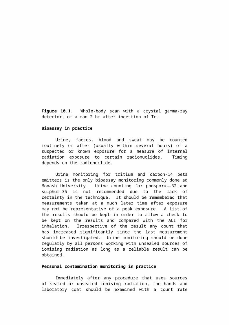
Figure 10.1. Whole-body scan with a crystal gamma-ray detector, of a man 2 hr after ingestion of Tc.
Bioassay in practice
Urine, faeces, blood and sweat may be counted routinely or after (usually within several hours) of a suspected or known exposure for a measure of internal radiation exposure to certain radionuclides. Timing depends on the radionuclide.
Urine monitoring for tritium and carbon-14 beta emitters is the only bioassay monitoring commonly done ad Monash University. Urine counting for phosporus-32 and sulphur-35 is not recommended due to the lack of certainty in the technique. It should be remembered that measurements taken at a much later time after exposure may not be representative of a peak exposure. A list of the results should be kept in order to allow a check to be kept on the results and compared with the ALI for inhalation. Irrespective of the result any count that has increased significantly since the last measurement should be investigated. Urine monitoring should be done regularly by all persons working with unsealed sources of ionising radiation as long as a reliable result can be obtained.
Personal contamination monitoring in practice
Immediately after any procedure that uses sources of sealed or unsealed ionising radiation, the hands and laboratory coat should be examined with a count rate meter. Such contamination is most serious as a source of internal exposure if it is ingested or absorbed.
Thyroid monitoring in practice
Monash University requires that all persons using radioactive iodine undergo regular thyroid monitoring. Radioactive iodine concentrates in the thyroid and persons routinely involved in handling radioiodine should monitor their thyroids at six monthly intervals at least. If radioactive iodine is being used regularly, monitoring must be done monthly and immediately following any major task using the isotope. In the case of persons irregularly using radioiodine, thyroid monitoring should be done before and after each round of radioiodine manipulation.
The thyroid monitor utilises a sodium iodide detector which is placed near to the throat and the result (in Becqerel) is comparable with the ALI.
10.2 Radiation area monitoring
PERSONAL AND AREA MONITORING 13
All areas where radiation producing apparatus, sealed radioactive sources, or unsealed radioactive materials are used or stored, should be periodically monitored. Where an abnormal radiation level is detected action should be taken to reduce the level.
In case of unsealed radioactive materials radioactive contamination on surfaces in the air should be monitored. Radiation workers should monitor their hands and clothing after work involving unsealed radioactive material.
Monitoring radiation levels is done using a dose rate meter. Such an instrument usually incorporates an ionisation chamber, a geiger tube or a scintillation crystal.
Extensive monitoring should be done during and immediately after the installation and testing of new apparatus or after alterations to experiments and equipment. Once the pattern of radiation from x ray producing apparatus has been established, subsequent surveys may be less detailed. Where sealed or unsealed radioactive materials are used surveys should be frequent because the materials can be moved and the radiation levels in the area can easily change.
Particular problems arise with narrow beam x ray apparatus eg: x ray diffractometers, because the primary beam and beams or sheets of scattered radiation are generally very small in cross section. A small volume, sensitive detector should be used. Photographic methods may also be useful to detect presence and extent of the beam. Monitoring of such apparatus should be done with great care whenever experimental conditions are changed in any way, however trivial these changes may appear to be. High energy machines may present similar problems, complicated by the much wider energy spectrum and different types of particles.
Electron microscope and diffraction (as distinct from x ray diffraction) apparatus generally present fewer problems. A thorough initial survey when the equipment is installed is usually sufficient, additional shielding being added if necessary in places where there is a high dose rate.
10.3 Monitoring radioactive contamination on surfaces
Introduction
Surface contamination can be located by scanning with a sensitive detector, such as a thin end window Geiger counter. After finding a contaminated spot or area, a dose measuring instrument may be employed to measure the dose rate at some appropriate distance from the surface. The main hazard from surface contamination is transmission of the contamination from the surface into the body via inhalation or ingestion. To estimate this hazard, a smear test is performed to determine whether the surface contamination is fixed or loose, and therefore transmissible.
A smear survey, which is a systematic series of smears without first using a scanning instrument to detect the contamination, is often done in a work area that is subject to contamination, and where the background due to radiation sources is high enough to mask the activity due to contamination. It should be
PERSONAL AND AREA MONITORING 15
emphasized that a smear test is a qualitative, or at best a semiquanititative determination whose chief purpose is to allow an estimate to be made of the degree to which surface contamination is fixed. If significant transmissible contamination is found, and, if in the opinion of the RSO or deputy RSO this contamination may be hazardous, then prompt decontamination procedures should be instituted.
The instrument most commonly used to measure radioactive contamination on surfaces is a contamination monitor. Such a monitor may have a variety of probes. For most work with beta gamma emitters, a probe containing a glass walled geiger tube with a wall thickness of about 30 mg/cm2 is adequate. For some beta emitters, particularly those emitting low-energy particles, such as 14C and 35S a probe with a thin entrance window is needed. This may be either a geiger tube with a thin mica end window (about 2 mg/cm2 in thickness) or a special scintillation detector. There is no satisfactory simple instrument for the direct monitoring of tritium surface contamination.
Smear testing should be used in situations such as:
- Monitoring low energy beta-emitting surface contamination eg: 3H, 14C, 35S.
- An interfering high radiation background is present.
- Surface to be monitored is inaccessible to the probe of a contamination monitor.
- Direct monitoring underestimates degree of contamination because of self absorption effects.
- Degree of removable contamination is to be estimated.
The procedure for a wipe or smear test is as follows:
The instrument must be calibrated in derived working limits (DWL or DL) before undertaking the measurement.
Find the instrument efficiency (Ec) by placing source of known at the same distance from the detector as the measurement will be done. Do this calibration in an area of low background.
Counts measuredEc = x 100%
Activity expected (Bq)
This relates activity to count rate.
Assume Ec = 10% if you don't have a suitable source as a calibration standard.
calibrate: DL x Ec x Ad = counts per second (cps) per
DWL 100
PERSONAL AND AREA MONITORING 17
Where Ad = surface area of the detector (m2) - usually about 0.1 m2 is used.
Use a filter paper or tissue to wipe an area for concentration. Factors affection the integrity of the sample include:
- The pressure exerted when taking the smear.
- The area smeared.
- The condition of surface tested.
Try to smear at an area of at least 0.5-1m2.
The smear samples must then be counted in an area of known low background.
Contamination level (Bqm-2) = Cc x 100/Ec x 1/AS x 100/EE
AS = Area smeared (m2)Cc = Count rate corrected for backgroundEE = % contamination picked up a paper. (Assume 2% unless your
know otherwise).
Contamination level in Bqm-2 can then be compared directly with the DL.
10.4 Monitoring airborne contamination
If operations involving radioactive materials may produce airborne contamination in the breathing zone, some form of monitoring should be instituted. The limit used for comparison with air monitoring results is the derived air concentration (DAC).
1 DAC is the air concentration of the radionuclide that would result in a worker receiving 1 ALI through a single years exposure. A DAC is calculated as follows:
ALIDAC = Bqm-3
2.4 X 10³
The DAC is defined for a reference man (ICRP 23) doin light work for a total of 2000 hours per year. The ICRP has not yet published DAC values in line with the effective dose limit of 20 mSv annually. Limits based on the outdated 50 mSv effective dose limit appear in ICRP 30 but should not be used.
The public DAC is taken as 1/200 of the occupational one on the basis that a member of the public, in 24 hours, breathes 2.4 times the volume of air that a worker does in 8 hours. In addition the
PERSONAL AND AREA MONITORING 19
member of the public inhales the radioactive material for 365 days a year. Whereas the worker only inhales it for 250 days.
Air sampling is considered an important part of a survey where there is a possibility of significant atmospheric contamination. Allowable working levels of contaminated air involve quantities of radioactivity very much less than those which would be considered hazardous if the activity were in a sealed source and if the hazard were limited only to external radiation. Furthermore, even if only sealed sources are used, a program of air sampling is recommended if the nature of the source is such that radioactive gaseous or particulate matter could escape in the event that the source capsule develops a flaw. An air sample, in such a case, might detect the contamination and the leaky source before a significant amount of radioactivity has escaped.
An air sampling system consists of two basic elements:
(1)A source of suction (a vacuum pump) for drawing the air to be sampled through.
(2)A collecting device, which usually separates the contaminant from the air.
The exact type of the sampling system depends on the nature of the radioactive contaminant - mainly whether the contaminant is gaseous or particulate. Regardless of the nature of the contaminant, however, there are two main problems that are common to all types of contaminant (including non-radioactive contaminants):
(1)Obtaining a sample of air that is representative of the situation under investigation.
(2)Obtaining a sample that is large enough to give a reasonably accurate estimate of the mean concentration of dust particles in the air, and also large enough to meet the sensitivity requirements of the radioactivity detector.
10.5 Leak testing sealed sources
Sealed gamma ray, beta ray, and neutron sources are used in a wide variety of applications in medicine and in industry. In all cases, the radioactive material is permanently enclosed either in a capsule or another suitable container. Before being shipped from the supplier, all such sources must pass inspection for surface contamination and leakage. Either during transport from the supplier or in the course of time, however, the capsule may develop faults through which the radioactive source material may escape into the environment. Because of the serious consequences of such an escape, a sealed source must be tested before being put into use and periodically thereafter for surface contamination and leakage.
The testing cycle depends on the nature of the source and on the kind of use to which it is put. However, it is usually recommended
PERSONAL AND AREA MONITORING 21
that such tests be performed at least once every six months. The following technique may be employed to perform these tests.
To test for surface contamination, wipe all exposed external surfaces of the source thoroughly with a piece of filter paper or a cotton swab moistened with an appropriate solvent, then measure the activity on the paper or the swab. The source is considered free of surface contamination if less than 3,700Bqm-2 alpha or less than 37,000Bqm-2 beta activity was wiped off.
To test for leakage, one of the following tests may be performed:
- Wipe the source with either a piece of wet filter paper or a cotton swab. Repeat at least 7 days later. If less than 200 Bq alpha or less than 2000 Bq beta activity was wiped off each time, then the source is considered to be free of leaks.
- For high activity sources such as those used in teletherapy, where wiping the source might be hazardous, accessible surfaces of the housing port or collimator may be wiped while the source is in the "off" position.
- Immerse the source in ethanediol, and reduce the pressure on the liquid to 100 mm Hg for a period of 30 sec. A leak is indicated if a stream of fine bubbles arise from the source. This method is reliable only for such sources where enough gas would be trapped to produce a stream of fine bubbles.
10.6 Calibration of monitoring instruments
All monitoring instruments should be calibrated when first taken into use and at annual intervals thereafter and following major repairs or service. Records should be maintained of the date and results of all calibrations and be kept for two years after disposal of the instrument.
Between annual calibrations constancy checks should be carried out. Where possible these should be made with the same type of radiation for which the instrument is designed. Some instruments are provided with a calibration source for this purpose.
There are two types of tests that are usually carried out on a radiation measurement instrument:-
- A full calibration to confirm that the reading is within its stated accuracy.
- A check that accuracy has not changed significantly after a particular period of use.
PERSONAL AND AREA MONITORING 23
The first of these tests should be carried out at a laboratory equipped with calibration facilities.
The second can usually be carried out by the user to ensure that all instruments used for the detection and measurement of ionising radiation are maintained in good working condition and properly used.
The procedure for a constancy check is:
- To place a source of known count rate (this is not the same as activity) or dose rate (whichever is appropriate to the instrument) for specified distance, at that distance from the detector. Where possible these should be made with the same type of radiation for which the instrument is designed.
- The subsequent readout on the instrument should be recorded for comparison with past and future constancy checks.
- Any reading that is more than 10% from previous ones or any trend in changing response calls for a complete service of the instrument.