Embed Size (px)
Citation preview
Early (and not so early) colorectal cancer:The pathologist’s point of viewDaniela E. Aust, Institute for Pathology, University Hospital Dresden,
Germany
www.pathologie-universitaetsmedizin-dresden.de
Institut für PathologieInstitut für Pathologie
Disclosure slide
I Member of advisory board for AMGEN, ROCHE, BOEHRINGER
I Speaker honoraria from FALK Pharma, Pfizer, Lilly and ROCHE
I Third party funds from MERCK for immunohistochemistry in a clinical
trial
www.pathologie-universitaetsmedizin-dresden.de
Institut für PathologieInstitut für Pathologie
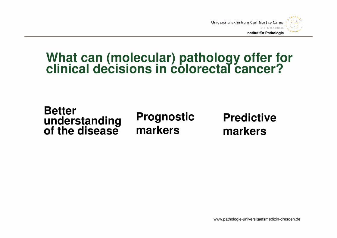
What can (molecular) pathology offer forclinical decisions in colorectal cancer?
Better understanding of the disease
Predictive markers
Prognostic markers
www.pathologie-universitaetsmedizin-dresden.de
Institut für PathologieInstitut für Pathologie
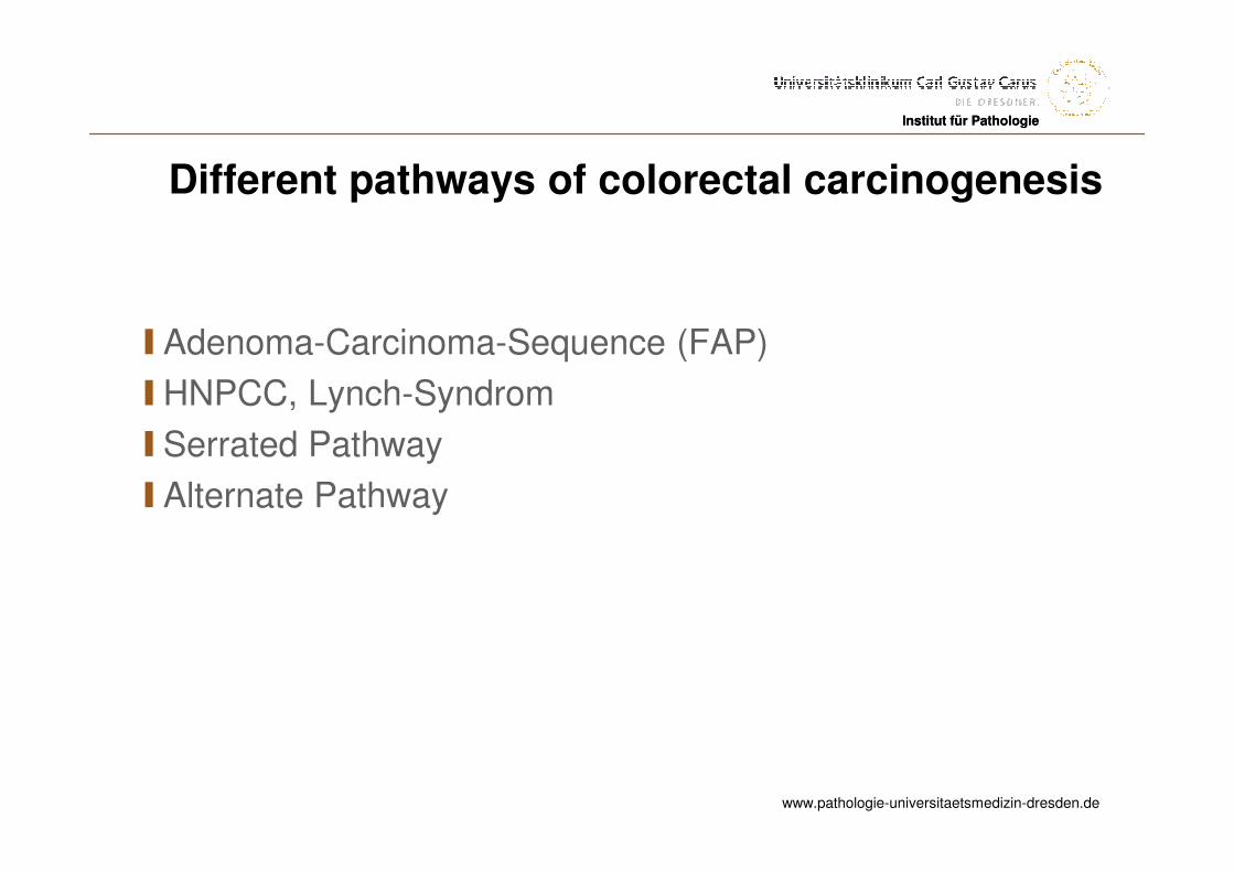
Different pathways of colorectal carcinogenesis
I Adenoma-Carcinoma-Sequence (FAP)
IHNPCC, Lynch-Syndrom
I Serrated Pathway
I Alternate Pathway
www.pathologie-universitaetsmedizin-dresden.de
Institut für PathologieInstitut für Pathologie
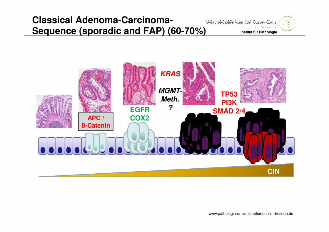
APC /ß-Catenin
KRAS
MGMT-
Meth.
?
TP53PI3K
SMAD 2/4EGFRCOX2
CIN
Classical Adenoma-Carcinoma-Sequence (sporadic and FAP) (60-70%)
www.pathologie-universitaetsmedizin-dresden.de
Institut für PathologieInstitut für Pathologie
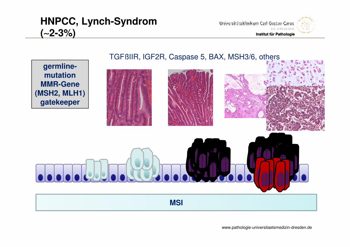
germline-mutation
MMR-Gene(MSH2, MLH1)
gatekeeper
TGFßIIR, IGF2R, Caspase 5, BAX, MSH3/6, others
MSI
HNPCC, Lynch-Syndrom(∼∼∼∼2-3%)
www.pathologie-universitaetsmedizin-dresden.de
Institut für Pathologie
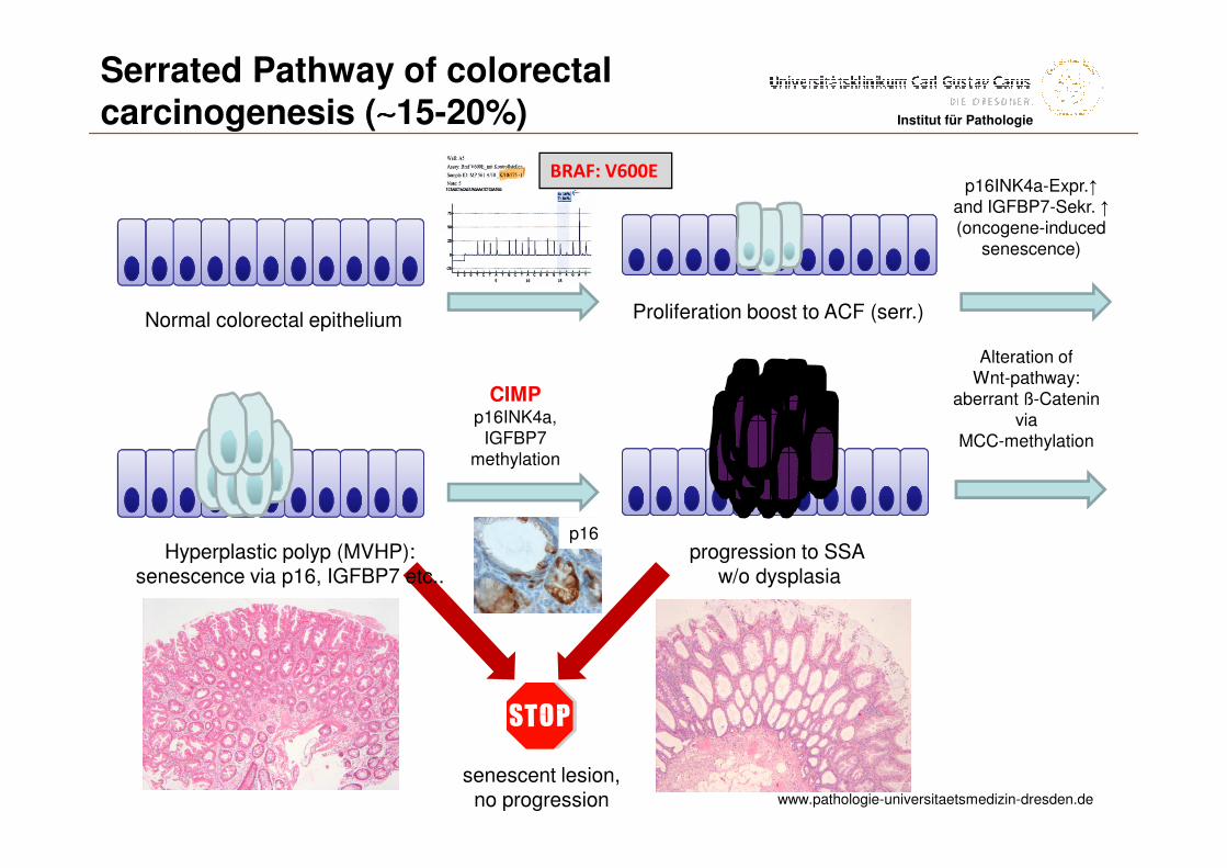
Alteration ofWnt-pathway:
aberrant ß-Cateninvia
MCC-methylation
senescent lesion,no progression
p16
Normal colorectal epithelium
BRAF: V600E
Proliferation boost to ACF (serr.)
p16INK4a-Expr.↑and IGFBP7-Sekr. ↑(oncogene-induced
senescence)
Hyperplastic polyp (MVHP): senescence via p16, IGFBP7 etc..
CIMPp16INK4a,
IGFBP7methylation
progression to SSA w/o dysplasia
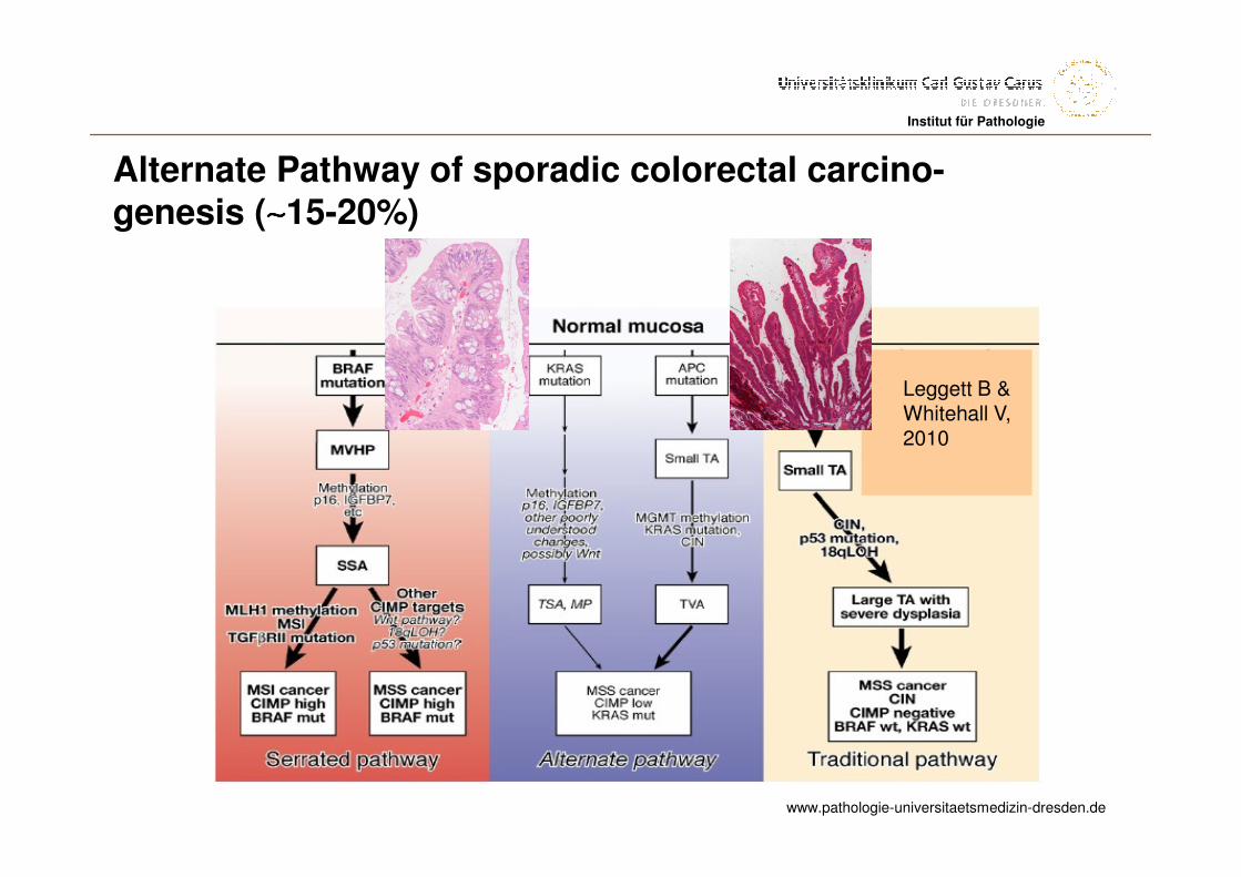
Serrated Pathway of colorectalcarcinogenesis (∼∼∼∼15-20%)
www.pathologie-universitaetsmedizin-dresden.de
Institut für Pathologie
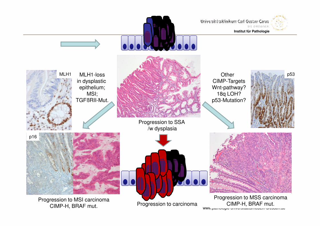
Other CIMP-TargetsWnt-pathway?
18q LOH?p53-Mutation?
Progression to MSS carcinomaCIMP-H, BRAF mut.
p53MLH1-lossin dysplasticepithelium;
MSI;TGFßRII-Mut.
Progression to MSI carcinomaCIMP-H, BRAF mut.
MLH1
p16
Progression to SSA /w dysplasia
Progression to carcinoma
www.pathologie-universitaetsmedizin-dresden.de
Institut für Pathologie
Leggett B & Whitehall V, 2010
Alternate Pathway of sporadic colorectal carcino-genesis (∼∼∼∼15-20%)
www.pathologie-universitaetsmedizin-dresden.de
Institut für PathologieInstitut für Pathologie
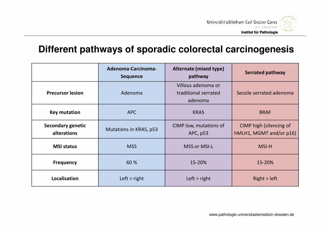
Different pathways of sporadic colorectal carcinogenesis
Adenoma-Carcinoma-
Sequence
Alternate (mixed type)
pathwaySerrated pathway
Precursor lesion Adenoma
Villous adenoma or
traditional serrated
adenoma
Sessile serrated adenoma
Key mutation APC KRAS BRAF
Secondary genetic
alterationsMutations in KRAS, p53
CIMP low, mutations of
APC, p53
CIMP high (silencing of
hMLH1, MGMT and/or p16)
MSI status MSS MSS or MSI-L MSI-H
Frequency 60 % 15-20% 15-20%
Localisation Left > right Left > right Right > left
www.pathologie-universitaetsmedizin-dresden.de
Institut für Pathologie
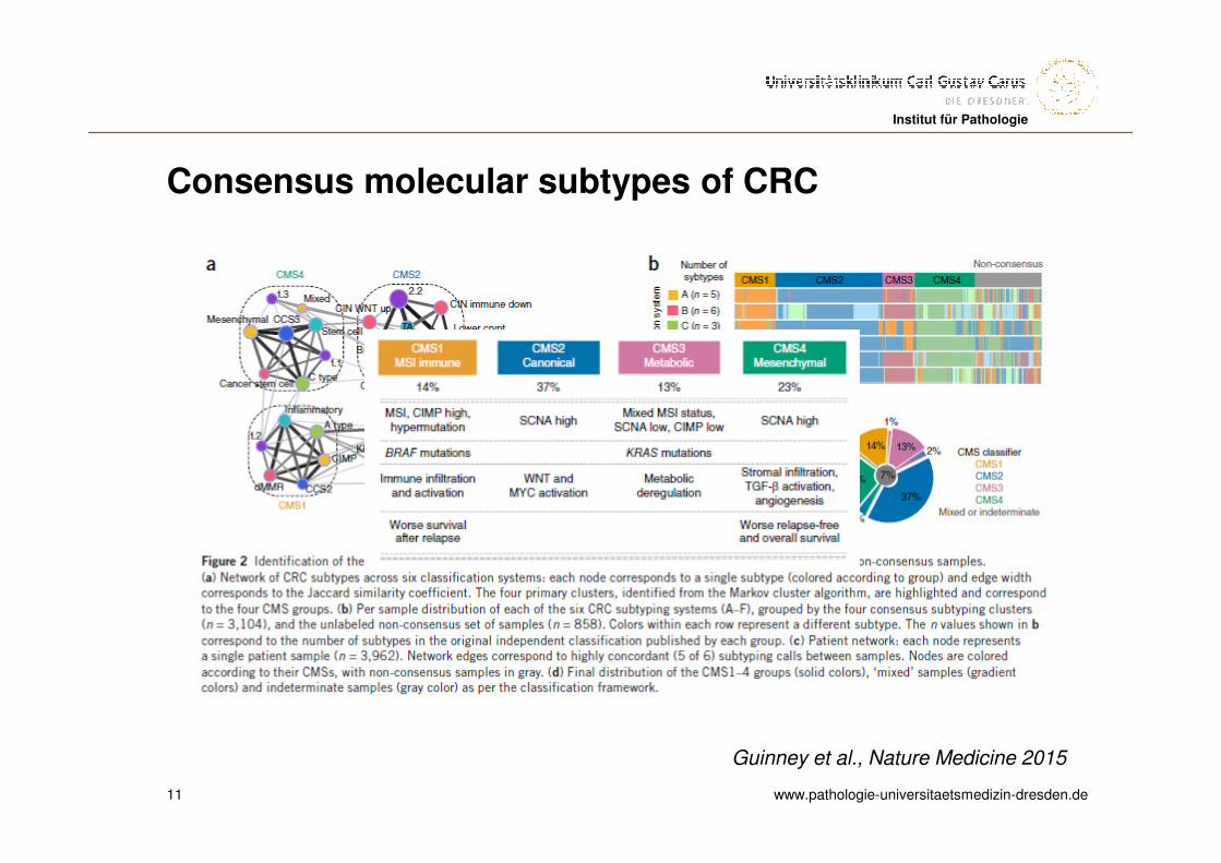
Consensus molecular subtypes of CRC
11
Guinney et al., Nature Medicine 2015
www.pathologie-universitaetsmedizin-dresden.de
Institut für PathologieInstitut für Pathologie
Different pathways of colorectal carcinogenesis
IColorectal cancer is not one disease, it consists of different subentities, developed through different pathways of carcinogenesis
ICertain mutations may be present as either drivers orpassengers and thus may have different prognostic valuein different pathways
www.pathologie-universitaetsmedizin-dresden.de
Institut für PathologieInstitut für Pathologie
Prognostic markers in colorectal cancer
I pTNM
IMicrosatellite instability
I BRAF
I Surgery
IConflicting data: p53, loss of 18q, 17p, gain of 20q13, KRAS, etc.
www.pathologie-universitaetsmedizin-dresden.de
Institut für PathologieInstitut für Pathologie
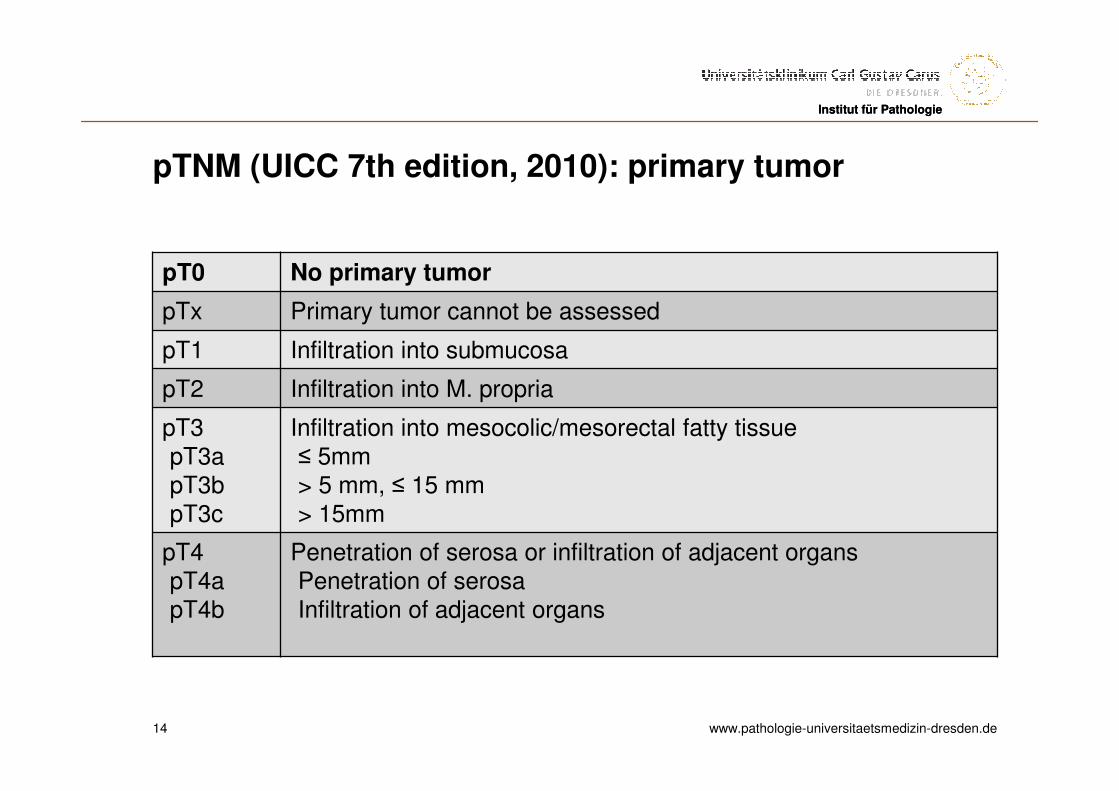
pTNM (UICC 7th edition, 2010): primary tumor
pT0 No primary tumor
pTx Primary tumor cannot be assessed
pT1 Infiltration into submucosa
pT2 Infiltration into M. propria
pT3pT3apT3bpT3c
Infiltration into mesocolic/mesorectal fatty tissue≤ 5mm> 5 mm, ≤ 15 mm> 15mm
pT4pT4apT4b
Penetration of serosa or infiltration of adjacent organsPenetration of serosaInfiltration of adjacent organs
14
www.pathologie-universitaetsmedizin-dresden.de
Institut für PathologieInstitut für Pathologie
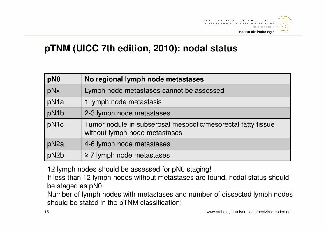
pTNM (UICC 7th edition, 2010): nodal status
pN0 No regional lymph node metastases
pNx Lymph node metastases cannot be assessed
pN1a 1 lymph node metastasis
pN1b 2-3 lymph node metastases
pN1c Tumor nodule in subserosal mesocolic/mesorectal fatty tissuewithout lymph node metastases
pN2a 4-6 lymph node metastases
pN2b ≥ 7 lymph node metastases
15
12 lymph nodes should be assessed for pN0 staging!If less than 12 lymph nodes without metastases are found, nodal status shouldbe staged as pN0!Number of lymph nodes with metastases and number of dissected lymph nodesshould be stated in the pTNM classification!
www.pathologie-universitaetsmedizin-dresden.de
Institut für PathologieInstitut für Pathologie
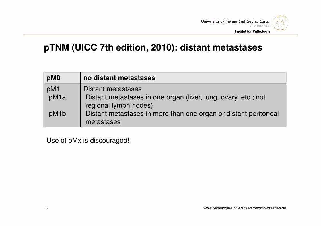
pTNM (UICC 7th edition, 2010): distant metastases
pM0 no distant metastases
pM1pM1a
pM1b
Distant metastasesDistant metastases in one organ (liver, lung, ovary, etc.; not regional lymph nodes)Distant metastases in more than one organ or distant peritonealmetastases
16
Use of pMx is discouraged!
www.pathologie-universitaetsmedizin-dresden.de
Institut für PathologieInstitut für Pathologie
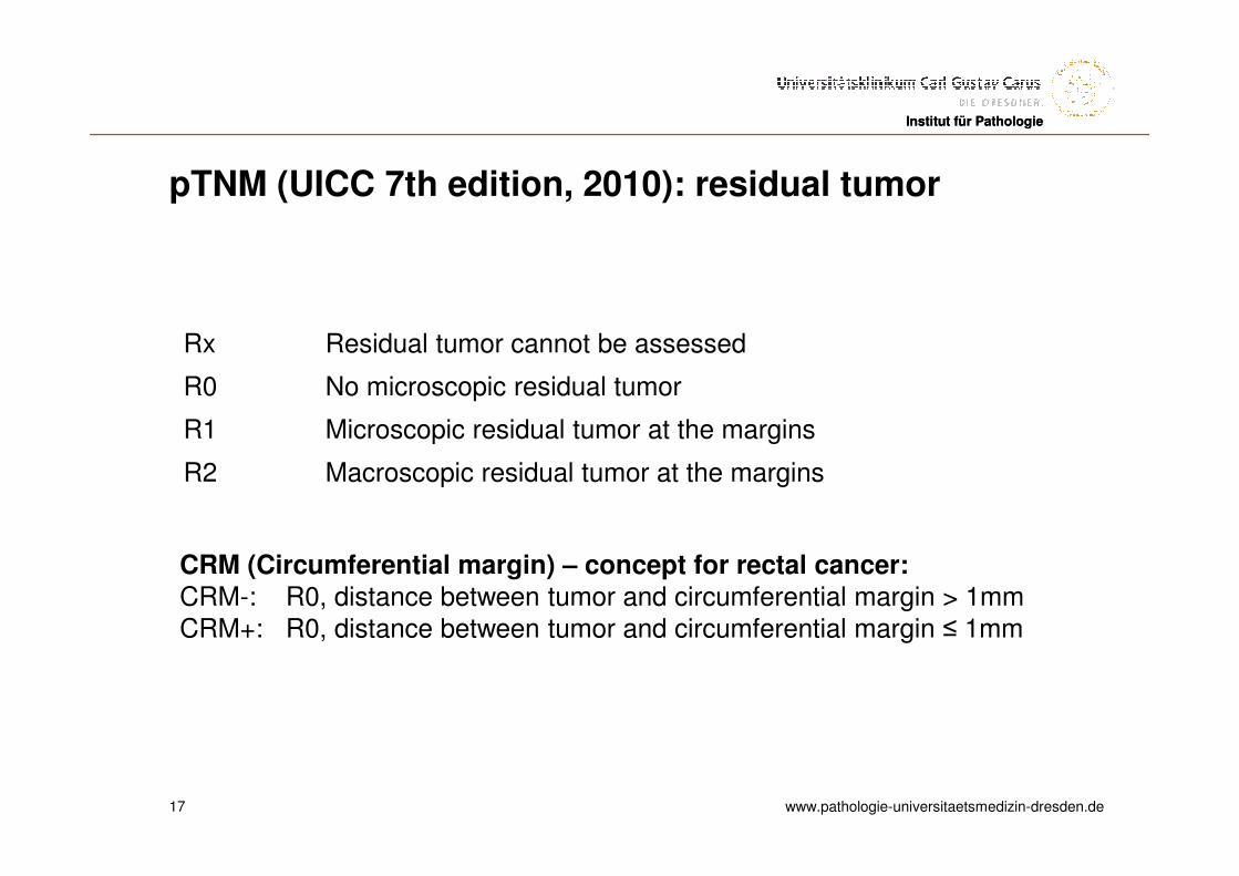
pTNM (UICC 7th edition, 2010): residual tumor
17
Rx Residual tumor cannot be assessed
R0 No microscopic residual tumor
R1 Microscopic residual tumor at the margins
R2 Macroscopic residual tumor at the margins
CRM (Circumferential margin) – concept for rectal cancer:CRM-: R0, distance between tumor and circumferential margin > 1mmCRM+: R0, distance between tumor and circumferential margin ≤ 1mm
www.pathologie-universitaetsmedizin-dresden.de
Institut für PathologieInstitut für Pathologie
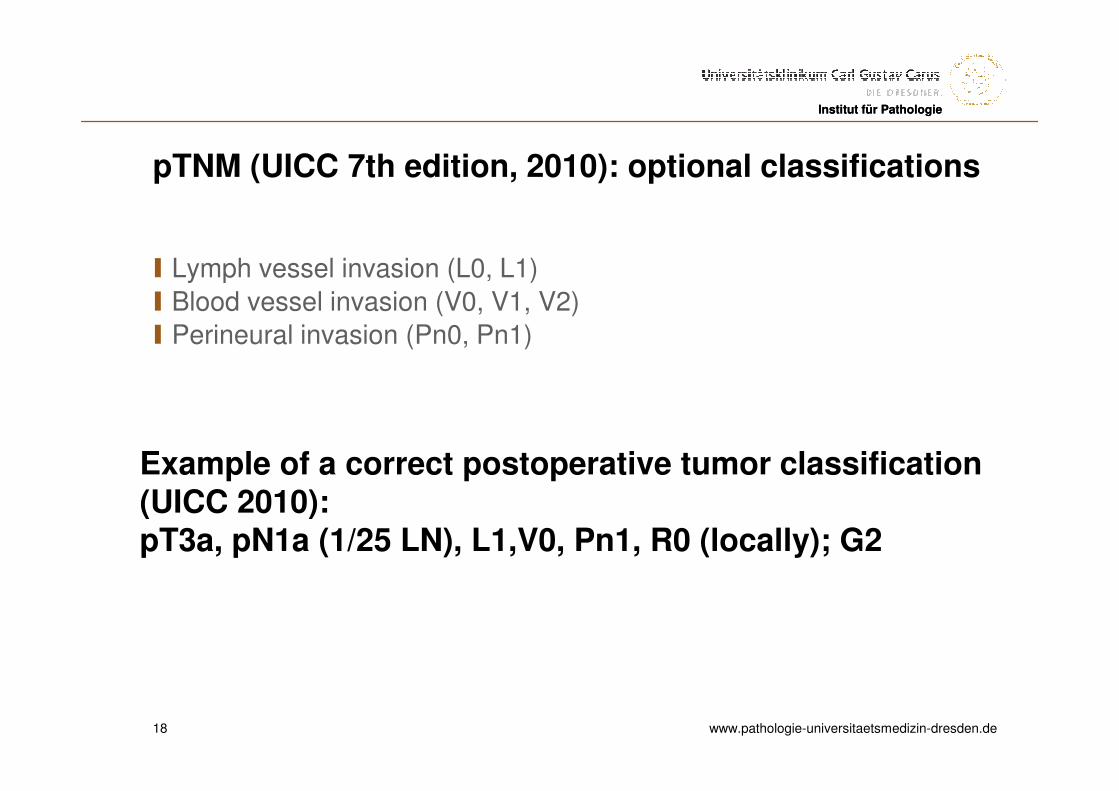
pTNM (UICC 7th edition, 2010): optional classifications
I Lymph vessel invasion (L0, L1)
I Blood vessel invasion (V0, V1, V2)
I Perineural invasion (Pn0, Pn1)
18
Example of a correct postoperative tumor classification(UICC 2010):pT3a, pN1a (1/25 LN), L1,V0, Pn1, R0 (locally); G2
www.pathologie-universitaetsmedizin-dresden.de
Institut für PathologieInstitut für Pathologie
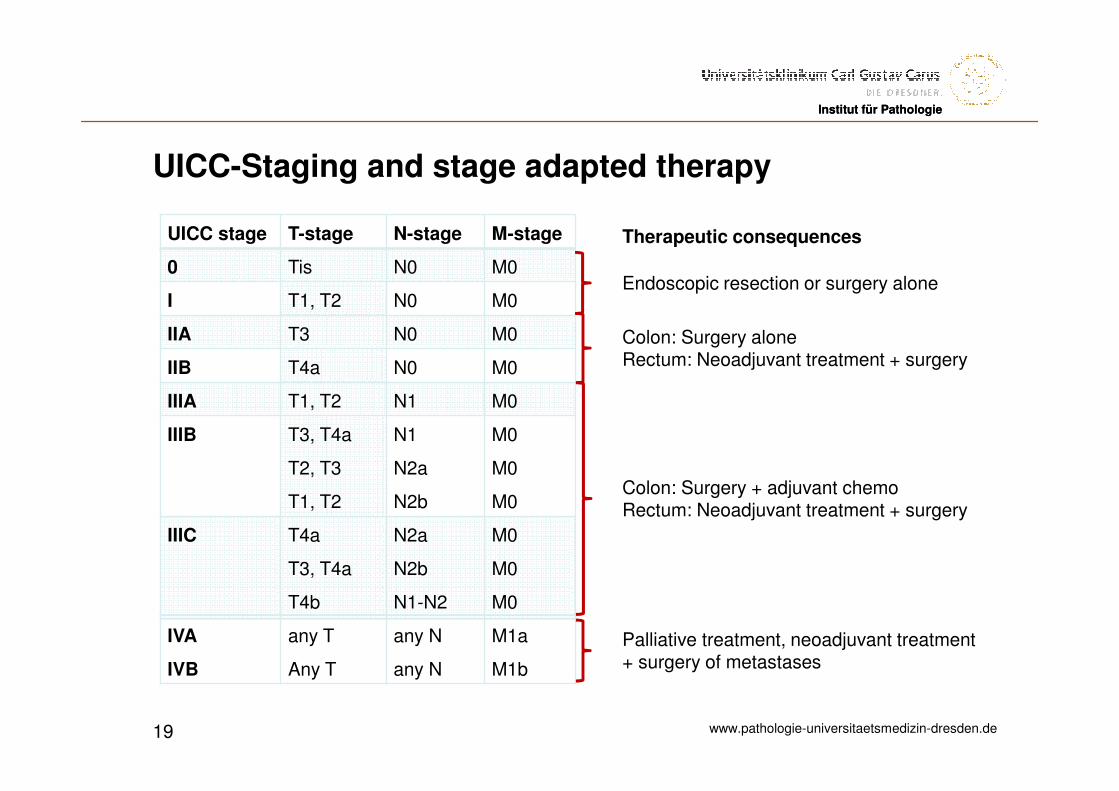
UICC-Staging and stage adapted therapy
UICC stage T-stage N-stage M-stage
0 Tis N0 M0
I T1, T2 N0 M0
IIA T3 N0 M0
IIB T4a N0 M0
IIIA T1, T2 N1 M0
IIIB T3, T4a
T2, T3
T1, T2
N1
N2a
N2b
M0
M0
M0
IIIC T4a
T3, T4a
T4b
N2a
N2b
N1-N2
M0
M0
M0
IVA
IVB
any T
Any T
any N
any N
M1a
M1b
19
Endoscopic resection or surgery alone
Palliative treatment, neoadjuvant treatment+ surgery of metastases
Colon: Surgery aloneRectum: Neoadjuvant treatment + surgery
Colon: Surgery + adjuvant chemoRectum: Neoadjuvant treatment + surgery
Therapeutic consequences
www.pathologie-universitaetsmedizin-dresden.de
Institut für PathologieInstitut für Pathologie
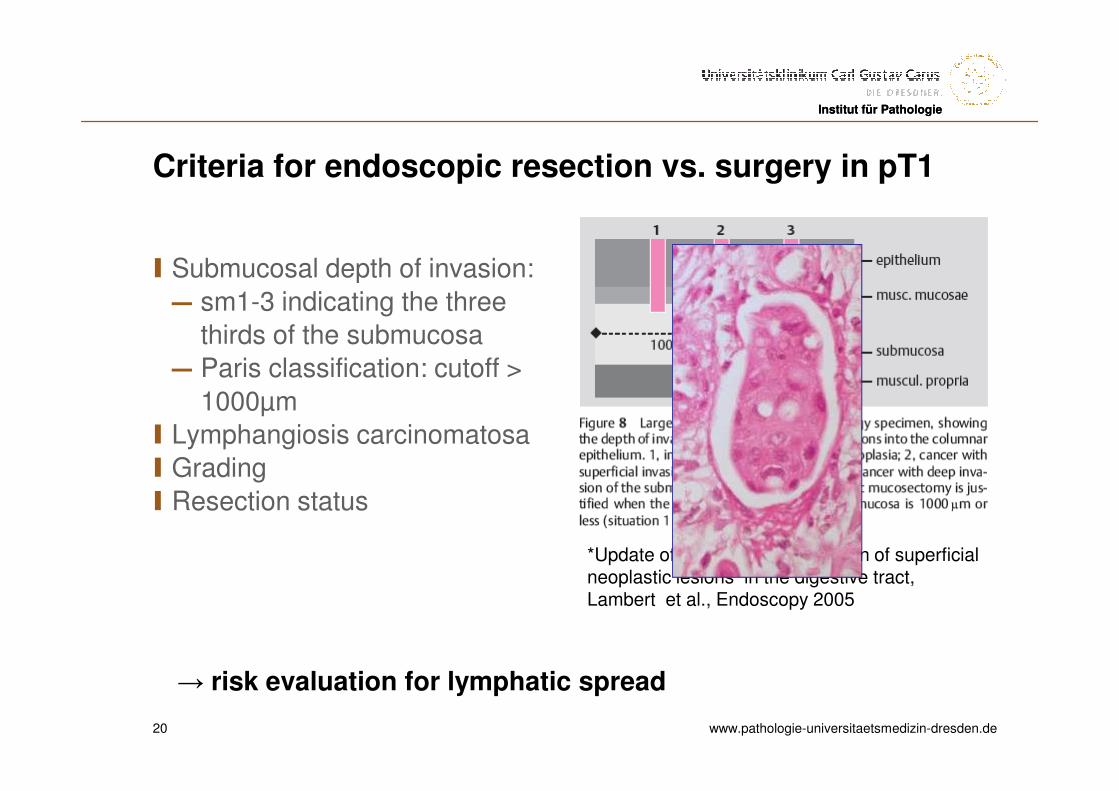
Criteria for endoscopic resection vs. surgery in pT1
I Submucosal depth of invasion:
― sm1-3 indicating the three
thirds of the submucosa
― Paris classification: cutoff >
1000µm
I Lymphangiosis carcinomatosa
I Grading
I Resection status
20
*Update of the Paris-Classification of superficialneoplastic lesions in the digestive tract, Lambert et al., Endoscopy 2005
→ risk evaluation for lymphatic spread
www.pathologie-universitaetsmedizin-dresden.de
Institut für PathologieInstitut für Pathologie
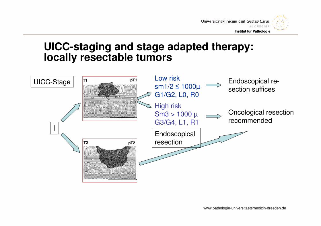
UICC-staging and stage adapted therapy: locally resectable tumors
UICC-Stage Endoscopical re-section suffices
I
Low risksm1/2 ≤ 1000µG1/G2, L0, R0
High riskSm3 > 1000 µG3/G4, L1, R1
Oncological resectionrecommended
Endoscopicalresection
www.pathologie-universitaetsmedizin-dresden.de
Institut für Pathologie
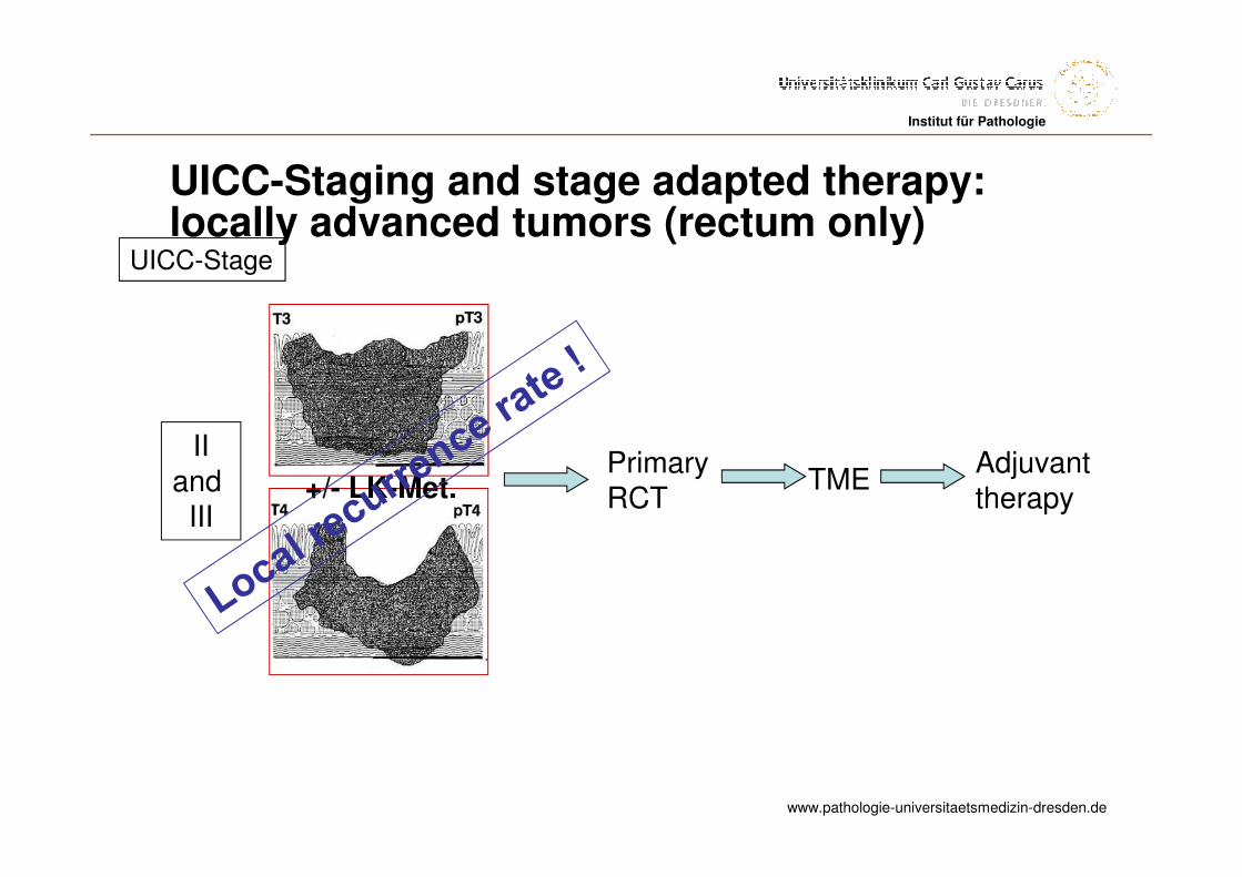
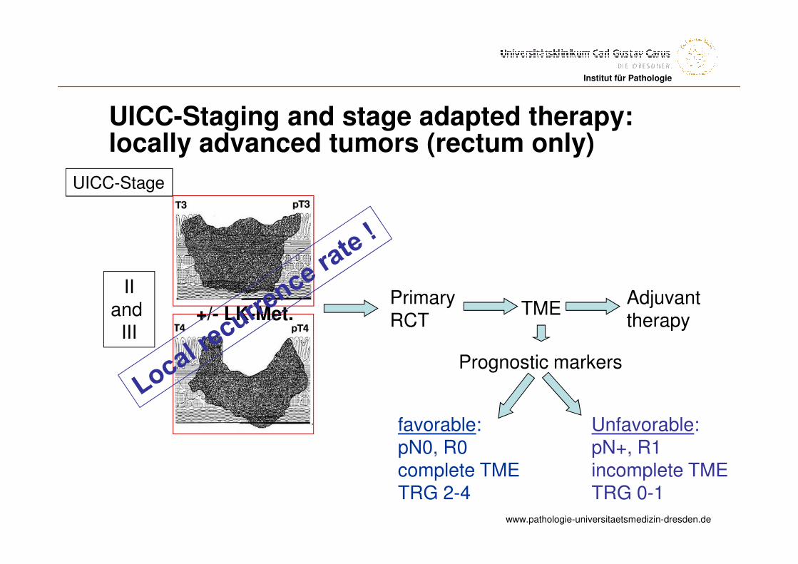
UICC-Staging and stage adapted therapy: locally advanced tumors (rectum only)
UICC-Stage
IIand
III+/- LK-Met.
Primary RCT
TMEAdjuvanttherapy
www.pathologie-universitaetsmedizin-dresden.de
Institut für PathologieInstitut für Pathologie
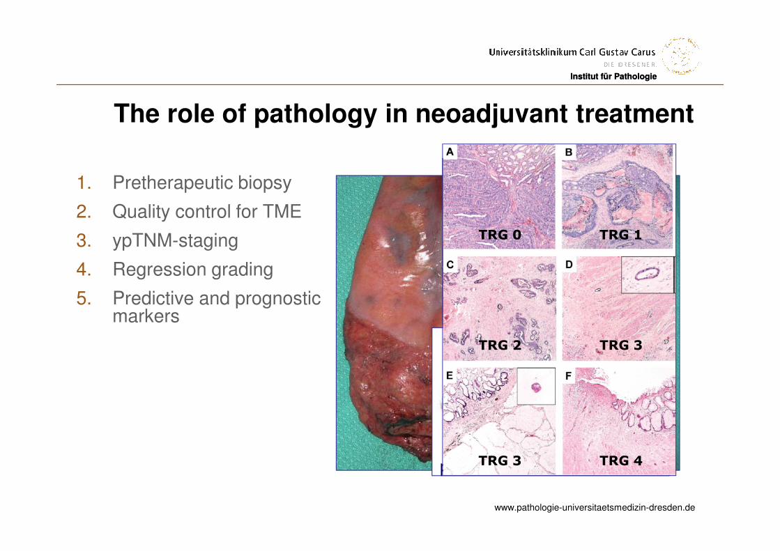
The role of pathology in neoadjuvant treatment
1. Pretherapeutic biopsy
2. Quality control for TME
3. ypTNM-staging
4. Regression grading
5. Predictive and prognosticmarkers
pT3
TUMOR
FIBROSE
TRG 0
TRG 2
TRG 1
TRG 3
TRG 3 TRG 4
www.pathologie-universitaetsmedizin-dresden.de
Institut für Pathologie
UICC-Staging and stage adapted therapy: locally advanced tumors (rectum only)
UICC-Stage
IIand
III+/- LK-Met.
Primary RCT
TMEAdjuvanttherapy
Prognostic markers
favorable:pN0, R0complete TMETRG 2-4
Unfavorable:pN+, R1incomplete TMETRG 0-1
www.pathologie-universitaetsmedizin-dresden.de
Institut für PathologieInstitut für Pathologie
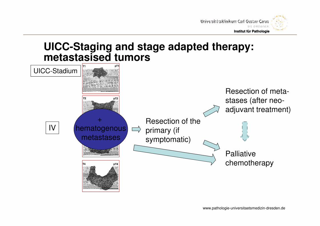
UICC-Staging and stage adapted therapy: metastasised tumors
UICC-Stadium
IV+
hematogenousmetastases
Resection of theprimary (ifsymptomatic)
Resection of meta-stases (after neo-adjuvant treatment)
Palliative chemotherapy
www.pathologie-universitaetsmedizin-dresden.de
Institut für PathologieInstitut für Pathologie
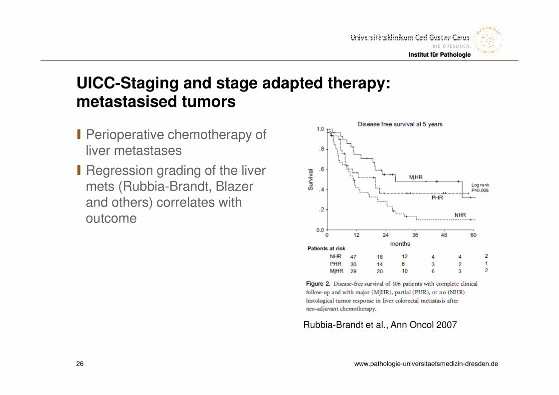
UICC-Staging and stage adapted therapy: metastasised tumors
I Perioperative chemotherapy ofliver metastases
I Regression grading of the livermets (Rubbia-Brandt, Blazer and others) correlates withoutcome
26
Rubbia-Brandt et al., Ann Oncol 2007
www.pathologie-universitaetsmedizin-dresden.de
Institut für PathologieInstitut für Pathologie
Prognostic markers in colorectal cancer
I pTNM
IMicrosatellite instability
I BRAF
www.pathologie-universitaetsmedizin-dresden.de
Institut für Pathologie
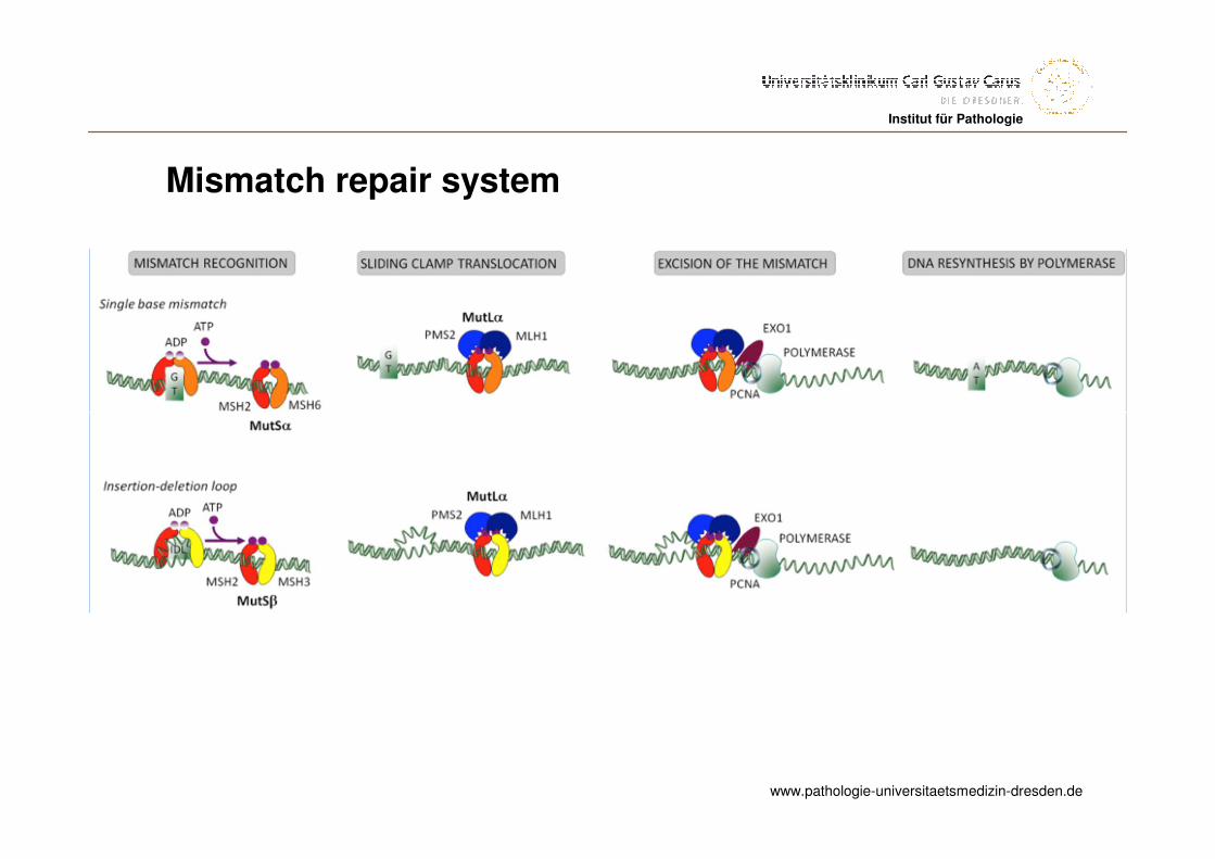
Mismatch repair system
www.pathologie-universitaetsmedizin-dresden.de
Institut für Pathologie
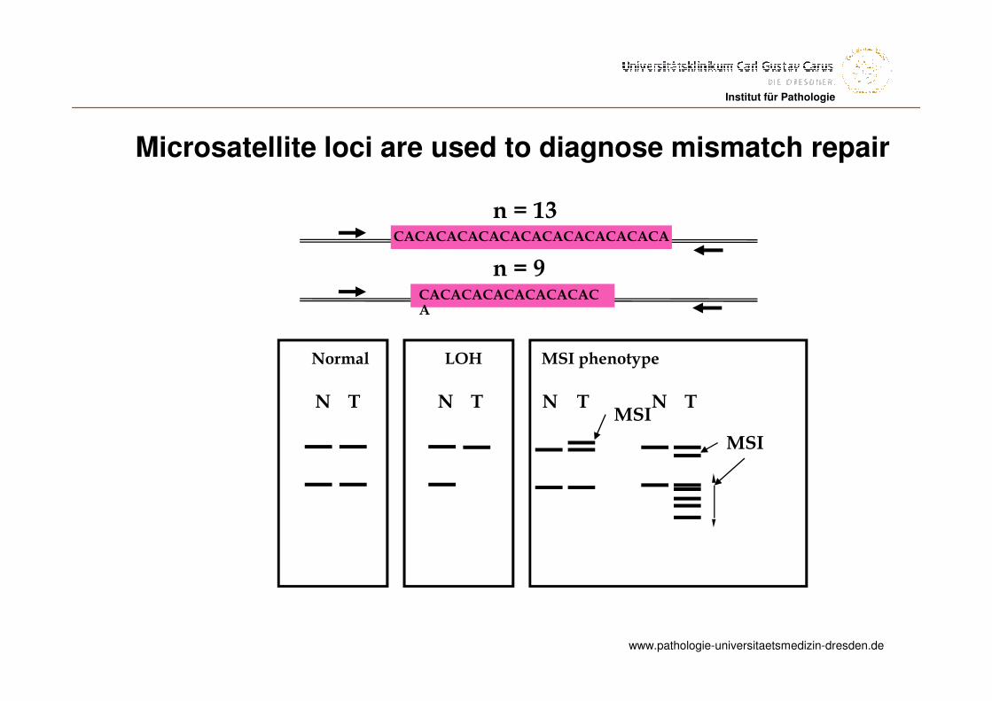
Microsatellite loci are used to diagnose mismatch repair
N T TN
MSI phenotype
TN
Normal
TN
LOH
n = 13
n = 9
CACACACACACACACACACACACACA
CACACACACACACACACA
MSI
MSI
www.pathologie-universitaetsmedizin-dresden.de
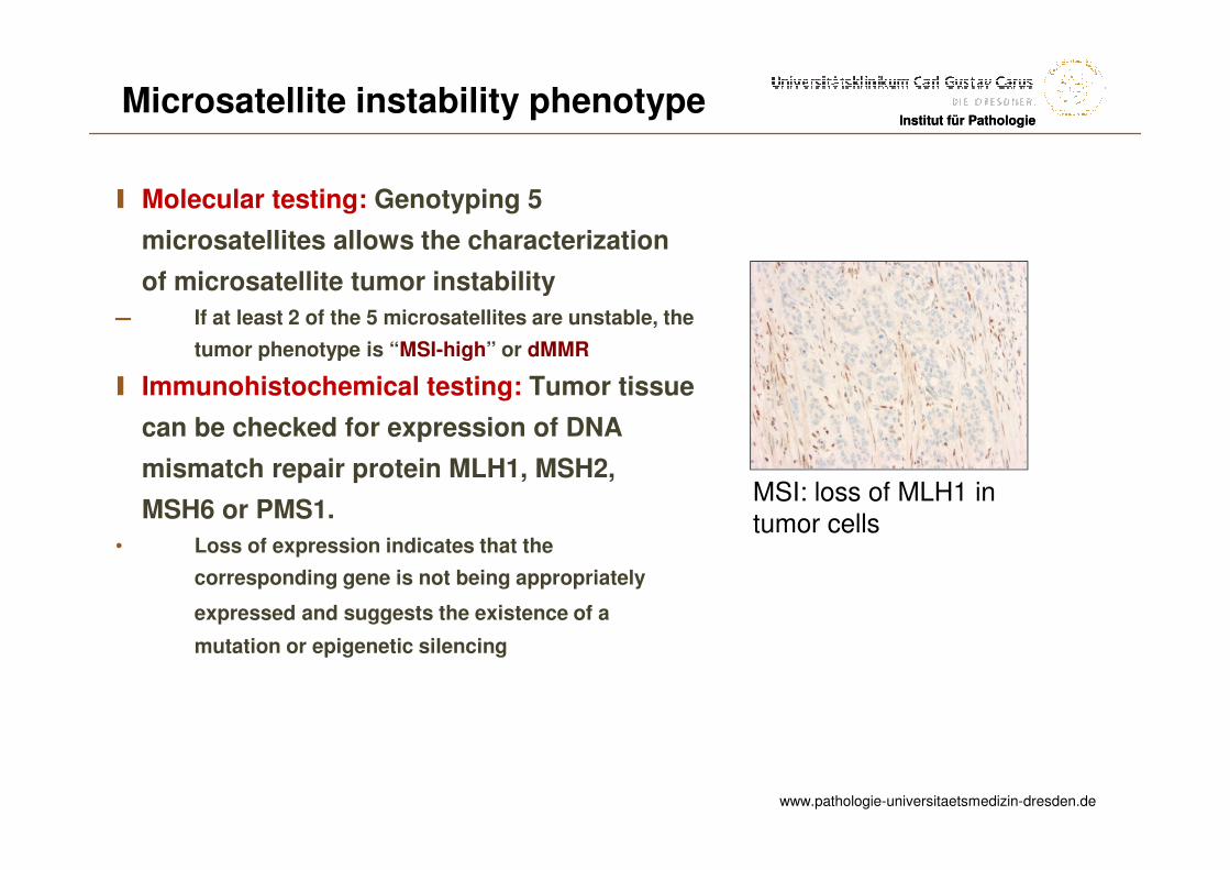
Institut für PathologieInstitut für PathologieMicrosatellite instability phenotype
I Molecular testing: Genotyping 5
microsatellites allows the characterization
of microsatellite tumor instability
― If at least 2 of the 5 microsatellites are unstable, the
tumor phenotype is “MSI-high” or dMMR
I Immunohistochemical testing: Tumor tissue
can be checked for expression of DNA
mismatch repair protein MLH1, MSH2,
MSH6 or PMS1.
• Loss of expression indicates that the
corresponding gene is not being appropriately
expressed and suggests the existence of a
mutation or epigenetic silencing
MSI: loss of MLH1 in tumor cells
www.pathologie-universitaetsmedizin-dresden.de
Institut für Pathologie
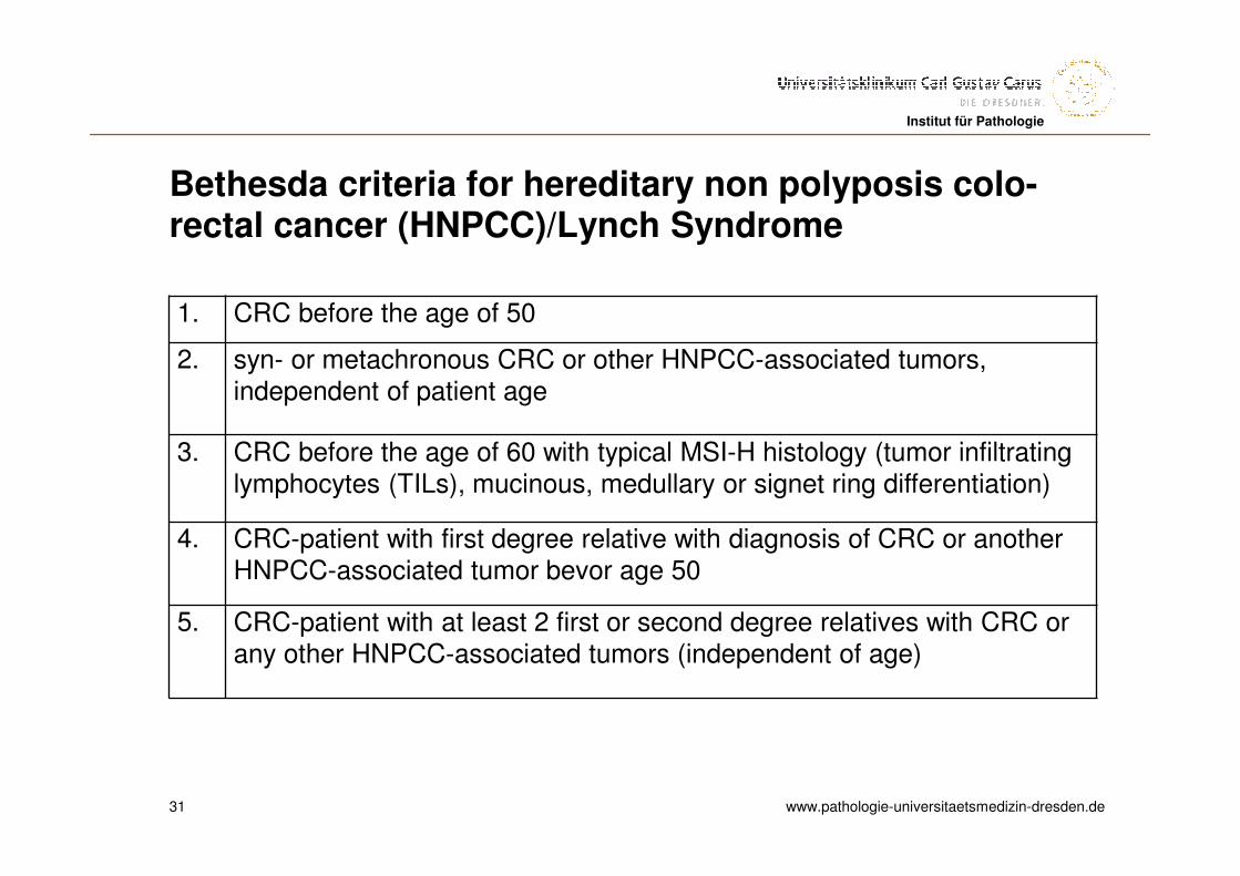
Bethesda criteria for hereditary non polyposis colo-rectal cancer (HNPCC)/Lynch Syndrome
31
1. CRC before the age of 50
2. syn- or metachronous CRC or other HNPCC-associated tumors,independent of patient age
3. CRC before the age of 60 with typical MSI-H histology (tumor infiltratinglymphocytes (TILs), mucinous, medullary or signet ring differentiation)
4. CRC-patient with first degree relative with diagnosis of CRC or anotherHNPCC-associated tumor bevor age 50
5. CRC-patient with at least 2 first or second degree relatives with CRC orany other HNPCC-associated tumors (independent of age)
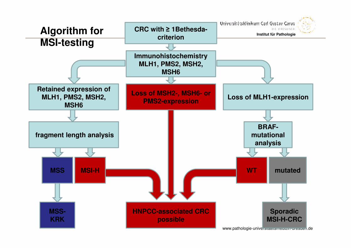
www.pathologie-universitaetsmedizin-dresden.de
Institut für PathologieCRC with ≥ 1Bethesda-
criterion
ImmunohistochemistryMLH1, PMS2, MSH2,
MSH6
Loss of MSH2-, MSH6- orPMS2-expression
HNPCC-associated CRC possible
Retained expression ofMLH1, PMS2, MSH2,
MSH6 Loss of MLH1-expression
fragment length analysis
MSS MSI-H
BRAF-mutational
analysis
WT mutated
MSS-KRK
SporadicMSI-H-CRC
Algorithm forMSI-testing
www.pathologie-universitaetsmedizin-dresden.de
Institut für Pathologie
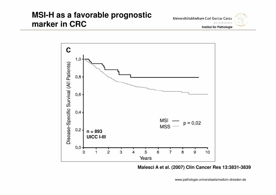
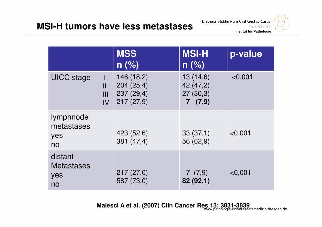
Malesci A et al. (2007) Clin Cancer Res 13:3831-3839
MSI-H as a favorable prognosticmarker in CRC
n = 893UICC I-III
www.pathologie-universitaetsmedizin-dresden.de
Institut für Pathologie
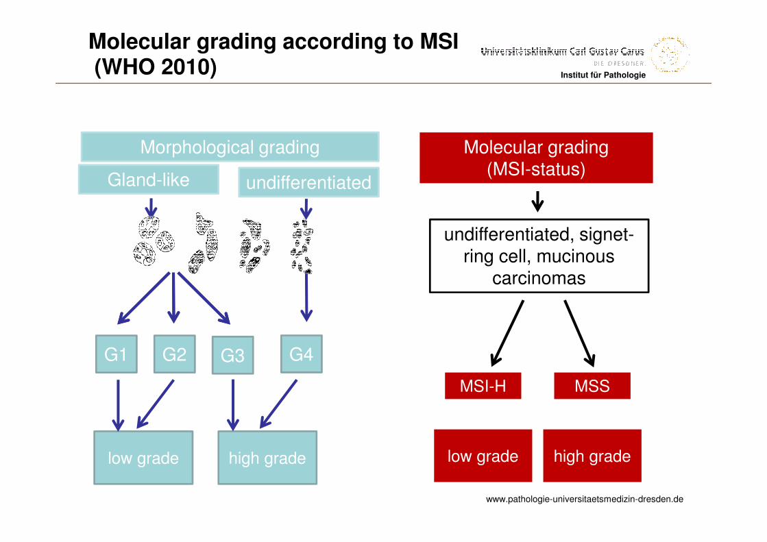
Morphological grading
G1 G2 G3
low grade high grade
undifferentiated, signet-ring cell, mucinous
carcinomas
Molecular grading(MSI-status)
low grade high grade
MSI-H MSS
Gland-like undifferentiated
G4
Molecular grading according to MSI(WHO 2010)
www.pathologie-universitaetsmedizin-dresden.de
Institut für Pathologie
MSSn (%)
MSI-Hn (%)
p-value
UICC stage I
IIIIIIV
146 (18,2)204 (25,4)237 (29,4)217 (27,9)
13 (14,6)42 (47,2)27 (30,3)7 (7,9)
<0,001
lymphnodemetastasesyesno
423 (52,6)381 (47,4)
33 (37,1)56 (62,9)
<0,001
distantMetastasesyesno
217 (27,0)587 (73,0)
7 (7,9)82 (92,1)
<0,001
Malesci A et al. (2007) Clin Cancer Res 13: 3831-3839
MSI-H tumors have less metastases
www.pathologie-universitaetsmedizin-dresden.de
Institut für PathologieInstitut für Pathologie
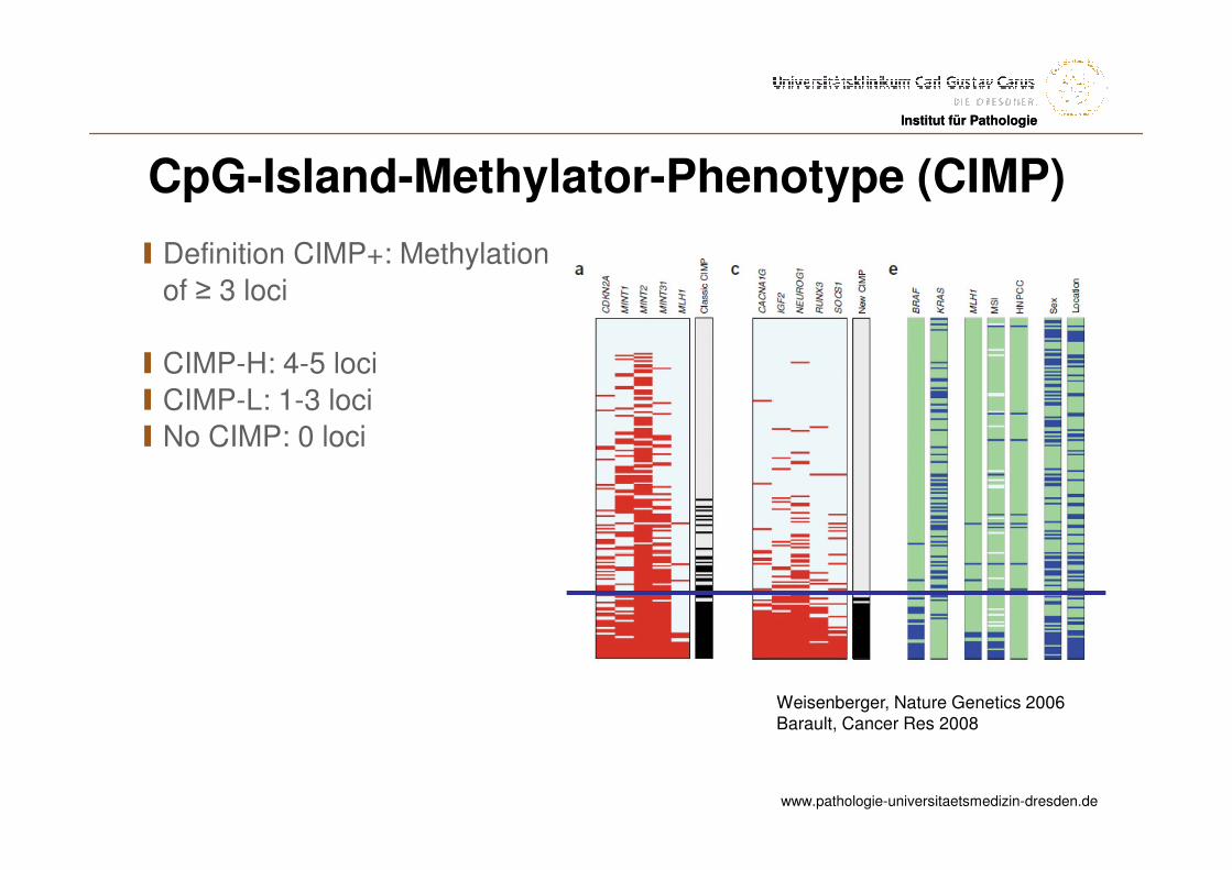
CpG-Island-Methylator-Phenotype (CIMP)
I Definition CIMP+: Methylation
of ≥ 3 loci
I CIMP-H: 4-5 loci
I CIMP-L: 1-3 loci
I No CIMP: 0 loci
Weisenberger, Nature Genetics 2006Barault, Cancer Res 2008
www.pathologie-universitaetsmedizin-dresden.de
Institut für Pathologie
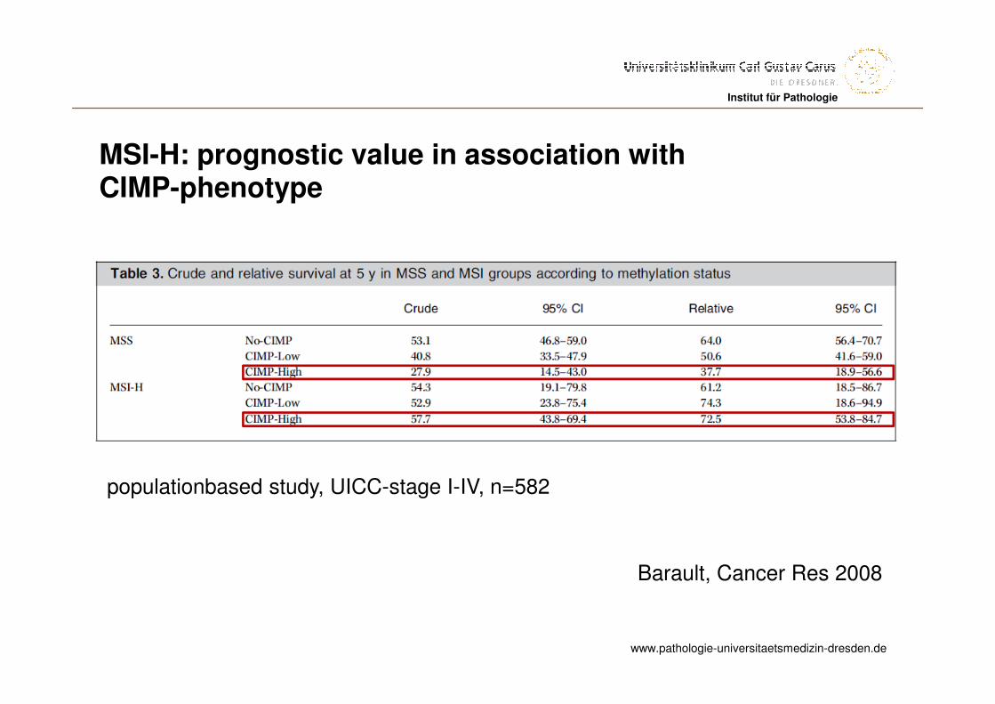
Barault, Cancer Res 2008
populationbased study, UICC-stage I-IV, n=582
MSI-H: prognostic value in association withCIMP-phenotype

www.pathologie-universitaetsmedizin-dresden.de
Institut für PathologieInstitut für Pathologie
Prognostic markers in colorectal cancer
I pTNM
IMicrosatellite instability
I BRAF
www.pathologie-universitaetsmedizin-dresden.de
Institut für Pathologie
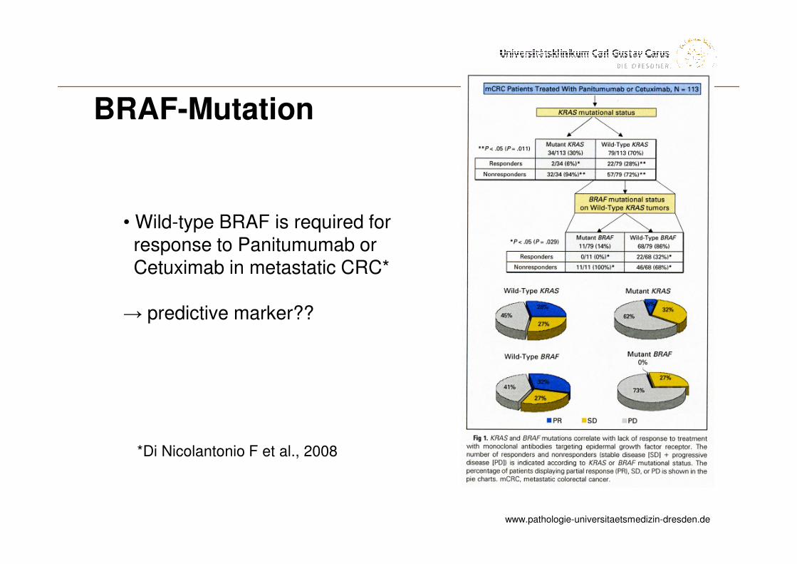
*Di Nicolantonio F et al., 2008
• Wild-type BRAF is required forresponse to Panitumumab orCetuximab in metastatic CRC*
→ predictive marker??
BRAF-Mutation
www.pathologie-universitaetsmedizin-dresden.de
Institut für Pathologie
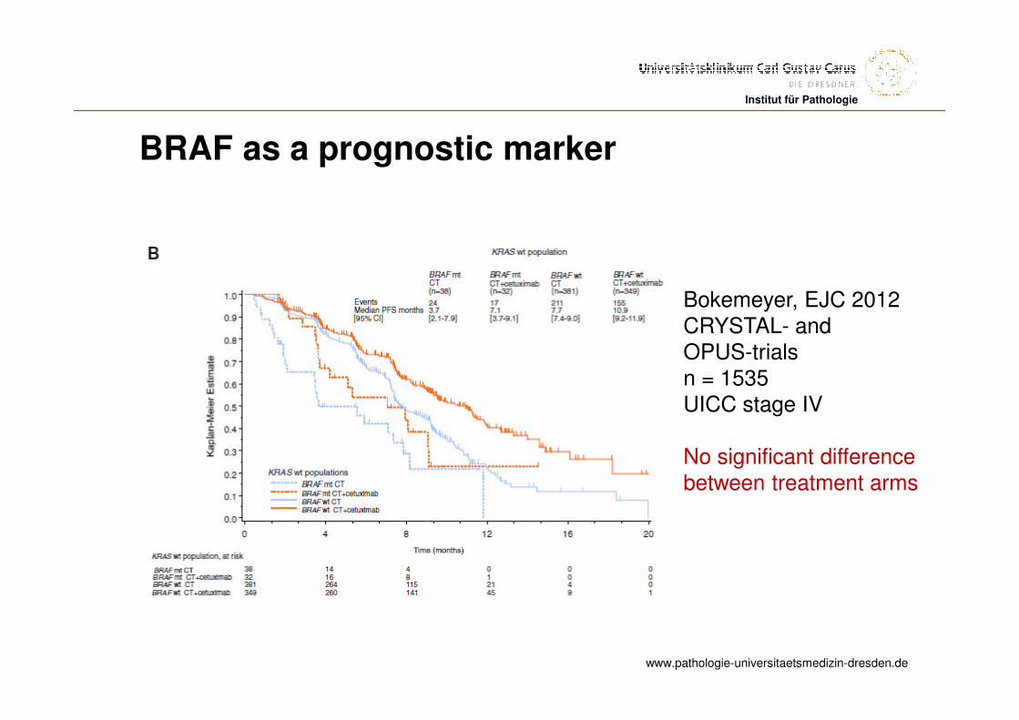
BRAF as a prognostic marker
Bokemeyer, EJC 2012CRYSTAL- andOPUS-trialsn = 1535 UICC stage IV
No significant differencebetween treatment arms
www.pathologie-universitaetsmedizin-dresden.de
Institut für Pathologie
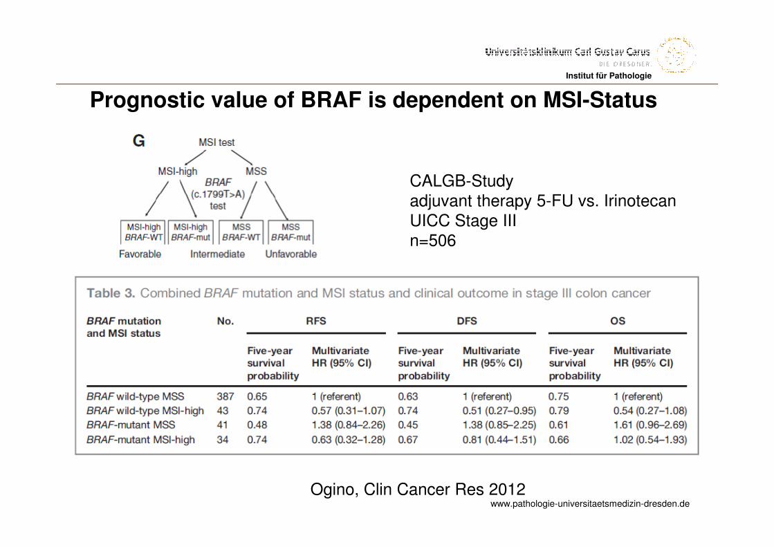
Prognostic value of BRAF is dependent on MSI-Status
Ogino, Clin Cancer Res 2012
CALGB-Studyadjuvant therapy 5-FU vs. IrinotecanUICC Stage IIIn=506
www.pathologie-universitaetsmedizin-dresden.de
Institut für Pathologie
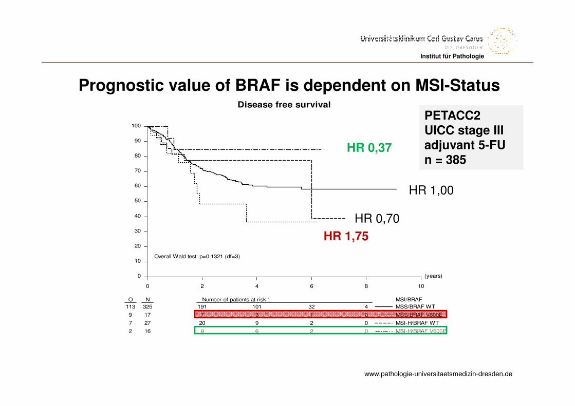
PETACC2 UICC stage IIIadjuvant 5-FUn = 385
(years)
0 2 4 6 8 10
0
10
20
30
40
50
60
70
80
90
100
O N Number of patients at risk : MSI/BRAF
113 325 191 101 32 4
9 17 7 3 1 0
7 27 20 9 2 0
2 16 9 6 2 0
MSS/BRAF WT
MSS/BRAF V600E
MSI-H/BRAF WT
MSI-H/BRAF V600E
Disease free survival
Overall Wald test: p=0.1321 (df=3)
HR 0,37
HR 1,00
HR 0,70
HR 1,75
Prognostic value of BRAF is dependent on MSI-Status
www.pathologie-universitaetsmedizin-dresden.de
Institut für PathologieInstitut für Pathologie
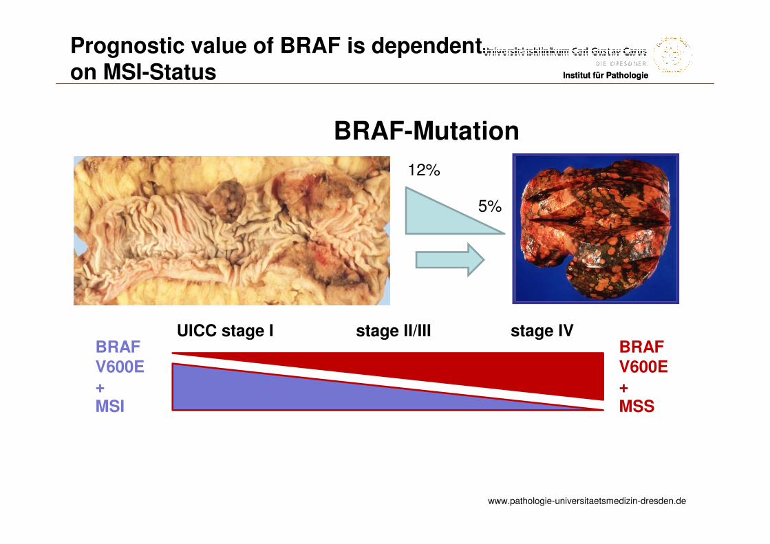
BRAF-Mutation
UICC stage I stage II/III stage IVBRAFV600E+MSS
BRAFV600E+MSI
12%
5%
Prognostic value of BRAF is dependenton MSI-Status
www.pathologie-universitaetsmedizin-dresden.de
Institut für Pathologie
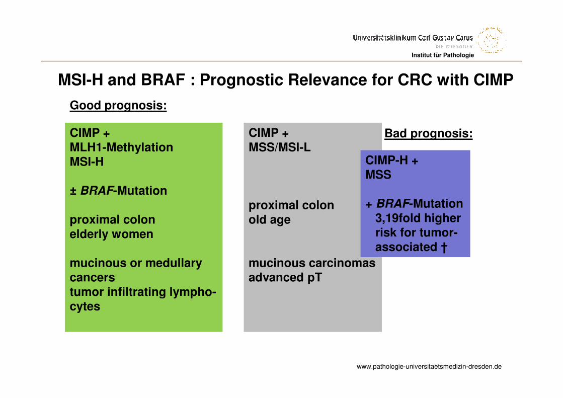
CIMP +MLH1-Methylation MSI-H
± BRAF-Mutation
proximal colonelderly women
mucinous or medullarycancerstumor infiltrating lympho-cytes
Good prognosis:
CIMP + MSS/MSI-L
proximal colonold age
mucinous carcinomasadvanced pT
CIMP-H +MSS
+ BRAF-Mutation3,19fold higherrisk for tumor-associated †
Bad prognosis:
MSI-H and BRAF : Prognostic Relevance for CRC with CIMP
www.pathologie-universitaetsmedizin-dresden.de
Institut für PathologieInstitut für Pathologie
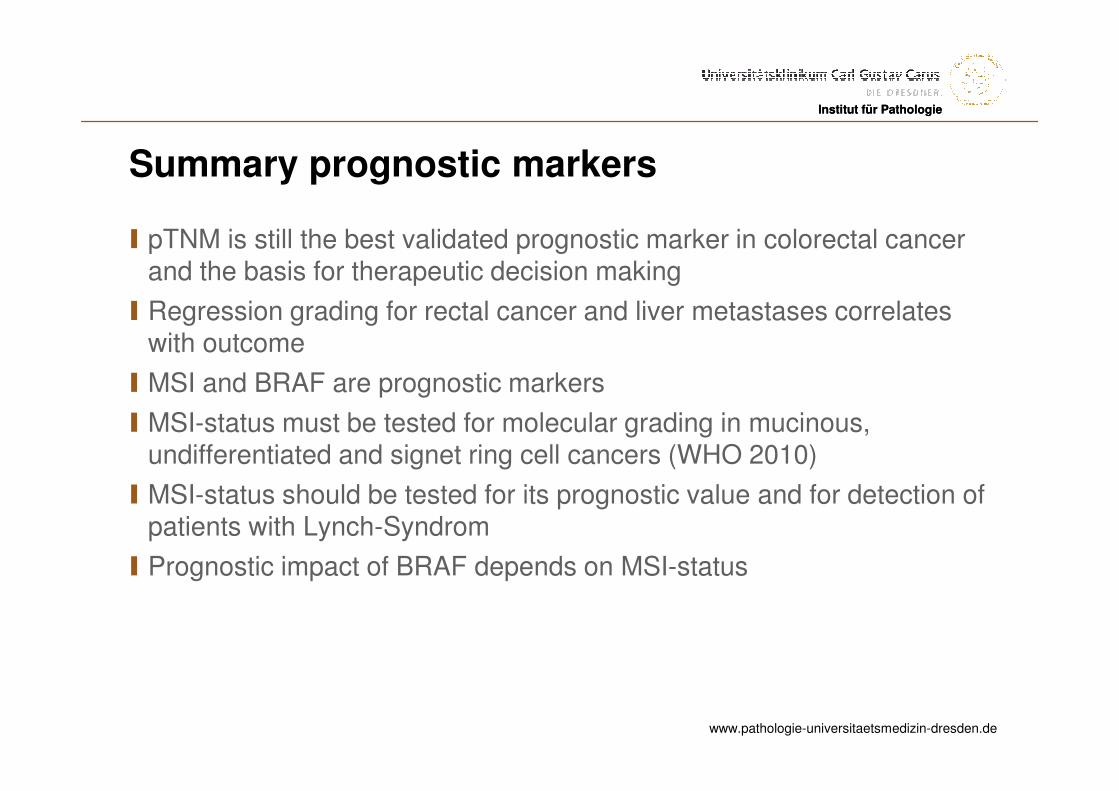
Summary prognostic markers
I pTNM is still the best validated prognostic marker in colorectal cancerand the basis for therapeutic decision making
I Regression grading for rectal cancer and liver metastases correlateswith outcome
I MSI and BRAF are prognostic markers
I MSI-status must be tested for molecular grading in mucinous, undifferentiated and signet ring cell cancers (WHO 2010)
I MSI-status should be tested for its prognostic value and for detection of patients with Lynch-Syndrom
I Prognostic impact of BRAF depends on MSI-status