Embed Size (px)
Citation preview
11. NEUROIMMUNOLOGY:IMMUNOGLOBULINS AND THEINTRATHECAL POLYSPECIFIC IMMUNERESPONSE IN ACUTE, SUBACUTE ANDCHRONIC NEUROLOGICAL DISEASES
by Professor Hansotto Reiber, Ph.D.,Neurochemisches Labor derNeurologischen Klinik,Universität Göttingen, Robert-KochStrasse 40, 37075 Göttingen, Germany
Acute, subacute and chronic inflammatory diseases of the centralnervous system (CNS) are often accompanied by humoral immuneresponse characterized by the following methods:
• The disease-related immunoglobulin class pattern (1)
• The specific antibodies against the causative antigen (IgG-, IgA-,IgM-class) (2)
• Oligocloncal IgG (IgG-class) (3)
• Polyspecific immune response against a large variety of non-causative antigens (4)
Intrathecally synthesized antibodies, detected in cerebrospinal fluid(CSF), can have two different sources: a causative persisting antigenor a polyspecific concomitant immune response. The classical viewof the immune response is the clonal selection of a B lymphocyteclone producing specific antibodies against the invadingmicroorganism. As always several clones are found to fit thespecific antigen, we speak about an “oligoclonal” immune reaction.In addition to the oligoclonal antibodies against the specificcausative antigen each immune reaction produces a wide spectrumof different antibody-species not connected with the causativeantigen. This “polyspecific immune response” does not depend onthe presence of a corresponding persisting antigen (13) and is oflower intensities than for the causative antigen (4, 5).
The detection of oligoclonal immunoglobulin G (IgG) in CSF is abasic part of a laboratory supported diagnosis of MS (3). Anunexplained high frequency of combined intrathecal measles,rubella and varicella zoster virus (VZV) antibodies in CSF ofpatients with (MS) (4) or autoimmune diseases with involvement ofthe CNS (6) allows the diagnosis of a chronic inflammatory process(autoimmune type) at the time of the first clinical manifestation (1,4). This is also important for diagnosis of cases with amonosymptomatic start of the disease like an optical neuritis or anuveitis intermedia and periphlebitis retinae (7).
It was a long way to understand that antibodies, which aresynthesized in brain and in blood, are not only directed against a
causative antigen (clonal selection) but also against other antigens,which are not involved in the cause of the disease. The first detectionof intrathecal measles antibody synthesis (8) in multiple sclerosis(MS) led to the hypothesis of a virus aetiology of MS. Laterobservations showed a polyspecific antibody synthesis againstrubella, varicella zoster, herpes simplex, mumps viruses (9) in thesingle MS-patient. Meanwhile there are also reports about theintrathecal synthesis of autoantibodies against ds-DNA, observed ina fraction of MS patients (4, 6).
It is the recognition of an immunological network (10), which givesthe clue to understand the polyspecific, oligoclonal immuneresponse.
The detection of intrathecal antibody synthesis in CSF has a longtradition. The linear Goldmann-Witmer-Index (11) frequently usedin ophthalmology (GW-I= Qspec / QIgG) is improved by thecorrected “Antibody-Index”, AI (2), established in CSF analysis toavoid a false negative interpretation (7) in cases with a strongintrathecal IgG synthesis.
The AI presents a relative value for the quantity of intrathecallysynthesized specific antibodies. With the invention of themeasurement of absolute antibody concentrations (5) theevaluation of quantitative intrathecal antibody synthesis becamepossible. With an improved calculation of the specific antibodyfraction in CSF, Fs, a virus-driven antibody synthesis can now bediscriminated from a polyspecific, network- related immuneresponse.
These methodological improvements are based on the evaluation ofimmunoglobulin quotients QIgG, QIgA, QIgM with a nonlinear,hyperbolic discrimination function, QLim (2), which allows thesensitive discrimination between blood- and brain- derivedimmunoglobulin fractions (i.e., intrathecal synthesis of IgG, IgA andIgM) in CSF. This replaces the earlier linear approaches, like IgG-Index (Ref. in (1)), which lead to false interpretations in cases of ablood/CSF barrier dysfunction as demonstrated in detail (1).
The polyspecific, oligoclonal IgG response is seen in many chronic,acute and subacute inflammatory diseases of the CNS, but there arebasic differences in quantity (Tab. 1) and the frequency of anintrathecally synthesized antibody species (4, 7).
In MS (4) as well as in autoimmune diseases with involvement of theCNS (6), i.e., in chronic inflammatory diseases, we observe a highfrequency of measles-, rubella- and VZV intrathecal antibodysynthesis among antibodies against other virotropic viruses orother microorganisms, like toxoplasma gondii (4), or chlamydiapneumonia (14). This frequency is not seen in any other acute orsubacute chronic disease.
The frequency of measles antibodies in MS is 78%, for rubella 60%,for VZV 55% and for HSV 28%. In other acute or subacuteinflammatory diseases (HSV-E, SSPE, neurosyphilis,
Page 82eJIFCC2004Vol15No3pp082-085

neurotuberculosis) we find the antibody response for these singlespecies with the frequency below 5% and for a combination of twoantibody species (e.g., measles and rubella) below 0.1%.
In MS patients we observed with an increasing intrathecal IgGfraction an increasing frequency of the combination of all threeantibody species (measles, rubella, zoster = M, R, Z) compared withthose patients who had a low intrathecal IgG response with only oneor a combination of two of these three antibodies (4). Withincreasing intrathecal IgG synthesis there is also an increasingamount of antibodies produced for these MRZ species, indicated byan increasing AI (4).
With the invention of the quantitation, i.e., characterization of theintensity of the intrathecal antibody response (5) and theimprovement of the calculation (7) for FS (Methods), we get newperspectives for detection of intrathecally synthesized antibodies,regarding its diagnostic as well as its pathophysiological relevance.
Table 1. Mean intensity of the intrathecal virus-specific antibodysynthesis in acute, subacute and chronic inflammatory diseasesnof the CNS. Comparison of the Antibody-index (AI) and thespecific intrathecal antibody fraction (FS) against the causativeantigen in subacute sclerosing panencephalitis (SSPE), herpessimplex encepahalitis (HSV-E) and the Fuchs heterochromiccyclitis of the eye (FHC) besides multiple sclerosis (MS) with apolyspecific immune reaction against non-causal antigens.
1 Data in the aqueous humor of the eye in the Fuchs heterochromiccyclitis (7)2 Correspondingly, in MS patients with uveitis or periphlebitis thefollowing data are found in aqueous humor: Rubella-AI = 3.0 (0.7-35); Rubella-FS = 0.06 (0.01-0.25)%
In Table 1 we compare directly the Antibody-Index (AI) with thespecific antibody fraction (FS) for chronic, acute and subacutediseases. From these data we learn that also in acute or subacutediseases with a persistent causative antigen, only less than 30% ofthe intrathecally synthesized total IgG represents the specificantibodies (20% measles antibodies in SSPE, 9% herpes simplexantibodies in HSV-E, or 2.6% rubella antibodies in aqueous humor ofthe Fuchs heterochromic cyclitis (FHC) of the eye). But it is clear bythese data that the intensity of antibody synthesis against thecausative antigen is up to 60-fold higher than the intensity of thepolyspecific antibody synthesis in a chronic inflammatory process,like MS (FS < 0.5%). The specific fraction, FS, allows a betterdiscrimination between these both causes of the antibody responsethan the AI. AI is a relative value depending on the ratio of theamounts of the antibodies in CSF, which derive from blood andbrain FS contributes an important diagnostic aspect to discriminate
the polyspecific antibody synthesis from the antibody synthesisagainst a causative antigen. Nevertheless, for the general diagnosticapproach, AI remains the most sensitive parameter to detect anintrathecal antibody synthesis.
As a second important information from these data in Tab. 1, welearn that also in case of a specific antibody response against thecausative antigen the larger fraction of the intrathecally synthesizedIgG represents a polyspecific antibody response against the non-causative antigens, which do not need the persistence of an antigenin the immune system (13).
As a particular clue of these investigations, e.g., about the rubellaantibody synthesis in the eye of the FHC patients, we get a biologicalexample for theoretical approaches (15), which try to explain thedynamics, which in the immune response can lead to a chronicinflammatory process.
With these approaches we are on the way to a new understanding ofan immunological network-based immune response in chronicinflammatory diseases.
11.2 Methods
11.2.1 Oligoclonal IgG
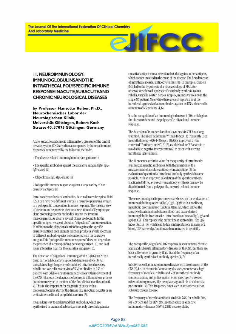
According to the international consensus, oligoclonal IgG isdetected with isoelectric focussing and immune detection(Andersson et al. 1994). As interpretation criteria the 5 typesshown in figure 1 are accepted as state of the art.
Figure 1. Isoelectric focussing on agarose gels with immunoblot:The figure represents the classical types 1 – 5 (Andersson et al.1994):
• Type 1: No bands in CSF and serum.
• Type 2: Oligoclonal IgG-bands in CSF, not in serum.Interpretation: Intrathecal IgG-synthesis.
• Type 3: Oligoclonal bands in CSF (like type 2) and additionalidentical oligoclonal bands in CSF and serum (like type 4).Interpretation: Intrathecal IgG-synthesis
Page 83eJIFCC2004Vol15No3pp082-085
• Type 4: Identical oligoclonal bands in CSF and serum.Interpretation: No intrathecal IgG-synthesis but systemic immunereaction.
• Type 5: Monoclonal bands in CSF and serum. Interpretation:Systemic paraproteinaemia.
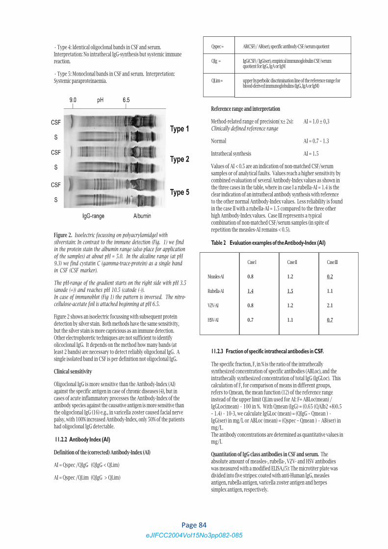
Figure 2. Isoelectric focussing on polyacrylamidgel withsilverstain: In contrast to the immune detection (Fig. 1) we findin the protein stain the albumin range (also place for applicationof the samples) at about pH = 5.0. In the alcaline range (at pH9.3) we find cystatin C (gamma-trace-protein) as a single bandin CSF (CSF marker).
The pH-range of the gradient starts on the right side with pH 3.5(anode (+)) and reaches pH 10.5 (catode (-)).In case of immunoblot (Fig 1) the pattern is inversed. The nitro-cellulose-acetate foil is attached beginning at pH 6.5.
Figure 2 shows an isoelectric focussing with subsequent proteindetection by silver stain. Both methods have the same sensitivity,but the silver stain is more capricious as an immune detection.Other electrophoretic techniques are not sufficient to identifyolicoclonal IgG. It depends on the method how many bands (atleast 2 bands) are necessary to detect reliably oligoclonal IgG. Asingle isolated band in CSF is per definition not oligoclonal IgG.
Clinical sensitivity
Oligoclonal IgG is more sensitive than the Antibody-Index (AI)against the specific antigen in case of chronic diseases (4), but incases of acute inflammatory processes the Antibody-Index of theantibody species against the causative antigen is more sensitive thanthe oligoclonal IgG (16) e.g., in varicella zoster caused facial nervepalsy, with 100% increased Antibody-Index, only 50% of the patientshad oligoclonal IgG detectable.
11.2.2 Antibody Index (AI)
Definition of the (corrected) Antibody-Index (AI)
AI = Qspec /QIgG (QIgG < QLim)
AI = Qspec /QLim (QIgG > QLim)
Reference range and interpretation
Method-related range of precision( x± 2s): AI = 1.0 ± 0,3Clinically defined reference range
Normal AI = 0.7 – 1.3
Intrathecal synthesis AI = 1.5
Values of AI < 0.5 are an indication of non-matched CSF/serumsamples or of analytical faults. Values reach a higher sensitivity bycombined evaluation of several Antibody-Index values as shown inthe three cases in the table, where in case I a rubella-AI = 1.4 is theclear indication of an intrathecal antibody synthesis with referenceto the other normal Antibody-Index values. Less reliability is foundin the case II with a rubella-AI = 1.5 compared to the three otherhigh Antibody-Index values. Case III represents a typicalcombination of non-matched CSF/serum samples (in spite ofrepetition the measles-AI remains < 0.5).
Table 2 Evaluation examples of the Antibody-Index (AI)
11.2.3 Fraction of specific intrathecal antibodies in CSF.
The specific fraction, F, in % is the ratio of the intrathecallysynthesized concentration of specific antibodies (ABLoc), and theintrathecally synthesized concentration of total IgG (IgGLoc). Thiscalculation of F, for comparison of means in different groups,refers to Qmean, the mean function (12) of the reference rangeinstead of the upper limit QLim used for AI: F= ABLoc(mean) /IgGLoc(mean) • 100 in %. With Qmean (IgG) = (0.65 (QAlb2 +8)0.5– 1.4) • 10-3, we calculate IgGLoc (mean) = (QIgG – Qmean ) •IgG(ser) in mg/L or ABLoc (mean) = (Qspec – Qmean ) • AB(ser) inmg/L.The antibody concentrations are determined as quantitative values inmg/l.
Quantitation of IgG class antibodies in CSF and serum. Theabsolute amount of measles-, rubella-, VZV- and HSV antibodieswas measured with a modified ELISA,(5): The microtiter plate wasdivided into five stripes: coated with anti-Human IgG, measlesantigen, rubella antigen, varicella zoster antigen and herpessimplex antigen, respectively.
Qspec =
QIg =
QLim =
AB(CSF) / AB(ser), specific antibody-CSF/serum quotient
IgG(CSF) / IgG(ser), empirical immunoglobulin CSF/serumquotient for IgG, IgA or IgM
upper hyperbolic discrimination line of the reference range forblood-derived immunoglobulins (IgG, IgA or IgM)
Measles-AI
Rubella-AI
VZV-AI
HSV-AI
Case III
0.2
1.1
2.1
0.7
Case II
1.2
1.5
1.2
1.1
Case I
0.8
1.4
0.8
0.7
Page 84eJIFCC2004Vol15No3pp082-085

References
1. Reiber H and Peter JB. Cerebrospinal fluid analysis – disease-related data patterns and evaluation programs. J Neurol Sci2001;184:101-122.
2. Reiber H, Lange P. Quantification of virus-specific antibodies incerebrospinal fluid and serum: sensitive and specific detection ofantibody synthesis in brain. Clin Chem 1991;37:1153-1160.
3. Andersson M, Alvarez-Cermeno J, Bernardi G, Cogato I, FredmanP, Fredriksen J, Fredriksen S, Gallo P et al. Cerebrospinal fluid inthe diagnosis of multiple sclerosis: a consensus report. J NeurolNeurosurg Psychiatr 1994;57:897-902.
4. Reiber H, Ungefehr St, Jacobi Chr (1998). The intrathecal,polyspecific and oligoclonal immune response in multiplesclerosis. Multiple Sclerosis 1998;4:111-117
5. Conrad A J, Chiang E Y, Andeen L E, Avolio C, Walker S M,Baumhefner R W, Mirzayan R, Tourtellotte W W. Quantitation ofintrathecal measles virus antibody synthesis rate: subacutesclerosing panencephalitis and multiple sclerosis. J Neuroimmunol1994;54:99-108.
6. Graef IT, Henze T und Reiber H. Polyspezifische Immunreaktionim ZNS bei Autoimmun-erkrankungen mit ZNS-Beteiligung.Zeitschrift für ärztl Fortbildung 1994;88:587-591.
7. Quentin CD, Reiber H. Fuch’s heterocyclic Cyclitis – rubella virusantibodies and genome in aqueous humor AJO 2004; 138: 46-54
8. Adams JM, Imagawa DT. Measles antibodies in multiple sclerosis.Proc. Soc. Exp. Biol. Med. 1962; 111: 46-54
9. Vandvik B, Norrby E. Nordal HJ. Optic neuritis: local synthesis inthe CNS of oligoclonal antibodies to measles, mumps, rubella andherpes simplex viruses. Acta Neurol Scand 1979; 60: 204-213
10. Varela FJ, Coutinho A. Second generation immune networks.Immunology Today, 1991; 12: 159-166.
11. Goldmann H, Witmer R. Antikörper im Kammerwasser.Ophthalmologica 1954; 127: 159-166
12. Reiber H. Flow rate of cerebrospinal fluid (CSF)- a conceptcommon to normal blood-CSF barrier function and to dysfunctionin neurological diseases. J Neurol Sci 1994;122:189-203.
13. Godec MS, Asher DM, Murray RS, Shin ML, Greenham LW, GibbsCJ, Gajdusek DC. Absence of measles, mumps, and rubella viralgenomic sequences from multiple sclerosis brain tissue bypolymerase chain reaction. Ann Neurol 1992;32:401-404.
14. Rostasy K, Reiber H, Pohl D, Lange P, Ohlenbusch A, Eiffert H,Maass M, Hanefeld F. Chlamydia pneumoniae in children with MS:Frequency and quantity of intrathecal antibodies. Neurology; inpress 2003.
15. Mayer H, Zaenker KS, an der Heiden U. A basic mathematicalmodel of the immune response. Chaos 1995; 5: 155-161
16. Felgenhauer K, Reiber H. The diagnostic significance of antobodyspecificity indices in multiple sclerosis brain tissue by polymerasechain reaction. Ann. Neurol. 1992; 32: 401-404. Reportingcerebrospinal fluid data – knowledge base and interpretationsoftware. Clin Chem Lab Med 2001;39:324-332.
Page 85eJIFCC2004Vol15No3pp082-085