Embed Size (px)
Citation preview
1104/20/23
Antigen Processing & Presentation
Hugh B. Fackrell
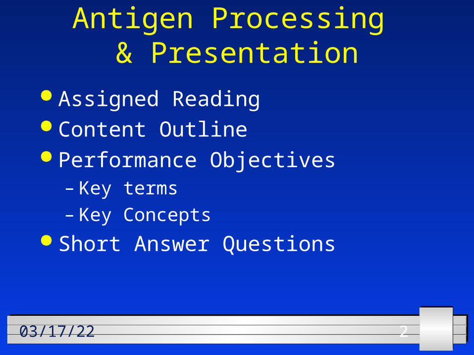
2204/20/23
Antigen Processing & Presentation
Assigned Reading Content Outline Performance Objectives
– Key terms– Key Concepts
Short Answer Questions
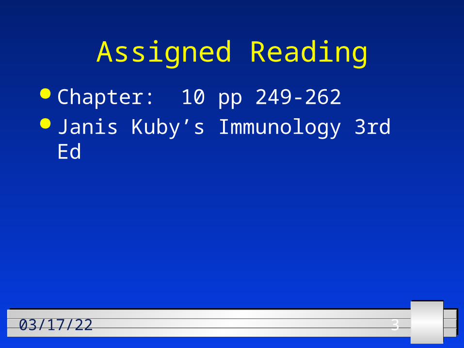
3304/20/23
Assigned Reading Chapter: 10 pp 249-262 Janis Kuby’s Immunology 3rd Ed
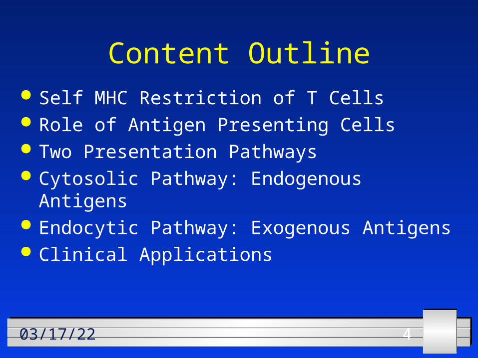
4404/20/23
Content Outline Self MHC Restriction of T Cells Role of Antigen Presenting Cells Two Presentation Pathways Cytosolic Pathway: Endogenous Antigens Endocytic Pathway: Exogenous Antigens Clinical Applications
5504/20/2353
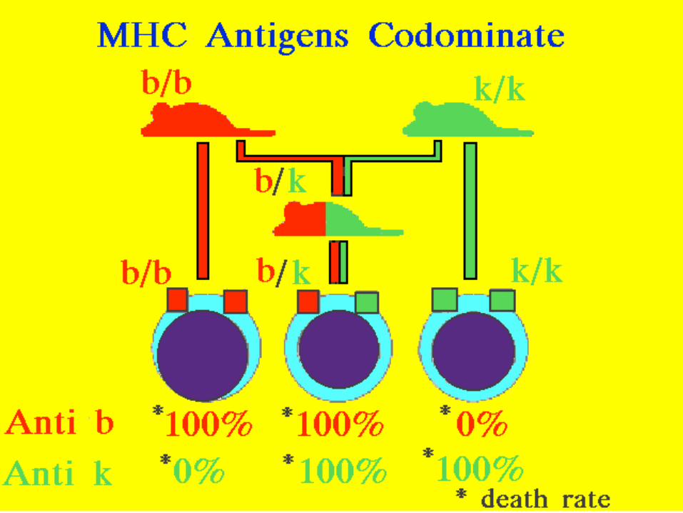
MHC antigens codominate
6604/20/2338
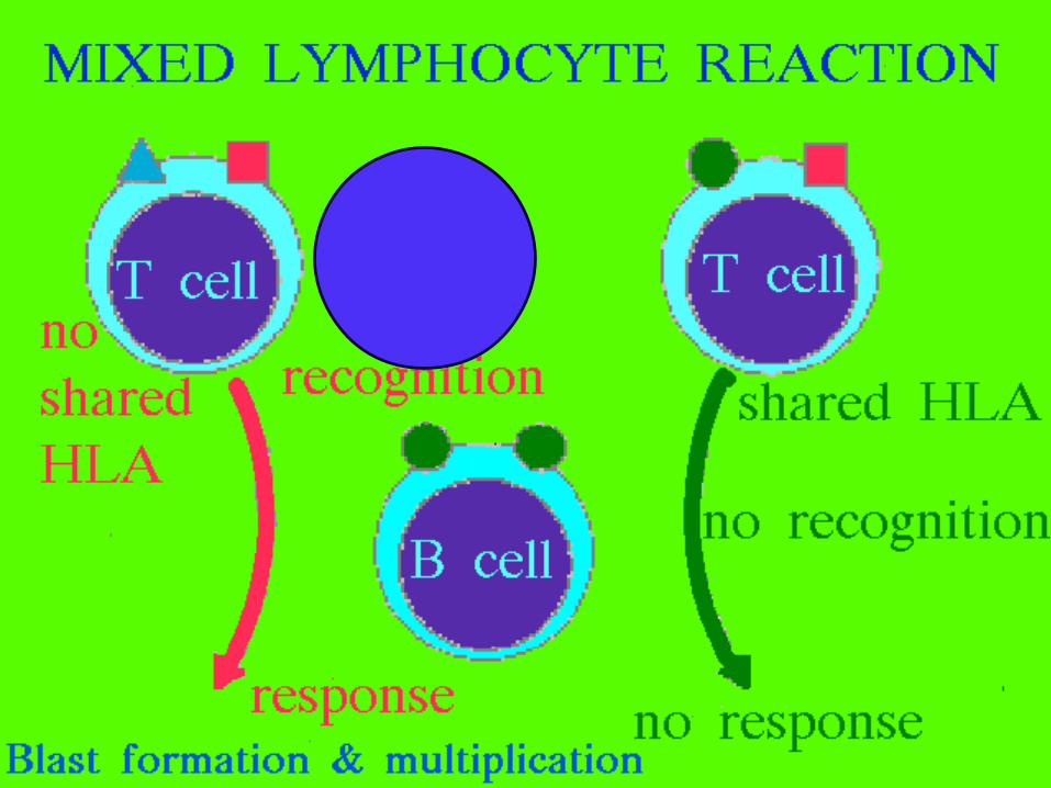
Mixed Lymphocyte Reaction
7704/20/23
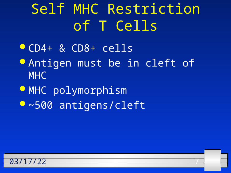
Self MHC Restriction of T Cells
CD4+ & CD8+ cells Antigen must be in cleft of MHC MHC polymorphism ~500 antigens/cleft
8804/20/2356
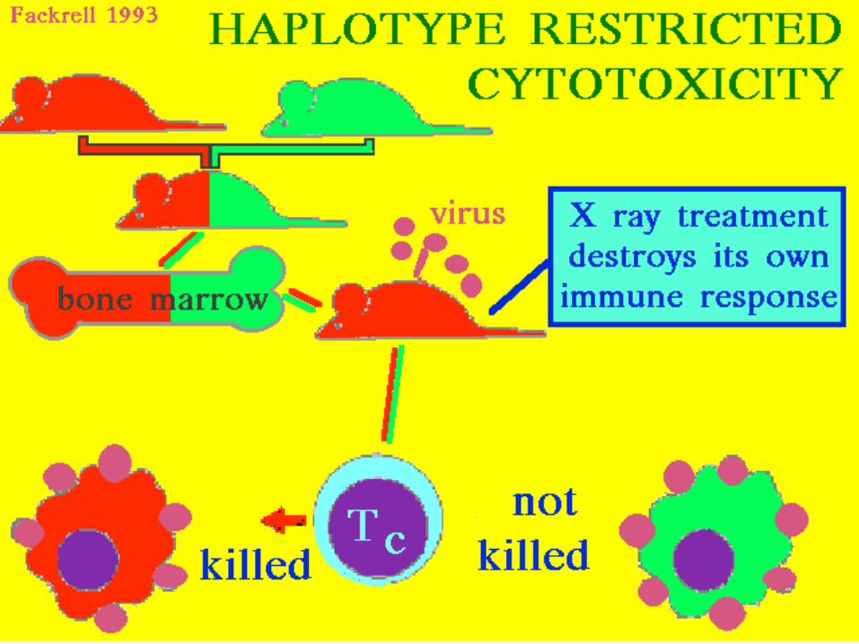
Haplotype Restricted Cytotoxicity
9904/20/23
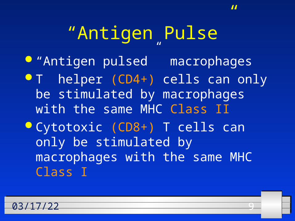
“Antigen Pulse” “Antigen pulsed” macrophages T helper (CD4+) cells can only be
stimulated by macrophages with the same MHC Class II
Cytotoxic (CD8+) T cells can only be stimulated by macrophages with the same MHC Class I
101004/20/23
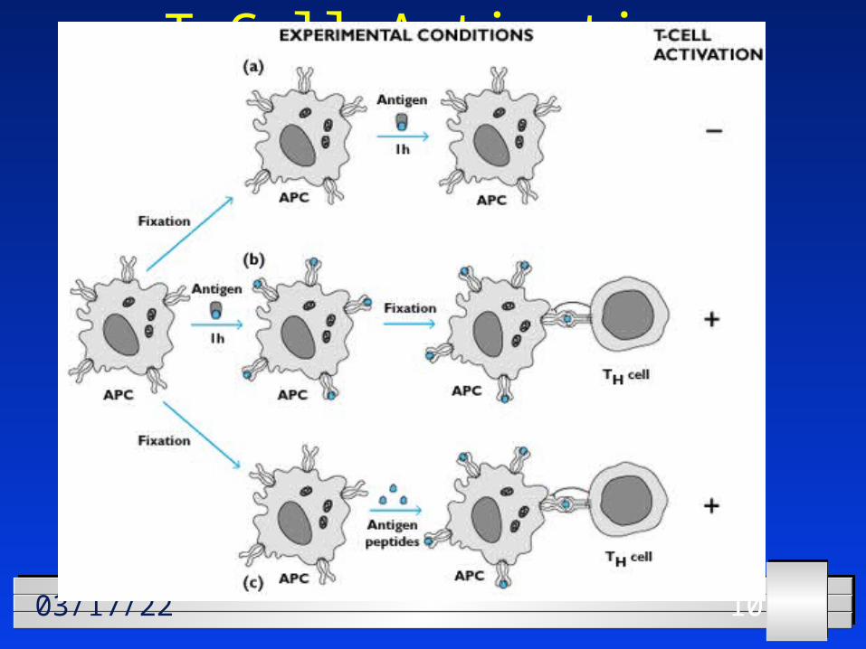
T Cell Activation
111104/20/2344
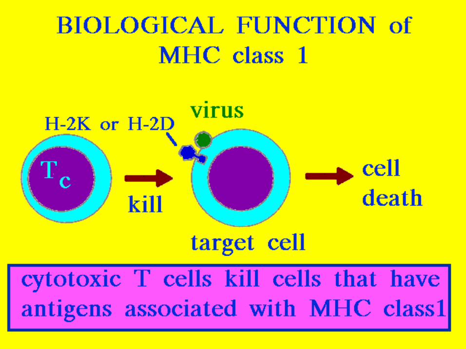
MHC I: Biological function
121204/20/2347
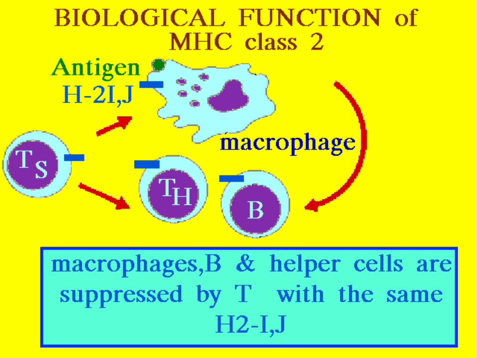
MHC II: Biological function
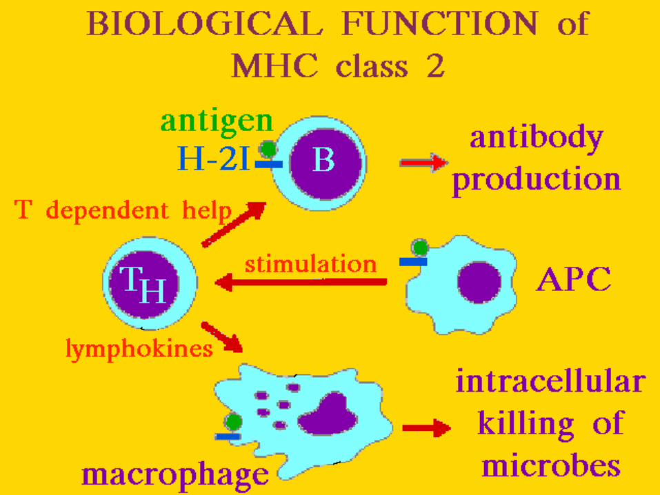
131304/20/2346
MHCII: Biologogical function (2)

141404/20/2350
MHCII: Function
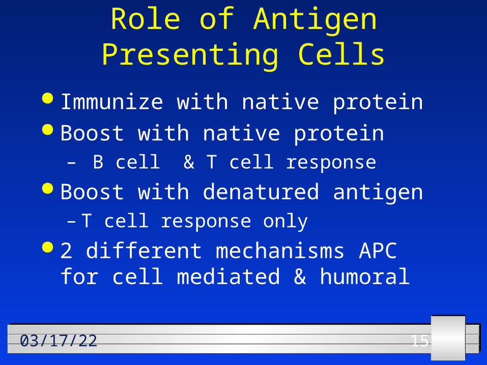
151504/20/23
Role of Antigen Presenting Cells
Immunize with native protein Boost with native protein
– B cell & T cell response Boost with denatured antigen
– T cell response only 2 different mechanisms APC for
cell mediated & humoral
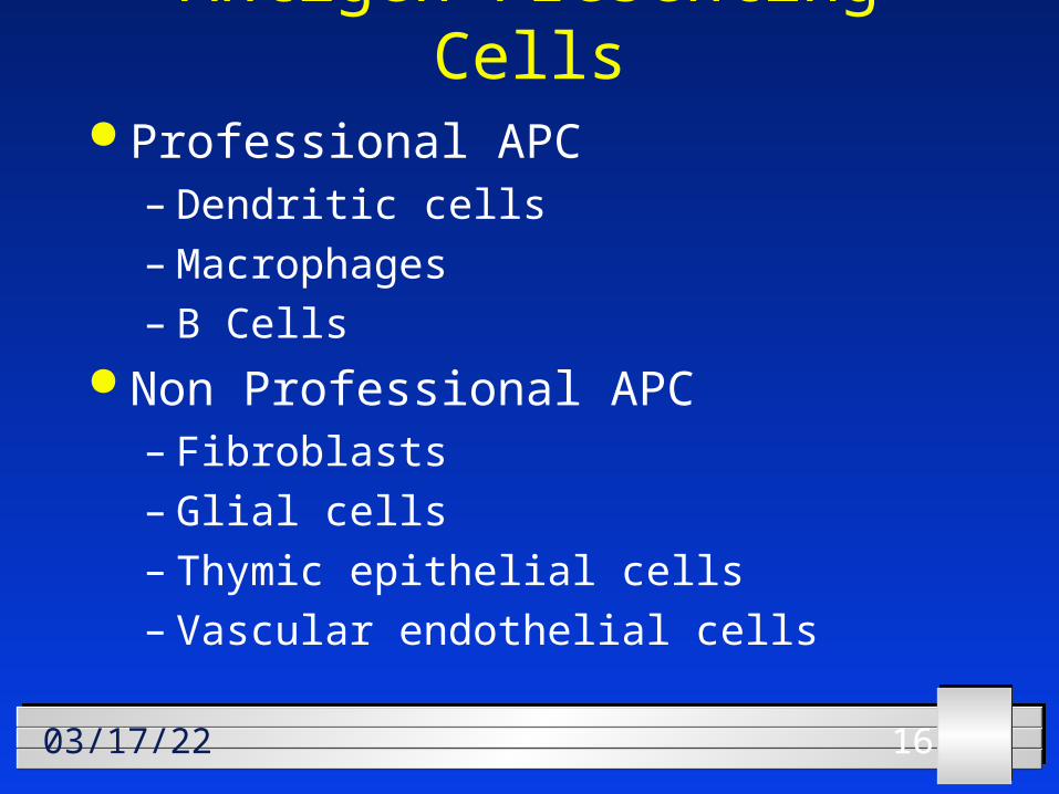
161604/20/23
Antigen Presenting Cells Professional APC
– Dendritic cells– Macrophages– B Cells
Non Professional APC– Fibroblasts– Glial cells– Thymic epithelial cells– Vascular endothelial cells
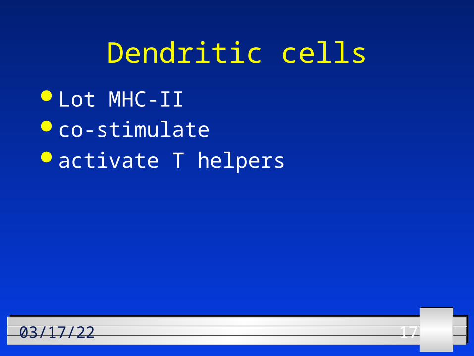
171704/20/23
Dendritic cells Lot MHC-II co-stimulate activate T helpers
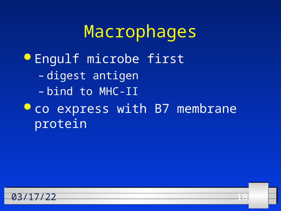
181804/20/23
Macrophages Engulf microbe first
– digest antigen– bind to MHC-II
co express with B7 membrane protein
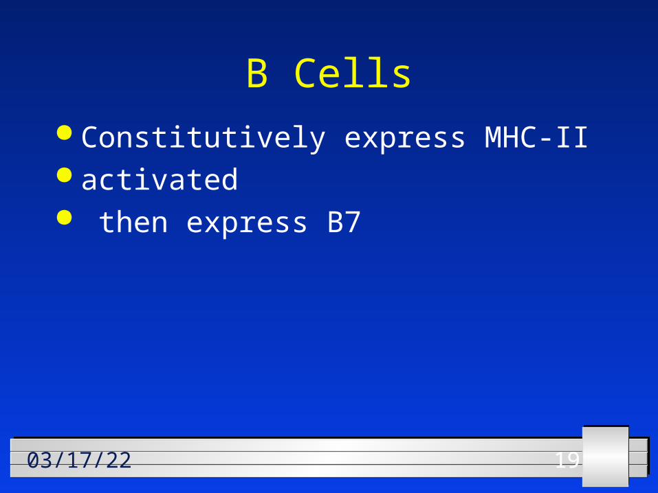
191904/20/23
B Cells Constitutively express MHC-II activated then express B7
202004/20/23
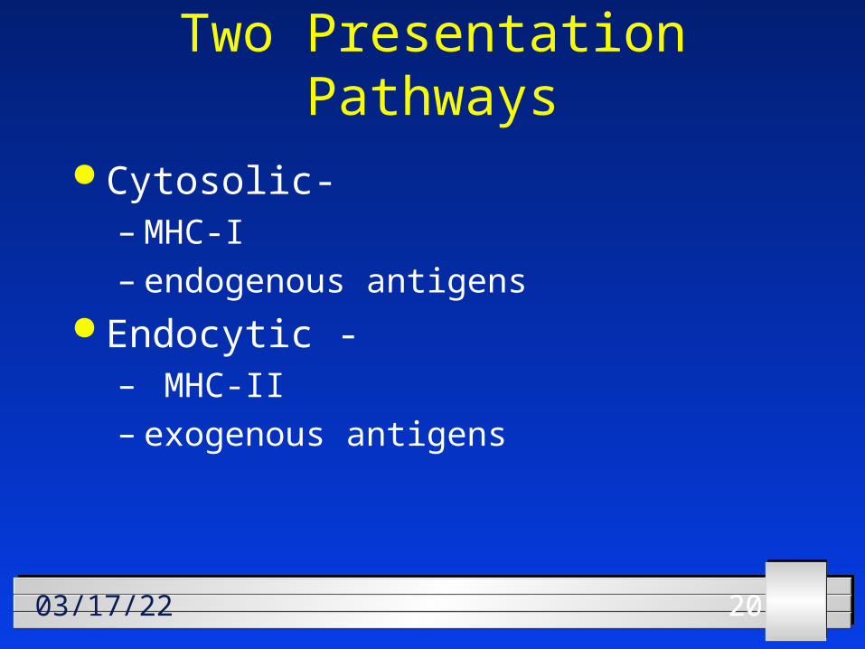
Two Presentation Pathways
Cytosolic- – MHC-I– endogenous antigens
Endocytic -– MHC-II– exogenous antigens
212104/20/23
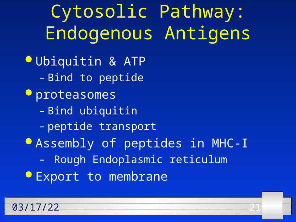
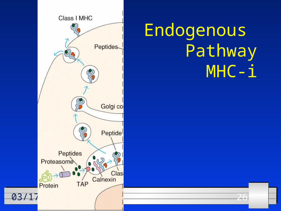
Cytosolic Pathway: Endogenous Antigens
Ubiquitin & ATP– Bind to peptide
proteasomes– Bind ubiquitin– peptide transport
Assembly of peptides in MHC-I– Rough Endoplasmic reticulum
Export to membrane
222204/20/23
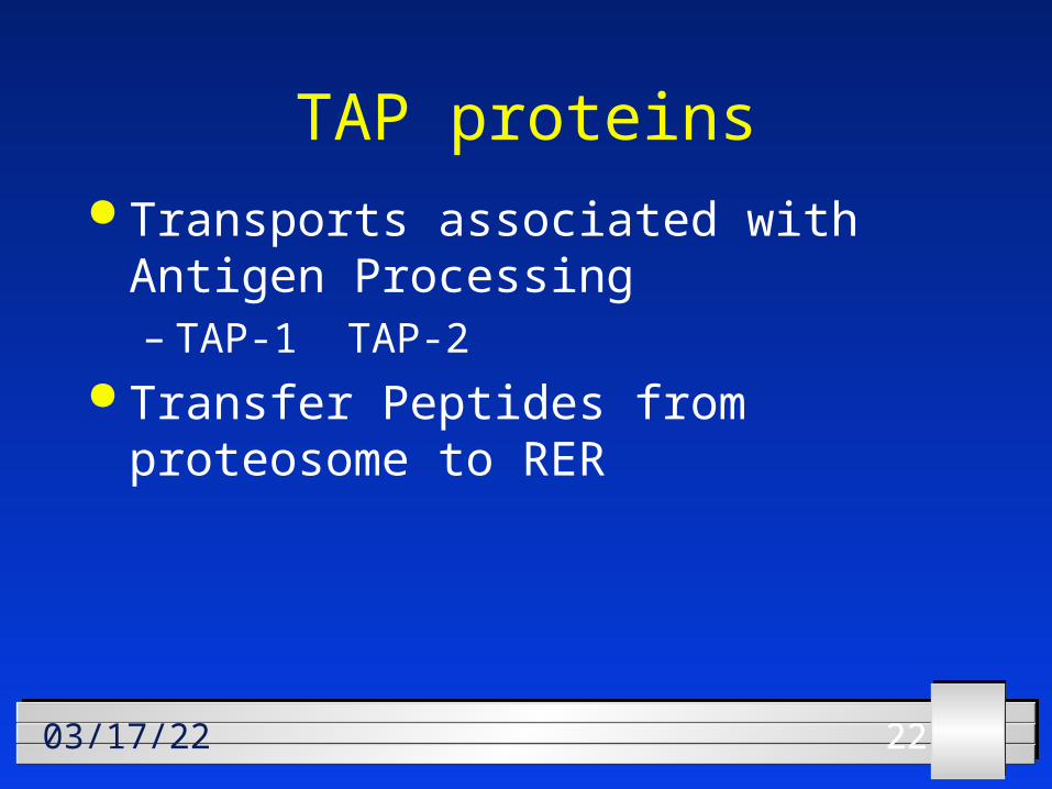
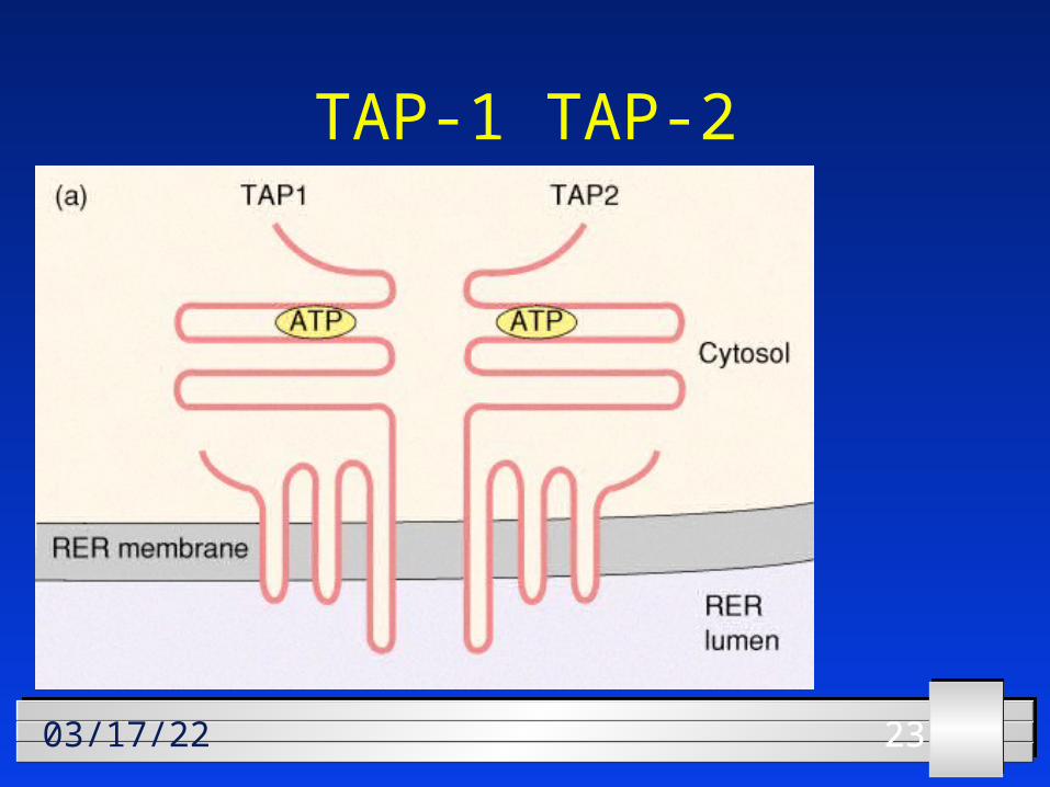
TAP proteins Transports associated with Antigen
Processing– TAP-1 TAP-2
Transfer Peptides from proteosome to RER
232304/20/23
TAP-1 TAP-2
242404/20/23
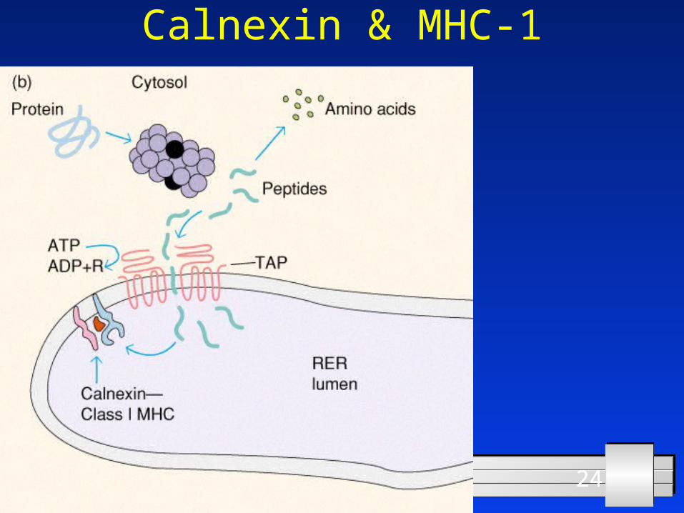
Calnexin & MHC-1
252504/20/23
Assembly in RER TAP-1 TAP-2 transport into ER Bind to Calnexin MHC-I Add peptide to MHC-I Release calnexin Transport to Gogli
262604/20/23
Endogenous Pathway
MHC-i
272704/20/23
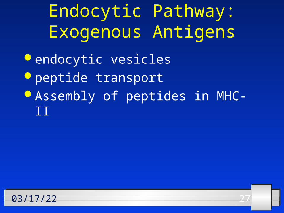
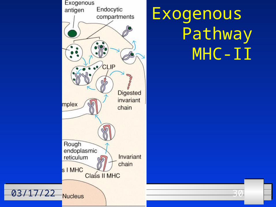
Endocytic Pathway: Exogenous Antigens
endocytic vesicles peptide transport Assembly of peptides in MHC-II
282804/20/23
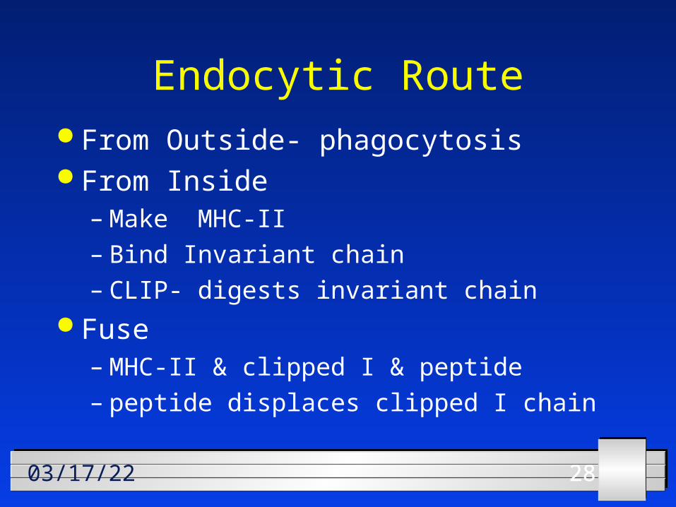
Endocytic Route From Outside- phagocytosis From Inside
– Make MHC-II– Bind Invariant chain– CLIP- digests invariant chain
Fuse– MHC-II & clipped I & peptide– peptide displaces clipped I chain
292904/20/23
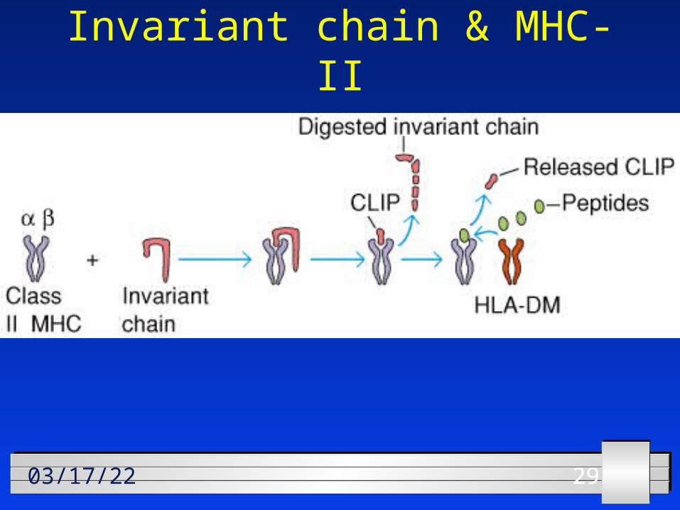
Invariant chain & MHC-II
303004/20/23
Exogenous Pathway
MHC-II
313104/20/23
Clinical Applications
323204/20/23
Performance Objectives
333304/20/23
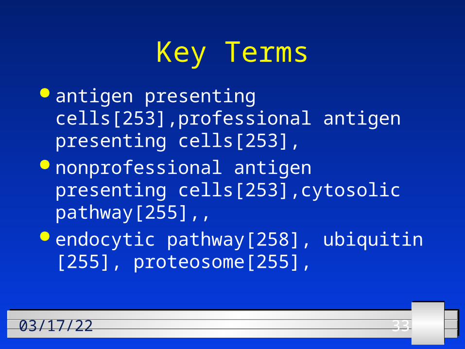
Key Terms antigen presenting
cells[253],professional antigen presenting cells[253],
nonprofessional antigen presenting cells[253],cytosolic pathway[255],,
endocytic pathway[258], ubiquitin [255], proteosome[255],
343404/20/23
Transportors associated with antigen processing (TAP) [256],
molecular chaperones[257], calnexin[ 257], invariant (Ii) chain [258],
CLIP [259],
353504/20/23
Key Concepts Describe self MHC restriction. Compare the cytosolic pathway for
antigen presentation with the endocytic pathway for antigen presentation
Compare professional with non professional antigen presenting cells.
363604/20/23
Draw a flow diagram comparing cytosolic and endocytic pathways for processing antigens [Fig 10-4]
Describe peptide generation by proteasomes.
Describe transport of antigenic peptides from the cytosol to the Rough Endoplasmic reticulum
373704/20/23
Draw a concept map for the separate antigen presenting pathways for endogenous and exogenous antigens.
Describe the assembly of MHC-II molecules within the rough endoplasmic reticulum.
Describe the development of a viral vaccine that uses the cytosolic pathway.
383804/20/23
Short Answer Questions
393904/20/23
Describe the development of a vaccine that used the endocytic pathway.
Name thee types of professional APCs. For each type indicate whether it
expresses MHC-II molecules and a co-stimulatory signal constitutively or must be activated before doing so.
Describe the role of ubiquitin in the processing of endogenous antigens.
404004/20/23
Describe the role of calnexin in the assembly of MHC-I.
T cells can react only with protein fragments.– What is this process called?– How does it occur?
Which pathway leads to antigen interaction with MHC-I molecules?
Which pathway leads to interaction with MHC-II molecules?
414104/20/23
DONE!!!DONE!!!