Embed Size (px)
Citation preview

13. Minisymposium „Infektion und Immunabwehr“ Burg Rothenfels,
12. – 14. März 2009

13. Symposium "Infektion und Immunabwehr" Burg Rothenfels, 12. – 14. März 2009
2
SCIENTIFIC PROGRAM
13. Minisymposium „Infektion und Immunabwehr“
Burg Rothenfels,
12. – 14. März 2009
PROGRAMM
Organisation: C. Hölscher, Borstel
D. Schlüter, Magdeburg
G. van Zandbergen, Ulm
Donnerstag, 12.3.08
14.45 Abfahrt Bus-Shuttle Würzburg Hauptbahnhof (Vorplatz)
15.30 – 16.30 Ankunft, Zimmerverteilung, Kaffee und Gebäck
16.30 – 17.40 Session 1:
Signalling and effector molecules
Chair: Alexander Dalpke
1 Murine 65 kDa GBPs : important effector molecules in Toxoplasma infection.
Daniel Degrandi et al., Düsseldorf
2 Identification of a novel subcellular localization pattern of Suppressor of Cytokine Signalling – 1 (SOCS-1)
Julia Strebovsky et al., Heidelberg
3 SOCS-1 enhances Pasteurella multocida Toxin induced STAT3 activity
Katharina F. Kubatzky et al., Heidelberg

13. Symposium "Infektion und Immunabwehr" Burg Rothenfels, 12. – 14. März 2009
3
4 The role of Frizzled1 in M. tuberculosis infection : Modes of induction and first
insights into function
Jan Neumann et al., Kiel
5 Wnt ligands differentially regulate the inflammatory response of macrophages upon mycobacterial infection
Kolja Schaale et al., Kiel
6 Influence of optineurin on adenovirus E3-14.7K mediated TNF-resistance Wulf Schneider-Brachert et al., Regensburg
7 Downregulation of AP-1 proteins in Chlamydia pneumoniae infected host cells
Christiane Jugert et al., Luebeck
17.40-18.00 Pause
18.00 – 19.00 Session 2:
T and B cells
Chair: Andreas Limmer
1 Delayed type hypersensitivity versus humoral immune response – Influence of high and low antigen doses on the cytokine expression within spleenic compartments
Claudia Stamm et al., Luebeck
2 Injury – induced Th cell suppression after injury is restored through targeting dendritic cells in the regenerating tissue
Florian Wirsdörfer et al., Essen
3 Apoptotic Leishmania major mediate a Th2 response in BALB/c mice
Julia Barthelmann et al., Luebeck
4 Bacterial DRiPs, Yes or No
Silke Grauling – Halama et al., Heidelberg

13. Symposium "Infektion und Immunabwehr" Burg Rothenfels, 12. – 14. März 2009
4
5 In E.coli Nissle 1917 monocolonized Rag1 deficient mice CD4+ T cells are essential for the protection against dissemination and septic shock
Ute Eberle et al., Tuebingen
6 The regulatory T cell response during acute viral infection is locally defined and controls the magnitude and duration of the virus-specific cytotoxic T cell response
Kirsten Dietze et al., Essen
19.00 – 20.00 Abendessen
20.00 Key note lecture 1
Introduction: Dirk Schlüter
Frank Kirchof, Ulm: Mechanisms of HIV-1 immune evasion
ab ca. 21 Uhr Chill out
Freitag, 13.3.08
9.00 – 10.30 Session 3:
TLRs and innate immune responses
Chair: Christian Bogdan
1 TLR against R848 inhibits differentiation of mDCs and leads to differentiaton of tolerogenic APc from CD14+ monocytes
Sabine Woelfle et al., Heidelberg
2 Improvement of host defense in the lung
Thomas Tschernig et al., Hannover

13. Symposium "Infektion und Immunabwehr" Burg Rothenfels, 12. – 14. März 2009
5
3 The phosphatidylserine binding protein annexin A5 can interfere with
chlamydial infection.
Lisa Pfleiderer et al., Ulm
4 Characterization of dual production of IFN-beta and IL12/23 p40 on a single
cell basis after TLR stimulation.
Magdalena Kocur et al., Düsseldorf
5 Antiretroviral effects of Toll-like receptor ligands
Kathrin Gibbert et al., Essen
6 Functional analysis of cells from IFIT-2 knockout mice
Alexandra Siegfried et al., Tuebingen
7 The role of cytokines for NK cell activation in visceral leishmaniasis
Simone Haeberlein et al., Erlangen
8 Acute treatment against cutaneous leishmaniasis with a two component gel developing nitric oxide
Beate Lorenz et al., Mainz
9 The role of Natural killer (NK) T cells for protection against Leishmania major
infection
Klaus Griewank et al., Mainz
10:30 – 11:00 Kaffeepause
11.00 – 12.30 Session 4a:
Cytokines
Chair: Stefan Ehlers
1 Tumor-derived Prostaglandin E2 and TGF-beta synergize to inhibit PDC-derived IFN-alpha
Marijo Parcina et al., Heidelberg
13. Symposium "Infektion und Immunabwehr" Burg Rothenfels, 12. – 14. März 2009
6
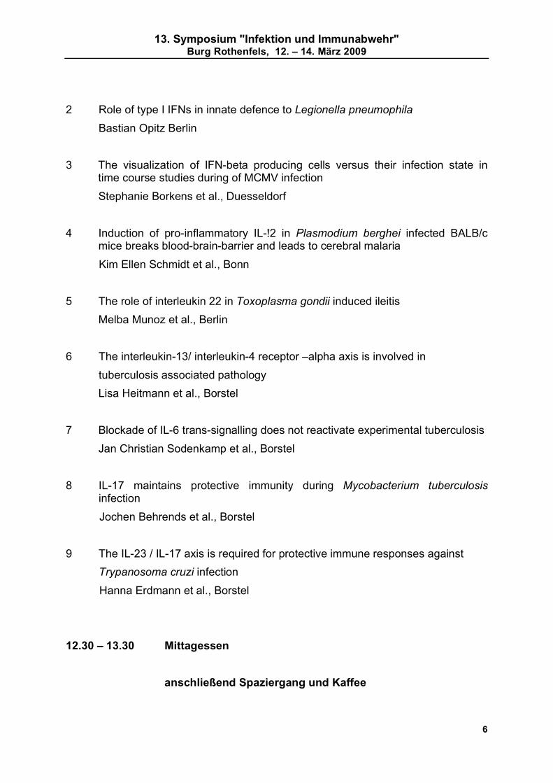
2 Role of type I IFNs in innate defence to Legionella pneumophila
Bastian Opitz Berlin
3 The visualization of IFN-beta producing cells versus their infection state in time course studies during of MCMV infection
Stephanie Borkens et al., Duesseldorf
4 Induction of pro-inflammatory IL-!2 in Plasmodium berghei infected BALB/c mice breaks blood-brain-barrier and leads to cerebral malaria
Kim Ellen Schmidt et al., Bonn
5 The role of interleukin 22 in Toxoplasma gondii induced ileitis
Melba Munoz et al., Berlin
6 The interleukin-13/ interleukin-4 receptor –alpha axis is involved in
tuberculosis associated pathology
Lisa Heitmann et al., Borstel
7 Blockade of IL-6 trans-signalling does not reactivate experimental tuberculosis
Jan Christian Sodenkamp et al., Borstel
8 IL-17 maintains protective immunity during Mycobacterium tuberculosis infection
Jochen Behrends et al., Borstel
9 The IL-23 / IL-17 axis is required for protective immune responses against
Trypanosoma cruzi infection
Hanna Erdmann et al., Borstel
12.30 – 13.30 Mittagessen
anschließend Spaziergang und Kaffee
13. Symposium "Infektion und Immunabwehr" Burg Rothenfels, 12. – 14. März 2009
7
15.30 – 17.00 Session 4b:
Dendritic cells and macrophages
Chair: Christoph Hölscher
1 Molecular mechanisms involved in aberrant type 1 interferon-induction by
S.aureus protein A.
Sibel Durlanik et al., Heidelberg
2 Detrimental role of dendritic cells during Y.enterocolitica infection in vivo
Philipp Warnke et al., Tübingen
3 Leishmania amastigote propagation in human host cells
Elena Bank et al., Ulm
4 Role of Langerin+ skin-derived DC in Leishmania major infection
Kordula Kautz-Neu et al., Mainz
5 Opsonization of L.major with cross-reactive anti-phospholipid antibodies promotes phagocytosis by dendritic cells (DC) and induction of protective immunity
Susanna Lopez Kostka et al., Mainz
6 Role of hypoxia and HIF-1 alpa in dendritic cell immunobiology.
Jonathan Jantsch et al., Erlangen
7 Conventional and plasmacytoid bone marrow-derived dendritic cells contribute to Toll like receptor-independent IFN-alpha/ beta production in response to inactivated parapoxvirus ovis
Gottfried Alber et al., Leipzig
8 Activation of macrophages by the mycobacterial cord factor (TDM) and its synthetic analogue (TDB)
Hanne Schoenen et al., Erlangen

13. Symposium "Infektion und Immunabwehr" Burg Rothenfels, 12. – 14. März 2009
8
9 Interaction of Mycobacterium tuberculosis and human macrophages under hypoxic conditions.
Daniel Nickel et al., Ulm
17.00–17.30 Fachgruppenaktivitäten
Ger van Zandbergen, Dirk Schlüter Vorsitzende der Fachgruppe/des Arbeitskreises
17.30 Keynote lecture 2
Introduction: Ger van Zandbergen
Freddy Frischknecht, Heidelberg: Imaging motile pathogens
ca. 18.30 Uhr Buffet
Samstag, 14.3.08
9.00 – 10.10 Session 5a:
Immunomodulation
Chair: Norbert Reiling
1 Split tolerance after oral vaccination of mice with recombinant Escherichia coli Nissle 1917 expressing fimbrial adhesion K88
Katharina A. Remer et al., Würzburg
2 Acute graft-versus –host-disease after reduced intensity conditioning is
mediated by MyD88 mediated TLR9 sensing of bacterial DNA and can be
modulated by administration of TLR antagonist.
Rita Plickert et al., Berlin

13. Symposium "Infektion und Immunabwehr" Burg Rothenfels, 12. – 14. März 2009
9
3 Long –term effect of sepsis –The influence of bacteremia and bacterial translocation on systemic adaptive immune responses
Timo Schwandt et al., Bonn
4 Systemic bacterial infection alters differentiation of dendritic cells in the bone marrow and mediates chronic dendritic cell dysfunction.
Eva Pastille et al., Essen
5 Increased susceptibility to infection with Aspergillus fumigatus in graft-versus-host disease is not due to impaired pathogen clearance
Bernd Echtenacher et al., Regensburg
6 Modulation of host macrophage apoptosis by Leishmania infection
Stefanie Enzenmüller et al., Ulm
7 Protective effect of filarial infection inhibiting malaria outcome in mice
Susanne Deininger et al., Bonn
10.10 – 10.30 Kaffepause
10.30 – 12.00 Session 5b:
Organ-specific regulation of immune responses
Chair: Gottfried Alber
1 Immune regulatory functions of alveolar type II epithelial cells
Dunja Bruder et al Braunschweig
2 Airway epithelial cells modify immune responses
Lotte M. Schmidt et al., Heidelberg
3 Corneal inflammation in response to filarial antigens
Katrin Gentil et al., Bonn

13. Symposium "Infektion und Immunabwehr" Burg Rothenfels, 12. – 14. März 2009
10
4 The contribution of the innate, placental immune system to defend the fetus from infections
Diana Friedrich et al., Erlangen
5 A crucial role of the spleen in the induction of pathogenic host responses towards P. berghei ANKA infection
Beatrix Schumak et al., Bonn
6 The role of the intestinal microflora in infection with Citrobacter rodentium
Ulrich Steinhoff et al., Berlin
7 IL-4/ IL-13-dependent alternative activation of macrophages but not microglial cells is associated with uncontrolled cerebral cryptococcosis
Werner Stenzel et al., Berlin
8 Neuron and astrocyte –specific function of IKK-2 and NEMO in Toxoplasma encephalitis
Ulrike Händel et al., Magdeburg
9 Toxoplasma gondii induces behavioural changes in infected mice
M. Fahad Haroon et al., Magdeburg
12:00 Preisverleihung
12:05 Ende der Tagung
Lunchpaket / Mittagessen
13.00 Abfahrt Bus (pünktlichst!)

13. Symposium "Infektion und Immunabwehr" Burg Rothenfels, 12. – 14. März 2009
11
ABSTRACTS (in alphabetical order of presenting authors)
Conventional and plasmacytoid bone marrow-derived dendritic cells contribute to
Toll-like receptor-independent IFN-alpha/beta production in response to inactivated
parapoxvirus ovis
Sabine Siegemund1, Andrea Hartl2, Franziska Dautel1, Ruediger Raue3, Marina A.
Freudenberg4, Mathias Buettner5, Gabriele Koehler6, Carsten J. Kirschning2, Tim
Sparwasser2, Gottfried Alber1
1Institute of Immunology, College of Veterinary Medicine, University of Leipzig, Germany; 2Institute of Microbiology, Technische Universitaet Muenchen, Germany; 3Pfizer Animal
Health, Kent, United Kingdom; 4Max Planck Institute for Immunobiology, Freiburg, Germany; 5Bayerisches Landesamt für Gesundheit und Lebensmittelsicherheit, Oberschleissheim,
Germany; 6Gerhard-Domagk-Institute for Pathology, Universitaetsklinikum Muenster,
Germany
Parapoxvirus ovis (PPVO) is a member of the Poxviridae family and belongs to the genus
Parapoxvirus. It displays only limited homology with orthopoxviruses and has some
molecular features such as an unusual high GC content distinct from orthopoxviruses.
Inactivated parapoxvirus ovis (iPPVO) has strong immunostimulatory capacities mediating
antiviral activity in vivo. The role of dendritic cells and the pattern recognition receptors
responsible for recognition of iPPVO are unknown. We demonstrate that bone marrow-
derived plasmacytoid dendritic cells (BM-pDC) and bone marrow-derived conventional
dendritic cells (BM-cDC) secrete IFN-alpha/beta in response to iPPVO. Furthermore, iPPVO
induces TNF-alpha and IL-12/23p40 release and MHC-II, MHC-I and CD86 up-regulation by
bone marrow-derived dendritic cells (BMDC). After engulfment, iPPVO was located in
endosomal compartments and in the cytosol of BMDC. Although iPPVO is a double-stranded
DNA virus, the DNA-recognizing toll-like receptor (TLR) 9 is not involved in iPPVO-induced
release of IFN-alpha/beta by BMDC. Moreover, we demonstrate that iPPVO elicits IFN-
alpha/beta by TLR-independent pathways, since IFN-alpha/beta release does not require
myeloid differentiation primary response gene 88 (MyD88) or TIR-domain containing adaptor
protein inducing interferon (TRIF). In contrast, iPPVO-induced TNF-alpha and IL-12/23p40
release and enhanced expression of MHC-I and CD86 but not MHC-II by BMDC partially
depends on MyD88 but not on TLR2, TLR4 or TLR9. These results provide first evidence
that iPPVO mediates its immunostimulatory properties by TLR-independent and -dependent
pathways and demonstrate an important role of cDC for IFN-alpha/beta production.

13. Symposium "Infektion und Immunabwehr" Burg Rothenfels, 12. – 14. März 2009
12
Leishmania amastigote propagation in human host cells
Elena Bank1, Alexander Wenzel1 and Ger van Zandbergen1.
1Instiute for Medical Microbiology and Hygiene, University Clinic of Ulm
Leishmania major (L. major) is an obligate intracellular parasite elegantly misusing the
apoptotic cell clearance system for disease development. This parasite enters
polymorphonuclear neutrophil granulocytes (PMN) in its promastigote form. Hiding inside
Annexin A5 (AnxA5)-positive PMN the parasite transfers into macrophages (MF), where it
multiplies in its amastigote form. We found that the presence of AnxA5-binding population of
promastigotes is responsible for the survival and infectivity of viable promastigotes inside
PMN. Still relatively little is known about macrophage infection by the amastigote form of L.
major propagating the disease.
We developed a novel in vitro method to culture the amastigote form of L. major and
compared these amastigotes with promastigotes We found them to be higly infectious for
different types of human MF. To study amastigote development and propagation in more
detail, we generated eGFP expressing L. major. The eGFP marker is upregulated in the
amastigotes stage. Using these transfectants we infected type I (inflammatory) and typ II
(anti-inflammatory) human MF. We found that type II MF take up more parasites as
compared to type I MF. Using FACS analyses we could detect the development of
amastigotes by a higher expression level of eGFP as compared to the eGFP expression of
intraphagocytic promastigotes. Focussing on amastigote uptake using timelapse imaging we
found that dying eGFP-negative and AnxA5-positive amastigotes enter MF first followed by
viable eGFP-positive parasites. These new tools will enable us to examine L. major
amastigote propagation in human host cells.

13. Symposium "Infektion und Immunabwehr" Burg Rothenfels, 12. – 14. März 2009
13
Apoptotic Leishmania major mediate a Th2 response in BALB/c mice
Julia Barthelmann, Juergen Westermann, Kathrin Kalies
Institute of Anatomy, University of Luebeck
It has recently been reported that the virulent inoculum of Leishmania major (L. major)
promastigotes conventionally used for infection experiments contains about 50% of apoptotic
parasites. The depletion of apoptotic parasites lead to a reduced infectivity in BALB/c mice in
vivo.
In order to analyse the detrimental effect of apoptotic leishmania on the immune response in
the lymph node, we separated viable and apoptotic L. major by magnetic cell separation. We
studied T-cell proliferation and cytokine mRNA expression in the draining lymph node after
infection of susceptible BALB/c mice with viable L. major.
We found that the lack of apoptotic parasites in the inoculum leads to a delay of disease
progression and decreased IL-4 and IL-10 mRNA levels 6 weeks after infection, while
IFN-gamma levels remain unchanged. Furthermore, Th2-associated total serum IgG1 levels
are reduced. Disease, cytokine mRNA production and total serum IgG1 after 6 weeks could
be restored by adding apoptotic leishmania to the inoculum, thereby indicating that apoptotic
L. major support the establishment of a Th2 immune response.
Although the initial composition of the inoculum determines the disease development, the
response in the lymph node is not defined 3 days after infection: Time point and magnitude
of initial T-cell proliferation do not differ compared to conventional L. major infections and the
cytokine pattern does not correlate with the final disease outcome.
Overall, the removal of apoptotic parasites in the inoculum decreases the Th2 response, but
does not explicitly support the establishment of a Th1 phenotype in susceptible BALB/c.

13. Symposium "Infektion und Immunabwehr" Burg Rothenfels, 12. – 14. März 2009
14
IL-17 maintains protective immunity during Mycobacterium tuberculosis infection
Jochen Behrends,1 Dominik Rueckerl,1 Manuela Heßmann,1 Uwe Mueller,2 Gottfried Alber,2
Yoichiro Iwakura,3 Stefan Ehlers4,5 & Christoph Hoelscher1
1Infection Immunology, Research Center Borstel, Germany; 2University of Leipzig, Germany; 3University of Tokyo, Japan; 4Microbial Inflammation Research, Research Center Borstel,
Germany; 5Molecular Inflammation Medicine, Christian-Albrechts-University, Kiel, Germany
Because a variety of autoimmune disorders have now been shown to depend on interleukin
(IL)-17-producing T helper (TH)17 cells, therapeutic blockade of TH17 development may
provide a novel approach to avoid adverse consequences of anti-inflammatory strategies
such as reactivation of latent tuberculosis (TB). To evaluate the potential risk of interfering
with IL-17-dependent inflammation, we analyzed the outcome of experimental TB in IL-17-
deficient (-/-) mice after infection with Mycobacterium tuberculosis (Mtb). IL-17 was important
for the induction of neutrophil chemokines after Mtb infection, but was not involved in
granuloma formation and protection during the first three months of Mtb infection. Mtb-
infected IL-17-/- mice efficiently generated interferon-gamma (IFN )-producing T cells and
IFNgamma-dependent effector responses. However, IL-17-/- mice were not able to control
mycobacterial replication during the chronic phase of experimental TB and died significantly
earlier than corresponding wildtype mice. This breakdown of immune protection in IL-17-/-
mice was associated with a drop in the frequency of IFN -producing CD4+ T cells. Our
findings reveal that IL-17 is essential for maintaining CD4+ T cell-dependent protection during
chronic stages of TB. Hence, interfering with IL-17-dependent pathways as an anti-
inflammatory therapeutic approach will possibly incur the danger of reactivating latent TB.
(Supported by the Inflammation Research Excellence Cluster)

13. Symposium "Infektion und Immunabwehr" Burg Rothenfels, 12. – 14. März 2009
15
The visualization of IFN-beta producing cells versus their infection state in time
course studies during of MCMV infection
Stephanie Borkens1, Vu Thuy Khanh Le2, Stefanie Scheu1
1Institute of Medical Microbiology and Hospital Hygiene, University of Duesseldorf 2Institute of Virology, University of Duesseldorf
Type I interferons are a family of multiple IFN-alphas and a single IFN-beta which were
initially identified on the basis of their antiviral activities. Previous findings identified pDCs as
the major IFN-beta producing cells in the spleen. However, which cells are actually infected
with MCMV remains unclear, although it has been suggested that at certain stages of
infection MCMV can actively replicate in macrophages and conventional DCs.
For a detailed analysis of the initial phase of MCMV infection, we used a MCMV strain which
expresses EGFP under the immediate-early promoter (MIEP) to infect IFN-beta/YFP reporter
knockin mice. This experimental approach allows for the simultaneous visualization of the
IFN-beta response and the infection status of MCMV in vivo.
In initial analyses at 24h post infection. we identified splenic pDCs as well as few CD11b+
cells expressing IFN-beta. Furthermore, we could show that the IFN-beta producing pDCs
are not directly infected with MCMV and that mainly CD11b+/c+ cells expressed EGFP as a
marker for MCMV infection.
Current studies aim at the clarification of IFN-beta-production versus the infection status of
individual cells in this simultaneous analysis during several time points of infection.

13. Symposium "Infektion und Immunabwehr" Burg Rothenfels, 12. – 14. März 2009
16
Immune regulatory functions of alveolar type II epithelial cells
Marcus Gereke, Harro Frauendorf, Dunja Bruder
Immune regulation group, Helmholtz Center for Infection Research, Braunschweig
Whereas the lung epithelium was long time related primarily with physical barrier and gas
exchange functions, the contribution of alveolar type II epithelial cells (AECII) in respiratory
immune regulation has become increasingly appreciated. However, their precise function in
the induction and regulation of T cell reactivity to self antigen remains poorly understood.
Utilizing a transgenic mouse model for CD4+ T cell mediated pulmonary inflammation we
found that MHC class II expressing AECII present self-antigen to CD4+ T cells resulting in
functional activation of lung-reactive T cells and finally autoimmunity. Importantly, we
unravelled a previously unknown immunological attribute of AECII in re-establishment of
peripheral T cell tolerance in the lung. Upon inflammation, AECII secrete a broad variety of
soluble factors including transforming growth factor-beta (TGF-beta), Platelet Factor-4 and
Surfactant Protein-A and -D, which suppress T cell proliferation and induce in a partially
TGF-beta dependent mechanism Foxp3 expression in lung-reactive CD4+ T cells. As a part
of the innate immune system AECII thus exhibit so far underestimated immune regulatory
function and synergize with adaptive immune mechanisms to re-establish tolerance to self-
antigen in the lung. Preliminary data suggest that upon influenza infection alveolar self
antigen recognition by CD4+ T cells does not result in increased abundance of Foxp3
expressing regulatory T cells thus indicating that the capability to respond to and to eradicate
a pathogen dominates about the necessity to protect against potentially harmful autoimmune
reactions. Currently we are investigating the contribution of AECII in balancing tolerogenic
and pathogen-specific immunity in the lung.

13. Symposium "Infektion und Immunabwehr" Burg Rothenfels, 12. – 14. März 2009
17
Murine 65 kDa GBPs: important effector molecules in Toxoplasma infection.
Daniel Degrandi, Carolin Konermann, Cornelia Beuter-Gunia, Sarah Lahme, Anna Fischer,
Elisabeth Kravets, Klaus Pfeffer
Medizinische Mikrobiologie und Krankenhaushygiene. Heinrich-Heine-Universität Düsseldorf
The 65 kDa guanylate-binding proteins comprise a growing family of highly conserved
GTPases, found in mice, humans, and many other species. In mice, 11 members of the GBP
family have been described. In recent studies, we could show that several members of the
mGBP family colocalize with the parasitophorous vacuole (PV) of avirulent Toxoplasma
gondii (Tg) strains, suggesting a direct antiparasitic function of the mGBP proteins.
Interestingly, highly virulent Tg strains are able to modulate the cellular response to the
infection and suppress the recruitment of mGBP proteins to the PV. The mechanisms
involved in the recruitment of mGBP proteins to the PV and their molecular and biochemical
activity are the major focus of our current studies. First results indicate, that GTP-binding and
hydrolysis are needed for normal localization of the proteins in infected and uninfected cells.
Furthermore, the recruitment of mGBP proteins towards the Toxoplasma PV is dependent on
the microtubular network, since cells treated with Paclitaxel and other microtubule interacting
drugs showed no colocalization of mGBPs with the PV.
Studies in mGBP2-/- mice showed an increased susceptibility towards infections with Tg.
Also, mGBP2 deficient cells show a significantly reduced capability to control Tg growth in
vitro. These data indicate, that the family of 65 kDa mGBPs play crucial roles in the cell
autonomous defense against intracellular pathogens.

13. Symposium "Infektion und Immunabwehr" Burg Rothenfels, 12. – 14. März 2009
18
Protective effect of filarial infection inhibiting malaria outcome in mice
Susanne Deininger1, Daniel Fernández-Ruiz1, Gwydion Brennan1, Bettina Dubben1, Achim
Hoerauf1, Sabine Specht1
1Institute for Medical Microbiology, Immunology and Parasitology, University Hospital Bonn,
Bonn, Germany
Malaria is a serious tropical disease with a high risk of mortality to date. Chronic helminth
infections and often co-endemic with malaria and both might alter the immune response
against another. By use of a co-infection model of the filarial nematode Litomosoides
sigmodontis and the malaria pathogen Plasmodium berghei ANKA in Balb/c mice we
discovered that a preceding filarial infection inhibited a subsequent infection with P. berghei
sporozoites in one-third of mice. Interestingly, this sterile protection was correlated with
presence of microfilariae, which are the first stage larvae of the filarial parasite. We observed
increased levels of activated T cells in spleen and liver of microfilaremic mice. Furthermore,
phagocytotic cells were increased in the spleens of these mice. In order to investigate the
underlying mechanism, we analysed cytokine profiles in these mice. We found that worm-
infected mice produced higher levels of IL-10, IFN-gamma and granzyme B and their CD4+
and CD8+ T cells were found to be in an activated status (CD69+) secreting IFN-gamma,
perforin and granzyme B.
Both candidates the cytotoxic T cells and the phagocytotic cells may contribute to the
mechanisms responsible for blocking parasitaemia of malaria pathogens in a part of filarial-
infected mice. A better knowledge about the protective mode of a patent filarial infection on
the outcome on malaria may provide some tools to influence the course of malaria in favour
for the host.

13. Symposium "Infektion und Immunabwehr" Burg Rothenfels, 12. – 14. März 2009
19
The regulatory T cell response during acute viral infection is locally defined and
controls the magnitude and duration of the virus-specific cytotoxic T cell response
Kirsten Dietze1, Gennadiy Zelinskyy1, Ulf Dittmer1
1Institut fuer Virologie, Universitaetsklinikum Essen
Cytotoxic T-cells (CTL) facilitate control of acute viremia in many viral infections, including
retroviruses like HIV or HTLV. However, viruses that establish chronic infections have
developed mechanisms to evade destruction by CTL. We have used the Friend Virus (FV)
model to investigate these mechanisms. In the acute infection FV induces a strong CTL
response but the mice become persistently infected. However, regulatory CD4+ T cells (Treg)
that expand in the spleen of infected mice suppress the production of cytotoxic molecules in
CD8+ T cells and the cytotoxic function of CTL. The aim of our current work was to analyse
the compartmentalisation of the Treg response and the subsequent local suppression of
CD8+ T cells by Tregs during an ongoing retroviral infection. We found, that expansion of
effector CD8+ T cells, production of cytotoxic molecules and degranulation was directly linked
to viral loads in lymphatic organs. Consequently the expansion of induced Treg correlated
with the number and function of virus-specific CD8+ T cells. For the expansion of Treg the
presence of CTL was obligatory, what was shown by CD8 depletion experiments.
Furthermore, depleting Treg in DEREG mice resulted in enhanced expansion of effector
CD8+ T cells and improved the production of cytotoxic molecules leading to reduced viral
loads in lymphatic organs. In summary, during acute retroviral infection Treg downregulated
the expansion and function of virus-specific CTL. The immunosuppressive activity of Tregs
was locally defined to the organs in which efficient viral replication followed by a strong CD8+
effector cell response took place.

13. Symposium "Infektion und Immunabwehr" Burg Rothenfels, 12. – 14. März 2009
20
Molecular mechanisms involved in aberrant type I interferon-induction by S. aureus
protein A
Isabelle Bekeredjian-Ding1, Sibel Durlanik1, Marijo Parcina1, Sandra Ammann1, Wulf
Schneider-Brachert2 Klaus Heeg1
1Hygiene-Institute, Dept. of Med. Microbiology and Hygiene, UniversityHospital Heidelberg 2Institute for Med. Microbiology und Hygiene, University of Regensburg
Protein A (SpA), an immunoglobulin (Ig)-binding protein, is a major virulence factor of
Staphylococcus aureus and well-known as an immunostimulatory protein. We previously
observed that certain strains of S. aureus induce IFN-alpha secretion from plasmacytoid
dendritic cells (pDC). The aim of the project presented was therefore, to investigate the role
of SpA in type I interferon induction from pDC. Remarkably, IFN-a induction correlated with
bacterial levels of SpA expression, and subsequent experiments showed that recombinant
SpA induces IFN-alpha secretion from pDC. Moreover, IFN-I induction could be blocked by
saturating Ig or Fc fragments, indicating that the Ig-binding domain may be essential for IFN-I
induction. Most importantly, DNAse and RNAse digestion of recombinant SpA did not alter its
ability to induce IFN-I. Recently, SpA has been shown to bind TNFR1 on respiratory
epithelial cells. Here we show that anti-TNF-R mAb neutralisation can inhibit the SpA-
induced type I interferon secretion. We further show that SpA colocalizes with the TNF-R2 in
transfected HEK293 cells. Thus, SpA may represent a novel TLR-independent stimulus for
pDC activation.

13. Symposium "Infektion und Immunabwehr" Burg Rothenfels, 12. – 14. März 2009
21
In E. coli Nissle 1917 monocolonized Rag1 deficient mice CD4+ T cells are essential for
the protection against dissemination and septic shock.
Ute Eberle*1, Kerstin Fink*1, Martina Müller1, Frank Leithäuser2, Thomas A. Ölschläger3, Ingo
B. Autenrieth1, Julia-Stefanie Frick1
1Institute of Medical Microbiology and Hygiene, University of Tuebingen, Germany 2Institute of Pathology University of Ulm, Germany 3Institute of Molecular Biology of Infection, University of Wuerzburg, Germany *equal contribution
E. coli Nissle 1917 (EcN) is a well defined probiotic E. coli strain, which is effective in
maintaining remission in ulcerative colitis. The aim of this study was to explore safety
aspects of this frequently used probiotic strain. Therefore, germfree Rag1-/- mice were
monocolonized with EcN. Upon CD4+ T cell transfer germfree Rag1-/- mice showed a high
mortality rate due to dissemination of EcN. In peripheral organs high bacterial loads of EcN
were detected correlated with high levels of TNF in the serum. In contrast, SPF Rag1-/- mice
colonized with EcN showed no dissemination of EcN in peripheral organs even though the
numbers of bacteria in the intestine did not differ. Furthermore, EcN monocolonized C57Bl/6
mice showed no increased mortality rate, indicating that CD4+ T cells were essential either
for the inhibition of translocation or for the clearance of EcN from the peripheral organs. The
translocation and dissemination are an EcN specific effect, as it was not observed in
germfree Rag1-/- mice monocolonized with E. coli mpk, a commensal E. coli stain isolated
from the murine intestine. Additionally, the dissemination of EcN in monocolonized Rag1-/-
mice was not flagella dependent. As the translocation of EcN Dflic and DflgE to peripheral
organs was comparable to the translocation of wildtype EcN. Therefore, an unknown
bacterial factor seems to account for the dissemination of EcN in monocolonized Rag1-/-
mice, and both the immature intestinal barrier and the immature mucosa associated immune
system of germfree animals seem to be essential for the translocation of EcN.

13. Symposium "Infektion und Immunabwehr" Burg Rothenfels, 12. – 14. März 2009
22
Increased susceptibility to infection with Aspergillus fumigatus in graft-versus-host
disease is not due to impaired pathogen clearance
Bernd Echtenacher1, Kristina Doser 2, Matthias Edinger2 and Petra Hoffmann2
1Institute of Immunology, University Regensburg and 2Dept. of Hematology and Oncology,
University Hospital Regensburg, Regensburg, Germany
Graft-versus-host disease (GvHD) is a frequent and life-threatening complication after
allogeneic bone marrow transplantation (alloBMT) that is initiated by mature alloreactive T
cells within the graft. These show excessive proliferation and pro-inflammatory cytokine
secretion upon interaction with host antigen-presenting cells, which leads to a dysregulated
cytokine milieu and finally results in tissue damage and target organ destruction (gut, liver
and skin). Patients after alloBMT are severely immunocompromised and therefore
particularly prone to opportunistic bacterial and fungal infections. While immunosuppressive
medication for GVHD prophylaxis clearly raises the risk of infection, the contribution of GVHD
itself to this increased susceptibility is much less understood. To evaluate the impact of
GVHD on the host's defence against fungal infections, allo-transplanted BALB/c mice with or
without GVHD were infected intratracheally or intravenously with the clinically relevant
pathogen Aspergillus fumigatus. Mortality was significantly higher in GVHD animals (only
20% survival after 2wks) as compared to the non-GVHD group (75% survival for more than
5wks). Interestingly, clearance of the fungus from the lung after i.t. infection, or from spleen
and liver after i.v. infection, was rapid and comparable in both groups and no live fungus was
detectable in moribund animals. However, when lymphocytes isolated from spleen and liver
of infected animals were restimulated in vitro with germinating conidia, cells from GVHD
animals secreted significantly more pro-inflammatory TNF and IL-6 than those from control
mice. This suggests that an uncontrolled inflammatory immune response contributes to the
high morbidity and mortality of opportunistic infections in GVHD.

13. Symposium "Infektion und Immunabwehr" Burg Rothenfels, 12. – 14. März 2009
23
Modulation of host macrophage apoptosis by leishmania infection
Stefanie Enzenmüller1, Cordula Schropp2, Simone Fulda1, Ger van Zandbergen2 and Silke
Fischer1,2
1University Children's Hospital, Ulm. 2Institute for Medical Microbiology and Hygiene,
University Clinic of Ulm
Leishmania are obligate intracellular parasites which undergo a promastigote flagellated
stage in the sandfly and an aflagellated amastigote stage in vertebrate hosts. They cause a
spectrum of human diseases, ranging from self-limiting cutaneous infections to visceral
leishmaniasis. The infection of mammalian hosts is initiated by promastigotes that are
phagocytosed by macrophages either directly or after infection of neutrophils initially
recruited to the sandfly bite. In host cells Leishmania replicate and differentiate to
amastigotes. Like all intracellular parasites, Leishmania have evolved specialized strategies
to evade immune destruction and to interact with multiple apoptotic systems to complete their
life cycle. The repression of host cell apoptosis is one type of strategy to protect the host
macrophage in which the intracellular parasites replicate and persist.
Our main research interest is focused on how Leishmania major interferes with the host cell
apoptotic machinery in infected cells. Therefore we investigate how Leishmania modulate the
different signalling pathways of apoptosis. We used two different cell lines (monocyte-derived
macrophages THP1 and murine macrophage-like Raw 264.7). Cells were infected with L.
major in vitro and subsequently treated with external apoptosis-inducing stimuli such as UV-
light or staurosporine. First results are indicating that L. major inhibits caspase activation,
cytochrome c release from mitochondria and modulates the level of BH3-only proteins within
the infected macrophage cells.

13. Symposium "Infektion und Immunabwehr" Burg Rothenfels, 12. – 14. März 2009
24
The IL-23/IL-17 axis is required for protective immune responses against
Trypanosoma cruzi infection
Hanna Erdmann,1 Caroline Rossnagel,1 Nico Ghilardi,2 Yoichiro Iwakura,3 Thomas Jacobs,4
Christoph Hoelscher1
1Infection Immunology, Research Center Borstel, Germany; 2Genentech, South San
Francisco CA USA; 3 Center for Experimental Medicine, Institute of Medical Science,
University of Tokyo, Japan; 4Bernhard-Nocht-Institute for Tropical Medicine and Hygiene,
Hamburg, Germany
Interleukin (IL)-12 is a potent inducer of interferon-gamma (IFN )-producing T helper (TH)1
cells and promotes a protective cell-mediated immune response after infection with the
protozoan parasite Trypanosoma cruzi, the causative agent of Chagas disease. IL-23 is
structurally closely related to IL-12 and shares the IL-12/23p40 subunit and the
corresponding IL-12 receptor 1 subunit. However, IL-23 is not as potent as an inducer of
IFN -production than IL-12. Instead IL-23, but not IL-12, promotes the proliferation of IL-17-
producing TH17 cells. To analyze the role of IL-23 for protective immune responses during
experimental Chagas disease, IL-23p19 deficient (-/-) mice were infected with T. cruzi.
Compared to wild-type mice, IL-23p19-/- mice developed a higher parasitemia and an
increased mortality. However, this susceptibility was not due to an impaired TH1 immune
response. Because IL-23 supports the development of IL-17-secreting TH17 cells, we
infected IL-17-/- mice with T. cruzi to study the relevance of IL-17 for protective immune
responses. Like IL-23-/- mice, IL-17-/- mice exhibited a higher parasitemia, an elevated
mortality and an altered liver pathology. Moreover, TH1 immune responses were not affected
by the absence of endogenous IL-17. Together, we suggest here that in addition to TH1
cells, IL-23-dependent TH17 cells are required for a successful resolution of T. cruzi
infection. One important effector cytokine that mediates this IFN -independent arm of the
protective immune response during experimental Chagas disease appears to be IL-17.
Downstream effector mechanisms induced by IL-17 are currently under investigation.
(Supported by the Inflammation Research Excellence Cluster).

13. Symposium "Infektion und Immunabwehr" Burg Rothenfels, 12. – 14. März 2009
25
The contribution of the innate, placental immune system to defend the fetus from
infections
Diana Friedrich, André Gessner, Daniela Klaffenbach, Jörg Dötsch, and Markus Schnare
Institut für Mikrobiologie - Klinische Mikrobiologie, Immunologie und Hygiene des
Universitätsklinikums Erlangen, Wasserturmstr. 3-5, 91054 Erlangen
The placenta establishes an anatomical barrier between the maternal side and the
developing fetus. Nevertheless, it is a potential origin where pathogens coming from the
maternal tissue might traverse to the fetal itssue. Therefore, we hypothesized that the
placenta upon pathogen encounter is able to orchestrate an immune response by releasing
inflammatory chemokines and cytokines. Furthermore the placenta might be able to express
a broad repertoire of antimicrobial effector proteins to directly fight against the incoming
bacterial threat.
The stimulation of isolated placental cells with TLR-ligands or complete bacteria enhanced
the secretion of the chemokine IL-8, the proinflammatory cytokines IL-6 and TNF as well as
the anti-inflammatory cytokine IL-10 in a stimulus dependent manner. In addition purified
placental cells expressed the antimicrobial effector proteins bactericidal/permeability-
increasing (BPI), beta-defensin 2, secretory leucocyte protease inhibitor and acyloxyacyl
hydrolase. Group B streptococci were potent stimulators of these products.
Because the synthesized spectrum of AMPs and cytokines identified from isolated placenta
cells is similar to what can be observed from neutrophilic granulocytes, we separated
trophoblasts from lymphocytes by MACS. Indeed by flow cytometry we could demonstrate
that granulocytes were present in the isolated trophoblast fraction. After the separation no
lymphocytes could be detected in the purified cell fraction. Via confocal scanning laser
microscopy we were able to detect intracellular expression of BPI in purified trophoblasts.
This separation step will enable us to determine the specific contribution of the purified
trophoblasts and the associated hematopoietic cells for the expression of the cytokines or
antimicrobial effector proteins.

13. Symposium "Infektion und Immunabwehr" Burg Rothenfels, 12. – 14. März 2009
26
Corneal inflammation in response to filarial antigens
Katrin Gentil1, Eric Pearlman2, Achim Hoerauf1
1 Institute for Medical Microbiology, Immunology and Parasitology, University Clinic Bonn 2 Department of Ophthalmology and Center for Global Health and Diseases, Case Western
Reserve University, Cleveland, OH, USA
River blindness caused by the filarial nematode Onchocerca volvulus is the second leading
cause of blindness in the developing world. Infections with O. volvulus cause corneal
inflammation eventually leading to sclerosing keratitis and blindness.
Our group has been using a mouse model of filarial infection to investigate the mechanisms
of neutrophil migration to the cornea. We were investigating the role of Toll-like receptors
(TLRs) in the generation of adaptive immune responses and migration of granulocytes to the
corneal stroma. We found decreased IFNgamma production by splenocytes of immunized
TLR2-/- mice when compared with immunized C57BL/6 mice. Similarly, we found decreased
CXC chemokine production in the cornea and decreased neutrophil infiltration into the
corneal stroma in TLR2-/- mice. In contrast, IL-5 production and eosinophil migration were
unaffected in TLR2-/- mice.
Further analysis revealed decreased neutrophil migration in IFNgamma-/- mice. We could
demonstrate that IFNgamma primes macrophages for activation by filarial extracts by
upregulation of TLR2 expression on the cell surface leading to increased production of pro-
inflammatory cytokines. These in turn induce CXC chemokine production by corneal
fibroblasts that attract neutrophil migration to the cornea.
These findings demonstrate that although parasites typically induce strong Th2 responses,
the low levels of Th1 responses present during filarial infection are potent inducers of
neutrophil migration and corneal inflammation.
13. Symposium "Infektion und Immunabwehr" Burg Rothenfels, 12. – 14. März 2009
27
Antiretroviral effects of Toll-like receptor ligands
Kathrin Gibbert1, Ulf Dittmer1
1Institut für Virologie; Universitaetsklinikum Essen
The Toll-like receptor (TLR) system plays an important role in the recognition of infectious
pathogens and their signals induce the coordinated activation of innate and adaptive immune
response. Nothing was known about the simultaneous activation of the TLR 3, 7 and 9
ligands which induce various cytokines like Type I Interferons or Interleukin-12 in acute
retroviral infection. In the current study, we analyzed the expression of inflammatory
cytokines by BM-derived DCs stimulated with different combinations of TLR ligands. A
simultaneous incubation with the TLR 3 ligand Poly I:C and the TLR 7 ligand Resiquimod
(R848) led to a stronger induction of IL-12 compared to the stimulation with just one of those
ligands. We used the Friend retrovirus model to get more insight into the role of the
combined TLR ligand stimulation in an acute retroviral infection. Treatment of FV-infected
mice with Poly I:C and R848 alone or in combination led to a strong reduction in viral loads in
the blood and the spleen. An expansion of NK cells and a strong activation of B cells was
found in the TLR ligand treated animals. We could show that the depletion of CD8+ T cells in
Poly I:C-treated, FV-infected animals led to an increase of viral loads in the spleen and the
bone marrow whereas the depletion of NK cells resulted in higher viral loads in the bone
marrow but not in the spleen. In summary Poly I:C and R848 showed a very strong antiviral
activity in vivo. Thus, the use of TLR ligands with strong antiviral activity might be a good
feature for the treatment of retroviral infections.

13. Symposium "Infektion und Immunabwehr" Burg Rothenfels, 12. – 14. März 2009
28
Bacterial DRiPs, Yes or No?
Silke Grauling-Halama and Gernot Geginat
Institut für Medizinische Mikrobiologie und Hygiene, Fakultät für Medizin Mannheim der
Universität Heidelberg, Theodor-Kutzer-Ufer 1-3, 68167 Mannheim
The rapid recognition of infected cells by CD8 T cells requires fast processing of pathogen-
derived antigens in the cytoplasm of the host cell followed by subsequent presentation of
antigenic peptides in the context of MHC class I molecules on the surface of infected cells. A
still unsolved paradox is the discrepancy between a rather long half-live of antigens in the
presence of rapid presentation of pathogen-derived antigenic peptides. Studies with virally
infected cells suggest that instable structurally altered, so-called defective ribosomal
products “DRiPs” are the main source of antigen in infected cells. As viral DRiPs can’t be
studied independent of host cells we took advantage of the facultative intracellular bacterium
Listeria monocytogenes which enabled us to characterize T cell antigens in infected cells as
well as independent of host cells. Our data indicate that similar to virally infected cells the
rapid processing of L. monocytogenes derived T cell antigens also depends on an instable
form of antigen. In contrast to viral DRiPs, however, the instable form of antigen is not
defined by structural alterations of antigens but is a temporary state that antigens are
exposed to in their statu nascendi. In summary, our studies suggest a general antigen
processing model that does not necessitate the assumption of defective protein synthesis as
basis of rapid antigen processing and presentation of stable T cell antigens.
13. Symposium "Infektion und Immunabwehr" Burg Rothenfels, 12. – 14. März 2009
29
The role of Natural killer (NK) T cells for protection against Leishmania major infection
Klaus Griewank, Beate Lorenz, Michael Fischer, Susanna Lopez Kostka, Esther von Stebut-
Borschitz
Dept of Dermatology, Johannes Gutenberg-University, Mainz
Leishmaniasis is a serious disease affecting 12 million people worldwide and causing tens of
thousands of deaths every year. Infection with Leishmania is also a classic immunological
model with resistant mouse strains developing Th1 immunity and susceptible strains initiating
a Th2 response leading to failure to contain the infection and eventually death. Here we
studied the influence of NKT cells on the development of immunity against L. major using a
physiological low dose infection model. Our initial results showed that NKT cell-deficient
(CD1d-/- or Ja18-/-) C57BL/6 mice were better able to contain Leishmania infections than
their wildtype counterparts. Lesions and parasite burdens in CD1d-/- mice were at least 2-
fold smaller than in C57BL/6 mice (lesions ~2-fold difference wk3, 5 and 8, p<0.05 to 0.0001,
parasite burden ear wk5 p<0.05). Application of 100 ng of the NKT cell stimulating glycolipid
alpha-Galactosyl-Ceramide (aGalCer) led to a more severe course of disease in C57BL/6
mice associated with higher parasite burdens in infected lesions and spleens (>10 fold; p<0.1
ear, p<0.05 spleen). In contrast, aGalCer administration to susceptible BALB/c mice
improved their ability to contain Leishmania infections as measured by differences in lesion
sizes and parasite burdens ( 5–fold; p< 0.05 wk5 and 8). In summary, our findings show that
in low dose infections with L. major, NKT cells can alter the immune response against
Leishmania, most likely by influencing the cytokine milieu early on. Modulating NKT cell
responses, for instance with glycolipid ligands, could be a successful approach to develop a
long awaited vaccine against this important human pathogen.

13. Symposium "Infektion und Immunabwehr" Burg Rothenfels, 12. – 14. März 2009
30
The role of cytokines for NK cell activation in visceral leishmaniasis
Simone Haeberlein, Heidi Sebald, Ulrike Schleicher and Christian Bogdan
Microbiology Institute - Clinical Microbiology, Immunology and Hygiene; University Clinic of
Erlangen, Wasserturmstraße 3-5, 91054 Erlangen
The innate immune response in C57BL/6 mice infected with the intracellular protozoan
parasite Leishmania (L.) infantum, an agent of visceral leishmaniasis, is characterized by the
stimulation of natural killer (NK) cells. NK cells activated in vivo show enhanced cytotoxic
activity and secrete interferon (IFN)-gamma which is crucial to induce production of
Ieishmanicidal nitric oxide by infected macrophages.
We previously demonstrated that the activation of NK cells in L. infantum-infected mice
requires the presence of myeloid dendritic cells that are able to phagocytose the parasites
and sense them by toll-like receptor 9. This stimulates the production of interleukin (IL)-12,
which was shown to be essential for the induction of NK cell effector functions, whereas type
I interferons were dispensable. In our ongoing studies we now identified IL-18 as an
additional signal for NK cell activation during L. infantum infection. The IFN-gamma secretion
and the anti-tumor cytotoxic activity of splenic NK cells from infected C57BL/6 IL-18-/- mice
was significantly reduced compared to wild-type controls. Surprisingly, the degranulation of
NK cells measured by CD107a surface expression, which is assumed to correlate positively
with cytotoxicity, was in fact increased in IL18-/- mice, despite the decreased cytolytic activity
in the chromium-release assay.
In conclusion, priming of NK cells by IL-18 is crucial to achieve full NK cell activation in L.
infantum-infected mice. Subsequent work will further investigate the differential effects of IL-
18 and analyse whether additional cytokines such as IL-15 are also involved in the process
of NK cell activation in experimental leishmaniasis.

13. Symposium "Infektion und Immunabwehr" Burg Rothenfels, 12. – 14. März 2009
31
Neuron- and astrocyte-specific function of IKK-2 and NEMO in Toxoplasma
encephalitis
Ulrike Händel,1 Martina Deckert,2 Dirk Schlüter 1
Institut für Medizinische Mikrobiologie, OvG Universität Magdeburg, 39120 Magdeburg,
Germany; 2 Abteilung für Neuropathologie, Universität zu Köln, 50937 Köln, Germany;
The obligate intracellular parasite Toxoplasma gondii induces a chronic persisting
encephalitis and is controlled by T and B cells. Although T. gondii infects brain resident cells
including astrocytes, neurons and microglia, the function of these cells in Toxoplasma
encephalitis (TE) is much less well defined compared to T and B cells. Since the transcription
factor NF- B is a major regulator of immune responses and intracellular survival of T. gondii
may be dependent on a manipultation of the NF-kB pathway, we studied the role of neuronal
and astrocytic IKK-2 and NEMO, two members of the NF-kB signalling cascade, in TE.
After infection of mice deficient in neuronal or astrocytic expression of IKK-2 and NEMO,
respectively, with T. gondii, all mouse strains developed a TE with only slight differences in
survival and parasite control as compared to control mice. However, neuronal loss was more
pronounced in inflammatory lesions of mice with neuronal deletion of IKK2. In addition,
cultured primary IKK2-deficient neurons exhibited an increased cell death after infection with
T. gondii as compared to neurons from control mice.
These findings suggest that IKK-2 is important for an optimal survival of neurons in TE but
that a topographally restricted loss of neurons close to inflammatory infiltrates does not result
in an increased mortality.

13. Symposium "Infektion und Immunabwehr" Burg Rothenfels, 12. – 14. März 2009
32
Toxoplasma gondii induces behavioural changes in infected mice.
M. Fahad Haroon1, F. Angenstein4, H. Lison2, W. Wetzel2, J. Goldschmidt3, H. Scheich2, E.
Budinger2, D. Schlüter1
1 Institute of Medical Microbiology, Universitätsklinikum Magdeburg, Leipziger Straße 44,
39120 Magdeburg 2 Department Auditory Learning and Speech, Leibniz Institute for Neurobiology (IfN),
Brenneckestraße 6, 39118 Magdeburg 3 Special Laboratory Non-invasive Brain Imaging, Leibniz Institute for Neurobiology (IfN),
Brenneckestraße 6, 39118 Magdeburg 4 Special Laboratory Behavioural Pharmacology, Leibniz Institute for Neurobiology (IfN),
Brenneckestraße 6, 39118 Magdeburg
It has been previously reported that Toxoplasma gondii-infected mice exhibit an altered
behaviour and loose their natural fear of cat urine. However, it is unclear whether these
behavioural changes are due to (i) infection of brain areas important for behaviour, e.g.
amygdala, (ii) active alteration of neuronal functions by the parasite or (iii) the ensuing
immune response. To address these questions we infected BALB/c mice with T. gondii cysts
(DX strain) and analysed these animals at days 30 and 60 pi. MRI and histology depicted
large, randomly distributed inflammatory lesions in the infected mice without any predilection
for brain areas modulating behaviour. These findings suggest that a preferential anatomical
location of parasite-induced inflammation is not responsible for behavioural changes of mice.
Immunohistochemistry showed that T. gondii infected mainly glial cells and neurons and that
T. gondii cysts persisted in neurons. Interestingly, cyst harbouring neurons had frequently
axonal contact to non-infected neurons and both, the axon and the non-infected neuron,
stained positive for amorphous T. gondii antigen. This may suggest that the persisting
obligate intracellular parasite might actively influence the neuronal activity of non-infected
neurons. Furthermore, thallium uptake experiment revealed that cyst harbouring neurons
showed no dynamic influx of potassium, which indicates that they were electrically silent and
functionally inactive. This might additionally contribute to an altered neuronal activity and
behavioural changes of mice.

13. Symposium "Infektion und Immunabwehr" Burg Rothenfels, 12. – 14. März 2009
33
The interleukin-13/interleukin-4 receptor-alpha axis is involved in tuberculosis-
associated pathology
Lisa Heitmann,1 Tanja Schreiber,1 Thorsten Thye,2 Andrew N. J. Mckenzie,3 Frank
Brombacher,4 Rolf D. Horstmann,2 Christian G. Meyer,2 Stefan Ehlers,5,6 & Christoph
Hoelscher1
1Infection Immunology, Research Center Borstel, Germany; 2Bernhard Nocht Institute for
Tropical Medicine, Hamburg, Germany; 3MRC Laboratory of Molecular Biology, Cambridge,
UK; 4University of Cape Town, South Africa; 5Molecular Inflammation Medicine, Research
Center Borstel, Germany; 6Molecular Inflammation Medicine, Christian-Albrechts-University,
Kiel, Germany.
Reactivation and granuloma necrosis (GN) are pivotal events of post-primary tuberculosis
(TB). To date, there is no clear evidence whether interleukin (IL)-4 and IL-13 are involved in
the pathogenesis of the disease. Therefore, we genotyped polymorphisms in IL-4, IL-13 and
the IL-4 receptor alpha-chain (IL-4R ) in patients with pulmonary TB from Ghana. A
structural variant of the IL-4R , which has been shown to be associated with an increased
transcription of the gene, was found to be significantly associated with increased cavity size
indicating a prominent role of the IL-4R in human GN. In Mtb-infected mice, however, IL-4
and IL-13 are only moderately induced and necrotizing granulomas are not apparent. To
address the question how IL-4R -mediated mechanisms affect the outcome of murine TB,
IL-13-overexpressing (IL-13tg) mice were infected with Mycobacterium tuberculosis (Mtb).
Aerosol infection of IL-13tg mice resulted in a profound induction of arginases concomitant
with increased bacterial loads at chronic stages of the disease very much resembling
reactivation TB. Importantly, in IL-13tg mice Mtb infection resulted in tissue pathology similar
to GN in human TB. From our data we conclude that IL-4R -dependent mechanisms are
involved in the pathogenesis of post-primary TB. (Supported by a BMBF Collaborative Grant
and the Inflammation Research Excellence Cluster).

13. Symposium "Infektion und Immunabwehr" Burg Rothenfels, 12. – 14. März 2009
34
Role of hypoxia and HIF-1alpha in dendritic cell immunobiology
Jonathan Jantsch1, Johannes Schödel2, Melanie Volke1,2 , Nadine Turza3, Ulrike Schleicher1,
Alexander Steinkasserer3, Christian Bogdan1, Markus Schnare 1, Carsten Willam 2
1Microbiology Institute – Clinical Microbiology, Immunology and Hygiene, 2Department of
Nephrology and Hypertension, and 3 Department of Dermatology, University Hospital
Erlangen, Friedrich-Alexander University of Erlangen-Nuremberg, Erlangen, Germany
Dendritic cells play a key role in linking innate and adaptive immunity. In inflamed tissues
oxygen tensions are thought to be low. The role of hypoxia and of the key player of hypoxic
gene regulation, the transcription factor hypoxia inducible factor 1alpha (HIF-1 alpha), are
largely unkown. Therefore, we investigated the effects of hypoxia and HIF-1alpha on murine
DC activation and function in the presence or absence of lipopolysaccharide (LPS).
Hypoxia alone did not activate murine DC, whereas hypoxia combined with LPS led to
augmented DC activation compared with LPS alone. Interestingly even under normoxic
conditions, DC activation was accompanied by accumulation of HIF-1alphaprotein levels,
induction of glycolytic HIF target genes, and enhanced glycolytic activity. Predominantly, this
LPS induced stabilisation of HIF-1alpha under normoxic conditions relies on a MyD88-
dependent NF-kappaB activity. Using RNA interference techniques, knockdown of
HIF-1alpha significantly reduced glucose use in DC, inhibited their maturation, and led to an
impaired capacity of DC to stimulate allogeneic T cells.
Altogether, our data indicate that HIF-1 alpha and hypoxia play a crucial role for DC
activation in inflammatory states.

13. Symposium "Infektion und Immunabwehr" Burg Rothenfels, 12. – 14. März 2009
35
Downregulation of AP-1 proteins in Chlamydia pneumoniae infected host cells
Christiane Jugert1, Matthias Klinger2, Werner Solbach1, Ger van Zandbergen3,
Jan Rupp1
1Institut für Med. Mikrobiologie und Hygiene, Ratzeburger Allee 160, 23538 Luebeck 2Institut für Anatomie, Ratzeburger Allee 160, 23538 Luebeck 3Institut für Med. Mikrobiologie und Hygiene, Albert-Einstein-Allee 11, 89081 Ulm
The development of Chlamydia pneumoniae (Cp) depends on the regulation of cell type
specific transcription factors of the host cells. The protein families of the transcription factor
AP-1 are known to determine cell life and death in response to extracellular stimuli.
Chlamydia-infected host cells are resistant to apoptosis induced by a wide spectrum of
proapoptotic stimuli. Our work focuses on the regulation of AP-1 proteins in Chlamydia
infected host cells.
HEp2-cells were infected with Cp for up to 72 hours. Nuclear proteins were extracted and the
DNA binding activity of AP-1 was measured by EMSA. To analyse the expression of different
AP-1 proteins, Western blot analysis were performed.
Whereas AP-1 activity was upregulated in Cp infected HEp2-cells in the early phase of the
developmental cycle, AP-1 proteins were downregulated in the late phase. Degradation of
c-Jun protein could also be shown in a degradation assay with cell extracts.
The overexpression of c-Jun through transient transfection resulted in smaller inclusion
bodies and lower recovery indicating that down regulation of c-Jun is necessary for rapid
development of Cp.
Summing up, our data indicate that AP-1 proteins are differentially regulated in early and late
stages of Cp infected HEp2-cells. We assume that Cp downregulates the expression of AP-1
proteins through degradation to inhibit host cell apoptosis and to fulfil its developmental
cycle.

13. Symposium "Infektion und Immunabwehr" Burg Rothenfels, 12. – 14. März 2009
36
Role of Langerin+ skin-derived DC in Leishmania major infections
Kordula Kautz-Neu1, Madelon Noordegraaf2,3, Stephanie Dinges1, Clare L. Bennett2,4, Björn
E. Clausen2,3, Esther von Stebut1
1Dept. of Dermatology, Johannes-Gutenberg University, 55131 Mainz, Germany; 2Dept. of
Cell Biology and Histology, Academic Medical Center, University of Amsterdam, 1105 AZ
Amsterdam, The Netherlands; 3Dept. of Immunology, Erasmus University Medical Center,
3015 GE Rotterdam, The Netherlands; 4Dept. of Haematology, University College London,
Royal Free Hospital, London, NW3 2PF, United Kingdom
Langerhans cells (LC) constitute a unique subset of dendritic cells (DC) in the epidermis.
These DC are characterized by the expression of MHCII, CD11c and Langerin. In addition to
LC, the skin contains a second type of Langerin+ DC, the Langerin+ dermal DC (dDC).
Epidermal LC and Langerin+ dDC migrate to skin draining lymph nodes under both steady
state and inflammatory conditions. The exact DC subtype responsible for the induction of
protective immunity against L. major has not been identified so far. In this study we analyzed
the role of Langerin+ skin-derived DC in the physiologically relevant low dose Leishmania
infection model. We utilized a diphtheria toxin (DT)-based system that allows the inducible in
vivo ablation of Langerin+ cells. In knock-in mice expressing a DT receptor (DTR) cDNA
under the control of the Langerin gene (Langerin-DTR mice) the entire population of
epidermal LC was eliminated 48 h after a single injection of DT. Interestingly, in low-dose
infections with L. major (1x10³ parasites), DT-treated Langerin-DTR mice developed
significantly smaller lesions, increased IFNgamma/IL-4 ratios and decreased parasite loads
compared to control mice. Since repopulation of skin with dDC is much faster than with LC,
selective depletion of LC only showed that LC, not dDC, were responsible for this effect. In
re-infection experiments, no difference was observed between DT- and PBS treatment
suggesting that Langerin+ skin-derived DC are not required for development of an intact
memory response. Interestingly, the number of CD4+ Foxp3+ regulatory T cells (Treg) in
infected ears was reduced in DT-treated Langerin-DTR mice in comparison to control mice.
In contrast to prior studies, our data clearly reveal a suppressive role of epidermal LC in the
course of L. major infection via induction of Treg, as the depletion of these cells leads to a
better disease outcome.

13. Symposium "Infektion und Immunabwehr" Burg Rothenfels, 12. – 14. März 2009
37
Characterization of dual production of IFN-beta and IL12/23 p40 on a single cell basis
after TLR stimulation
Magdalena Kocur1, Stefanie Scheu1
1Institute of Medical Microbiology and Hospital Hygiene, Heinrich-Heine-University
Düsseldorf
Type I Interferons and IL-12/23 p40 play important roles in the response to bacterial and viral
infections and link innate and adaptive immunity. Their expression is activated in response to
TLR ligand binding mainly on antigen presenting cells. It has been shown that IFN-beta
contributes to the regulation of p40 expression, but data concerning its possible negative or
positive effect remain controversial. To determine the localization and kinetics of the
production of these cytokines in vivo, a double reporter mouse system for IFN-beta/YFP and
IL12/23 p40/GFP was established. Previous findings showed that after injection of the TLR9
ligand CpG in vivo IFN-beta is mainly expressed in plasmacytoid dendritic cells, but to a very
low extent additionally by conventional dendritic cells. For the expression of IL12/23 p40 only
the in vivo expression after injection of the TLR4 ligand LPS was determined to be restricted
to conventional dendritic cells until now. Here we characterize phenotypically cells, which
show dual versus single expression of IFN-beta and IL12/23 p40 after systemic stimulation
with several TLR-ligands. In vitro analysis of GM-CSF DCs will determine a possible co- or
counterregulation of both cytokines after TLR stimulation in individual cells. The
characterization of the effect of combined versus single TLR stimulation is facilitated by the
analysis of the frequency of secreted cytokine amounts by ELISA versus quantification of
cytokine producing cells as identified by the respective reporter allele.

13. Symposium "Infektion und Immunabwehr" Burg Rothenfels, 12. – 14. März 2009
38
SOCS-1 enhances PASTEURELLA MULTOCIDA Toxin induced STAT3 activity
Dagmar Hildebrand, Klaus Heeg and Katharina F. Kubatzky
Hygiene Institute, Dept. of Hygiene and Medical Microbiology, Im Neuenheimer Feld 324,
69120 Heidelberg
Pasteurella multocida Toxin (PMT) is a mitogenic protein toxin that causes respiratory
diseases in wild and domestic animals. PMT is able to perturb mammalian signalling
cascades through activation of heterotrimeric G proteins and downstream signalling events.
Recently we showed that PMT induces signal transducers and activators of transcription
(STAT) activity through Galphaq mediated activation of Janus kinases (JAK). Activation of
the JAK-STAT pathway is persistent, as PMT does not induce expression of suppressor of
cytokine signalling (SOCS) proteins.
Overexpression of SOCS-1 in HEK293 cells, however, did not decrease STAT3 activity, but
enhanced it significantly. SOCS proteins also act as E3 ubiquitin ligases that target proteins,
such as JAKs, to proteasomal degradation. Function as an E3 ligase, however, is dependent
on the phosphorylation status of SOCS-1 and serine phosphorylation of SOCS-1 was shown
to abrogate proteasomal degradation. We therefore determined the expression levels of the
tyrosine kinase JAK2 and found them to be elevated in the presence of SOCS-1, eventually
causing hyperphosphorylation of JAK2 and STAT3. The serine/threonine kinases of the Pim
family are STAT dependent genes and the protein was shown to interact with SOCS-1
previously. Stimulation of cells with PMT induced expression of Pim-1 that accumulated over
time. In addition, we found that SOCS-1 is heavily threonine phosphorylated after PMT-
stimulation but not after stimulation with IL-6. We hypothesise that persistent expression of
Pim-1 leads to phosphorylation of SOCS-1 which protects JAK2 from proteasomal
degradation. JAK2 accumulates, leading to hyperactivation of STAT3 and enhanced
transforming potential.
13. Symposium "Infektion und Immunabwehr" Burg Rothenfels, 12. – 14. März 2009
39
Opsonization of L. major with cross-reactive anti-phospholipid antibodies promotes
phagocytosis by dendritic cells (DC) and induction of protective immunity
Susanna Lopez Kostka, J. Hurst1, Filiz Demircik2, Philipp von Landenberg1, Mark C. Udey3,
Ari Waisman2, and Esther von Stebut.
Department of Dermatology, 1Institute of Clinical Chemistry and Laboratory Medicine, 21st
Medical Department, Johannes Gutenberg-University Mainz, Germany, and 3Dermatology
Branch, NCI, NIH, Bethesda, USA.
DC acquire L. major through FcgR/IgG-mediated phagocytosis. Thus, both B cell- and Fcg-
deficient mice were more susceptible to leishmaniasis which was directly attributable to a
failure of DC to prime T cells efficiently and to reduced production of IFNg. We now
investigated how initial B cell responses to the parasite itself develop. L. major parasites
display phospholipids on their surface. Here, parasites were opsonised by incubation with
normal mouse serum (NMS), immune serum (IS) from infected mice or serum containing
anti-phospholipid IgG (PhAk-S). Binding of Ab to Leishmania was detected by FACS.
Second, both IS as well as PhAk-S significantly enhanced phagocytosis of L. major by DC as
compared to unopsonized controls or NMS (35±5 and 27±5% vs. 13±1 and 18±3% infected
DC, n 5). Next, mice were infected with parasites opsonised with NMS, IS or PhAk-S. Both
Leishmania-specific IgG (IS) as well as cross-reactive PhAk-S significantly improved disease
outcome. Finally, genetically modified C57BL/6 mice which produce membrane-only IgM+ B
cells and have no immunoglobulins (“IgMi”) displayed increased susceptibility as compared
to wild type mice. Interestingly, the IgMi phenoptye was normalized upon reconstitution with
NMS, but if IS was used for adoptive transfer, the mice showed a significantly improved
disease outcome (smaller lesions/faster resolution). In conclusion, our findings suggest that
cross-reactive antibodies are found in NMS which bind to pathogens to facilitate
phagocytosis of L. major leading to DC-/IL-12-mediated induction of protective immunity.
Prior B cell-priming does not seem to be absolutely required to facilitate clearance of this
important human pathogen in vivo.

13. Symposium "Infektion und Immunabwehr" Burg Rothenfels, 12. – 14. März 2009
40
Treatment against cutaneous leishmaniasis with a two component gel developing
nitric oxide
Beate Lorenz, Alfred Goldinger1, and Esther von Stebut.
Department of Dermatology, and 1Central Pharmacy, Johannes Gutenberg-University Mainz,
Germany.
Nitric oxide (NO) released by e.g. activated MF exerts powerful effects against L. major. We
have now investigated the therapeutic potential of a two-component gel containing ascorbic
acid 5% and sodium nitrite 5%; once mixed the gel releases exogeneous NO. Next,
Leishmania-resistant C57BL/6 and -susceptible BALB/c mice were infected intradermally
with physiological doses of 103 L. major. Mice were treated early on, or – similar to the
clinical situation – on fully developed lesions. Treatment was performed 2x/week (200 mg
gel). Lesions were significantly smaller in all treatment groups reaching maximal differences
starting 2 weeks post treatment (e.g. C57BL/6 4.5±0.8 early treatment groups vs. 10.3±1.2
mm3 in controls, n 16). In addition, we observed a significant reduction of the parasite load
both after early and delayed treatment as compared to untreated controls (C57BL/6 early:
0.8±0.2 versus 9±2x10⁴ parasites/wk6; late: 1±1 versus 10±3 x10⁴ parasites/wk9, n 8). In
addition, visceralisation of parasites into spleen was significantly inhibited in all treatment
groups. Finally, antigen-specific cytokine release in draining lymph node cells revealed
strongly decreased IL-4 and increased amounts of IFNg associated with higher IL-12p40
release in all treatment groups as compared to controls, whereas IL-10 levels remained
unaltered. In conclusion, our data indicate that local application of NO donors may be useful
in treating newly developed as well as established lesions of L. major infection with no
severe side effects. Together with a direct parasite killing effect by exogeneous NO, the
mechanism of action is an alteration of the cytokine profile towards Th1-associated protective
immunity.

13. Symposium "Infektion und Immunabwehr" Burg Rothenfels, 12. – 14. März 2009
41
The role of interleukin 22 in Toxoplasma gondii induced ileitis.
Melba Muñoz1, Markus Heimesaat1, Robert Sabat2, Christoph Hoelscher3, Oliver Frey4,
Thomas Kamradt4, Wenjun Ouyang5, Oliver Liesenfeld1
1Institute of Microbiology and Hygiene, Campus Benjamin Franklin, University Hospital
Charité Berlin, Germany. 2Interdisciplinary Group of Molecular Immunopathology,
Dermatology/Medical Immunology, Campus Mitte, University Hospital Charité Berlin,
Germany. 3Infection immunology, Research Center Borstel, Borstel, Germany, 4Institue of
Immunology, Friedrich Schiller University Jena, Germany, 5Immunology Department,
Genentec Inc., South San Francisco, CA USA.
Peroral infection in susceptible C57BL/6 mice with Toxoplasma gondii leads to the
development of small intestinal inflammation with massive necrosis (pan-ileitis) dependent on
Th1 cytokines. The role of Th17 cells in ileitis is unknown. We have previously shown that IL-
23 but not IL-17 mediates T. gondii-induced small intestinal immunopathology. Therefore, we
investigated the role of IL-22 in the development of T. gondii-induced ileitis. IL-23 dependent
upregulation of IL-22 was essential for the development of T. gondii-induced small intestinal
immunopathology whereas IL-17 was downregulated and dispensable. IL-22-/- mice did not
develop small intestinal necrosis although they harbored the same number of parasites as
both Wild-type and IL-17-/- mice. Commensal gut flora was also important in the upregulation
of IL-22 in the ileum of infected mice. Interestingly, IL-22 was not exclusively produced by
CD4+ T cells but also by a non-T non-B cell population in the small intestinal lamina propria.
In conclusion, IL-22 induced by IL-23 but not IL-17 is a key mediator of immunopathology in
the small intestine.

13. Symposium "Infektion und Immunabwehr" Burg Rothenfels, 12. – 14. März 2009
42
The role of Frizzled1 in M. tuberculosis infection: Modes of induction and first insights
into function
Jan Neumann1, Stefan Ehlers2,3, Norbert Reiling1
1Microbial Interface Biology, 2Microbial Inflammation Research, Research Center Borstel,
23845 Borstel, 3Molecular Inflammation Medicine, Christian-Albrechts University, 24118 Kiel,
Germany.
Frizzleds represent seven-pass transmembrane receptors for a family of secreted
glycoprotein ligands termed "Wnts". Multiple Wnt and Frizzled (Fzd) homologs conduct
Wnt/Fzd signaling events through three known pathways: the beta-catenin pathway, the
planar cell polarity (PCP) pathway and the Ca2+-dependent pathway. These signaling events
assign key functions in embryonic development, stem cell homeostasis and very recently
also in inflammation. In the current study we have analysed the regulation of the Wnt/beta-
catenin pathway in experimental M.tb infection. We observed that the Wnt/beta-catenin
signaling is drastically down-regulated in lung homogenates of M.tb-infected mice. In contrast
Frizzled1 (Fzd1), a Frizzled homolog associated with beta-catenin signaling, was identified to
be up-regulated during M.tb infection. In macrophages Fzd1 mRNA expression is triggered
by mycobacteria via TLR2 and MyD88. To gain insights into Fzd1 function in the context of
M.tb infection, we analysed the cellular response of macrophages to its ligand Wnt3a, which
we find constitutively expressed in lung homogenates of M.tb infected mice. Wnt3a is able to
induce beta-catenin signaling in macrophages in a Fzd1-dependent manner. Currently we
investigate the functional relevance of Wnt3a and Fzd1 in mycobacteria induced immune
responses.

13. Symposium "Infektion und Immunabwehr" Burg Rothenfels, 12. – 14. März 2009
43
Interaction of Mycobacterium tuberculosis and human macrophages under hypoxic
conditions
Daniel Nickel1, Steffen Stenger1
1Institute for Medical Microbiology and Hygiene, University Hospital of Ulm, Germany
Mycobacterium tuberculosis (MTb) is an intracellular pathogen that has adapted to the milieu
of human lung tissue. Most strikingly mycobacteria manage to survive within a defined
cellular conglomerate termed granuloma. Recent studies indicate that oxygen tension in
granulomatous areas is far lower as compared to the environmental atmosphere even
though exact figures are not available. Due to technical obstacles this physiological situation
has not been sufficiently considered in the past when immunological analyses were
performed. Therefore we established a model to co-culture human macrophages and virulent
MTb under hypoxic conditions. Using a hypoxia chamber integrated in our high safety air flow
system we demonstrated that bacterial uptake, cytokine release (IL-10, CCL5, TNF),
intracellular growth and cellular viability were virtually identical at 20% O2 and 3% O2.
Therefore the interaction of human macrophages and MTb appears to be stable across a
broad range of oxygen tension. Western Blotting of macrophages for HIF-1alpha, a surrogate
marker of anaerobic metabolism, revealed that this molecule is not yet stabilized at 3%, the
condition used in our experiments. However, an oxygen tension of 1% induces anaerobic
metabolism, i.e. expression of HIF-1 alpha, without disturbing cellular viability. Ongoing
studies are investigating whether further restriction of oxygen availability to 1% or lower will
alter the immunological features of the host pathogen interaction in human tuberculosis.
13. Symposium "Infektion und Immunabwehr" Burg Rothenfels, 12. – 14. März 2009
44
Role of type I IFNs in innate defence to Legionella pneumophila
Bastian Opitz
Department of Internal Medicine/Infectious Diseases and Pulmonary Medicine, Charité
Universitätsmedizin Berlin, Augustenburger Platz 1, 13353 Berlin, Germany
Legionella pneumophila, a Gram-negative facultative intracellular bacterium, is the causative
agent of a severe pneumonia known as Legionnaires´ disease. Classically, type I IFNs (IFN-
alpha/beta) have been associated with antiviral immunity. IFN-alpha/beta signal through the
IFN-alpha/beta receptor (IFNAR) leading to induction of hundreds of IFN-stimulated genes
(ISGs), many of which have anti-microbial activities. Recently, it was demonstrated that type
I IFNs are also produced in host cells infected with (intracellular) bacteria or stimulated with
cytosolic DNA. These responses were mediated by a so far unidentified cytosolic pattern
recognition receptor, and independent of the Toll-like receptors, NOD-like receptors as well
as RIG-like receptors.
Here we show by RNAi and overexpression assays that L. pneumophila-infected host cells
produced IFN-beta dependent on IRF3, but independent of the recently suggested pattern
recognition receptor ZBP1 (DLM-1/DAI). We observed enhanced L. pneumophila replication
in mouse macrophages lacking IFNAR and human cells after IRF3 knock-down, suggesting
that endogenously produced IFN-beta activates a cell-autonomous defence against
Legionella. Moreover, IFN-beta treatment restricts Legionella replication in human and
murine host cells. Type I IFNs appear not to influence the establishment of the Legionella-
containing vacuole (LCV) or fusion of the LCV with the lysosome. Moreover, type I IFN-
mediated cell-autonomous defence was independent of autophagy and pyroptosis. We thus
hypothesize a crucial involvement of antibacterially acting ISGs. Ongoing studies focus on
the role of IFN-induced immunity-related GTPases (IRGs).

13. Symposium "Infektion und Immunabwehr" Burg Rothenfels, 12. – 14. März 2009
45
Tumor-derived Prostaglandin E2 and TGF-beta synergize to inhibit PDC-derived IFN-
alpha
Isabelle Bekeredjian-Ding1, Meike Schäfer2, Evelyn Hartmann3, Marijo Parcina1, Stefan
Endres2, Barbara Wollenberg4, Gunther Hartmann2,3
1Hygiene-Institute, Dept. of Medical Microbiology and Hygiene, UniversityHospital Heidelberg 2Dept. of Internal Medicine, Division of Clinical Pharmacology, University of Munich 3Institute for Clinical Biochemistry and Pharmacology, University Hospital Bonn 4Department of Otorhinolaryngology, University of Schleswig-Holstein
Due to their capacity to produce high levels of type I interferons (IFN-I) plasmacytoid
dendritic cells (PDC) are the major targets for adjuvant therapies with CpG ODN. However,
PDC infiltrating tumor tissues often lose their IFN-secreting capacity. The tumor environment
is characterized by the presence of high concentrations of immunosuppressory mediators
such as PGE2, IL-10 and TGF-b. Here we show that prostaglandin E2 (PGE2) and TGF-b
increase IL-6 and IL-8 and synergistically inhibit IFN-a and TNF production upon TLR7- or
TLR9-mediated activation of human PDC. Receptor expression studies showed that the
effects of PGE2 are most likely mediated by EP2 receptor engagement. Further phenotypical
characterization of PGE2 and TGFb-treated PDC revealed a down-regulation of CD40
paralleled by an up-regulation of CD86. PDC interaction with other infiltrating immune cells
may thus be skewed to a tolerogeneic action at the tumor site. Moreover, PGE2 and TGF-b
increase CXCR4 and decrease CCR7 in TLR-stimulated PDC suggesting that PDC lose their
ability to migrate to the lymph node and may be retained in the periphery. We therefore
speculate that cyclooxygenase inhibitors and TGF-b antagonists may improve TLR7 and
TLR9-based immunotherapy in diseases associated with immunosupression.

13. Symposium "Infektion und Immunabwehr" Burg Rothenfels, 12. – 14. März 2009
46
Systemic bacterial infection alters differentiation of dendritic cells in the bone marrow
and mediates chronic dendritic cell dysfunction
Eva Pastille1, Yang Zhang2, Nadine Kroell1, F. Ulrich Schade1, Stefanie B. Flohé1,
1Surgical Research, Department of Trauma Surgery, University Hospital Essen, Essen,
Germany; 2Department of Molecular Biology, University Hospital Essen, Essen, Germany
Polymicrobial sepsis rapidly causes a dysfunction of dendritic cells (DC) that is maintained
even after resolution of the infection. This dysfunction is characterized by an impaired
capacity of DC to secrete the Th1-polarizing cytokine IL-12 and to induce a protective Th1
cell response, while the production of IL-10 is enhanced. The long-lasting DC dysfunction
causes an enhanced susceptibility to secondary infections that represent life-threatening
complications for sepsis patients. We investigated whether DC dysfunction is mediated by a
defective differentiation of DC from bone marrow. Sepsis was induced in mice by cecal
legation and puncture (CLP) and 4 d after CLP or sham operation, when mice recovered
from sepsis, DC differentiation from bone marrow was analyzed in vitro. Upon stimulation
with CpG and CD40 ligand, bone marrow-derived dendritic cells (BMDC) from septic mice
released extended levels of IL-10 but similar levels of IL-6 compared to sham-BMDC. When
injected subcutaneously, BMDC from septic mice showed a reduced capacity to promote
Th1-cell polarization. Intranasal application of BMDC from septic mice along with
Pseudomonas aeruginosa into naïve mice impaired bacteria clearance in the lung. Lungs of
mice treated with BMDC from septic mice contained less IL-12 and IFN-gamma in
comparison to lungs of mice injected with sham-BMDC. Co-cultures of BMDC and naive
peritoneal macrophages showed an enhanced level of IL-10 upon Pseudomonas aeruginosa
stimulation when BMDC derived from septic mice. Thus, during systemic bacterial infection
DC follow an altered developmental pathway and mediate chronic immunosuppression
through interference of the innate and adaptive immune system.

13. Symposium "Infektion und Immunabwehr" Burg Rothenfels, 12. – 14. März 2009
47
The phosphatidylserine binding protein annexin A5 can interfere with chlamydial
infection
Lisa Pfleiderer1, Jan Rupp2, Martin Herrmann3 and Ger van Zandbergen1
1Institute for Medical Microbiology and Hygiene, University Clinic of Ulm. 2Institute for Medical
Microbiology and Hygiene, Center for Structural and Cell Biology in Medicine, University of
Lübeck. 3Department for Internal Medicine 3 University Hospital Erlangen
Chlamydophila pneumoniae (Cp) is an obligate intracellular pathogen causing respiratory
disease. Cp can transfer itself between host cells without being detected by the immune
system. It has been suggested that Cp can be released from infected cells hiding in apoptotic
bodies. These dying cell parts, present phosphatidylserine (PS) as an apoptotic eat me
signal for phagocytes. Recognition of PS induces a non-inflammatory uptake-mechanism
and can be blocked by the PS-binding protein annexin-A5 (AnxA5).
In this study we focus on chlamydial release of dying infected epithelial cells. In addition we
investigated the role of PS on infected apoptotic granulocytes for chlamydial transfer to
macrophages.
First we used HeLa cells transgenic for GFP on the endoplasmatic reticulum (ER), since it
was suggested that apoptotic bodies consist of ER-membrane. Chlamydia infected
transgenic HeLa cells were monitored using timelapse imaging. We found that 4 days after
infection chlamydia.was present outside the HeLa cells in ER-GFP apoptotic bodies. To
study the effect of AnxA5 on chlamydial infection we used 3 day old Cp infected polymorph
nuclear neutrophil granulocytes (PMN), one of the first host cells in the lung. We transferred
chlamydia from these apoptotic PS-positive cells to either macrophages or alveolar
macrophages, as other potential lung host cells. Co-incubation with AnxA5 resulted in a
lower infection rate and the presence of smaller Cp. inclusions.
Our data suggest that chlamydia can hide in PS-positive apoptotic bodies, in part consisting
of ER-membrane. Recognition of PS is an important step in the silent propagation of
chlamydia.

13. Symposium "Infektion und Immunabwehr" Burg Rothenfels, 12. – 14. März 2009
48
Acute graft-versus-host-disease after reduced intensity conditioning is mediated by
MyD88 mediated TLR9 sensing of bacterial DNA and can be modulated by
administration of TLR9 antagonist
Rita Plickert,1 Markus M. Heimesaat,1 Axel Nogai,2 Stefan Bereswill,1 André Fischer,1
Christoph Loddenkemper,3 Ulrich Steinhoff,4 Eckhard Thiel,2 Marina Freudenberg,5 Ulf B.
Göbel1 und Lutz Uharek2
1Institut für Mikrobiologie und Hygiene, Charité - Universitätsmedizin Berlin, Charité Centrum
5, Campus Benjamin Franklin, Hindenburgdamm 27, D-12203 Berlin, Germany 2Medizinische Klinik III (Hämatologie, Onkologie und Transfusionsmedizin), Charité -
Universitätsmedizin Berlin, Charité Centrum 14, Campus Benjamin Franklin,
Hindenburgdamm 30, D-12200 Berlin, Germany 3Institut für Pathologie, Charité - Universitätsmedizin Berlin, Campus Benjamin Franklin,
Hindenburgdamm 30, D-12200 Berlin, Germany 4Max Planck Institute of Infection Biology, Schumannstrasse 20-21, 10117 Berlin, Germany 5Max Planck Institut für Immunbiologie, Stübeweg 51, D-79108 Freiburg, Germany
Graft-versus-host-disease (GvHD) is a major cause for morbidity and mortality after
allogeneic stem cell transplantation. Acute intestinal GvHD is mediated by bacterial sensing
via toll-like-receptors (TLR), but the immunobiology in situ is poorly understood. In a murine
stem cell transplantation model based on reduced intensity conditioning, we investigated the
impact of bacterial sensing via TLRs on intestinal GvHD. Following transplantation, wild type
mice displayed intestinal tissue damage, accompanied by elevated numbers of T-cells,
neutrophils, apoptotic and proliferating cells in the colon. In TLR9-/- mice the numbers of T-
cells, neutrophils and apoptotic cells after transplantation in the colon were significantly
reduced, resulting in amelioration of the tissue damage and in improved survival of TLR9
deficient mice. Similar changes were observed in MyD88-/- mice, supporting our results in
TLR9-/- mice.
The administration of a TLR9 antagonist in wild type mice after transplantation showed
similar results as demonstrated in TLR9-/- and MyD88-/- mice, indicating that intestinal GvHD
can be influenced by the modulation of TLR9 sensing.
These results indicate an important role of TLR9 in the pathomechanism of GvHD and may
lead to a therapeutic approach in the future.
13. Symposium "Infektion und Immunabwehr" Burg Rothenfels, 12. – 14. März 2009
49
Split tolerance after oral vaccination of mice with recombinant Escherichia coli Nissle
1917 expressing fimbrial adhesin K88
Katharina A. Remer, Marita Bartrow, Bianca Roeger, Heidrun Moll,
Tobias A. Oelschlaeger
University of Würzburg, Institute for Molecular Infection Biology, Würzburg, Germany
Probiotic bacteria are live, non-pathogenic micro-organisms which are beneficial for the
health of the host. One of these, the Escherichia coli strain Nissle 1917 (EcN) is used as a
probiotic agent for the treatment of infectious and chronic inflammatory diseases of the
intestine. Since it has been shown that EcN can serve as a safe carrier for targeted delivery
of recombinant molecules to the intestinal mucosa, we constructed the recombinant strain
EcN pMut2-kanK88 (EcN-K88) stably expressing the determinant for the K88 fimbrial
adhesin on the bacterial surface. K88 is a key virulence factor of enterotoxigenic E. coli
(ETEC), which are the cause of one of the economically most important diseases in swine
husbandry. Massive efforts have therefore been made to develop a vaccine for the induction
of protective mucosal immunity against ETEC.
After oral application of EcN-K88 to mice for one week, EcN-K88 as well as wild-type EcN
and EcN mock-transformed with the plasmid vector only could be detected in faecal samples
for a prolonged period of time after the last feeding, indicating that EcN can transiently
colonise the murine intestine. Oral application of EcN-K88 resulted in significant IgG serum
titers against K88 as early as seven days after the initial feeding with EcN-K88, but no
significant IgA titers. In contrast, we failed to detect any specific T cell responses towards the
K88 antigen both in spleen and mesentheric lymph nodes. Although dendritic cells readily
up-regulated maturation and activation markers in response to K88 stimulation, accompanied
by secretion of interleukin (IL)-12, IL-6, IL-10 and tumour necrosis factor, restimulation of T
cells from mice having received EcN-K88 with K88-loaded dendritic cells did not result in
detectable T cell proliferation and IL-2 secretion, but rather induced an IL-10 bias. While the
serum antibody responses clearly demonstrate that K88 is recognized by the humoral
immune system, our findings indicate that oral application of probiotic EcN expressing the
K88 fimbrial adhesin induces a selective T cell tolerance towards the antigen.
13. Symposium "Infektion und Immunabwehr" Burg Rothenfels, 12. – 14. März 2009
50
Wnt ligands differentially regulate the inflammatory response of macrophages upon
mycobacterial infection
Kolja Schaale1, Stefan Ehlers2,3 and Norbert Reiling1
1Microbial Interface Biology, 2Microbial Inflammation Research, Research Center Borstel,
23845 Borstel, 3Molecular Inflammation Medicine, Christian-Albrechts University, 24118 Kiel,
Germany.
Wnt proteins are secreted palmitoylated glycoproteins with multiple functions in cell
proliferation, migration as well as tissue organization. They are best known for their role in
embryonic development and tissue homeostasis. Deregulation of Wnt signaling has been
shown to promote carcinogenesis. Recently we identified Wnt signaling to be involved in the
regulation of inflammatory processes: Wnt5a is induced in human macrophages in response
to mycobacteria and conserved bacterial structures and contributes to the regulation of the
proinflammatory cytokines IL-12 and IFN-gamma. To gain deeper insights into Wnt mediated
modulation of inflammatory processes we now used murine bone marrow derived
macrophages and analyzed the effects of the addition of exogenous Wnt homologs. We
monitored Wnt-mediated activation of primary macrophages by measuring the activation of
signaling pathways and transcription factors, analyzed the expression of target genes by
Real-Time PCR and measured the secretion of inflammatory cytokines by ELISA.
Exogenous Wnt5a - but not Wnt3a - was able to induce cytokine expression in primary
macrophages. In infection experiments Wnt5a promoted the mycobacteria-induced
macrophage activation and enhanced the expression of inflammatory mediators in murine
macrophages. In contrast, addition of Wnt3a reduced the expression of inflammatory
mediators upon mycobacterial infection. These data corroborate our previous findings and
further support the notion that TLR/NF-kappaB and Wnt signaling, both being evolutionary
highly conserved pathways, are functionally interconnected.

13. Symposium "Infektion und Immunabwehr" Burg Rothenfels, 12. – 14. März 2009
51
Induction of pro-inflammatory IL-12 in Plasmodium berghei infected BALB/c mice
breaks blood-brain-barrier and leads to cerebral malaria
Kim Ellen Schmidt1, Beatrix Schumak2, Sabine Specht1, Bettina Dubben1, Frank
Juengerkes2, Volker Schmitz3, Andreas Limmer2 and Achim Hoerauf1
1Institute of Medical Microbiology, Immunology and Parasitology, University Hospital Bonn 2Institute of Molecular Medicine and Experimental Immunology, University Hospital Bonn 3Department of Internal Medicine I, University Hospital Bonn,
Infection with Plasmodium parasites causes malaria and often results in development of
severe complications like severe malaria anaemia or cerebral malaria, which are mainly
attributed to overwhelming inflammatory immune reactions of the host. Murine models that
differ in the development of experimental cerebral malaria (ECM) allow detailed studies of
host responses towards infection with the rodent malaria parasite Plasmodium berghei ANKA
(PbA). Here we show that BALB/c mice, which are known to be resistant against
experimental cerebral malaria can be driven into interferon-gamma (IFN-gamma) dependent
ECM pathology and subsequent death if they received CpG after Plasmodium infection. CpG
application resulted in production of pro-inflammatory cytokines as well as neuro-pathological
symptoms, like hemiplegia, convulsions, coma leading to increased mortality. Importantly,
CpG application caused permeabilization of the blood brain barrier, comparable to ECM-
susceptible PbA infected C57 BL/6 mice. Experiments in genetically deficient mice revealed
that ECM development in BALB/c mice after CpG application was dependent on the
presence of interleukin 12. The pathological consequences of CpG application could be
ascribed to interleukin 12 as similar results were obtained with injection of recombinant
Adenovirus expressing interleukin 12. Taken together our data suggest that the induction of
pro-inflammatory mediators provokes ECM development in BALB/c mice in an IL-12
dependent manner.

13. Symposium "Infektion und Immunabwehr" Burg Rothenfels, 12. – 14. März 2009
52
Airway epithelial cells modify immune responses
Lotte M. Schmidt, Magdalena Dahmen, Fabian Fey and Alexander H. Dalpke
Department of Hygiene and Medical Microbiology, University of Heidelberg, Germany
Airway epithelial cells are lining the body’s largest surface and represent the first line of
defence against invading pathogens. Moreover, they are also playing an important role in
innate mucosal immunity by secretion of antibacterial proteins. Nowadays, it turns out that
the airway epithelium also has simple immune functions, mediated in part by the expression
of TLRs. However, as upper airways are not an entirely sterile compartment the question
was raised how local immunity and homeostasis is regulated. We speculated that epithelial
cells within the airway microenvironment are regulators of professional immune cells. We
show that epithelial-cell conditioned dendritic cells display properties of alternative activation
with increased expression of arginase-1 and interleukin 10 and reduced TLR-mediated
upregulation of CD40, CD86 and pro-inflammatory cytokines. These “alternative “activated
dendritic cells also showed a reduced capacity of induction of T-cell proliferation. Modulation
of T-cell proliferation by epithelial cells was mediated in two different ways, first directly via
TGF-beta and second indirectly via the influence on antigen presenting cells. Modified T cells
showed increased differentiation towards interleukin 10 producing Tr1 cells.
Our results indicate that bronchial epithelial cells are capable of inducing a non-inflammatory
microenvironment. Furthermore we now hypothesize that during infections this inhibition is
transiently down-regulated resulting in a “release of inhibition”. First results with supernatant
showed that the infection of epithelial cells in the absence of further immune cells is not
sufficient to convert this anti-inflammatory microenvironment into an immune activated state.
This hints towards a role of migratory immune cells invading the lung during infection.

13. Symposium "Infektion und Immunabwehr" Burg Rothenfels, 12. – 14. März 2009
53
Influence of optineurin on adenovirus E3-14.7K mediated TNF-resistance
Laura Klingseisen; G.Knoll; M.Kurz; Wulf Schneider-Brachert
Institute for Med. Microbiology; University of Regensburg
Recently we have shown, that adenovirus E3-14.7K (14.7K) expressing cells are protected
from TNF-mediated apoptosis by inhibition of TNF-Receptor-1 (TNFR1) internalization. In the
presence of 14.7K the recruitment of essential adaptorproteins involved in both apoptosis
signalling (TRADD, FADD, and caspase-8) and clathrin-mediated endocytosis (rab5 and
dynamin2) to the activated TNFR1 is blocked. The 14.7K interacting protein optineurin
seems to link 14.7K not only to the apoptotic pathway but also to the membrane trafficking
network via its additional interacting partners rab8 and huntingtin. Therefore we investigated
wether the interaction of 14.7K with optineurin is essential to prevent TNF-mediated
apoptosis.
We generated several mutants of 14.7K and characterized their ability to protect cells from
apoptosis and to interact with optineurin. Finally 14.7K expressing cells were treated with
optineurin targeting siRNA to examine the importance of optineurin for 14.7K mediated
inhibition of apoptosis.
The 14.7K mutants defined three groups: (i) ones, which interacted with optineurin and
protect cells from cell death, (ii) others that interacted with optineurin but failed to protect,
and those (iii) that were incapable for interaction and inhibition of apoptosis. Interestingly the
suppression of optineurin by siRNA transfection did not revert the TNF-resistant phenotype.
Collectively, these data suggest that optineurin might not be essentially involved in 14.7K
mediated inhibition of TNFR1 induced apoptosis.
Additional work is under progress to address the effect of optineurin on TNFR1-
internalization.

13. Symposium "Infektion und Immunabwehr" Burg Rothenfels, 12. – 14. März 2009
54
Activation of macrophages by the mycobacterial cord factor (TDM) and its synthetic
analogue (TDB)
Hanne Schoenen7, Kerstin Werninghaus1,7, Anna Babiak1, Olaf Groß2, Christoph Hoelscher3,
Else Marie Agger4, Falk Nimmerjahn6, Juergen Ruland2, and Roland Lang1,7 1Institute of Medical Microbiology, Immunology and Hygiene, Technical University Munich, D-
81675 Munich, Germany; 2Department of Hematology, Technical University Munich, 80333
Munich, Germany; 3Division of Infection Immunology, Research Center Borstel, D-23845
Borstel, Germany; 4Adjuvant Research, Department of Infectious Disease Immunology,
Statens Serum Institute, 2300 Copenhagen, Denmark; 5Semmelweis University School of
Medicine, Budapest H-1089, Hungary; 6Molecular Medicine, 7Institute of Clinical
Microbiology, University Hospital Erlangen, 91054 Erlangen, Germany; 8Division of
Immunology, CLS, Institute for Infectious Diseases and Molecular Medicine, University of
Cape Town, 7925 Cape Town, South Africa
Mycobacterium tuberculosis (MTB) causes nearly two million deaths per year. The live
vaccine M. bovis (BCG) does not prevent pulmonary tuberculosis and can not be used in
immunocompromised people. Immunization with recombinant subunit vaccines is an
attractive new vaccination strategy. These subunit vaccines require adjuvants that trigger
innate immunity which then activate the adaptive immune system. In mouse vaccination
studies the mycobacterial cord factor Trehalose-6, 6-dimycolate (TDM) and its synthetic
analogue Trehalose-6,6-dibehenate (TDB) have shown to be efficient adjuvants. Injection of
the recombinant protein H1 derived from MTB in combination with TDB as adjuvant lead to a
strong Th1 and Th17 T-cell response in mice. Furthermore, macrophages and DCs respond
with cytokine and NO production in vitro. We have addressed the mechanism of APC
activation by TDB and TDM utilizing different techniques. Using knock-out mice it was shown
that the response to the glycolipids was independent on the Toll-like receptor associated
signalling molecule MyD88 in vitro. Microarray analysis revealed that the stimulation of
macrophages with TDB leads to upregulation of several C-type lectins including Dectin-1.
However the recognition of these glycolipids was independent of Dectin-1 and Dap12,
whereas the adaptor protein Fc-receptor gamma chain was essential. Furthermore, knock-
out and inhibition studies showed that the Syk-Card9-Bcl10-Malt1 pathway was required for
glycolipid recognition.
To get more detailed information about the mechanism of APC activation mediated by TDB
further investigations are required and will be focused on potential receptor candidates of the
C-type lectin family and the signalling events after glycolipid recognition.

13. Symposium "Infektion und Immunabwehr" Burg Rothenfels, 12. – 14. März 2009
55
A crucial role of the spleen in the induction of pathogenic host responses towards P.
berghei ANKA infection
Beatrix Schumak1, Kim Schmidt2, Sabine Specht2, Frank Juengerkes1, Günter J.
Hämmerling3, Achim Hoerauf2, Andreas Limmer1
1Institute of molecular Medicine and experimental Immunology, University Hospital Bonn; 2Institute of medical Microbiology, Immunology and Parasitology, University Hospital Bonn; 3German Cancer Research Center, Department of Tumor Immunology, Heidelberg
Cerebral malaria is a severe complication of Plasmodium infection and is generally assumed
to be an immune-mediated pathology as a result of excessive inflammation. Interferon
gamma and activated T cells are among the few factors that are known to be essential for
the development of experimental CM (ECM) in susceptible mouse strains upon infection with
P. berghei ANKA. It has been long speculated where pathogenic T cells and inflammatory
cytokines causing ECM are generated. Among direct effects on the brain, the spleen has
been taken into consideration. We show that splenectomized animals were protected against
ECM; in addition, our data demonstrate the relevance of an intact micro-architecture of the
spleen. PbA infected RAG-deficient mice developed ECM only if they received effector cells
from PbA infected wildtype donors but not after transfer of lymphocytes from naïve donors.
Furthermore, we determined a crucial role for dendritic cells in the detection of Plasmodium
parasites and subsequent induction of detrimental parasite specific immune responses. Mice
depleted of DCs or deficient in IL-12p35 were protected against ECM and lacked PbA
specific T cells, which emphasizes the importance of these antigen presenting cells in the
induction of parasite-specific T cells responses. In addition, support of T cell priming and
induction of interferon gamma belong to the most prominent biological functions of IL-12, a
cytokine which is almost exclusively produced by DCs. Taken together, our data show the
importance of the spleen in the induction of pathological host responses towards blood stage
PbA infection in susceptible mice.

13. Symposium "Infektion und Immunabwehr" Burg Rothenfels, 12. – 14. März 2009
56
Long-term effect of sepsis - The influence of bacteremia and bacterial translocation on
systemic adaptive immune responses
Timo Schwandt1, Frank Juengerkes1, Beatrix Schumak1, Gerrit Gielen*, Joerg Kalff2,
Percy Knolle1, Bernhard Holzmann3, Andreas Limmer1
1Institute of Molecular Medicine and Experimental Immunology, University of Bonn,
Germany; 2Department of Surgery, University of Bonn, Germany; 3Department of Surgery,
Technical University Munich, Germany
Bacterial translocation is a possible risk of abdominal surgery and could be the cause of life-
threatening consequences such as organ failure and septic shock. Patients surviving septic
shock often suffer from opportunistic infections as well as defects in adaptive immunity.
Here, we mimicked abdominal surgery by intestinal manipulation (IM) in mice. To study
septic conditions, mice received E.coli expressing luciferase either intravenously or per os
prior to subjection to CASP as well as IM. Translocation of gut-derived bacteria was
monitored by in vivo imaging (Xenogen) and additional microbiological assays. We detected
comparable amounts of bacteria in lung, liver and spleen of mice that underwent CASP or
were injected i.v. with E.coli. In contrast, bacteria were detected in lung and liver but not in
spleen of mice that were subjected to IM or received E.coli by injection into the hepatic portal
vein. Depletion experiments imply that Kupffer cells as well as soluble mediators such as
Tumor necrosis factor play an important role in trapping and clearance of translocated
bacteria in liver and lung.
Our experiments demonstrated that translocation of bacteria did not cause immune
suppression as long as they did not reach the spleen in high numbers. We suggest that liver
and lung fulfill a filter function to prevent systemic distribution of gut-derived bacteria. Failure
of or bypassing these barriers might enable bacteria to access the spleen and thus cause
systemic suppression of adaptive immunity.

13. Symposium "Infektion und Immunabwehr" Burg Rothenfels, 12. – 14. März 2009
57
Functional analysis of cells from IFIT-2 knockout mice
Alexandra Siegfried, Susanne Berchtold, Birgit Manncke, Ingo B. Autenrieth, Erwin Bohn
Institut für Medizinische Mikrobiologie und Hygiene, UKT Tuebingen
Interferon-induced tetratricopeptide repeat protein (IFIT)-2 is a type I interferon induced gene
which we found to be highly expressed during Y. enterocolitica infection as well as in a
mouse model for IBD. The biological functions of IFIT-2 are so far elusive. It is only known
that IFIT-2 may interact with the translation initiation factor eIF3c and may affect protein
translation. In addition, hIFIT-2 may interact with cytokeratin and may influence tumor cell
migration. In this study IFIT-2 knockout mice were generated to further assess the biological
roles of IFIT-2. The effects of mouse IFIT-2 on cell composition, T cell proliferation, B cell
maturation, LPS induced cytokine secretion and first results on its regulatory properties will
be discussed.

13. Symposium "Infektion und Immunabwehr" Burg Rothenfels, 12. – 14. März 2009
58
Blockade of IL-6 trans-signalling does not reactivate experimental tuberculosis
Jan Christian Sodenkamp,1 Georg H. Waetzig,2 Juergen Scheller,3 Dirk Seegert,2 Stefan
Rose-John,3 Stefan Ehlers,4,5 Christoph Hoelscher1
1Division of Infection Immunology, Research Center Borstel, Borstel, Germany; 2CONARIS
Research Institute AG, Kiel, Germany; 3Department of Biochemistry, Christian-Albrechts-
University, Kiel, Germany; 4Molecular Inflammation Medicine, Research Center Borstel,
Borstel, Germany; 5Molecular Inflammation Medicine, Christian-Albrechts-University, Kiel,
Germany
Therapeutic approaches for chronic inflammatory conditions, such as rheumatoid arthritis
and Crohn's disease, using e.g. TNF targeted biologics are associated with an increased risk
of infectious complications, especially tuberculosis (TB). The pro-inflammatory cytokine
interleukin (IL)-6 can bind to gp130 on cells lacking the IL-6 receptor (IL-6R) when it forms a
complex with the soluble IL-6R (sIL-6R) (IL-6 trans-signalling). Trans-signalling is
counteracted by soluble gp130 (sgp130). In mice, specific neutralization of the IL-6/sIL-6R
complex in vivo by sgp130Fc fusion proteins with 10- to 100-fold higher inhibitory activity
than natural sgp130 has been shown to suppress inflammatory bowel disease and
ameliorate rheumatoid arthritis. Hence, blockade of IL-6 trans-signalling by sgp130Fc
provides a novel approach for the treatment of a range of chronic inflammatory diseases.
However, the effect of sIL-6R neutralization on the outcome of experimental TB has not been
investigated so far. To evaluate the potential risk of interfering with IL-6 trans-signalling, we
analyzed the outcome of experimental TB under treatment with an optimized sgp130Fc
fusion protein. C57BL/6 mice were infected with Mycobacterium tuberculosis via the aerosol
route. At the onset of experimental TB or during an already established infection, mice were
treated with an anti-TNF antibody and with various doses of sgp130Fc for five weeks.
Whereas neutralization of TNF resulted in a strikingly impaired cell-mediated immune
response followed by pulmonary granuloma desintegration and exacerbated bacterial loads,
protective immune responses were unaffected after treatment with sgp130Fc. Our finding
that blockade of IL-6 trans-signalling does not reactivate experimental TB implies the
treatment of chronic inflammatory diseases with sgp130Fc as a superior approach to avoid
adverse consequences of anti-inflammatory strategies.
(Supported by the Inflammation Research Excellence Cluster)
13. Symposium "Infektion und Immunabwehr" Burg Rothenfels, 12. – 14. März 2009
59
Delayed type hypersensitivity versus humoral immune response – Influence of high
and low antigen doses on the cytokine expression within splenic compartments
Claudia Stamm, Juergen Westermann, Kathrin Kalies
Institute of Anatomy, University of Luebeck
Immunisations with different antigen doses provoke different immune responses. Thus,
injection of a low dose of sheep red blood cells (SRBC) (1x10⁵) induces a delayed type
hypersensitivity (DTH) reaction after reexposure to the antigen, whereas the injection of a
high dose (1x10⁹) induces a humoral immune response and suppresses a DTH. In order to
examine these mechanisms, we investigate the sequence of events occurring in the
compartments of the spleen from 1 h after immunisation up to 10 days.
After primary injection of a low dose, SRBC are hardly detectable in the spleen. Moreover,
the isolation of individual splenic compartments followed by an exponential amplification
during real-time RT-PCR is not sensitive enough to detect any changes in cytokine
expression before T-cell proliferation takes place after 2-3 days. Interestingly, IFN-gamma
expressing cells appear in the B-cell follicles between 3-6 days after immunisation.
In contrast after primary injection of a high dose, SRBC localize in the marginal zone and in
the red pulp. Increased IL12p40 transcription is found in the marginal zone 4 h after injection
and is followed by an increase of IL12p40 and IL2 in the T-cell zone. Surprisingly, compared
to the low-dose injection a twofold higher T-cell proliferation is observed after 2-3 days.
Overall, Th1-cytokines are usually associated with DTH. As the high-dose immunisation
doesn’t induce DTH but unexpectedly upregulates Th1-inducing cytokines, we hypothesize
that antigen-specific Th1 cells are induced but unable to reach the skin.

13. Symposium "Infektion und Immunabwehr" Burg Rothenfels, 12. – 14. März 2009
60
The role of the intestinal microflora in infection with Citrobacter rodentium
Stephen T. Reece, Anne Kohler, Petra Krienke, Marcus Heimessat, Ulrich Steinhoff
Max-Planck Institute für Infektionsbiologie and Charité Universitätsmedizin, Institut für
Mikrobiologie und Hygiene, Berlin
Citrobacter rodentium (C. rodentium) is a gram negative enteric pathogen that triggers
intestinal inflammation and hyperplasia. During infection, the bacillus attaches to the colonic
epithelium and forms attaching and effacing (A/E) lesions, analogous to the groups of human
gastrointestinal pathogens Enterohaemorraghic Escherichia coli (EHEC) and
Enteropathogenic E. coli (EPEC). After infection via the oral route, the bacterium rapidly
colonises the host colonic epithelium, induces an acute colonic inflammatory response that
peaks between day 7 and 14 post infection and susceptible mouse strains and certain
genetic KO mice succumb at this timepoint. Resistant mouse strains rapidly clear C.
rodentium from the colon with very low or absent bacterial titres present by day 28 post
infection. It has been postulated that C. rodentium utilises A/E lesion formation to out
compete normal flora to attach to the colonic epithelium and A/E lesion formation is critical
for both colonisation of the host and the local inflammatory response. However, the role of
normal colonic microflora during C. rodentium infection has not yet been addressed. In the
present study we show that despite being a carrier of C. rodentium, infection is not lethal for
germfree mice. Interestingly, the association of normal flora to previously germfree mice that
have been infected C. rodentium mice will lead to bacterial clearance in these animals. Our
data indicate that bacterial resistance is not due to antibodies but to the regulation of genes
of the fat-metabolism which are induced by the intestinal microflora.

13. Symposium "Infektion und Immunabwehr" Burg Rothenfels, 12. – 14. März 2009
61
IL-4/IL-13-dependent alternative activation of macrophages but not microglial cells is
associated with uncontrolled cerebral cryptococcosis
Werner Stenzel1, Uwe Müller2,4, Gabriele Köhler3, Frank L. Heppner1, Manfred
Blessing2,4, Andrew N.J. McKenzie5, Frank Brombacher6, and Gottfried Alber2
1Department of Neuropathology, Charité Universitätsmedizin, Berlin, Germany; 2Institute of
Immunology, College of Veterinary Medicine, University of Leipzig, Leipzig, Germany; 3Gerhard Domagk Institute for Pathology, University of Münster, Münster, Germany; 4Molecular Pathogenesis, College of Veterinary Medicine, Center for Biotechnology and
Biomedicine, University of Leipzig, Leipzig, Germany; 5Medical Research Council Laboratory
of Molecular Biology, Cambridge, United Kingdom; 6Institute of Infectious Disease and
Molecular Medicine and International Centre for Genetic Engineering and Biotechnology,
University of Cape Town, Cape Town, South Africa
IL-4- and IL-13-dependent Th2-mediated immune mechanisms exacerbate murine
Cryptococcus neoformans-induced bronchopulmonary disease. To study the roles of IL-4
and IL-13 in cerebral cryptococcosis, interleukin (IL)-4 receptor a-deficient (IL-4Ra-/-), IL-4-
deficient (IL-4-/-), IL-13-deficient (IL-13-/-), IL-13 transgenic (IL-13T/+), and wild-type (WT) mice
were infected intranasally. IL-13T/+ mice displayed higher fungal brain burden than WT mice,
whereas the brain burden of IL-4Ra-/-, IL-4-/-, and IL-13-/- mice was significantly lower as
compared to WT mice. Upon infection 68 % of WT mice, and 88 % of IL-13-overexpressing
IL-13T/+ mice developed cerebral lesions. In contrast, IL-4Ra-/-, IL-4-/-, and IL-13-/- were
essentially protected from CNS invasion by the fungus. Furthermore, IL-13T/+ mice harbored
large pseudocystic lesions in the CNS parenchyma, bordered by voluminous foamy
alternatively activated macrophages (aaMph) containing intracellular cryptococci. In WT
mice, also aaMph tightly bordered pseudocystic lesions. In resistant IL-4-/-, IL-13-/-, and IL-
4Ra-/- mice, no aaMph were discernible. Microglial cells of all mouse genotypes neither
internalized cryptococci nor were found to express markers of alternative activation although
they displayed similar IL-4Ra expression as macrophages. These data provide first evidence
of the development of aaMph in a CNS infectious disease model and point to distinct roles of
macrophages vs. microglial cells in the CNS immune response against C. neoformans.
13. Symposium "Infektion und Immunabwehr" Burg Rothenfels, 12. – 14. März 2009
62
Identification of a novel subcellular localization pattern of Suppressor of Cytokine
Signaling-1 (SOCS-1)
Julia Strebovsky, Andrea Baetz, Christian Koelsche, Alexander H. Dalpke
Dept. of Hygiene and Medical Microbiology, University Heidelberg
Suppressor of cytokine signaling (SOCS) proteins are inducible feedback inhibitors of janus
kinases and signal transducer and activator of transcription (STAT) signaling pathways.
SOCS proteins are induced by various cytokines as well as Toll-like receptor (TLR) ligands
and are supposed to act inhibitory at the level of cytokine receptor phosphorylation, janus
kinase activation or STAT recruitment.
We surprisingly observed that SOCS-1, but no other members of the SOCS family,
predominantly localizes to the nucleus which could be attributed to a novel bipartite nuclear
localization signal (NLS) located between the SH2 domain and the SOCS box. Using a
mutational approach the NLS could be narrowed down to several pivotal arginine amino
acids. Although primarily expressed within the nucleus a fraction of GFP-SOCS-1 was also
observed within the cytoplasm being ligated to the plasma membrane. GFP-SOCS-1 was
demonstrated to clearly suppress IFN-alpha mediated STAT1 phosphorylation. GFP-SOCS-1
proteins bearing mutations in the NLS evidently showed a loss of nuclear localization and
were still able to interfere with STAT activation, however were less inhibitory in STAT-
dependent reporter assays. To further analyse the role of nuclear SOCS-1 inducible cell-lines
have been established. Using photoactivable SOCS-1 constructs we could confirm that
translocation from the cytoplasm to the nucleus occurs very rapidly and that SOCS-1 is
highly mobile within the nucleus.
Thus, we claim a new role for SOCS-1 which is independent from feedback inhibition of
Jak/STAT pathways but dependent on its nuclear availability.
13. Symposium "Infektion und Immunabwehr" Burg Rothenfels, 12. – 14. März 2009
63
Improvement of host defense in the lung
Thomas Tschernig, Felicitas Hecke, Tanja Kerber-Momot, Ulrich Maus, Reinhard Pabst.
Medizinische Hochschule Hannover, Carl-Neuberg-Str. 1, 30625 Hannover
MALP-2 is a synthetic lipopeptide and a specific ligand of TLR-2/6. It stimulates the host
defense by induction of chemokines and proinflammatory cytokines. In cocultures with rat
alveolar macrophages and M. bovis BCG MALP-2 led to an enhanced killing of the bacilli.
After intratracheal infection of mice with M. bovis BCG and simultaneous MALP-2 application
decreased numbers of the bacteria were found in the BAL and the lung homogenate at
different times of analyses. In an acute model of lung infection caused by P. aeruginosa
MALP-2 lead to reduced numbers of bacteria in the lung tissue and to a better clinical course
of the infection when the application was performed one day prior to the infection. Thus, local
stimulation of the pulmonary immune system via the TLR-2/6 improves host defense to
bacteria in the lung.

13. Symposium "Infektion und Immunabwehr" Burg Rothenfels, 12. – 14. März 2009
64
Detrimental role of dendritic cells during Y. enterocolitica infection in vivo
Philipp Warnke1, Tanja Linzer2, Sandra Schmitz1, Ingo B. Autenrieth1, Stella Autenrieth1,2
1Institute of Medical Microbiology and Hygiene, University Hospital of Tübingen, Tübingen,
Germany; 2Department of Immunology, University of Tübingen, Tübingen, Germany
CD4 T cells have been demonstrated to be required for control of Yersinia enterocolitica
infection in mice. However, virulence factors of Y. enterocolitica may inhibit T cell priming by
targeting dendritic cells (DC) in several ways: inhibition of maturation and antigen uptake as
well as by the induction of cell death as demonstrated in bone marrow-derived DC. Here we
investigated the role of DC in severe Y. enterocolitica infection in vivo using C57BL/6 wild
type and transgenic mice expressing the diphtheria toxin (DT) receptor under the control of
the CD11c promoter (CD11c.DOG mice) in order to deplete DC in vivo. Repetitive application
of DT led to significant ablation of DC. Upon Y. enterocolitica infection of wild type and DC
depleted CD11c.DOG mice we observed less bacterial burden in the spleen one to 7 days
post infection in CD11c.DOG mice compared to wild type mice and a better survival of the
CD11c.DOG mice compared to the wild type mice. We also observed less viable intracellular
Y. enterocolitica in CD11b+ and Gr-1+ cells one day post infection in CD11c.DOG mice
compared to wild type mice. No difference in the cell populations in the spleen could be
observed one day post infection between the two mouse strains. However, the number of
CD11b+ and Gr-1+ cells was increased in the spleen of CD11c.DOG mice compared to wild
type mice at later time points. One day post infection CCL2, CCL4, CCL7, and IFN-gamma
mRNA expression was increased 12 to 35 fold in wild type mice compared to CD11c.DOG
mice. Adoptive transfer of CFSE-labeled OVA-specific CD4+ T cells into CD11c.DOG mice
treated with DT and infected with Y. enterocolitica revealed reduced proliferation rates
compared to control mice. Taken together, our data suggest that DC may play an ambiguous
role in Y. enterocolitica infection: DC contribute to increased cytokine and chemokine
production and T cell priming, but upon high dose Y. enterocolitica infection may contribute
to increased bacterial burden and mortality.

13. Symposium "Infektion und Immunabwehr" Burg Rothenfels, 12. – 14. März 2009
65
Injury-induced Th cell suppression after injury is restored through targeting dendritic
cells in the regenerating tissue
Florian Wirsdörfer, Jörg Martin Bangen, Michaela Bak, F. U. Schade, Stefanie Flohé
Surgical Research, Department of Trauma Surgery, University Hospital Essen,
Virchowstr.171, D-45147 Essen
Severe injury causes a broad deterioration of the immune system that leads to an enhanced
susceptibility to infectious complications. In parallel, repair/regeneration processes are
initiated in order to restore tissue integrity. We investigated whether dendritic cells (DC) that
appear in the regenerating tissue after skeletal muscle injury might modulate the immune
response to a microbial antigen. A blunt soft tissue trauma of the gastrocnemius muscles of
mice was induced through a single drop-mass impact. One, 4, or 7 d later, leukocyte
infiltration in the muscles and specific T cell responses to ovalbumin/LPS as model antigen in
the popliteal lymph nodes (LN) were characterized. DC in the damaged tissue increased in
number from day 4 after trauma and continuously increased the expression of MHC class II,
CD40, and CD86. After intramuscular application of FITC-labelled ovalbumin, the percentage
of ovalbumin-FITC positive DC was higher in draining LN from trauma than from sham mice
indicating migration of DC from the muscle into the LN. Ovalbumin-specific Th-cells in the LN
of trauma mice secreted higher levels of IFN-gamma than cells from sham mice when
ovalbumin/LPS was applied into the gastrocnemius muscle on day 7 but not earlier after
injury. The proliferation of antigen-specific T cells remained unaffected. In contrast, upon
application of ovalbumin/LPS distant to the site of injury, namely into the footpad, the
ovalbumin-specific IFN-gamma secretion in the popliteal LN was suppressed. Thus, Th cell
responses are impaired after skeletal muscle injury but can be restored through targeting DC
in the regenerating tissue.

13. Symposium "Infektion und Immunabwehr" Burg Rothenfels, 12. – 14. März 2009
66
TLR agonist R848 inhibits differentiation of mDCs and leads to differentiation of
tolerogenic APC from CD14+ monocytes
Sabine Woelfle1, Aline Gierschke1, Klaus Heeg1
1Dept. of Hygiene and Medical Microbiology, University of Heidelberg, Germany
We have shown previously that TLR agonists interfere with GMCSF and IL4 (G4) driven
differentiation of immature mDC from CD14+ monocytes in vitro. Among the TLR agonists
the TLR7/8 agonist R848 is the strongest inhibitor of mDC differentiation. Stimulation of
CD14+ monocytes with G4 and R848 resulted within 5 days in a cell type that is
characterized by an increased PDL-1 and a decreased CD1a expression. The induced
deviant APCs preferentially induce CD25+ FoxP3+ T regulatory cells. We have started to
analyse the mechanisms of action. Therefore we determined cytokine secretion and MAP-
kinases activation: In comparison to G4 stimulated monocytes, R848 plus G4 stimulated
monocytes released high IL-6 amounts and TNF, IL-10 and IL-12p40. Because of the ability
of IL-6 to prevent upregulation of CD1a we blocked the IL-6 signalling pathway and
investigated STAT-3 phosphorylation. IL-6 alone was not responsible for the decreased
CD1a expression. However STAT-3 phosphorylation was enhanced by R848 plus G4. There
were also differences in the MAPK activation pattern. Upon stimulation with R848 and G4 a
strong JNK phosphorylation was observed which was not seen with G4 alone. P38 was also
stronger phosphorylated in R848 plus G4 stimulated cells. P44/42 phosphorylation was
higher with R848 and G4 within the first 15 min. However, at later time points p44/42
phosphorylation was lower. Inhibition of p44/42 lead to downregulation of PD-L1 as STAT-3
inhibition did. We suggest a link between p44/42 and STAT-3 phosphorylation. Here we
present first hints, that this could be indeed the case.
13. Symposium "Infektion und Immunabwehr" Burg Rothenfels, 12. – 14. März 2009
67
LIST OF PARTICIPANTS
Name Institute Email
1. Alber, Gottfried Universität Leipzig, Veterinärmedizinische Fakultät, Immunologie [email protected]
2. Bang, Claudia Universität Lübeck, Institut für Anatomie [email protected]
3. Bank, Elena Universitätsklinikum Ulm; Medizinische Mikrobiologie und Hygiene
4. Bart, Thomas Universität Regensburg, Institut für Immunologie [email protected]
5. Barthelmann, Julia Universität Lübeck; Institut für Anatomie [email protected]
6. Bauer, Judith
Universität Heidelberg, Institut für Hygiene und Medizinische Mikrobiologie
7. Behrends, Jochen Forschungszentrum Borstel; Infektionsimmunologie [email protected]
8. Bekeredjian-Ding, Isabelle Universität Heidelberg; Medizinische Mikrobiologie [email protected]
9. Bogdan, Christian Universität Erlangen; Medizinische Mikrobiologie [email protected]
10. Bohn, Erwin
Universität Tübingen, Institut für Medizinische Mikrobiologie und Hygiene
11. Borkens, Stephanie Universität Düsseldorf; Medizinische Mikrobiologie [email protected]
12. Bruder, Dunja Helmholtz-Zentrum für Infektionsforschung, Braunschweig [email protected]
13. Chittappen Kandiyil, Prajeeth
Universität Erlangen, Institut für Klinische Mikrobiologie, Immunologie und Hygiene
14. Dalpke, Alexander
Universität Heidelberg, Institut für Hygiene und Medizinische Mikrobiologie
15. Degrandi, Daniel Universität Düsseldorf; Medizinische Mikrobiologie [email protected]
16. Deininger, Susanne
Universitätsklinikum Bonn, Institut für Medizinische Mikrobiologie, Immunologie und Parasitologie
17. Didovic, Sonja
Universität Düsseldorf, Düsseldorf Institut für umweltmedizinische Foschung
Sonja. [email protected]
18. Dietze, Kirsten Universitätsklinikum Essen, Institut für Virologie [email protected]
19. Dresing, Phillip Universität Düsseldorf; Medizinische Mikrobiologie [email protected]
20. Durlanik, Sibel
Universität Heidelberg, Institut für Hygiene und Medizinische Mikrobiologie
21. Eberle, Florian Universität Heidelberg; Medizinische Mikrobiologie [email protected]
22. Echtennacher, Bernd Universität Regensburg; Immunologie [email protected]
23. Ehlers, Stefan Forschungszentrum Borstel, Molekulare Entzündungsmedizin [email protected]
24. Enzenmüller, Stefanie
Universitätsklinikum Ulm, Medizinische Mikrobiologie und Hygiene
25. Erdmann, Hanna Forschungszentrum Borstel, Infektionsimmunologie [email protected]
26. Fischer, Anna Universität Düsseldorf, Institut für medizinische Mikrobiologie [email protected]
27. Fischer, Michael Universität Mainz, Institut für Dermatologie [email protected]
28. Flohe, Stefanie Universitätsklinikum Essen; Klinik und Poliklinik für Unfallchirurgie [email protected]
29. Florian, Christian Universität Regensburg, Institut für Immunologie [email protected]
30. Friedrich, Diana
Universität Erlangen, Institut für Klinische Mikrobiologie, Immunologie und Hygiene
31. Geginat, Gernot
Universität Mannheim, Institut für Medizinische Mikrobiologie und Hygiene
32. Gentil, Katrin
Universitätsklinikum Bonn, Institut für Medizinische Mikrobiologie, Immunologie und Parasitologie
33. Gibbert, Kathrin Universitätsklinikum Essen, Institut für Virologie [email protected]
34. Grauling-Halama, Silke Institut für Medizinische Mikrobiologie und Hygiene, Mannheim [email protected]
35. Griewank, Klaus Universität Mainz, Institut für Dermatologie [email protected]
36. Häberlein, Susanne
Universität Erlangen, Institut für Klinische Mikrobiologie, Immunologie und Hygiene
37. Händel, Ulrike Universität Magdeburg, Institut für Medizinische Mikrobiologie [email protected]
38. Haroon, Fahad Universität Magdeburg, Institut für Medizinische Mikrobiologie [email protected]
39. Heeg, Klaus
Universität Heidelberg, Institut für Hygiene und Medizinische Mikrobiologie
40. Heitmann, Lisa Forschungszentrum Borstel, Infektionsimmunologie [email protected]
41. Hölscher, Christoph Forschungszentrum Borstel, Infektionsimmunologie [email protected]
42. Holz, Kristian Forschungszentrum Borstel, Infektionsimmunologie [email protected]
43. Jantsch, Jonathan
Universität Erlangen, Institut für Klinische Mikrobiologie, Immunologie und Hygiene
44. Jugert, Christiane Universität zu Lübeck; Medizinische Mikrobiologie und Hygiene [email protected]
45. Kalies, Kathrin Universität Lübeck; Institut für Anatomie [email protected]
46. Kaschel, Mariel Esther
Universitä Heidelberg, Institut für Hygiene und Medizinische Mikrobiologie

13. Symposium "Infektion und Immunabwehr" Burg Rothenfels, 12. – 14. März 2009
68
Name Institute Email
47. Kautz-Neu, Kordula Universität Mainz; Institut für Dermatologie [email protected]
48. Kocur, Magdalena Universität Düsseldorf; Medizinische Mikrobiologie [email protected]
49. Kravets, Elisabeth Universität Düsseldorf, Institut für medizinische Mikrobiologie [email protected]
50. Kubatzky, Katharina
Universität Heidelberg, Institut für Hygiene und Medizinische Mikrobiologie
51. Lang, Roland Universität Erlangen; Medizinische Mikrobiologie [email protected]
52. Limmer, Andreas Universitätsklinik Bonn; Molekulare Medizin und Experimentelle Immunologie
53. Lopez, Susanne Universität Mainz; Institut für Dermatologie [email protected]
54. Lorenz, Beate Universität Mainz; Institut für Dermatologie [email protected]
55. Luhn, Kerstin
University of Oxford, John Radcliffe Hospital, MRC Human Immunology Unit
56. Masic, Anita Universität Würzburg, Institut für Molekulare Infektionsbiologie [email protected]
57. Metzdorf, Daniela
Universität Heidelberg, Institut für Hygiene und Medizinische Mikrobiologie
58. Minsinger, Andreas
Universität Heidelberg, Institut für Hygiene und Medizinische Mikrobiologie
59. Mostböck, Sven Universität Regensburg, Institut für Immunologie [email protected]
60. Munoz, Melba Charite Berlin; Institut für Mikrobiologie [email protected]
61. Neumann, Jan Forschungszentrum Borstel; Mikrobielle Grenzflächenbiologie [email protected]
62. Nickel, Daniel
Universitätsklinikum Ulm, Medizinische Mikrobiologie und Hygiene
63. Nippe, Nadine Universität Münster, Institut für Immunologie [email protected]
64. Opitz, Bastian Charite Berlin, Med. Klinik m. S. Infektiologie und Pneumologie, [email protected]
65. Parcina, Marijo
Universität Heidelberg, Institut für Hygiene und Medizinische Mikrobiologie
66. Pastille, Eva Universität Duisburg-Essen; Klinik und Poliklinik für Unfallchirurgie
67. Pfleiderer, Lisa
Universitätsklinikum Ulm, Medizinische Mikrobiologie und Hygiene
68. Plickert, Rita Charite Berlin; Institut für Mikrobiologie [email protected]
69. Raczkowski, Friederike Universitätsklinikum Hamburg Eppendorf, Institut für Immunologie [email protected]
70. Reiling, Norbert Forschungszentrum Borstel; Mikrobielle Grenzflächenbiologie [email protected]
71. Remer, Katharina Universität Würzburg; Molekulare Infektionsbiologie [email protected]
72. Ritter, Uwe Universität Regensburg, Institut für Immunologie [email protected]
73. Schale, Kolja Forschungszentrum Borstel, Mikrobielle Grenzflächenbiologie [email protected]
74. Scheu, Stefanie Universität Düsseldorf; Medizinische Mikrobiologie [email protected]
75. Schleicher, Ulrike Universität Erlangen; Medizinische Mikrobiologie [email protected]
76. Schlüter, Dirk Universität Magdeburg; Medizinische Mikrobiologie [email protected]
77. Schmidt, Kim Universitätsklinik Bonn; Molekulare Medizin und Experimentelle Immunologie
78. Schmidt, Lotte Universität Heidelberg; Medizinische Mikrobiologie [email protected]
79. Schnare, Markus
Universität Erlangen, Institut für Klinische Mikrobiologie, Immunologie und Hygiene
80. Schneider, Bianca LSHTM London; Infectious and Tropical Diseases [email protected]
81. Schneider, Michael Universitätsklinikum Hamburg Eppendorf, Institut für Immunologie [email protected]
82. Schneider, Wulf
Universität Regensburg, Institut für Medizinische Mikrobiologie und Hygiene
83. Schoenen, Hanna
Universität Erlangen, Institut für Klinische Mikrobiologie, Immunologie und Hygiene
84. Schumak, Beatrix Universitätsklinik Bonn; Molekulare Medizin und Experimentelle Immunologie
85. Schwandt, Timo
Universitätsklinikum Bonn, Institut für Molekulare Medizin und Experimentelle Immunologie
86. Schwonberg, Kirsten Universität Mainz, Institut für Dermatologie [email protected]
87. Siegfried, Alexandra
Universität Tübingen, Institut für Medizinische Mikrobiologie und Hygiene
88. Sodenkamp, Jan Christian Forschungszentrum Borstel; Infektionsimmunologie [email protected]
89. Stamm, Claudia Universität Lübeck; Institut für Anatomie [email protected]
90. Steinhoff, Ulrich MPI Infektionsbiologie [email protected]
91. Stenzel, Werner
Klinikum Virchow, Campus Charite, Berlin, Institut für Neuropathologie
92. Strebovsky, Julia
Universität Heidelberg, Institut für Hygiene und Medizinische Mikrobiologie
93. Stutz, Andrea
Universität Heidelberg, Institut für Hygiene und Medizinische Mikrobiologie

13. Symposium "Infektion und Immunabwehr" Burg Rothenfels, 12. – 14. März 2009
69
Name Institute Email
94. Tschernig, Thomas Medizinische Hochschule Hannover, Institu für Anatomie [email protected]
95. Van Laak, Vincent Charite Berlin, Med. Klinik m. S. Infektiologie und Pneumologie [email protected]
96. Van Zandbergen, Ger Universitätsklinikum Ulm; Medizinische Mikrobiologie und Hygiene
97. Von Stebut, Esther Universität Mainz, Institut für Dermatologie [email protected]
98. Warnke, Philip
Universität Tübingen, Institut für Medizinische Mikrobiologie und Hygiene
99. Weighardt, Heike Universität Düsseldorf, Institut für umweltmedizinische Foschung [email protected]
100. Wirsdörfer, Florian Universitätsklinikum Essen, Klinik für Unfallchirurgie [email protected]
101.Wölfle, Sabine
Universität Heidelberg, Institut für Hygiene und Medizinische Mikrobiologie