Embed Size (px)
Citation preview
Head to savemyexams.co.uk for more awesome resources
Question Paper 1
Neuronal Communication
Time allowed: 72 minutes
Score: /53
Percentage: /100
Grade Boundaries:
Level A Level
Subject Biology
Exam Board OCR
Module Communication, homeostasis and energy
Topic Neuronal communication
Booklet Question Paper 1
A* A B C D E
>69% 56% 50% 42% 34% 26%
1
Head to savemyexams.co.uk for more awesome resources
2
Which of the following diagrams, A to D, shows a sensory neurone?
A
C D
B
[Total: 1 ]
Question 1
Head to savemyexams.co.uk for more awesome resources
3
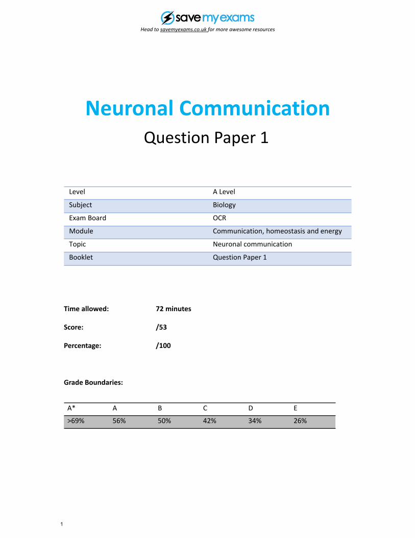
The table below shows the membrane potentials of different neurones at a cholinergic synapse. The data were recorded on five separate occasions, as shown in the five rows.
Membrane potential (mV)
Presynaptic neurone A
Presynaptic neurone B
Presynaptic neurone C
Postsynaptic neurone
1 +40 –70 –70 –70
2 –70 +40 –70 –70
3 –70 –70 +40 –70
4 +40 +40 –70 –70
5 +40 +40 +40 +40
Which of the following, A to D, explains these data?
A. divergence
B. hyperpolarisation
C. spatial summation
D. temporal summation
[Total: 1 ]
Question 2
Head to savemyexams.co.uk for more awesome resources
4
A
B
C
D
spatial summation
all or nothing response
temporal summation
cell signalling
[1]
Animals receive different stimuli from their environment. Their synapses can manage multiple stimuli, often resulting in one response (such as a muscle twitching).
This action of the synapse is an example of
Question 3

Head to savemyexams.co.uk for more awesome resources
5
B C D
Plasma membranes are very important for many processes involved in the functioning of cells, both in animals and plants.
(a) Fig. 4.1 represents part of the plasma (cell surface) membrane of an axon.
Structures labelled A to D represent the involvement of proteins in the movement of ions during depolarisation of the membrane.
D represents a voltage-gated protein.
inside of cell
A
outside of cell
Fig. 4.1
Identify, using the appropriate letter(s), which of the proteins A, B, C or D:
(i) need(s) ATP to function [1]
(ii) transport(s) potassium ions (K+) into the cell [1]
(iii) allow(s) potassium ions (K+) out of the cell [1]
(iv) allow(s) or transport(s) sodium ions (Na+) into or out of the cell. [1]
Question 4
Head to savemyexams.co.uk for more awesome resources
6
inside of cell
outside of celladrenaline molecule
L
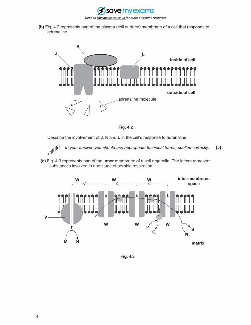
(b) Fig. 4.2 represents part of the plasma (cell surface) membrane of a cell that responds to adrenaline.
KJ
Fig. 4.2
Describe the involvement of J, K and L in the cell’s response to adrenaline.
In your answer, you should use appropriate technical terms, spelled correctly. [5]
(c) Fig. 4.3 represents part of the inner membrane of a cell organelle. The letters represent substances involved in one stage of aerobic respiration.
V
M N
WW
W W PQ R
matrix
inter-membrane space
SW
W
Fig. 4.3
Head to savemyexams.co.uk for more awesome resources
7
(i) In which organelle would this inner membrane be found? [1]
(ii) Identify V. [1]
(iii)
(iv)
Identify W.
Which letter from Fig. 4.3 represents a reduced coenzyme?
(v) Which letter from Fig. 4.3 represents ATP?
[Total: 14]
[1]
[1]
[1]

Head to savemyexams.co.uk for more awesome resources
8
(a) Fig. 1.1 represents a cross section through a myelinated neurone.
ASchwann cell
B
C
Fig. 1.1
(i) Identify A to C. [3]
(ii) Name the gap between two adjacent Schwann cells along the length of the neurone. [1]
(b) There are a number of differences between myelinated and non-myelinated neurones. One difference is the distribution of voltage-gated sodium ion channels in the membrane.
myelinated neurone
• voltage-gated sodium ion channels only occur at gaps between Schwann cells
• each gap is approximately 2 m long
• gaps occur at approximately 1000 m intervals
non-myelinated neurone
• voltage-gated sodium ion channels occur along the total length of the neurone
Use the information above to explain the difference in the speed of conduction of an action potential along the length of a myelinated neurone and a non-myelinated neurone. [4]
Question 5
Head to savemyexams.co.uk for more awesome resources
9
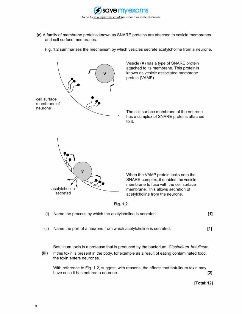
(c) A family of membrane proteins known as SNARE proteins are attached to vesicle membranes and cell surface membranes.
Fig. 1.2 summarises the mechanism by which vesicles secrete acetylcholine from a neurone.
cell surface membrane of neurone
acetylcholine secreted
V
V
Vesicle (V) has a type of SNARE protein attached to its membrane. This protein is known as vesicle associated membrane protein (VAMP).
The cell surface membrane of the neurone has a complex of SNARE proteins attached to it.
When the VAMP protein locks onto the SNARE complex, it enables the vesicle membrane to fuse with the cell surface membrane. This allows secretion of acetylcholine from the neurone.
Fig. 1.2
(i) Name the process by which the acetylcholine is secreted. [1]
(ii) Name the part of a neurone from which acetylcholine is secreted. [1]
(iii)Botulinum toxin is a protease that is produced by the bacterium, Clostridium botulinum.If this toxin is present in the body, for example as a result of eating contaminated food, the toxin enters neurones.
With reference to Fig. 1.2, suggest, with reasons, the effects that botulinum toxin may have once it has entered a neurone. [2]
[Total: 12]
Head to savemyexams.co.uk for more awesome resources
10
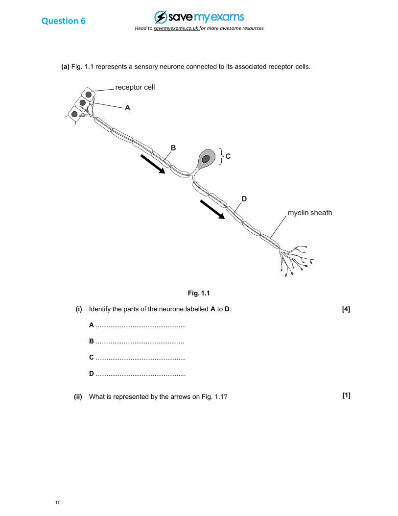
(a) Fig. 1.1 represents a sensory neurone connected to its associated receptor cells.
receptor cell
A
BC
D
myelin sheath
Fig. 1.1
(i) Identify the parts of the neurone labelled A to D.
A .................................................
B ................................................
C .................................................
D .................................................
[4]
(ii) What is represented by the arrows on Fig. 1.1? [1]
Question 6
Head to savemyexams.co.uk for more awesome resources
11
(b) Describe and explain how the resting potential is established and how it is maintained in a sensory neurone.
In your answer, you should use appropriate technical terms, spelled correctly.
[4]
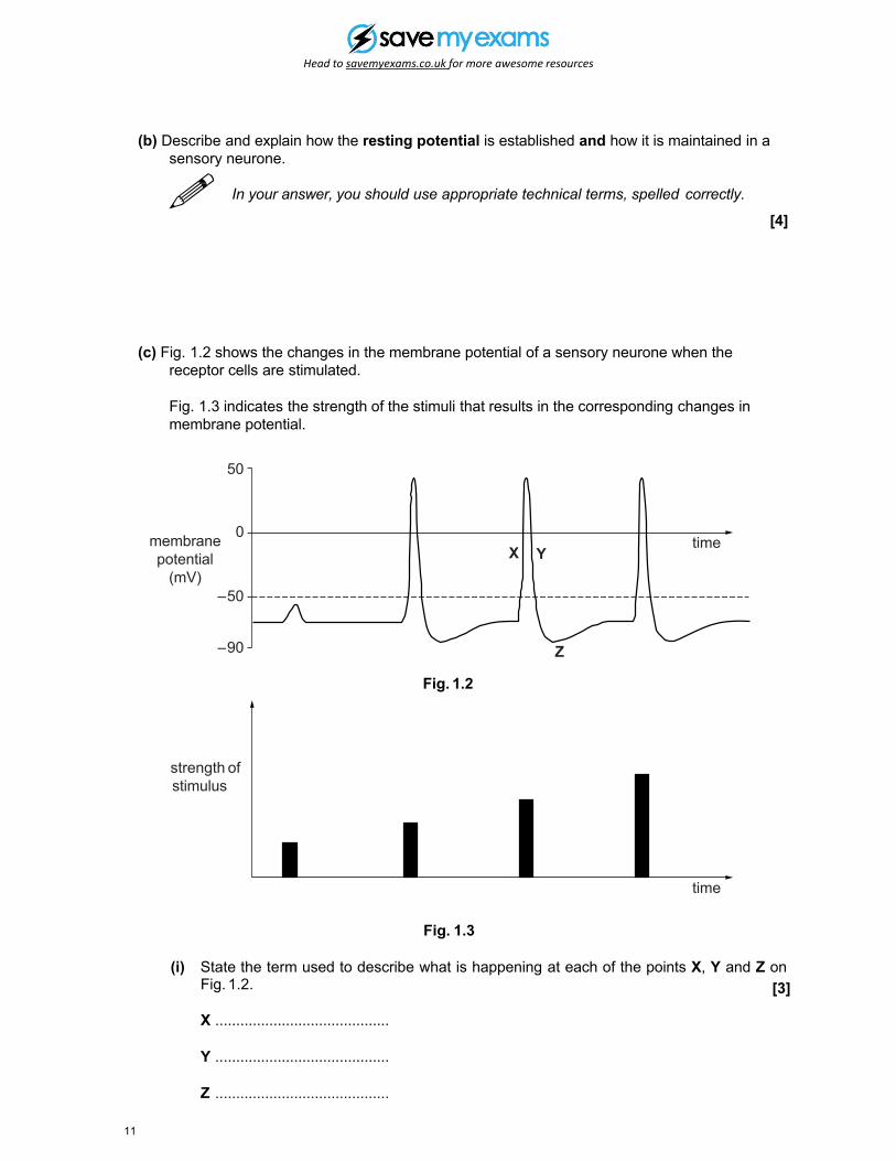
membrane potential
(mV)
strength of stimulus
(c) Fig. 1.2 shows the changes in the membrane potential of a sensory neurone when the receptor cells are stimulated.
Fig. 1.3 indicates the strength of the stimuli that results in the corresponding changes in membrane potential.
50
0
–50
–90
X Y
Z
time
time
Fig. 1.2
Fig. 1.3
(i) State the term used to describe what is happening at each of the points X, Y and Z onFig. 1.2.
X ..........................................
Y ..........................................
Z ..........................................
[3]
Head to savemyexams.co.uk for more awesome resources
12
(ii) What term is used to refer to the value of −50 mV on Fig.1.2? [1]
[Total: 15]
(iii) Comment on the relationship between the strength of a stimulus, as shown in Fig. 1.3, and the resulting action potential, as shown in Fig 1.2.
[2]
Head to savemyexams.co.uk for more awesome resources
13
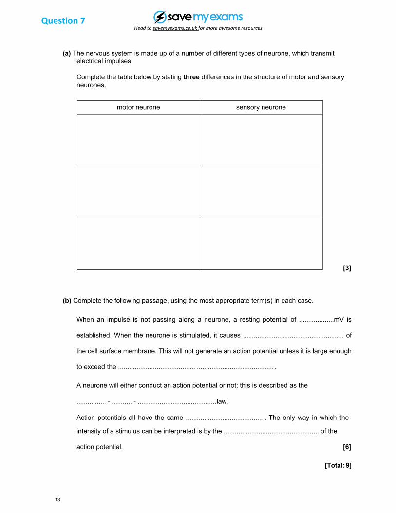
(a) The nervous system is made up of a number of different types of neurone, which transmit electrical impulses.
Complete the table below by stating three differences in the structure of motor and sensory neurones.
motor neurone sensory neurone
[3]
(b) Complete the following passage, using the most appropriate term(s) in each case.
When an impulse is not passing along a neurone, a resting potential of ...................mV is
established. When the neurone is stimulated, it causes ........................................................ of
the cell surface membrane. This will not generate an action potential unless it is large enough
to exceed the .......................................... .......................................... .
A neurone will either conduct an action potential or not; this is described as the
................ - ........... - ...........................................law.
Action potentials all have the same .......................................... . The only way in which the
intensity of a stimulus can be interpreted is by the .................................................... of the
action potential. [6]
[Total: 9]
Question 7