Embed Size (px)
Citation preview
209
18
Plasmodium malariae (Grassi and Feletti, 1890) SYNONYMS:
Haemamoeba malariae Feletti and Grassi, 1889; Plasmodium malariae M. and C. var. quartanae Celli and Sanfelice, 1891; Plasmodium malariae quartanae Kruse, 1892; Haemamoeba laverani var. quartanae Labbe, 1894; Haemosporidium tertianae Lewkowicz, 1897; ? Plasmodium rodhaini Brumpt, 1939.
There is little doubt that Laveran saw this parasite in fresh blood of patients in Algeria in 1880 because his illustrations show schizonts with 8 merozoites arranged in the typical rosette and a central body of pigment. In his opinion, the human malaria parasites belonged to one species and, possibly for that reason, he did not propose a name. He recognized the phenomenon of periodicity but rejected the idea that it might be a clue toward the taxonomy of the malarias.
Golgi in a short note to the Royal Academy of Medicine in Turin (1885) first recognized the periodic succession of fever attacks and in 1889 he clearly showed how to separate tertian and quartan fevers by linking the development of a particular brood of parasites to the fever episode. He was definite in pointing out that if only young forms are present in the circulating blood there will be 1 or 2 days, depending on the species, free of fever. Golgi was also able to demonstrate quotidian fevers in both tertian and quartan infections and postulated, correctly, that the phenomenon was due to double and triple broods of parasites. This important contribution to the biology of these parasites was more or less ignored at the time and then virtually forgotten until well after the turn of the century.
In attempting to arrive at the correct name and credit for the quartan parasite of man, one runs into considerable difficulties. Coatney and Young (1941) in reviewing the problem arrived at the conclusion that the name Plasmodium
malariae should be declared valid as the de facto name and credited to Grassi and Feletti, 1890. Part of this was accomplished, Opinion 283, under the Plenary Powers of the Commission on Zoological Nomenclature (Hemming, 1954), but credit for the name was given to Grassi and Feletti, 1889. It now appears that the 1889 pamphlet, which is said to have given the authorship in reverse, is no longer extant and therefore the order of authorship cannot be verified. In 1890, Grassi and Feletti gave malariae as the specific name for the quartan parasite in the genus Haemamoeba, and on the basis of priority, that date is valid. Following this, Grassi and Feletti (1892) described and illustrated the quartan parasite, as seen in the peripheral blood, and separated it from the other blood-inhabiting forms in man and birds under the name H. malariae Grassi and Feletti. Garnham (1966) holds that "in view of the authority of this paper, and the disappearance of all traces of the original 1889 pamphlet", the name should be credited to Grassi and Feletti 1892. The tenet that authority is a valid basis for assigning credit is unacceptable, and in view of the loss of the 1889 paper and the fact that the Regles had to be suspended in order to validate the de facto name, it appears to us, therefore, that the correct name for the human quartan parasite is Plasmodium malariae (Grassi and Feletti, 1890).
Plasmodium malariae is a cosmopolitan parasite which develops where the summer isotherm does not fall below 15° C (59° F). Its distribution is variable and spotty. Why this is true no one has been able to explain although numerous theories have been advanced; it is still, one of the unsolved problems in the biology of human quartan malaria.
One theory to account for its unique distribution was that because the parasite
210 PRIMATE MALARIAS
requires an extended sporogonic cycle, its greatest prevalence would be in areas where the vector was able to survive for the longest time. This might obtain in some areas, but it sometimes reaches its highest incidence in parts of the tropics where the vector has only a short life.
Another theory was that quartan malaria demands a special vector. It is true, that experimental infections in mosquitoes are difficult to produce and when obtained, the oocysts fail to develop at a uniform rate which probably accounts for the varied results obtained by such workers as Mayne (1932), Mer (1933), and others. However, the special vector theory hardly seems to cover the problem either because many species transmit the parasite: A. atroparvus, A. sacharovi, A. stephensi et al.
The third theory, rested on the presence of an animal reservoir in certain tropical regions. This might be applicable if one is concerned with certain areas in tropical Africa where chimpanzees harbor P. rodhaini. In man, that parasite takes on the attributes of P. malariae and, in some quarters, is considered to be P. malariae. The inui parasites of India and the Malaysian area are morphologically distinct from human quartan and so far only one strain has been established in man. The rest of the Old World is without any zoonotic connection to explain the persistence of quartan malaria. In the New World, the situation is altogether different than it is in Africa or the Far East, because it appears relatively clear that the quartan malaria of monkeys (P. brasilianum) came from man, hence an anthroponosis, rather than the other way around.
It may well be that one or more of these factors, along with changes in the environment which might facilitate transmission, play a part in the unique distribution of quartan malaria throughout the world. But whatever the explanation may be, it appears that P. malariae is probably the oldest parasite in terms of time, and although Knowles et al (1930) considered it a disappearing parasite, it might well be that instead of dying out, it has 'learned' during its long association with man how to cope with adversity and, when conditions permit, to enjoy prosperity.
This is illustrated in the work of Field and Shute (1956) where they site that in West Malaysia "quartan infections are not commonly found in hospital patients--less than five per cent of the admissions . . .--but there are localized areas where P. malariae is the dominant species." In areas of high transmission of all species, P. falciparum is dominant for a few months, whereupon, it is succeeded by P. vivax, which holds center stage for a time only to be replaced by P. malariae which has 'learned' to wait in the wings. Field and Shute call it a "residual infestation" which pretty well sums up the life-cycle of P. malariae. It is a highly successful parasite in that it can live in a host longer than any other malaria and without renewed activity from fixed tissue parasites, it causes the host relatively little inconvenience because of its symbiotic nature and, it is always ready, during periods of recrudescence, to gain access to a cooperative vector and, hence, perpetuate the species.

212 PRIMATE MALARIAS
PLASMODIUM MALARIAE 213
213
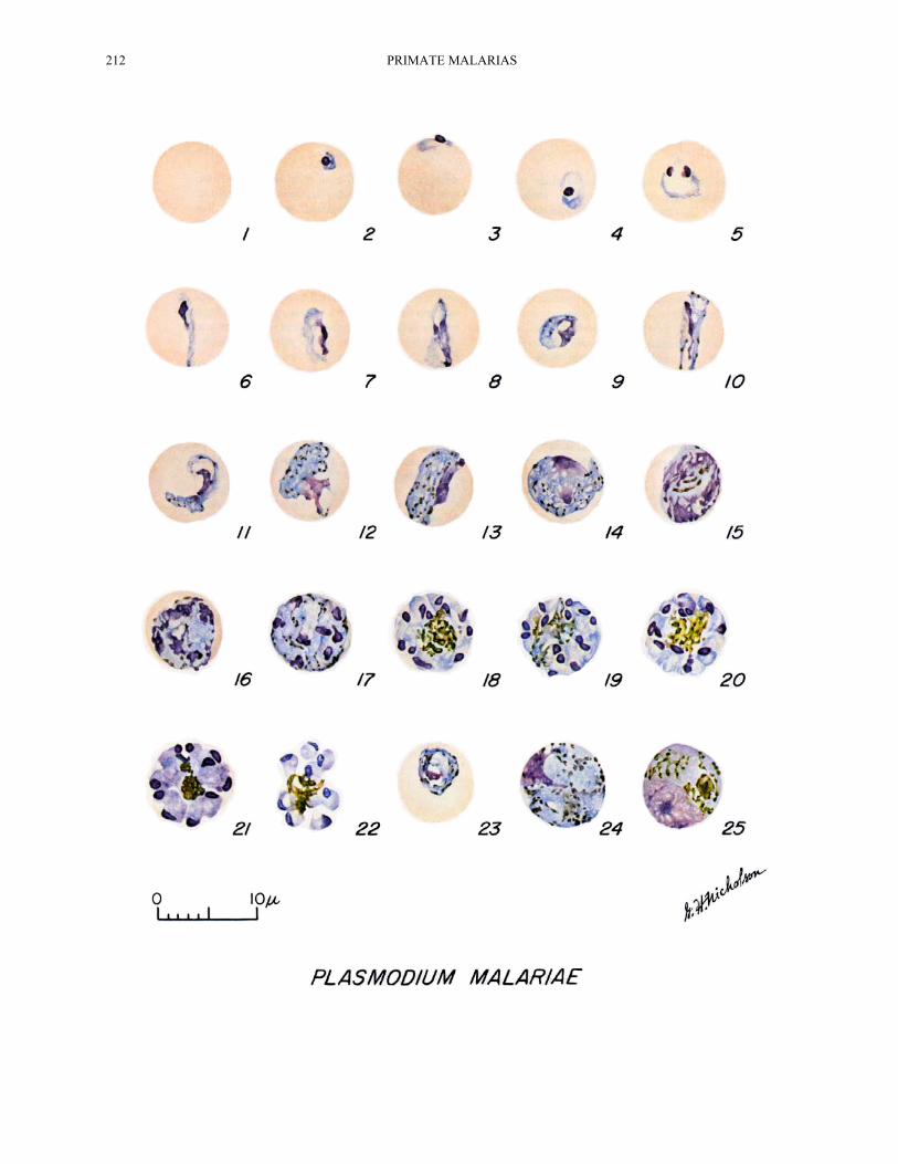
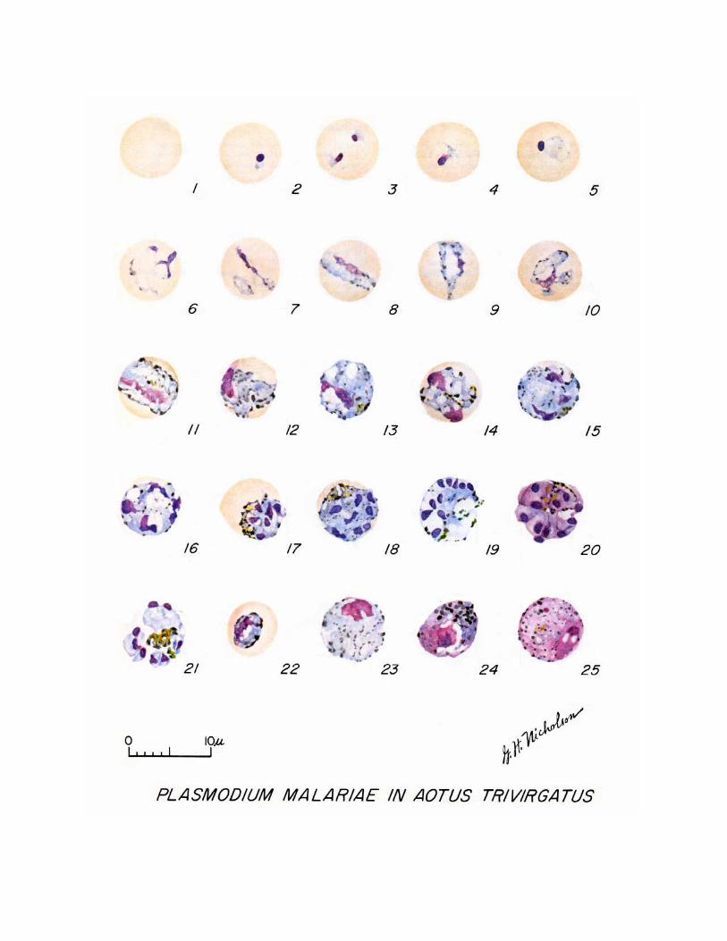
Cycle in the Blood PLATE XXXIII
The first stage to appear in the peripheral blood is the familiar ring-stage, not shown as a true ring on the plate, a circle of cytoplasm, and a spherical bit of chromatin enclosing a vacuole. It very shortly occupies one-fourth to one-third of the parasitized cell and sometimes exhibits an accessory chromatin dot (Figs. 4, 5). Although this parasite generally displays denser cytoplasm and chromatin than P. vivax, it is difficult to separate them at this stage because they are about the same size. The parasite grows more slowly than any of the other human malarias. The vacuole disappears after a few hours. In the living condition, the movement is sluggish. Pigment granules appear early in its growth and sometimes a granule may appear in the late ring- stage. The pigment increases rapidly; half grown parasites may exhibit 30 to 50 jet-black granules (Figs. 12, 13) in contrast to the rod-like pigment found in P. vivax.
As the parasite grows, it assumes various shapes. Some appear stretched out like a ribbon across the host cell and are known as band forms (Figs. 6, 10, 11). Band forms are found in other species, but are more frequent in P. malariae and hence are considered diagnostic. These forms may be seen at any time until the parasite virtually fills the host cell which is not enlarged or blanched. At about the 54th hour, segmentation begins, and by the 65th hour, the host cell is completely filled, or nearly so, and the parasite contains 5 to 6 chromatin masses; the pigment is scattered (Figs. 14-16). During further growth, the definitive number of nuclei are formed and with the final number, the cytoplasm divides to give each nucleus a small amount of cytoplasm. During this stage, the pigment may appear segregated and then
clumped in a loose mass in the center of the cell, surrounded by more or less symmetrically arranged merozoites, to give the "rosette" effect so characteristic of the species. The number of merozoites may be from 6 to 12, sometimes 14; the average is 8 (Figs. 17-22).
The young gametocytes are very similar to the asexual forms which makes it virtually impossible to distinguish them with certainty until about the 54th hour when the asexual forms begin segmentation (Fig. 23). The mature macrogametocyte exhibits a heavy deeply-staining blue cytoplasm with a small, eccentric, well-defined deep red-staining nucleus. The pigment is scattered. The parasite completely fills the host cell (Fig. 24). The cytoplasm of the adult microgametocyte takes a light bluish-pink stain. The pigment is limited to this area of the parasite. The nucleus is diffuse, takes a pinkish-blue stain, and may occupy about half the cell. The parasite fills the entire host cell (Fig. 25). Ordinarily, microgametocytes outnumber the macrogametocytes but this may vary with different strains.
The asexual cycle requires 72 hours.
Sporogonic Cycle PLATE XXXIV
Observations have been made by a number of workers concerning the sporogonic cycle of Plasmodium malariae, but Shute and Maryon (1952) carried out the first definitive studies. These investigators observed its development in Anopheles atroparvus mosquitoes incubated at a temperature of 25°C. They found that the pigment, which seldom consisted of more than 30 granules, was very dark brown and variable in size. During the first 7 to 8 days, the granules were distributed over the oocyst, but, from the
PLATE XXXIII.—Plasmodium malariae. Fig. 1. Normal red cell. Figs. 21, 22. Mature schizonts. Figs. 2-5. Young trophozoites. Fig. 23. Developing gametocyte. Figs. 6-11. Growing trophozoites. Fig. 24. Mature macrogametocyte. Figs. 12, 13. Nearly mature and mature trophozoites. Fig. 25. Mature microgametocyte. Figs. 14-20. Developing schizonts.
214 PRIMATE MALARIAS
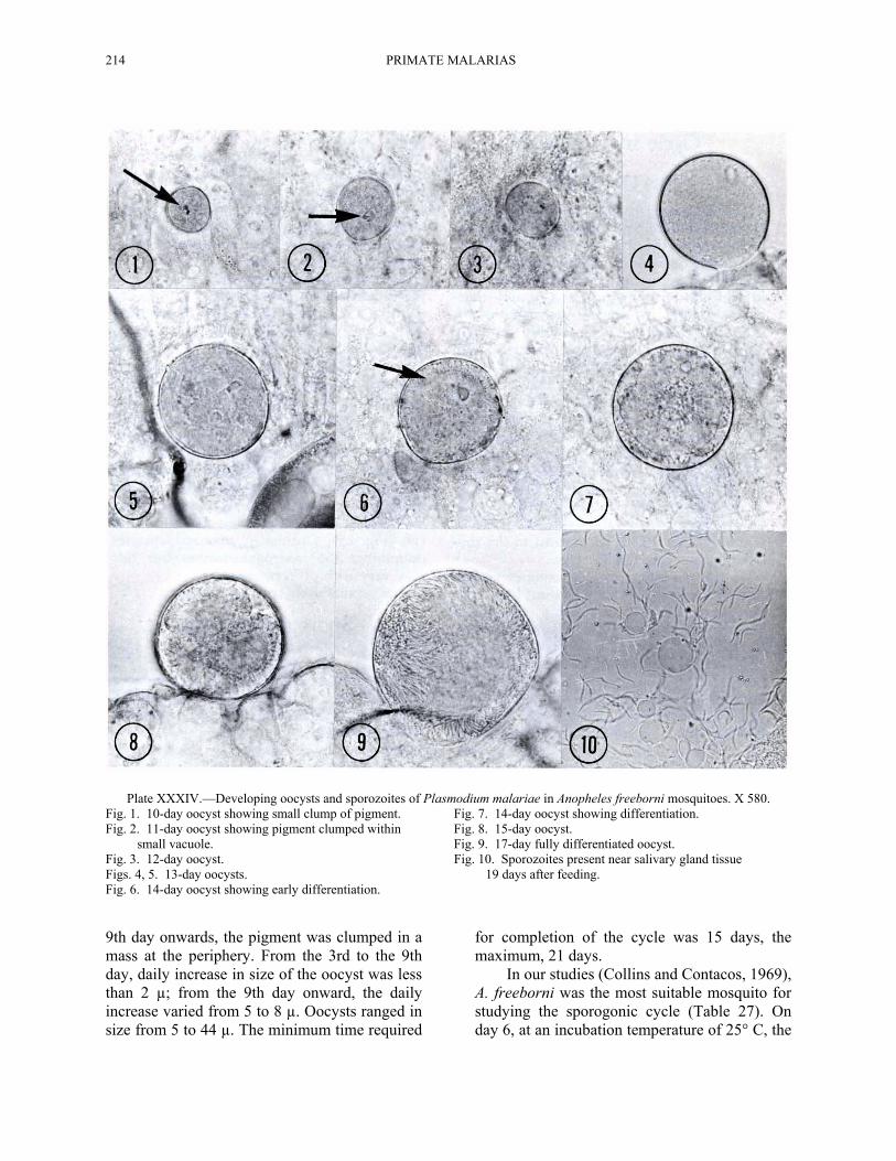
Plate XXXIV.—Developing oocysts and sporozoites of Plasmodium malariae in Anopheles freeborni mosquitoes. X 580. Fig. 1. 10-day oocyst showing small clump of pigment. Fig. 7. 14-day oocyst showing differentiation. Fig. 2. 11-day oocyst showing pigment clumped within Fig. 8. 15-day oocyst. small vacuole. Fig. 9. 17-day fully differentiated oocyst. Fig. 3. 12-day oocyst. Fig. 10. Sporozoites present near salivary gland tissue Figs. 4, 5. 13-day oocysts. 19 days after feeding. Fig. 6. 14-day oocyst showing early differentiation. 9th day onwards, the pigment was clumped in a mass at the periphery. From the 3rd to the 9th day, daily increase in size of the oocyst was less than 2 µ; from the 9th day onward, the daily increase varied from 5 to 8 µ. Oocysts ranged in size from 5 to 44 µ. The minimum time required
for completion of the cycle was 15 days, the maximum, 21 days.
In our studies (Collins and Contacos, 1969), A. freeborni was the most suitable mosquito for studying the sporogonic cycle (Table 27). On day 6, at an incubation temperature of 25° C, the
PLASMODIUM MALARIAE 215
215
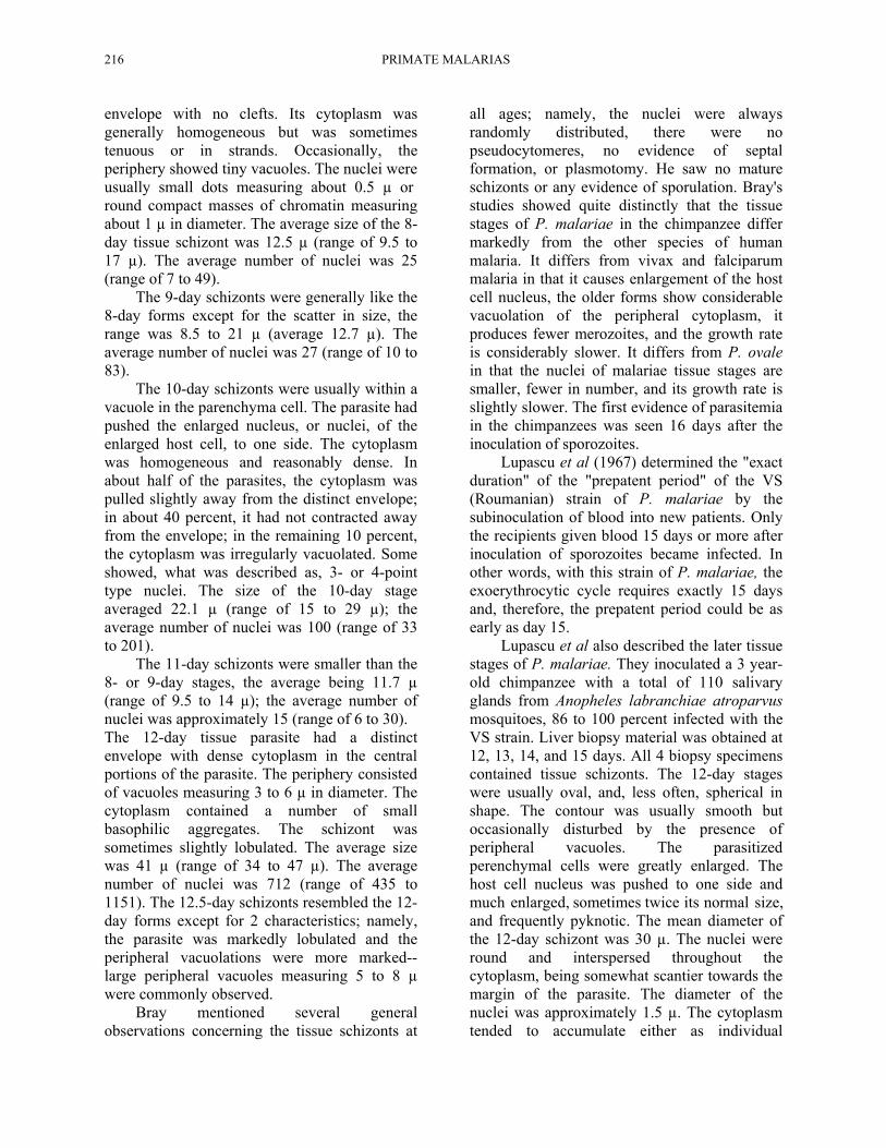
mean oocyst diameter was 12 µ with a range of 9 to 14 µ. The oocysts continued to grow so that by day 14, the mean size was 38 µ with a range of 20 to 65 µ. The first signs of oocyst differentiation were apparent by day 14; sporozoites were present in the salivary glands on day 17.
A comparison of the oocyst growth rate of P. malariae with that of P. cynomolgi (Fig. 45) points-up the marked difference between the 2 parasites. The mean oocyst diameter of P. cynomolgi on day 8 is about the same as that of P. malariae on day 15. Sporozoites appear in the salivary glands 6 days sooner with P. cynomolgi than with P. malariae. The comparison of the sporogonic cycle of this parasite with P. brasilianum is presented elsewhere (Chapter 19).
Cycle in the Tissue
The first tissue stages of Plasmodium malariae were seen and described by Bray (1959, 1960). Liver biopsy specimens (8, 9, 10, 11, 12, and 12.5 days) were taken from 3 different chimpanzees which had received 72 to 110 salivary glands of Anopheles Gambia; 33 to 50 percent of them were infected with sporozoites of a Liberian strain of P. malariae.
TABLE 27.—Oocyst diameters of Plasmodium malariae in Anopheles freeborni mosquitoes.
A. freeborni Days post
No. Range Mean*
6 7 8 9
10 11 12 13 14 15 16 17
104 105 185 169 146 268 218 195 260 260 348 168
9-14 11-19 12-21 14-27 13-32 13-45 14-51 19-59 20-65 17-77 19-88 15-86
12 14 17 20 21 27 29 37 38† 50† 48†
53†**
Totals 2426 9-88
* Measurements expressed in microns. † Oocyst differentiation. ** Sporozoites present in the salivary glands.
The 8-day tissue schizont was described as
lying in the liver parenchyma cell usually, but not always, in a vacuole. The host cell was enlarged and the cytoplasm pushed aside in a crescent-shape around the parasite. The host cell nucleus was enlarged and pushed to one side. In over 50 percent of the parasitized parenchymal cells, 2 or more enlarged nuclei were present. The tissue schizont usually had a distinct
FIGURE 45.—Range in oocyst diameters and the mean oocyst diameter curves of Plasmodium cynomolgi and P. malariae in Anopheles freeborni mosquitoes. (D = oocyst differentiation; SP = sporozoites present in the salivary glands).
216 PRIMATE MALARIAS
envelope with no clefts. Its cytoplasm was generally homogeneous but was sometimes tenuous or in strands. Occasionally, the periphery showed tiny vacuoles. The nuclei were usually small dots measuring about 0.5 µ or round compact masses of chromatin measuring about 1 µ in diameter. The average size of the 8-day tissue schizont was 12.5 µ (range of 9.5 to 17 µ). The average number of nuclei was 25 (range of 7 to 49).
The 9-day schizonts were generally like the 8-day forms except for the scatter in size, the range was 8.5 to 21 µ (average 12.7 µ). The average number of nuclei was 27 (range of 10 to 83).
The 10-day schizonts were usually within a vacuole in the parenchyma cell. The parasite had pushed the enlarged nucleus, or nuclei, of the enlarged host cell, to one side. The cytoplasm was homogeneous and reasonably dense. In about half of the parasites, the cytoplasm was pulled slightly away from the distinct envelope; in about 40 percent, it had not contracted away from the envelope; in the remaining 10 percent, the cytoplasm was irregularly vacuolated. Some showed, what was described as, 3- or 4-point type nuclei. The size of the 10-day stage averaged 22.1 µ (range of 15 to 29 µ); the average number of nuclei was 100 (range of 33 to 201).
The 11-day schizonts were smaller than the 8- or 9-day stages, the average being 11.7 µ (range of 9.5 to 14 µ); the average number of nuclei was approximately 15 (range of 6 to 30). The 12-day tissue parasite had a distinct envelope with dense cytoplasm in the central portions of the parasite. The periphery consisted of vacuoles measuring 3 to 6 µ in diameter. The cytoplasm contained a number of small basophilic aggregates. The schizont was sometimes slightly lobulated. The average size was 41 µ (range of 34 to 47 µ). The average number of nuclei was 712 (range of 435 to 1151). The 12.5-day schizonts resembled the 12-day forms except for 2 characteristics; namely, the parasite was markedly lobulated and the peripheral vacuolations were more marked--large peripheral vacuoles measuring 5 to 8 µ were commonly observed.
Bray mentioned several general observations concerning the tissue schizonts at
all ages; namely, the nuclei were always randomly distributed, there were no pseudocytomeres, no evidence of septal formation, or plasmotomy. He saw no mature schizonts or any evidence of sporulation. Bray's studies showed quite distinctly that the tissue stages of P. malariae in the chimpanzee differ markedly from the other species of human malaria. It differs from vivax and falciparum malaria in that it causes enlargement of the host cell nucleus, the older forms show considerable vacuolation of the peripheral cytoplasm, it produces fewer merozoites, and the growth rate is considerably slower. It differs from P. ovale in that the nuclei of malariae tissue stages are smaller, fewer in number, and its growth rate is slightly slower. The first evidence of parasitemia in the chimpanzees was seen 16 days after the inoculation of sporozoites.
Lupascu et al (1967) determined the "exact duration" of the "prepatent period" of the VS (Roumanian) strain of P. malariae by the subinoculation of blood into new patients. Only the recipients given blood 15 days or more after inoculation of sporozoites became infected. In other words, with this strain of P. malariae, the exoerythrocytic cycle requires exactly 15 days and, therefore, the prepatent period could be as early as day 15.
Lupascu et al also described the later tissue stages of P. malariae. They inoculated a 3 year- old chimpanzee with a total of 110 salivary glands from Anopheles labranchiae atroparvus mosquitoes, 86 to 100 percent infected with the VS strain. Liver biopsy material was obtained at 12, 13, 14, and 15 days. All 4 biopsy specimens contained tissue schizonts. The 12-day stages were usually oval, and, less often, spherical in shape. The contour was usually smooth but occasionally disturbed by the presence of peripheral vacuoles. The parasitized perenchymal cells were greatly enlarged. The host cell nucleus was pushed to one side and much enlarged, sometimes twice its normal size, and frequently pyknotic. The mean diameter of the 12-day schizont was 30 µ. The nuclei were round and interspersed throughout the cytoplasm, being somewhat scantier towards the margin of the parasite. The diameter of the nuclei was approximately 1.5 µ. The cytoplasm tended to accumulate either as individual
PLASMODIUM MALARIAE 217
217
flocculi or as larger masses. Numerous vacuoles of all sizes were present. The largest vacuoles were 10 µ. The parasite had a definite membrane.
The 13-day forms had an average size of 44 µ, indicating considerable growth in 24 hours. This rapid growth probably accounted for the pronounced lobulation. The internal structure of the parasite was much the same as that of the 12-day schizont except that the nuclei were "less loosely packed." Cytoplasmic clumps and vacuoles persisted and clefts were usually seen.
The 14-day schizonts reached a maximum mean diameter of 47 µ. They were more difficult to measure because of the lobulated surface. The most striking feature was the extensive cleavage of the cytoplasm in some of the schizonts. The cytoplasm was condensed into long strands, studded with nuclei, and sometimes smaller clefts were present throughout the parasite. The nuclei were "tightly packed" and measured about 2 µ in diameter; vacuoles were obvious. The limiting membrane of the parasite was distinct and occasionally thrown into pronounced folds. Only the 14-day schizonts exhibited merozoites.
The 15-day schizonts were considered mature and remnants of ruptured schizonts were readily observed. The mature forms had a mean diameter of about 51 µ. Their shape was mostly oval, although irregular processes (lobulations) were observed. The nuclei, of the near-mature schizonts, lost their spherical shape just prior to the final division, became triangular, and measured as much as 3.3 µ in their greatest dimension. The cytoplasm formed patterns although the nuclei did not enter this material and no cytomeres or any form of aposchizogony was observed. Large vacuoles were present but were less common than in the nearly-mature stage. The authors described curious reddish strands in the cytoplasm of some of the schizonts and considered this feature unique among tissue forms of malaria parasites. The mature schizont, except for the strands and a few vacuoles, consisted, almost entirely, of merozoites within the outer limiting membrane of the parasite. The largest mature schizont measured 41 by 85 µ, the average was 56 µ. The number of merozoites was directly dependent upon the size of the mature schizont. The largest
number of merozoites was estimated to be 18,650 and the smallest 7,500. The merozoite was a sphere measuring 2 µ in diameter, consisting of cytoplasm which indents the nucleus to render it characteristically crescentic in shape.
Lupascu et al (1967) in their summary gave the main characteristics of the tissue schizonts of the VS strain of P. malariae as: enlargement of the host cell nucleus, many peripheral and internal vacuoles, no cytomeres, large clefts, red-staining strands, and plaques in the mature schizonts.
Course of Infection
As mentioned earlier, the shortest prepatent period for P. malariae could be as early as 15 days, on the basis of the findings of Lupascu's subinoculation experiments. However, in actuality, the earliest reported prepatent period has been 16 days in a West African strain (Shute and Maryon, 1951).
Generally, the prepatent periods, with this species, have been longer. Boyd and Stratman-Thomas (1933) transmitted 2 strains in which the prepatent periods were 27, 32, and 37 days. Mer (1933) transmitted a Palestinian strain to 3 patients; prepatent periods were 26, 28, and 31 days. Prepatent periods of 23 to 26, days were reported by de Buck (1935) for 4 patients infected with a Vienna strain. Boyd and Stratman-Thomas (1936) reported a prepatent period of 37 days (thin smear examination); the incubation period was 40 days. This patient had only 5 paroxysms with 4 chills. The paroxysms subsided spontaneously although parasites were found as late as 173 days after exposure to infection. They also reported a prepatent period of 28 days and an incubation period of 30 days in another patient. Marotta and Sandicchi (1939) reported incubation periods of 23 and 29 days in 2 patients.
Boyd (1940) reported on 3 different strains: USPHS, Jones, and Weaver. The prepatent periods ranged from 28 to 37 days (median of 34 days) and the incubation periods from 30 to 49 days (median of 36 days). Siddons (1944) transmitted an Indian strain of P. malariae to a
218 PRIMATE MALARIAS
patient who exhibited a prepatent period of 30 days and an incubation period of 36 days. Young and Burgess (1947) observed prepatent periods of 29 and 59 days in 2 patients whose incubation periods were 28 and 69 days. These authors considered the prepatent period of 59 days to be due, possibly, to some immunity on the part of the patient which was substantiated by a short duration of parasitemia and extremely low parasite counts. Mackerras and Ercole (1948) reported a prepatent period of 24 days for a Melanesian strain. Kitchen (1949) reported mean prepatent periods of 32.2 days (range 27 to 37 days) and mean incubation periods of 34.8 days (range 29 to 40 days) for naturally induced infections of American strains of P. malariae.
Young and Burgess (1961) transmitted the USPHS strain of P. malariae to 2 patients and observed prepatent periods of 33 and 36 days and incubation periods of 28 and 43 days. Ciuca et al (1964) reported prepatent periods ranging from 18 to 25 days for a local Roumanian strain, now known as the VS strain. Lupascu et al (1968) reported incubation periods of 18 to 19 days with the VS strain of P. malariae. As these data show, there is a wide range in the length of the prepatent periods in naturally transmitted P. malariae (18 to 59 days) and an equally wide range in the incubation periods (28 to 69 days).
In our transmission studies with a Nigerian strain of P. malariae, involving 4 volunteers, we have observed prepatent periods ranging from 24 to 33 days (Contacos and Collins, 1969); with the VS strain, in 4 patients, the pre-patent periods ranged from 21 to 30 days.
Malariae malaria infections are considered to be relatively benign when compared to falciparum malaria but more severe than vivax infections. Probably its most peculiar characteristic is its pronounced chronicity, especially the unusually long period of time during which parasites can remain 'dormant' within the host. A less constant characteristic is the tendency to cause renal damage which was dealt with in some detail by Giglioli (1930).
The onset of P. malariae attacks is generally more gradual than observed with many vivax and falciparum infections. The initial remittent fever pattern characteristic of vivax infections is seen less frequently in malariae infections. Rather, they exhibit an intermittent
pattern (Kitchen, 1949) because the sporulation of this species is highly synchronous.
Young et al (1940) studied the periodic phenomenon of the asexual cycle of quartan malaria in Negro paretics. They found that their strain of malariae malaria (USPHS) exhibited a high degree of synchronicity, the asexual erythrocytic cycle repeating itself every 72 hours, but being more exact in some patients than in others. The length of time consumed by the different growth stages was: 54.2 hours for the trophozoites, 10.4 hours for the young schizonts, and 7.4 hours for the segmenters or late schizonts. The process of segmentation required roughly 6 hours. The rise in temperature closely followed the progress of segmentation and reached its height approximately at the end of the process.
Young et al (1940a) showed that modifying the external conditions of the host affected the time of sporulation of P. malariae in man when patients were placed under reversed conditions of activity; the segmenter-number peaks in one changed from the normal 9:00 a.m. hour to 9:00 p.m. In the other, the cycle was shortened until the segmenters peaked about 22 hours before normally expected. Two other patients were placed under reversed conditions, except lighted continuously. In these, the segmenter-peak time changed from 9:00 a.m. to 9 :00 p.m. When one of them was returned to a normal schedule, the segmenter-number peaks returned to the normal time, 9:00 a.m.
Young et al (1941) presented data on 420 paroxysms occurring in 15 patients infected with the USPHS strain of P. malariae. Chills were observed 102 times (24 percent). Some patients experienced chills more often than others; one had 9 chills in 14 paroxysms (64 percent), while another had only 1 chill in 45 paroxysms (2 percent). Temperatures at the beginning of chills were found to range from 97.6° F to 106.0° F rectally, with an average of 101.8° F. Temperatures at the end of the chills ranged from 97.8° F to 106.0° F, with an average of 103.5° F. The greatest increase in temperature during a chill was observed in a patient whose temperature rose from 98.0° F to 103.0° F during a chill lasting 45 minutes. Two patients experienced the opposite; in other words, a drop in temperature during a chill. One had a
PLASMODIUM MALARIAE 219
219
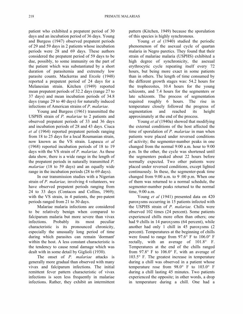
temperature of 106.0° F at the beginning and 105.0° F at the end of the chill which lasted 50 minutes. Another patient dropped from 104.2° F at the beginning of the chill to 103.2° F at the end of the chill which lasted 15 minutes. It is interesting that in both these cases, very high temperatures had already been reached by the time the chill began. The durations of all chills ranged from 13 to 195 minutes with an average of 53 minutes (Fig. 46).
According to Kitchen (1949), the malariae paroxysm is less often introduced by chills than are vivax paroxysms. Boyd (1940) found that the initiation of the malariae paroxysm by chills was extremely variable. Indeed, one of 5 naturally induced cases studied by him had no chills at all. In Kitchen's observations, on patients infected with the Trinidad strain, the temperature at the onset of the chill varied between 97.4°F and 104.4°F (mean of 100.3° F). The mean duration of the chills was 55.97 minutes. The mean temperature peak was 104.98° F reached, on an average, 2 hours, 54 minutes after the onset. The interval from peak temperature until the temperature returned to normal was 10 hours, 23 minutes.
The entire paroxysm, therefore, occupied a period of 13 hours and 17 minutes. Plasmodium
malariae paroxysms are longer in duration than those of P. vivax.
In the Young et al series, the average fever peak for the 420 paroxysms was 104.1°F rectally, with the highest temperature recorded, 106.4° F. The duration of the fevers ranged from 5 to 32 hours with an average of 10 hours, 58 minutes. The average time for the interval from the beginning of fever to the fever peak was 4 hours, 45 minutes and from the fever peak to the end of the fever was 6 hours, 13 minutes. These authors also observed that some paroxysms were introduced by a chill, while others were devoid of chills. When a chill accompanied the paroxysm, the fever was significantly higher (104.6° F as compared to 104.0° F without a chill) and the duration of the fever was 1 hour, 16 minutes shorter (10 hours duration for the paroxysms with chills as compared to 11 hours, 16 minutes for the paroxysms without chills). In addition, it was obvious that this shortening of the fever period occurred almost entirely in the period between the onset of the paroxysm and the peak of the fever. They believed that the chill exerted a definite influence on the character of the paroxysm. When a chill occurred, the duration of the fever was shorter (1 hour, 16 minutes) which was due, apparently, to the
FIGURE 46.—The temperature curve in relation to the chill in 102 paroxysms in Plasmodium malariae infections (after Young,
Coatney, and McLendon, 1941).
220 PRIMATE MALARIAS
shortening of the period when the temperature was rising, and, that the maximal temperature was significantly higher than in paroxysms without chills. The greatest proportionate increase in temperature occurred during the chilling period (Fig. 46).
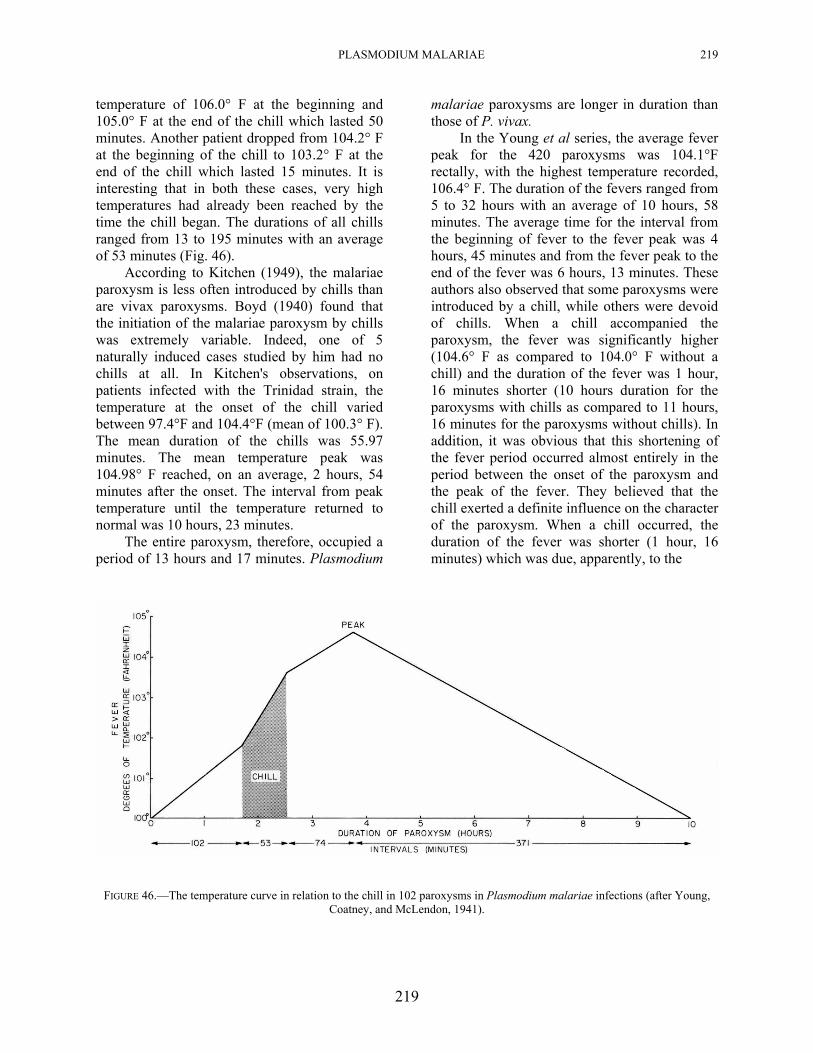
Generally, the degree of parasitemia is lower for P. malariae than for either vivax or falciparum malaria. Boyd (1940) reported mean parasite densities ranging up to 12,500 per mm3. However, there was one patient in his series, whose infection resulted in a fatal outcome, in whom the count exceeded 100,000 parasites per mm3. Young and Burgess (1947) observed parasites at least through 58 days in a patient having a maximum parasite count of 3,280 per mm3 of blood. Mackerras and Ercole (1948) did not observe parasitemias which exceeded 2,300 per mm3.
Kitchen (1949) stated that the parasitemias observed in his P. malariae infections were comparatively low. Counts above 20,000 per mm3 were unusual and counts above 50,000 per mm3 were rare. He indicated that most patients do not attain counts above 10,000 per mm3 during their entire attack. After the first few weeks, the counts tend to drop and remain below 5,000 per mm3. Young and Burgess (1961) reported parasitemias up to 17,580 parasites per mm3, with an average of 3,799 per mm3 in symptomatic cases and up to 4,920 per mm3, with an average of 999 per mm3 in
asymptomatic cases. Ciuca et al (1964) reported maximum parasitemias of 6,700 and 8,852 per mm3 for sporozoite-and parasitized blood-induced infections, respectively. In our studies with a Nigerian strain of P. malariae, a maximum parasite count of 11,200 per mm3 has been observed in a blood-induced infection (Collins and Contacos, 1969).
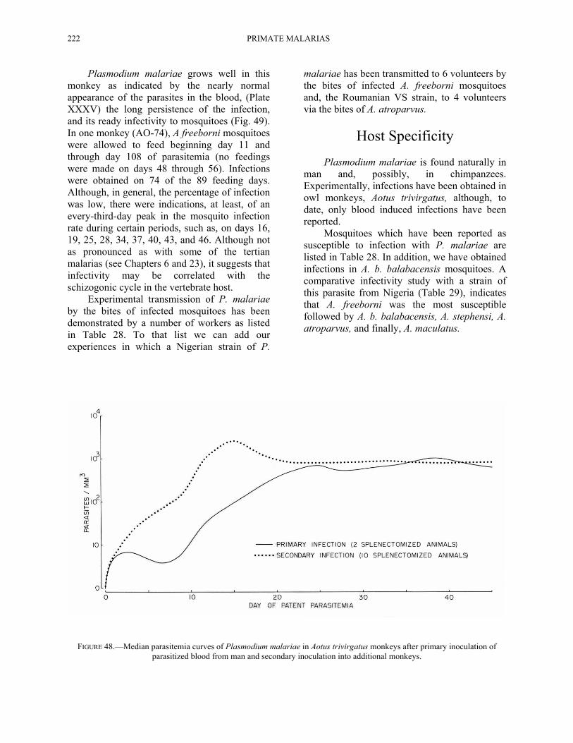
Figure 47 shows the median parasitemia curves for Caucasian and Negro patients infected with the USPHS strain of Plasmodium malariae, as well as curves for the minimum and maximum parasitemia. It can be readily seen that the median parasitemia curves for the Caucasian and Negro patients show only minor differences. Median peak parasitemia occurs some time between day 15 and day 20. The parasitemia subsequently persists at an almost constant level, tapering off very gradually during the next 40 days, in the Negro patients and only a very little, if at all, in the Caucasians. The median maximum parasitemias were approximately 5,000 per mm3 at day 25 and 6,000 at day 28 for the Negro and Caucasian patients, respectively. The maximum parasitemia attained by a Caucasian patient was 22,000 parasites per mm3 of blood at day 17, and, for Negro patients, 25,000 per mm3 at day 18, and 26,500 per mm3 at day 40. There were slight differences between the parasitemias in the Caucasian and Negro patients from day 30
FIGURE 47.—Minimum and maximum parasitemia curves in 54 human volunteers infected with Plasmodium malariae and median parasitemia curves for 29 Caucasian and 25 Negro volunteers, all infected by the inoculation of parasitized blood.
PLASMODIUM MALARIAE 221
on. By day 30, the median parasite count for the Caucasian patients was approximately 5,000 per mms whereas for the Negro patients, it was approximately 3,000 per mm3. Between day 30 and 50, the median parasite counts for the Caucasian patients ranged between 3,000 and 6,000 per mm3 whereas for the Negro patients, it was between 2,000 and 3,500 per mm3. After day 50, the median parasite counts for the Caucasian patients ranged between 3,000 and 4,000 per mm3 whereas in the Negro patients, the counts ranged between 1,000 and 3,000 per mm3. In other words, there appeared to be a slightly earlier trend toward chronicity in the Negro patients than in the Caucasian patients infected with the same strain.
The duration of P. malariae infections can be extremely long, especially in the form of subpatent parasitemias. Boyd and Stratman- Thomas (1936) observed malariae parasites as late as 173 days after exposure to infection. Boyd (1940) found that in infections induced by mosquito bites, P. malariae attacks, including recurrences, had a mean duration of 170 days in Caucasians and 76 days in Negroes. The mean duration of all naturally induced clinical attacks, regardless of race, was 132 days. Boyd (1947) reported that a naturally induced infection with P. malariae had persisted in a latent-chronic state for a period of 4,305 days (11 years, 9 months, 14 days); this was established when the patient, who had received no specific therapy, gave 250 ml of blood for a transfusion. Shute and Maryon (1955) reported "true relapse" of P. malariae infection in 2 patients 12 and 32 years after infection. What they reported, probably, were recrudescences since it is not clear whether the therapy given was adequate to totally eliminate all the blood stages. Ciuca et al (1956) published on a case in which parasites were observed after 18 years. Lentini and Tecce (1955) observed a patient whose infection relapsed (recrudesced) 45 years after leaving an endemic area. Guazzi and Grazi (1963) reported an infection which relapsed, subsequent to a splenectomy, approximately 53 years after the primary attack. Diamond (1966) found P. malariae parasites in a blood film from a patient who had immigrated from Antigua to England 12 years earlier and had no previous history of malaria.
Finally, a comment about true relapse activity in P. malariae. Ciuca et al (1964) did not observe relapse activity in any of their patients whose infections were induced by bites of infected mosquitoes. They concluded, therefore, that there was no secondary exoerythrocytic cycle. In this connection, we also have observed no relapse activity after adequate blood schizonticidal therapy in our volunteers exposed to infection with a Nigerian strain of P. malariae. It seems safe to say that, in this respect, P. malariae resembles P. falciparum more than it does the human tertian malarias which do relapse.
Plasmodium malariae can infect the chimpanzee (Rodhain, 1948; Garnham et al, 1956). Rodhain observed maximum parasitemias of 7,000 per mm3. Garnham et al observed a maximum parasitemia of 160,000 per mm3 in a splenectomized chimpanzee. Bray (1960) ob- served parasitemia of P. malariae in splenectomized chimpanzees, between 25,000 and 50,000 parasites per mm3 of blood. Some consider P. malariae of man and P. rodhaini in the chimpanzee to be one and the same species. Maybe they are one and the same but, in our opinion, adequate proof is lacking.
The first adaptation of Plasmodium malariae to Aotus trivirgatus monkeys was reported by Geiman and Siddiqui (1969). One hundred and thirty days after inoculation, the parasitemia reached a peak--of 97,920 per mm3 of blood. During the succeeding one year of observation, the parasitemia fluctuated between 200 and 26,000 per mm3. The infection was subsequently passaged to splenectomized and to intact A. trivirgatus monkeys; their parasitemias ranged from 100 to 23,000 per mm3.
In our own studies (Contacos and Collins, 1969; Collins and Contacos, 1969), we have been able to transfer the infection from man to splenectomized A. trivirgatus monkeys on 2 occasions (Fig. 48). The parasitemia was slow to rise and reached a mean parasite level of approximately 1,000 per mm3 on the 25th day of patent parasitemia. Subsequent passage to 10 other splenectomized monkeys resulted in a peak median parasitemia of approximately 2,500 per mm3 by day 15. Thereafter, the parasitemia was maintained at approximately the same level as that in the primary passages.
222 PRIMATE MALARIAS
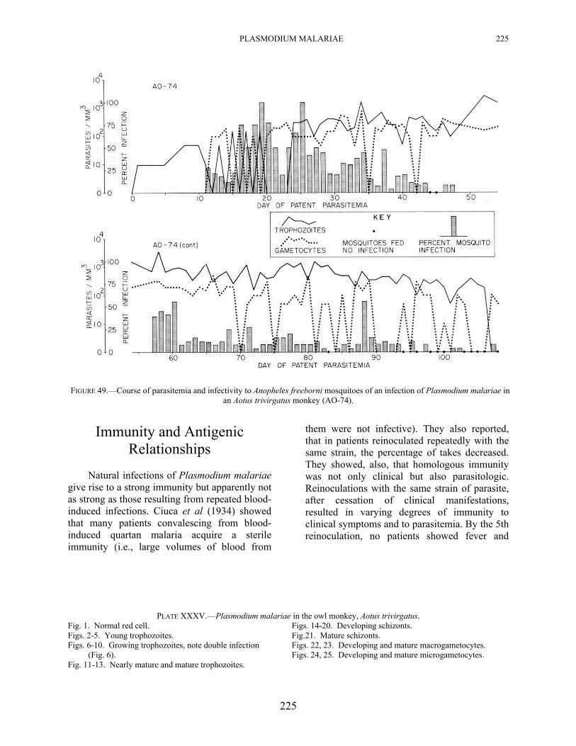
Plasmodium malariae grows well in this monkey as indicated by the nearly normal appearance of the parasites in the blood, (Plate XXXV) the long persistence of the infection, and its ready infectivity to mosquitoes (Fig. 49). In one monkey (AO-74), A freeborni mosquitoes were allowed to feed beginning day 11 and through day 108 of parasitemia (no feedings were made on days 48 through 56). Infections were obtained on 74 of the 89 feeding days. Although, in general, the percentage of infection was low, there were indications, at least, of an every-third-day peak in the mosquito infection rate during certain periods, such as, on days 16, 19, 25, 28, 34, 37, 40, 43, and 46. Although not as pronounced as with some of the tertian malarias (see Chapters 6 and 23), it suggests that infectivity may be correlated with the schizogonic cycle in the vertebrate host.
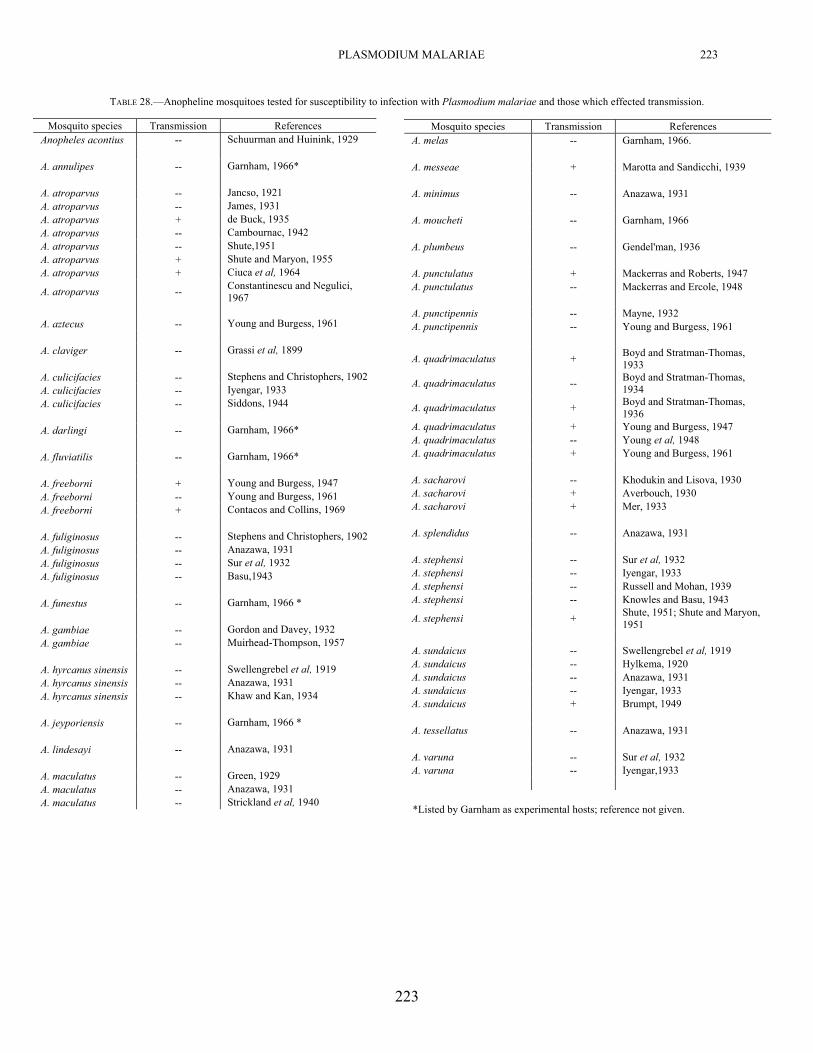
Experimental transmission of P. malariae by the bites of infected mosquitoes has been demonstrated by a number of workers as listed in Table 28. To that list we can add our experiences in which a Nigerian strain of P.
malariae has been transmitted to 6 volunteers by the bites of infected A. freeborni mosquitoes and, the Roumanian VS strain, to 4 volunteers via the bites of A. atroparvus.
Host Specificity
Plasmodium malariae is found naturally in man and, possibly, in chimpanzees. Experimentally, infections have been obtained in owl monkeys, Aotus trivirgatus, although, to date, only blood induced infections have been reported.
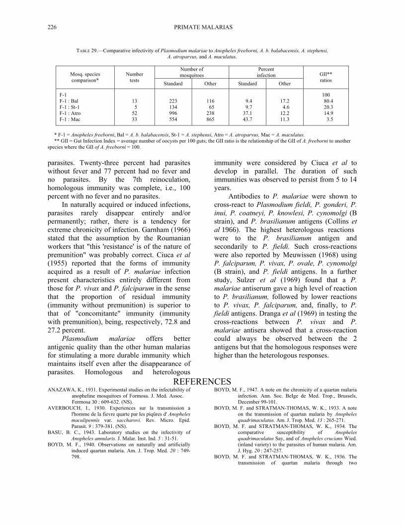
Mosquitoes which have been reported as susceptible to infection with P. malariae are listed in Table 28. In addition, we have obtained infections in A. b. balabacensis mosquitoes. A comparative infectivity study with a strain of this parasite from Nigeria (Table 29), indicates that A. freeborni was the most susceptible followed by A. b. balabacensis, A. stephensi, A. atroparvus, and finally, A. maculatus.
FIGURE 48.—Median parasitemia curves of Plasmodium malariae in Aotus trivirgatus monkeys after primary inoculation of parasitized blood from man and secondary inoculation into additional monkeys.
PLASMODIUM MALARIAE 223
223
TABLE 28.—Anopheline mosquitoes tested for susceptibility to infection with Plasmodium malariae and those which effected transmission.
Mosquito species Transmission References Anopheles acontius -- Schuurman and Huinink, 1929 A. annulipes -- Garnham, 1966* A. atroparvus -- Jancso, 1921 A. atroparvus -- James, 1931 A. atroparvus + de Buck, 1935 A. atroparvus -- Cambournac, 1942 A. atroparvus -- Shute,1951 A. atroparvus + Shute and Maryon, 1955 A. atroparvus + Ciuca et al, 1964
A. atroparvus -- Constantinescu and Negulici, 1967
A. aztecus -- Young and Burgess, 1961 A. claviger -- Grassi et al, 1899 A. culicifacies -- Stephens and Christophers, 1902 A. culicifacies -- Iyengar, 1933 A. culicifacies -- Siddons, 1944 A. darlingi -- Garnham, 1966* A. fluviatilis -- Garnham, 1966* A. freeborni + Young and Burgess, 1947 A. freeborni -- Young and Burgess, 1961 A. freeborni + Contacos and Collins, 1969 A. fuliginosus -- Stephens and Christophers, 1902 A. fuliginosus -- Anazawa, 1931 A. fuliginosus -- Sur et al, 1932 A. fuliginosus -- Basu,1943 A. funestus -- Garnham, 1966 * A. gambiae -- Gordon and Davey, 1932 A. gambiae -- Muirhead-Thompson, 1957 A. hyrcanus sinensis -- Swellengrebel et al, 1919 A. hyrcanus sinensis -- Anazawa, 1931 A. hyrcanus sinensis -- Khaw and Kan, 1934 A. jeyporiensis -- Garnham, 1966 * A. lindesayi -- Anazawa, 1931 A. maculatus -- Green, 1929 A. maculatus -- Anazawa, 1931 A. maculatus -- Strickland et al, 1940
Mosquito species Transmission References A. melas -- Garnham, 1966. A. messeae + Marotta and Sandicchi, 1939 A. minimus -- Anazawa, 1931
A. moucheti -- Garnham, 1966 A. plumbeus -- Gendel'man, 1936 A. punctulatus + Mackerras and Roberts, 1947 A. punctulatus -- Mackerras and Ercole, 1948 A. punctipennis -- Mayne, 1932 A. punctipennis -- Young and Burgess, 1961
A. quadrimaculatus + Boyd and Stratman-Thomas, 1933
A. quadrimaculatus -- Boyd and Stratman-Thomas, 1934
A. quadrimaculatus + Boyd and Stratman-Thomas, 1936
A. quadrimaculatus + Young and Burgess, 1947 A. quadrimaculatus -- Young et al, 1948 A. quadrimaculatus + Young and Burgess, 1961 A. sacharovi -- Khodukin and Lisova, 1930 A. sacharovi + Averbouch, 1930 A. sacharovi + Mer, 1933 A. splendidus -- Anazawa, 1931
A. stephensi -- Sur et al, 1932 A. stephensi -- Iyengar, 1933 A. stephensi -- Russell and Mohan, 1939 A. stephensi -- Knowles and Basu, 1943
A. stephensi + Shute, 1951; Shute and Maryon, 1951
A. sundaicus -- Swellengrebel et al, 1919 A. sundaicus -- Hylkema, 1920 A. sundaicus -- Anazawa, 1931 A. sundaicus -- Iyengar, 1933 A. sundaicus + Brumpt, 1949 A. tessellatus -- Anazawa, 1931 A. varuna -- Sur et al, 1932 A. varuna -- Iyengar,1933
*Listed by Garnham as experimental hosts; reference not given.
PLASMODIUM MALARIAE 225
225
FIGURE 49.—Course of parasitemia and infectivity to Anopheles freeborni mosquitoes of an infection of Plasmodium malariae in an Aotus trivirgatus monkey (AO-74).
Immunity and Antigenic
Relationships Natural infections of Plasmodium malariae
give rise to a strong immunity but apparently not as strong as those resulting from repeated blood-induced infections. Ciuca et al (1934) showed that many patients convalescing from blood-induced quartan malaria acquire a sterile immunity (i.e., large volumes of blood from
them were not infective). They also reported, that in patients reinoculated repeatedly with the same strain, the percentage of takes decreased. They showed, also, that homologous immunity was not only clinical but also parasitologic. Reinoculations with the same strain of parasite, after cessation of clinical manifestations, resulted in varying degrees of immunity to clinical symptoms and to parasitemia. By the 5th reinoculation, no patients showed fever and
PLATE XXXV.—Plasmodium malariae in the owl monkey, Aotus trivirgatus. Fig. 1. Normal red cell. Figs. 14-20. Developing schizonts. Figs. 2-5. Young trophozoites. Fig.21. Mature schizonts. Figs. 6-10. Growing trophozoites, note double infection Figs. 22, 23. Developing and mature macrogametocytes. (Fig. 6). Figs. 24, 25. Developing and mature microgametocytes. Fig. 11-13. Nearly mature and mature trophozoites.
226 PRIMATE MALARIAS
TABLE 29.—Comparative infectivity of Plasmodium malariae to Anopheles freeborni, A. b. balabacensis, A. stephensi, A. atroparvus, and A. maculatus.
Number of mosquitoes
Percent infection Mosq. species
comparison* Number
tests Standard Other Standard Other
GII** ratios
F-1 F-1 : Bal F-1 : St-1 F-1 : Atro F-1 : Mac
13 5 52 33
223 134 996 554
116 65 238 865
9.4 9.7 37.1 43.7
17.2 4.6 12.2 11.3
100 80.4 20.3 14.9 3.5
* F-1 = Anopheles freeborni, Bal = A. b. balabacensis, St-1 = A. stephensi, Atro = A. atroparvus, Mac = A. maculatus. ** GII = Gut Infection Index = average number of oocysts per 100 guts; the GII ratio is the relationship of the GII of A. freeborni to another
species where the GII of A. freeborni = 100. parasites. Twenty-three percent had parasites without fever and 77 percent had no fever and no parasites. By the 7th reinoculation, homologous immunity was complete, i.e., 100 percent with no fever and no parasites.
In naturally acquired or induced infections, parasites rarely disappear entirely and/or permanently; rather, there is a tendency for extreme chronicity of infection. Garnham (1966) stated that the assumption by the Roumanian workers that "this 'resistance' is of the nature of premunition" was probably correct. Ciuca et al (1955) reported that the forms of immunity acquired as a result of P. malariae infection present characteristics entirely different from those for P. vivax and P. falciparum in the sense that the proportion of residual immunity (immunity without premunition) is superior to that of "concomitante" immunity (immunity with premunition), being, respectively, 72.8 and 27.2 percent.
Plasmodium malariae offers better antigenic quality than the other human malarias for stimulating a more durable immunity which maintains itself even after the disappearance of parasites. Homologous and heterologous
immunity were considered by Ciuca et al to develop in parallel. The duration of such immunities was observed to persist from 5 to 14 years.
Antibodies to P. malariae were shown to cross-react to Plasmodium fieldi, P. gonderi, P. inui, P. coatneyi, P. knowlesi, P. cynomolgi (B strain), and P. brasilianum antigens (Collins et al 1966). The highest heterologous reactions were to the P. brasilianum antigen and secondarily to P. fieldi. Such cross-reactions were also reported by Meuwissen (1968) using P. falciparum, P. vivax, P. ovale, P. cynomolgi (B strain), and P. fieldi antigens. In a further study, Sulzer et al (1969) found that a P. malariae antiserum gave a high level of reaction to P. brasilianum, followed by lower reactions to P. vivax, P. falciparum, and, finally, to P. fieldi antigens. Dranga et al (1969) in testing the cross-reactions between P. vivax and P. malariae antisera showed that a cross-reaction could always be observed between the 2 antigens but that the homologous responses were higher than the heterologous responses.
REFERENCES ANAZAWA, K., 1931. Experimental studies on the infectability of
anopheline mosquitoes of Formosa. J. Med. Assoc. Formosa 30 : 609-632. (NS).
AVERBOUCH, I., 1930. Experiences sur la transmission a l'homme de la fievre quarte par les piqûres d' Anopheles maculipennis var. saccharovi. Rev. Micro. Epid. Parasit. 9 : 379-381. (NS).
BASU, B. C., 1943. Laboratory studies on the infectivity of Anopheles annularis. J. Malar. Inst. Ind. 5 : 31-51.
BOYD, M. F., 1940. Observations on naturally and artificially induced quartan malaria. Am. J. Trop. Med. 20 : 749-798.
BOYD, M. F., 1947. A note on the chronicity of a quartan malaria infection. Ann. Soc. BeIge de Med. Trop., Brussels, December 99-101.
BOYD, M. F. and STRATMAN-THOMAS, W. K., 1933. A note on the transmission of quartan malaria by Anopheles quadrimaculatus. Am. J. Trop. Med. 13 : 265-271.
BOYD, M. F. and STRATMAN-THOMAS, W. K., 1934. The comparative susceptibility of Anopheles quadrimaculatus Say, and of Anopheles crucians Wied. (inland variety) to the parasites of human malaria. Am. J. Hyg. 20 : 247-257.
BOYD, M. F. and STRATMAN-THOMAS, W. K., 1936. The transmission of quartan malaria through two
PLASMODIUM MALARIAE 227
REFERENCES—Continued consecutive human-anopheline passages. Am. J. Trop.
Med. 16 : 63-65. BRAY, R. S., 1959. Pre-erythrocytic stages of human malaria
parasites: Plasmodium malariae. Brit. Med. Jour. 2 : 679-680.
BRAY, R. S., 1960. Studies on malaria in chimpanzees. VIII. The experimental transmission and pre-erythrocytic phase of Plasmodium malariae, with a note on the host-range of the parasite. Am. J. Trop. Med. & Hyg. 9 : 455-465.
BRUMPT, E., 1939. Les parasites du paludisme des chimpanzes. C. R. Soc. BioI. 130 : 837-840.
BRUMPT, E., 1949. The human parasites of the genus Plasmodium. Malariology, Vol. I, edited by Mark F. Boyd. W. B. Saunders Co., Phila.
CAMBOURNAC, F. J. C., 1942. Sobre a epidemiologia do sezonismo em Portugal. Soc. Ind. de Tipografia, Lda., Lisbon.
CELLI, A. and SANFELICE, F., 1891. Sui parassiti del globulo rosso nell'uomo et negli animali. Quar. 1st. Igene Sper. Univ. Roma, 1 (N.S.) 33-63 and Ueber die Parasiten des Rothen Blutkorpercheres im Menschen und in Thieren. Fortsch. Med. 9 : 581-586.
CIUCA, M., BALLIF, L., and CHELARESCU-VIERU, M., 1934. Immunity in malaria. Trans. Roy. Sac. Trop. Med. & Hyg. 27 : 619-622.
CIUCA, M., CHELARESCU, M., SOFLETEA, A., CONSTANTINESCU, P., TERITEANU, E., CORTEZ, P., BALANOVSCHI, G., and ILIES, M., 1955. Experimentale a Petude de Pimmunite dans le paludisme. Editions De L'academie de la Republique Populaire Roumaine (Bucharest), 61-108.
CIUCA, M., RADACOVICI, E., CHELARESCU, M., ATANASIU, M., ISFAN, T., CONSTANTINESCU, P., TERlTEANU, E., GIMA, I., SCARLAT, M., CONSTANTINESCU, G., and TAUTU, L., 1956. Cercetari privind durata evolutiei infectiilor cu Plasmodium vivax, Plasmodium falciparum si Plasmodium malariae. Bull. stiint., sect., med. 8 : 549-564.
CIUCA, M., LUPASCU, G., NEGULICI, E., and CONSTANTINESCU, P., 1964. Recherches sur la transmission experimentale de P. malariae a Phomme. Arch. Roum. Path. Exp. Microbiol. 23 : 763-776.
COATNEY, G. R. and YOUNG, M. D., 1941. The taxonomy of the human malaria parasites with notes on the principal American strains. Am. Assoc. Adv. Sci. (No.15), 19-24.
COLLINS, W. E. and CONTACOS, P. G., 1969. Infectivityof Plasmodium malariae in the Aotus trivirgatus monkey to Anopheles freeborni mosquitoes. J. Parasit. 55 : 1253-1257.
COLLINS, W. E., JEFFERY, G. M., GUINN, E., and SKINNER, J. C., 1966. Fluorescent antibody studies in human malaria. IV. Cross-reactions between human and simian malaria. Am. J. Trop. Med. & Hyg. 15 : 11-15.
CONSTANTINESCU, P. and NEGULICI, E., 1967. The experimental transmission of Plasmodium malariae to Anopheles labranchiae atroparvus. Trans. Roy. Soc. Trop. Med. & Hyg. 61 : 182-188.
CONTACOS, P. G. and COLLINS, W. E., 1969. Plasmodium malariae: Transmission from monkey to man by mosquito bite. Science, 165 : 918-919.
DE BUCK, A., 1935. Infection experiments with quartan malaria. Ann. Trop. Med. Parasit. 29 : 171-175.
DIAMOND, M. P., 1966. Blood film containing Plasmodium malariae from patient who came to this country 12 years ago. Trans. Roy. Soc. Trop. Med. & Hyg. 60 : 425.
DRANGA, A., MARINOV, R., ROMANESCU, C., and TUDOSE, M., 1969. L'evolution des anticorps fluorescents dans le paludisme provogue par inoculation palustre primaire. (Personal communication and WHO /Mal/69.697.)
FELETTI, R. and GRASSI, B., 1889, 1890. Sui parassiti della malaria. Rif. Med. 6 : 62-64. (The 1889 paper was a preprint of the 1890 paper according to Hemming, 1954).
FIELD, J. W. and SHUTE, P. G., 1956. The microscopic diagnosis of human malaria. II. A morphological study of the erythrocytic parasites. Inst. Med. Res., Fed. Malaya, No.24. pp. 251.
GARNHAM, P. C. C., 1966. Malaria parasites and other haemosporidia; Blackwell Scientific Publications, Oxford. pp.1114.
GARNHAM, P. C. C., LAINSON, R., and GUNDERS, A. E., 1956. Some observations on malaria parasites in a chimpanzee, with particular reference to the presistence of Plasmodium reichenowi and Plasmodium vivax. Ann. Soc. Beige de Med. Trop. 36 : 811-821.
GEIMAN, Q. M. and SIDDIQUI, W. A., 1969. Susceptibility of a new world monkey to Plasmodium malariae from man. Am. J. Trop. Med. & Hyg. 18 : 351-354.
GENDLE'MAN, C. A., 1936. Sull'infezione sperimentale dell' Anopheles nigripes con plasmodi. Trydii Tropitchkogo Inst. Narkomzdrava Assr. Abhhazii 2 : 70. (NS).
GIGLIOLI, G., 1930. Malarial nephritis. J. & A. Churchill, London. pp. 164.
GOLGI, C., 1885. Sui infezione malarica. J. R. Accad Med. Torino 33 : 734-83.
GOLGI, C., 1889. Sui ciclo evolutive dei parassiti malarici nella febre terzana; diagnosi differenziale tra i parassiti endoglobulari malarici della terzana et quelli della quartana. Arch. Sc. Med. 13 : 173-195.
GORDON, R. M. and DAVEY, T. H., 1932. P. malariae in Freetown, Sierra Leone. Ann. Trop. Med. & Parasit. 26 : 65-84.
GRASSI, B. and FELETTI, R., 1890. Parasites malariques chez les oiseaux. Arch. Ital. de Biologie 13 : 297-300.
GRASSI, B. and FELETTI, R., 1892. Contribuzione allo studio die parassiti malarici. Atti Accad. Gioenia. Series 4,5 : 1-81.
GRASSI, B., BIGNAMI, A., and BASTIANELLI, G., 1899. Risoconto degli studi fatti sulla malaria durante il mese di Gennaio. Rend. Atti Accad. Lincei 8: 100-104.
GREEN, R., 1929. Observations on some factors influencing the infectivity of malarial gamete carriers in Malaya to Anopheles maculatus. Bull. Inst. Med. Res. Fed. Malay States 5 : 1-41.
GUAZZI, M. and GRAZI, S., 1963. Considerazioni su un caso di
228 PRIMATE MALARIAS
REFERENCES—Continued malaria quartana recidivnte dopo 53 anni di latenza. Riv. di Malariol. 42 : 55-59.
HEMMING, F., (Ed.), 1954. Opinions and declarations rendered by the International Commission on Zoological Nomenclature. Opinion 283, Vol. 7, London, pp. 225.
HYLKEMA, B., 1920. The development of the parasites of quartan malaria in the Myzomyia ludlowi and their trans- mission to man. Meded. v.d. Burg. Geneesk. d. Nederl.- Indie 6 : 51. (NS).
IYENGAR, M. O. T., 1933. Experimental infection of anopheline mosquitoes. Ind. J. Med. Res. 20 : 841-861.
JAMES, S. P., 1931. Some general results of a study of induced malaria in England. Trans. Roy. Sac. Trop. Med. & Hyg. 24 : 477-525.
JANCSO, N., 1921. Experimentelle Untersuchungen über die Malaria-infection des Anopheles und des Menschenbeeinin£Iussenden Umstiinde. Arch. f. Schiffs-u. Trop. -Hyg. 25 : 5. (NS).
KHAW, O. K. and KAN, N. C., 1934. Some observations on the prevalence of malaria in Nanking and its vicinity. Chinese Med. J. 48 : 109-123. (NS).
KHODUKIN, N. I. and LISOVA, A. I., 1930. Zur Frage über die Moglichkeit von Erkrankungen an Malaria im Winter (In Russian). Meditz. Muisl' Uzbekitana 1 : 76. (NS).
KITCHEN, S. F., 1949. Quartan malaria. Malariology, Vol. II, edited by Mark F. Boyd. W. B. Saunders Co.. Phila.
KNOWLES, R. and BASU, B. C., 1943. Laboratory studies on the infectivity of Anopheles stephensi. J. Malar. Inst. Ind. 5 : 1-29. (NS).
KNOWLES, R., SENIOR-WHITE, R., and DAS GUPTA, B. M., 1930. Studies in the parasitology of malaria. Ind. J . Med. Res. Memoir 18 : 1-436.
KRUSE, W., 1892. Der Gegenwartige Stand Unserer Kenntnisse von den Parasiten Protozoen. Hyg. Rdschr. 2: 357-380 and 453-500.
LABBE, A., 1894. Recherches zoologiques et biologiques sur les parasites endoglobulaires du sang des vertebrates. Arch. Zool. exp. 2 : 55-258.
LAVERAN, A., 1880. Note sur un nouveau parasite trouve dans le sang de plusieurs malades atteints de fievre palustre. Bull. Acad. Med. 9 : 1235-1236.
LENTINI, D. and TECCE, T., 1955. Recidiva a lunga scadenza di infezione malarica quartana. Riv. di Malariol. 34 : 259-265.
LEWKOWICZ, X., 1897. Ueber den Entwickelungsgang und die Einteilung der Malariaparasiten. Zentralbl. Bakt. 1 Abt. Orig. 21 : 130-133.
LUPASCU, G., CONSTANTINESCU, P., NEGULICI, E., GARNHAM, P. C. C., BRAY, R. S., KILLICK-KENDRICK, R., SHUTE, P. G., and MARYON, M., 1967. The late primary exoerythrocytic stages of Plasmodium malariae. Trans. Roy. Soc. Trop. Med. & Hyg. 61 : 482-489.
LUPASCU, G., CONSTANTINESCU, P., NEGULICI, E., SHUTE, P. G., and MARYON. M. E., 1968. Parasitological and clinical investigations on infections with the VS Romanian strain of Plasmodium malariae transmitted by Anopheles labranchiae atroparvus. Bull. Wid. Hlth. Org. 38 : 61-67.
MACKERRAS, M. J. and ERCOLE, Q. N ., 1948. Observations on the life-cycle of Plasmodium malariae. Australian J. Exper. Bioi. & Med. Sci. 26 : 515-519.
MACKERRAS, M. J. and ROBERTS, F. H. S., 1947. Experimental malarial infections in Australasian anophelines. Ann. Trop. Med. Parasit. 41 : 329-356.
MAROTTA, G. and SANDICCHI, G., 1939. Contributo all'infezione sperimentale di anofeli con Plasmodium malariae c inoculazione della malattia all'uomo. Riv. di Malariol. 18 : 89-94. (NS).
MAYNE, B., 1932. Note on experimental infection of Anopheles punctipennis with quartan malaria. Pub. Hlth. Rep. 47 : 1771-1773.
MER, G., 1933. Observations on the development of Plasmodium malariae Lav. in Anopheles elutus Edw. Ann. Trop. Med. Parasit. 27 : 483-488.
MEUWISSEN, J. H. E. TH., 1968. Antibody responses of patients with natural malaria to human and simian Plasmodium antigens measured by the fluorescent antibody test. Trop. geogr. Med. 20 : 137-140.
MUIRHEAD-THOMSON, R. C., 1957. Notes on the characters of P. malariae oocysts of possible value in mixed infections. Am. J. Trop. Med. & Hyg. 6 : 980-986.
RODHAIN, J., 1948. Contribution a I'etude des Plasmodiums des anthropoides Africains. Ann. Soc. Beige de Med. Trop. 28 : 39-49.
RUSSELL, P. F. and MOHAN, B. N., 1939. On experimental malaria infections in certain Anopheles of south-eastern Madras. J. Mal. Inst. Ind. 2 : 425-431.
SCHUURMAN, C. J. and HUININK, A. S. B., 1929. A malaria- problem in Java's South-coast. Meded. Dienst d. Volksgezondh. in Mederl.-Indie 18 : 120-142. (NS).
SHUTE, P. G., 1951. Mosquito infection in artificially induced malaria. Brit. Med. Bull. 8 : 56-63.
SHUTE, P. G. and MARYON, M., 1951. Studies in the transmission of Plasmodium malariae by Anopheles mosquitoes. Parasitology 41 : 292-300.
SHUTE, P. G. and MARYON, M., 1952. A study of human malaria oocysts as an aid to species diagnosis. Trans. Roy. Soc. Trop. Med. & Hyg. 46 : 275-292.
SHUTE, P. G. and MARYON, M., 1955. Transmission of Plasmodium malariae by laboratory-bred Anopheles maculipennis var. atroparvus Meigen. Ann. Trop. Med. Parasit. 49: 451-454.
SIDDONS, L. B., 1944. The experimental transmission of quartan malaria by Anopheles culicifacies Giles. J. Malar. Inst. Ind. 5: 361-373.
STEPHENS, J. W. W. and CHRISTOPHERS, S. R., 1902. The relation of species of anopheles to malarial endemicity. Rep. Malar. Comm. Roy. Soc. 7th series, p. 20.
STRICKLAND, C., ROY, D. N., and SEN GUPTA, S. C., 1940. Anopheles maculatus and malaria. Trans. Roy. Soc. Trop. Med. & Hyg. 33 : 639-652.
SULZER, A. J., WILSON, M., and HALL, E. C., 1969. Indirect fluorescent-antibody tests for parasitic diseases. V. An evaluation of a thick-smear antigen in the IF A test for malaria antibodies. Am. J. Trop. Med. & Hyg. 18 : 199-205.
SUR, S. N., SARKAR, H. P., and BANERJI, K. M., 1932. Plasmochin as a malarial gametocide. Ind. Med. Gaz.
PLASMODIUM MALARIAE 229
REFERENCES—Continued 67 : 490-493.
SWELLENGREBEL, N. H., SCHUFFNER, W., and SWELLENGREBEL-DE GRAAF, J. M. H., 1919. The susceptibility of anophelines to malarial-infections in Netherlands India. Meded. Burgerlijk. Geneesk. Dienst. Ned. Indie. 3 : 1-64. (NS).
YOUNG, M. D. and BURGESS, R. W., 1947. The transmission of Plasmodium malariae by Anopheles maculipennis freeborni. Am. J. Trop. Med. 27 : 39-40.
YOUNG, M. D. and BURGESS, R. W., 1961. The infectivity to mosquitoes of Plasmodium malariae. Am. J. Hyg. 73 : 182-192.
YOUNG, M. D., COATNEY, G. R., and MCLENDON, S. B., 1941. Studies on induced quartan malaria in Negro paretics. III. Measurements of the paroxysmal phases. Southern Med. Jour. 34 : 709-712.
YOUNG, M. D., STUBBS, T. H., and COATNEY, G. R., 1940. Studies on induced quartan malaria in Negro paretics. I. Periodic phenomena of the asexual cycle. Am. J. Hyg. 31 : 51-59.
YOUNG, M. D., COATNEY, G. R., and STUBBS, T. H., 1940a. Studies on induced quartan malaria in Negro paretics. II. The effect of modifying the external conditions. Am. J. Hyg. 32 : 63-70.
YOUNG, M. D., HARDMAN, N. F., BURGESS, R. W., FROHNE, W. C., and SABROSKY, C. W., 1948. The infectivity of native malarias in South Carolina to Anopheles quadrimaculatus. Am. J. Trop. Med. 28 : 303-311.
(NS) = Not seen.