Embed Size (px)
Citation preview
Status December 2014
Literature List
Electrical Impedance Tomography
1998 – 2013
2
INDEX
Authors Subject Publication Page
Durlak W, Kwinta P
Role of electrical impedance tomography in clinical practice in pediatric respiratory medicine
ISRN Pediatr 2013 Dec; 2013:529038, 5 pages
7
Mauri T et al. Regional distribution of air trapping in chronic obstructive pulmonary disease
Am J Respir Crit Care Med. 2013 Dec; 188(12); 1466-7
7
Yoshida T et al. Spontaneous effort causes occult pendelluft during mechanical ventilation
Am J Respir Crit Care Med. 2013 Dec; 188(12); 1420-7
8
Zhao Z et al. Regional airway obstruction in cystic fibrosis determined by electrical impedance tomography in comparison with high resolution CT
Physiol Meas. 2013 Nov; 34(11); N107-14 8
Bikker IG et al. Global and regional parameters to visualize the 'best' PEEP during a PEEP trial in a porcine model with and without acute lung injury
Minerva Anestesiol. 2013 Sep; 79(9); 983-92
9
Mauri T et al.
Topographic Distribution of Tidal Ventilation in Acute Respiratory Distress Syndrome: Effects of Positive End-Expiratory Pressure and Pressure Support
Crit Care Med. 2013 Jul; 41(7): 1664-73 10
Blankman P et al.
Ventilation distribution measured with EIT at varying levels of pressure support and Neurally Adjusted Ventilatory Assist in patients with ALI
Intensive Care Med. 2013 Jun; 39(6); 1057-62
11
Steinmann D et al.
Electrical impedance tomography for verification of correct endotracheal tube placement in paediatric patients: a feasibility study
Acta Anaesthesiol Scand. 2013 May; 57: 881-87
11
Wolf G et al.
Mechanical Ventilation Guided by Electrical Impedance Tomography Experimental Acute Lung Injury
Crit Care Med. 2013 May; 41(5); 1296-1304
12
Hemmes S et al.
From the Dark Side of Ventilation Toward a Brighter Look at Lungs
Crit Care Med. 2013 May; 41(5); 1376-77 12
Riera J et al. Effect of high-flow nasal cannula and body position on end-expiratory lung volume: a cohort study using electrical impedance tomography
Respir Care. 2013 Apr; 58(4); 589-96 13
Electrical Impedance Tomography (EIT) Literature List
3
Karsten J et al.
Electrical impedance tomography may optimize ventilation in a postpartum woman with respiratory failure
Int J Obstet Anesth. 2013 Jan; 22(1):67-71
13
Camporota L et al.
Assessment of regional lung mechanics with electrical impedance tomography can determine the requirement for ECMO in patients with ARDS
Intensive Care Med. 2012 Dec; 38(12); 2086-87
13
Leonhardt S, Lachmann B
Electrical Impedance Tomography: The Holy Grail of ventilation and Perfusion Monitoring?
Intensive Care Med 2012 Dec; 38(12); 1917-29
14
Pulletz S et al.
Dynamics of regional lung aeration determined by electrical impedance tomography in patients with acute respiratory distress syndrome
Multidisciplinary respiratory medicine 2012 Nov; 7(1); 44
14
Wolf GK et al. Reversal of dependent lung collapse predicts response to lung recruitment in children with early acute lung injury
Pediatr Crit Care Med. 2012 Sep; 13(5); 509-15
15
Vogt B et al.
Spatial and temporal heterogeneity of regional lung ventilation determined by electrical impedance tomography during pulmonary function testing
J Appl Physiol 2012 Aug; 113; 1154-61 16
Blankman P, Gommers D
Lung monitoring at the bedside in mechanically ventilated patients
Curr Opin Crit Care. 2012 Jun; 18(3); 261-66
16
Radke O et al.
Spontaneous Breathing During General Anesthesia Prevents the Ventral Redistribution of Ventilation as Detected by Electrical Impedance Tomography
Anesthesiology 2012 Jun; 116(6); 1227-34 17
Canet J, Gallart L
The Dark Side of the Lung. Unveiling Regional Lung Ventilation with Electrical Impedance Tomography
Anesthesiology 2012 Jun; 116(6); 1186-88 17
Hough J et al.
Effect of body position on ventilation distribution in preterm infants on continuous positive airway pressure
Pediatr Crit Care Med. 2012; 13(4); 446-51
18
Guervilly C et al.
Right ventricular function during high-frequency oscillatory ventilation in adults with acute respiratory distress syndrome
Crit Care Med. 2012 May; 40(5); 1539-45 19
Adler A et al.
Whither lung EIT: Where are we, where do we want to go and what do we need to get there?
Physiol Meas. 2012 May; 33(5):679-94 20
Luecke T, Corradi F, Pelosi P
Lung imaging for titration of mechanical ventilation Current Opinion 2012 Apr; 25(2); 131-40 20
Gómez-Laberge, Camille et al.
A Unified Approach for EIT Imaging of Regional Overdistension and Atelectasis in Acute Lung Injury
Medical Imaging 2012 Mar; 31(3); 834-42 20
Muders T et al.
Tidal recruitment assessed by electrical impedance tomography and computed tomography in a porcine model of lung injury
Crit Care Med. 2012 Mar;40(3):903-11 21
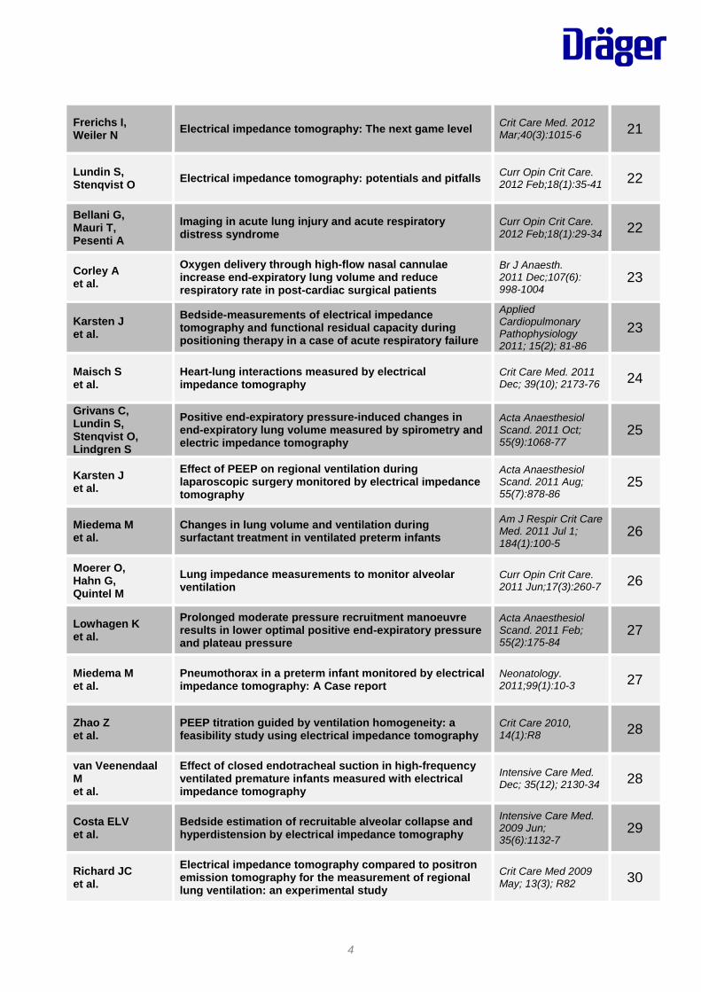
4
Frerichs I, Weiler N Electrical impedance tomography: The next game level Crit Care Med. 2012
Mar;40(3):1015-6 21
Lundin S, Stenqvist O Electrical impedance tomography: potentials and pitfalls Curr Opin Crit Care.
2012 Feb;18(1):35-41 22
Bellani G, Mauri T, Pesenti A
Imaging in acute lung injury and acute respiratory distress syndrome
Curr Opin Crit Care. 2012 Feb;18(1):29-34 22
Corley A et al.
Oxygen delivery through high-flow nasal cannulae increase end-expiratory lung volume and reduce respiratory rate in post-cardiac surgical patients
Br J Anaesth. 2011 Dec;107(6): 998-1004
23
Karsten J et al.
Bedside-measurements of electrical impedance tomography and functional residual capacity during positioning therapy in a case of acute respiratory failure
Applied Cardiopulmonary Pathophysiology 2011; 15(2); 81-86
23
Maisch S et al.
Heart-lung interactions measured by electrical impedance tomography
Crit Care Med. 2011 Dec; 39(10); 2173-76 24
Grivans C, Lundin S, Stenqvist O, Lindgren S
Positive end-expiratory pressure-induced changes in end-expiratory lung volume measured by spirometry and electric impedance tomography
Acta Anaesthesiol Scand. 2011 Oct; 55(9):1068-77
25
Karsten J et al.
Effect of PEEP on regional ventilation during laparoscopic surgery monitored by electrical impedance tomography
Acta Anaesthesiol Scand. 2011 Aug; 55(7):878-86
25
Miedema M et al.
Changes in lung volume and ventilation during surfactant treatment in ventilated preterm infants
Am J Respir Crit Care Med. 2011 Jul 1; 184(1):100-5
26
Moerer O, Hahn G, Quintel M
Lung impedance measurements to monitor alveolar ventilation
Curr Opin Crit Care. 2011 Jun;17(3):260-7 26
Lowhagen K et al.
Prolonged moderate pressure recruitment manoeuvre results in lower optimal positive end-expiratory pressure and plateau pressure
Acta Anaesthesiol Scand. 2011 Feb; 55(2):175-84
27
Miedema M et al.
Pneumothorax in a preterm infant monitored by electrical impedance tomography: A Case report
Neonatology. 2011;99(1):10-3 27
Zhao Z et al.
PEEP titration guided by ventilation homogeneity: a feasibility study using electrical impedance tomography
Crit Care 2010, 14(1):R8 28
van Veenendaal M et al.
Effect of closed endotracheal suction in high-frequency ventilated premature infants measured with electrical impedance tomography
Intensive Care Med. Dec; 35(12); 2130-34 28
Costa ELV et al.
Bedside estimation of recruitable alveolar collapse and hyperdistension by electrical impedance tomography
Intensive Care Med. 2009 Jun; 35(6):1132-7
29
Richard JC et al.
Electrical impedance tomography compared to positron emission tomography for the measurement of regional lung ventilation: an experimental study
Crit Care Med 2009 May; 13(3); R82 30
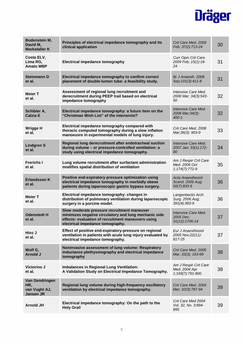
5
Bodenstein M, David M, Markstaller K
Principles of electrical impedance tomography and its clinical application
Crit Care Med. 2009 Feb; 37(2):713-24 30
Costa ELV, Lima RG, Amato MBP
Electrical impedance tomography Curr Opin Crit Care. 2009 Feb; 15(1):18-24
31
Steinmann D et al.
Electrical impedance tomography to confirm correct placement of double-lumen tube: a feasibility study.
Br J Anaesth. 2008 Sep;101(3):411-8 31
Meier T et al.
Assessment of regional lung recruitment and derecruitment during PEEP trail based on electrical impedance tomography
Intensive Care Med. 2008 Mar; 34(3):543-50
32
Schibler A, Calzia E
Electrical impedance tomography: a future item on the “Christmas Wish List” of the intensivist?
Intensive Care Med. 2008 Mar;34(3): 400-1
32
Wrigge H et al.
Electrical impedance tomography compared with thoracic computed tomography during a slow inflation manoeuvre in experimental models of lung injury.
Crit Care Med. 2008 Mar;36(3): 903-9 33
Lindgren S et al.
Regional lung derecruitment after endotracheal suction during volume – or pressure-controlled ventilation: a study using electrical impedance tomography.
Intensive Care Med. 2007 Jan; 33(1):172-80
34
Frerichs I et al.
Lung volume recruitment after surfactant administration modifies spatial distribution of ventilation
Am J Respir Crit Care Med. 2006 Oct 1;174(7):772-9
35
Erlandsson K et al.
Positive end-expiratory pressure optimization using electrical impedance tomography in morbidly obese patients during laparoscopic gastric bypass surgery.
Acta Anaesthesiol Scand. 2006 Aug; 50(7):833-9
36
Meier T et al.
Electrical impedance tomography: changes in distribution of pulmonary ventilation during laparoscopic surgery in a porcine model.
Langenbecks Arch Surg. 2006 Aug; 391(4):383-9
36
Odenstedt H et al.
Slow moderate pressure recruitment maneuver minimizes negative circulatory and lung mechanic side effects: evaluation of recruitment maneuvers using electrical impedance tomography.
Intensive Care Med. 2005 Dec; 31(12):1706-14
37
Hinz J et al.
Effect of positive end-expiratory-pressure on regional ventilation in patients with acute lung injury evaluated by electrical impedance tomography.
Eur J Anaesthesiol 2005 Nov;22(11): 817-25
37
Wolf G, Arnold J
Noninvasive assessment of lung volume: Respiratory inductance plethysmography and electrical impedance tomography
Crit Care Med. 2005 Mar; 33(3); 163-69 38
Victorino J et al.
Imbalances in Regional Lung Ventilation: A Validation Study on Electrical Impedance Tomography.
Am J Respir Crit Care Med. 2004 Apr 1;169(7):791-800.
38
Van Gendringen HR, van Vught AJ, Jansen JR
Regional lung volume during high-frequency oscillatory ventilation by electrical impedance tomography.
Crit Care Med. 2004 Mar; 32(3):787-94 39
Arnold JH Electrical impedance tomography: On the path to the Holy Grail
Crit Care Med 2004 Vol. 32, No. 3:894-895
39
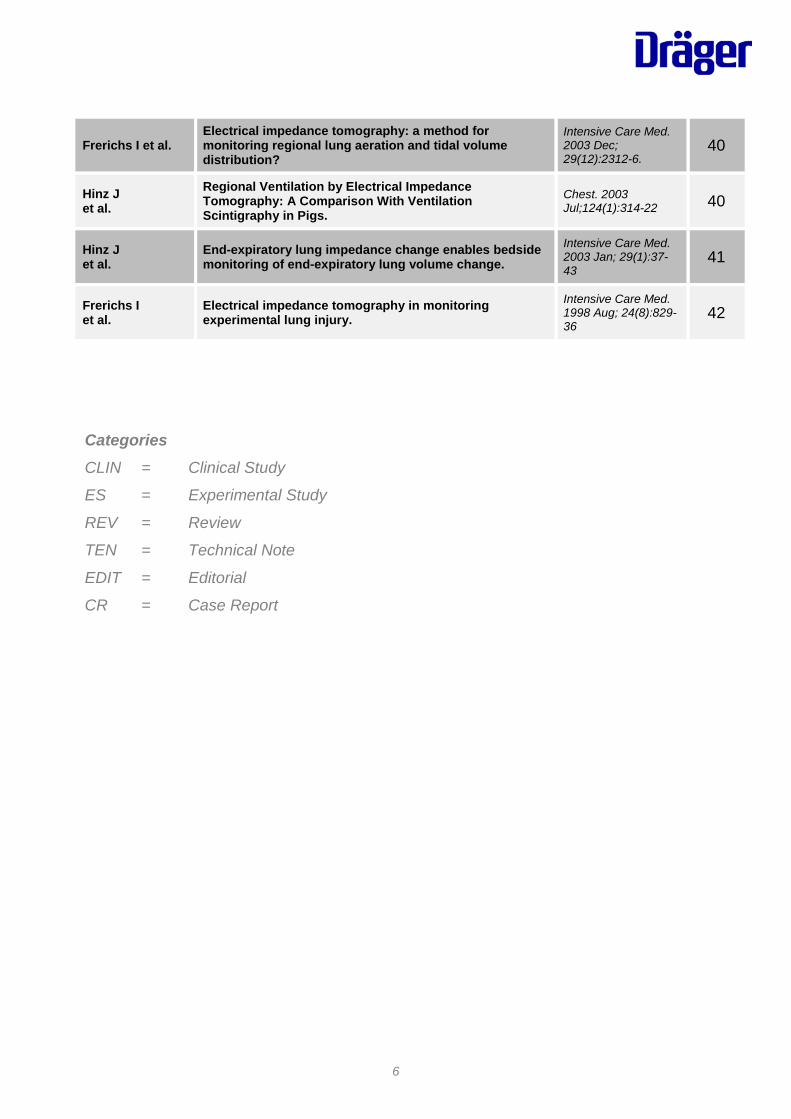
6
Frerichs I et al. Electrical impedance tomography: a method for monitoring regional lung aeration and tidal volume distribution?
Intensive Care Med. 2003 Dec; 29(12):2312-6.
40
Hinz J et al.
Regional Ventilation by Electrical Impedance Tomography: A Comparison With Ventilation Scintigraphy in Pigs.
Chest. 2003 Jul;124(1):314-22 40
Hinz J et al.
End-expiratory lung impedance change enables bedside monitoring of end-expiratory lung volume change.
Intensive Care Med. 2003 Jan; 29(1):37-43
41
Frerichs I et al.
Electrical impedance tomography in monitoring experimental lung injury.
Intensive Care Med. 1998 Aug; 24(8):829-36
42
Categories CLIN = Clinical Study
ES = Experimental Study
REV = Review
TEN = Technical Note
EDIT = Editorial
CR = Case Report

7
REV Durlak W, Kwinta P
Role of electrical impedance tomography in clinical practice in pediatric respiratory medicine
ISRN Pediatr 2013 Dec; 2013:529038, 5 pages
Abstract: This article summarizes current knowledge about electrical impedance tomography (EIT) and its present and possible applications in clinical practice in pediatric respiratory medicine. EIT is a relatively new technique based on real-time monitoring of bioimpedance. Its possible application in clinical practice related to ventilation and perfusion monitoring in children has gain increasing attention in recent years. Most of the currently published data is based on studies performed on small and heterogeneous groups of patients. Thus the results need to be corroborated in future well-designed clinical trials. Firstly a short theoretical overview summarizing physical principles and main advantages and disadvantages is provided. It is followed by a review of the current data regarding EIT application in ventilation distribution monitoring in healthy individuals. Finally the most important studies utilizing EIT in ventilation and perfusion monitoring in critically ill newborns and children are outlined. Summary: Electrical impedance tomography is a very promising technique for non-invasive, radiation free monitoring of lung function. It is widely applicable and safe for the patient, allowing real-time continuous monitoring of mechanical properties of the lungs over extended periods of time. Even though the physical principle of EIT is thirty years old, we have seen increasing number of publications suggesting its possible application in clinical practice in recent years’ literature. This is mainly due to constantly improving quality of hardware and data processing modalities. However most of these studies were based on small heterogeneous groups of patients. Larger, well designed trials are necessary to assess the role of electrical impedance tomography in every-day clinical practice.
CR Mauri T et al.
Regional distribution of air trapping in chronic obstructive pulmonary disease
Am J Respir Crit Care Med. 2013 Dec; 188(12); 1466-7
The authors present a case of a morbidly obese 51-year-old woman with chronic obstructive pulmonary disease (COPD) who was intubated and mechanically ventilated for respiratory failure and septic shock. The authors summarize that their data generate the new hypothesis that, in such patients, EIT might be a valid tool to image regional gas trapping and to guide titration of mechanical ventilation settings. In particular, this technique could prove useful if the purpose is to facilitate ventilation of the most disadvantaged dependent regions.
Electrical Impedance Tomography (EIT) Literature List
8
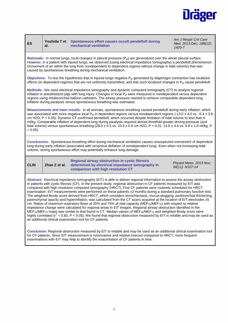
ES Yoshida T et al.
Spontaneous effort causes occult pendelluft during mechanical ventilation
Am J Respir Crit Care Med. 2013 Dec; 188(12); 1420-7
Rationale: In normal lungs, local changes in pleural pressure (Ppl) are generalized over the whole pleural surface. However, in a patient with injured lungs, we observed (using electrical impedance tomography) a pendelluft phenomenon (movement of air within the lung from nondependent to dependent regions without change in tidal volume) that was caused by spontaneous breathing during mechanical ventilation. Objectives: To test the hypotheses that in injured lungs negative Ppl generated by diaphragm contraction has localized effects (in dependent regions) that are not uniformly transmitted, and that such localized changes in Ppl cause pendelluft. Methods: We used electrical impedance tomography and dynamic computed tomography (CT) to analyze regional inflation in anesthetized pigs with lung injury. Changes in local Ppl were measured in nondependent versus dependent regions using intrabronchial balloon catheters. The airway pressure needed to achieve comparable dependent lung inflation during paralysis versus spontaneous breathing was estimated. Measurements and main results: In all animals, spontaneous breathing caused pendelluft during early inflation, which was associated with more negative local Ppl in dependent regions versus nondependent regions (-13.0 ± 4.0 vs. -6.4 ± 3.8 cm H2O; P < 0.05). Dynamic CT confirmed pendelluft, which occurred despite limitation of tidal volume to less than 6 ml/kg. Comparable inflation of dependent lung during paralysis required almost threefold greater driving pressure (and tidal volume) versus spontaneous breathing (28.0 ± 0.5 vs. 10.3 ± 0.6 cm H2O, P < 0.01; 14.8 ± 4.6 vs. 5.8 ± 1.6 ml/kg, P < 0.05). Conclusions: Spontaneous breathing effort during mechanical ventilation causes unsuspected overstretch of dependent lung during early inflation (associated with reciprocal deflation of nondependent lung). Even when not increasing tidal volume, strong spontaneous effort may potentially enhance lung damage.
CLIN Zhao Z et al. Regional airway obstruction in cystic fibrosis determined by electrical impedance tomography in comparison with high resolution CT
Physiol Meas. 2013 Nov; 34(11): N107-14
Abstract: Electrical impedance tomography (EIT) is able to deliver regional information to assess the airway obstruction in patients with cystic fibrosis (CF). In the present study, regional obstruction in CF patients measured by EIT was compared with high resolution computed tomography (HRCT). Five CF patients were routinely scheduled for HRCT examination. EIT measurements were performed on these patients ±2 months during a standard pulmonary function test. The weighted Brody score derived from HRCT, which considers bronchiectasis, mucus plugging, peribronchial thickening, parenchymal opacity and hyperinflation, was calculated from the CT scans acquired at the location of EIT electrodes ±5 cm. Ratios of maximum expiratory flows at 25% and 75% of vital capacity (MEF25/MEF75) with respect to relative impedance change were calculated for regional areas in EIT images. Regional airway obstruction identified in the MEF25/MEF75 maps was similar to that found in CT. Median values of MEF25/MEF75 and weighted Brody score were highly correlated (r2 = 0.83, P < 0.05). We found that regional obstruction measured by EIT is reliable and may be used as an additional clinical examination tool for CF patients. Conclusion: Regional obstruction measured by EIT is reliable and may be used as an additional clinical examination tool for CF patients. Since EIT measurement is noninvasive and relative lowcost compared to HRCT, more frequent examinations with EIT may help to identify the exacerbation of CF patients in time.
9
ES Bikker IG et al.
Global and regional parameters to visualize the 'best' PEEP during a PEEP trial in a porcine model with and without acute lung injury
Minerva Anestesiol. 2013 Sep; 79(9); 983-92
Background: Setting the optimal level of positive end-expiratory pressure (PEEP) in critically ill patients remains a matter of debate. "Best" PEEP is regarded as minimal lung collapse and overdistention to prevent lung injury. In this study, global and regional variables were evaluated in a porcine model to identify which variables should be used to visualize "best" PEEP. Methods: Eight pigs (28-31 kg) were studied during an incremental and decremental PEEP trial before and after the induction of acute lung injury (ALI) with oleic acid. Arterial oxygenation, compliance, lung volume, dead space, esophageal pressure and electrical impedance tomography (EIT) were recorded at the end of each PEEP step. Results: After ALI, "best" PEEP was comparable at 15 cmH2O between regional compliance of the dorsal lung region by EIT and the global indicators: dynamic compliance, arterial oxygenation, alveolar dead space and venous admixture. After ALI, the intratidal gas distribution was able to detect regional overdistention at 15 cmH2O PEEP. "Best" PEEP based on transpulmonary pressure was lower and no optimal level could be found based on lung volume measurements alone. In addition, the recruitment phase significantly improved end-expiratory lung volume, PaO2, venous admixture and regional and global compliance, both in ALI and the "healthy" lung. Conclusion: Most of the evaluated parameters indicate comparable 'best' PEEP levels. However, a combination of these parameters, and especially EIT-derived intraridal gas distribution, might provide additional information. The application of lung recruitment was beneficial in both ALI and the "healthy" lung.
10
CLIN Mauri T et al.
Topographic Distribution of Tidal Ventilation in Acute Respiratory Distress Syndrome: Effects of Positive End-Expiratory Pressure and Pressure Support
Crit Care Med. 2013 Jul; 41(7); 1664-73.
Objective: Acute respiratory distress syndrome is characterized by collapse of gravitationally dependent lung regions that usually diverts tidal ventilation toward nondependent regions. We hypothesized that higher positive end-expiratory pressure and enhanced spontaneous breathing may increase the proportion of tidal ventilation reaching dependent lung regions in patients with acute respiratory distress syndrome undergoing pressure support ventilation. Design: Prospective, randomized, cross-over study. Setting: General and neurosurgical ICUs of a single university-affiliated hospital. Subjects: We enrolled ten intubated patients recovering from acute respiratory distress syndrome, after clinical switch from controlled ventilation to pressure support ventilation. Interventions: We compared, at the same pressure support ventilation level, a lower positive end-expiratory pressure (i.e., clinical positive end-expiratory pressure = 7 ± 2 cm H2O) with a higher one, obtained by adding 5 cm H2O (12 ± 2 cm H2O). Furthermore, a pressure support ventilation level associated with increased respiratory drive (3 ± 2 cm H2O) was tested against resting pressure support ventilation (12 ± 3 cm H2O), at clinical positive end-expiratory pressure. Measurements and Main Results: During all study phases, we measured, by electrical impedance tomography, the proportion of tidal ventilation reaching dependent and nondependent lung regions (Vt%dep and Vt%nondep), regional tidal volumes (Vtdep and Vtnondep), and antero-posterior ventilation homogeneity (Vt%nondep/Vt%dep). We also collected ventilation variables and arterial blood gases. Application of higher positive end-expiratory pressure levels increased Vt%dep and Vtdep values and decreased Vt%nondep/Vt%dep ratio, as compared with lower positive end-expiratory pressure (p < 0.01). Similarly, during lower pressure support ventilation, Vt%dep increased, Vtnondep decreased, and Vtdep did not change, likely indicating a higher efficiency of posterior diaphragm that led to decreased Vt%nondep/Vt%dep (p < 0.01). Finally, Pao2/Fio2 ratios correlated with Vt%dep during all study phases (p < 0.05). Conclusions: In patients with acute respiratory distress syndrome undergoing pressure support ventilation, higher positive end-expiratory pressure and lower support levels increase the fraction of tidal ventilation reaching dependent lung regions, yielding more homogeneous ventilation and, possibly, better ventilation/perfusion coupling.
11
CLIN Blankmann P et al.
Ventilation Distribution of Tidal Ventilation in Acute Respiratory Distress Syndrome: Effects of Positive End-Expiratory Pressure and Pressure Support
Intensive Care Med. 2013 Jun; 39(6); 1057-62.
Background: The purpose of this study was to compare the effect of varying levels of assist during pressure support (PSV) and Neurally Adjusted Ventilatory Assist (NAVA) on the aeration of the dependent and non-dependent lung regions by means of Electrical Impedance Tomography (EIT). Methods: We studied ten mechanically ventilated patients with Acute Lung Injury (ALI). Positive-End Expiratory Pressure (PEEP) and PSV levels were both 10 cm H2O during the initial PSV step. Thereafter, we changed the inspiratory pressure to 15 and 5 cm H2O during PSV. The electrical activity of the diaphragm (EAdi) during pressure support ten was used to define the initial NAVA gain (100 %). Thereafter, we changed NAVA gain to 150 and 50 %, respectively. After each step the assist level was switched back to PSV 10 cmH2O or NAVA100 %to get a new baseline. The EIT registration was performed continuously. Results: Tidal impedance variation significantly decreased during descending PSV levels within patients, whereas not during NAVA. The dorsal- to-ventral impedance distribution, expressed according to the center of gravity index, was lower during PSV compared to NAVA. Ventilation contribution of the dependent lung region was equally in balance with the nondependent lung region during PSV 5 cm H2O, NAVA 50 and 100 % Conclusions: Neurally Adjusted Ventilatory Assist ventilation had a beneficial effect on the ventilation of the dependent lung region and showed less over-assistance compared to PSV in patients with ALI.
CLIN Steinmann D et al.
Electrical impedance tomography for verification of correct endotracheal tube placement in paediatric patients: a feasibility study
Acta Anaesthesiol Scand. 2013 May; 57; 881-87.
Background: Endotracheal tubes (ETTs) are frequently used in paediatric anaesthesia. Correct placement is crucial. The aim of this study was to evaluate electrical impedance tomography (EIT) for guiding and confirmation of paediatric ETT placement. In a retrospective analysis of stored EIT data, distribution of ventilation between left and right lung was used to verify correct paediatric ETT placement. Methods: Left and right lung ventilation was studied by EIT in 18 paediatric patients (median age: 53 months) requiring anaesthesia and endotracheal intubation. EIT was recorded before induction of anaesthesia, during mask ventilation, during ETT placement (including deliberate mainstem intubation), and after ETT repositioning according to the formula: ETT intubation depth (cm) = 3× ETT internal diameter (mm) or the mainstem intubation method (withdrawing the ETT 2 cm). Final ETT position was confirmed by fluoroscopy. Results: Following deliberate mainstem intubation, distribution of ventilation to the right lung was unequivocally demonstrated by EIT. Homogeneous distribution of ventilation between left and right lung monitored with EIT correlated in each patient with correct endotracheal ETT placement. The distribution of left and right lung ventilation differed significantly (P < 0.05) between the initial two-lung ventilation and subsequent right one-lung ventilation, and between right one-lung and subsequent two-lung ventilation according to auscultation and the final ETT position, respectively. In one patient, ETT was misplaced within the oesophagus which was also obvious from the EIT record. Conclusions: This study demonstrates that EIT enables noninvasive recognition of correct ETT placement. Homogeneous right-left-lung ventilation is an indicator for correct ETT placement.
12
ES Wolf G et al.
Mechanical Ventilation Guides by Electrical Impedance Tomography Experimental Acute Lung Injury
Crit Care Med. 2013 May; 41(5); 1296-1304.
Objectives: To utilize real-time electrical impedance tomography to guide lung protective ventilation in an animal model of acute respiratory distress syndrome. Design: Design: Prospective animal Setting: Animal research center. Subjects: Twelve Yorkshire swine (15 kg). Interventions: Lung injury was induced with saline lavage and augmented using large tidal volumes. The control group (n = 6) was ventilated using ARDSnet guidelines, and the electrical impedance tomography–guided group (n = 6) was ventilated using guidance with real-time electrical impedance tomography lung imaging. Regional electrical impedance tomography–derived compliance was used to maximize the recruitment of dependent lung and minimize overdistension of nondependent lung areas. Tidal volume was 6 mL/kg in both groups. Computed tomography was performed in a subset of animals to define the anatomic correlates of electrical impedance tomography imaging (n = 5). Interleukin-8 was quantified in serum and bronchoalveolar lavage samples. Sections of dependent and nondependent regions of the lung were fixed in formalin for histopathologic analysis. Measurements and Main Results: Positive end-expiratory pressure levels were higher in the electrical impedance tomography–guided group (14.3 cm H2O vs. 8.6 cm H2O, p < 0.0001), whereas plateau pressures did not differ. Global respiratory system compliance was improved in the electrical impedance tomography–guided group (6.9 mL/cm H2O vs. 4.7 mL/cm H2O, p = 0.013). Regional electrical impedance tomography–derived compliance of the most dependent lung region was increased in the electrical impedance tomography group (1.78 mL/cm H2O vs. 0.99 mL/cm H2O, p = 0.001). Pao2/Fio2 ratio was higher and oxygenation index was lower in the electrical impedance tomography–guided group (Pao2/Fio2: 388 mm Hg vs. 113 mm Hg, p < 0.0001; oxygenation index: 6.4 vs. 15.7, p =0.02) (all averages over the 6-hr time course). The presence of hyaline membranes (HM) and airway fibrin (AF) was significantly reduced in the electrical impedance tomography–guided group (HMEIT 42% samples vs. HMCONTROL 67% samples, p < 0.01; AFEIT 75% samples vs. AFCONTROL 100% samples, p < 0.01). Interleukin-8 level (bronchoalveolar lavage) did not differ between the groups. The upper and lower 95% limits of agreement between electrical impedance tomography and computed tomography were ± 16%. Conclusions: Electrical impedance tomography–guided ventilation resulted in improved respiratory mechanics, improved gas exchange, and reduced histologic evidence of ventilator-induced lung injury in an animal model. This is the first prospective use of electrical impedance tomography–derived variables to improve outcomes in the setting of acute lung injury.
EDIT Hemmes S et al.
From the Dark Side of Ventilation Toward a Brighter Look at Lungs
Crit Care Med. 2013 May; 41(5); 1376-77.
Editorial by Sabrine Hemmes, Frederique Paulus and Marcus Schultz that refers to the following publication: Wolf GK, Gómez-Laberge C, Rettig JS, et al: Mechanical Ventilation Guided by Electrical Impedance Tomography in Experimental Acute Lung Injury. Crit Care Med 2013; 41:1296–1304.
13
CLIN Riera J et al. Effect of high-flow nasal cannula and body position on end-expiratory lung volume: a cohort study using electrical impedance tomography
Respir Care. 2013 Apr; 58(4); 589-96
Background: Electrical impedance tomography measures changes in lung impedance, which are mainly related to changes in lung volume. We used electrical impedance tomography to investigate the effects of high-flow nasal cannula (HFNC) and body position on global and regional end-expiratory lung impedance variation (ΔEELI). Methods: Prospective study with 20 healthy adults. Two periods were defined: the first in supine position and the second in prone position. Each period was divided into 3 phases. In the first and the third phases the subjects were breathing ambient air, and in the second HFNC was implemented. Four regions of interest were defined: 2 ventral and 2 dorsal. For each respiratory cycle, global and regional ΔEELI were measured by electrical impedance tomography and were expressed as a function of the tidal variation of the first stable respiratory cycle (units). Results: HFNC increased global EELI by 1.26 units (95% CI 1.20-1.31, P < .001) in supine position, and by 0.87 units (95% CI 0.82-0.91, P < .001) in prone position. The distribution of ΔEELI was homogeneous in prone position, with no difference between ventral and dorsal lung regions (-0.01 units, 95% CI -0.01 to 0, P = .18), while in supine position a significant difference was found (0.22 units, 95% CI 0.21-0.23, P < .001) with increased EELI in ventral areas. Conclusions: HFNC increased global EELI in our population, regardless of body position, suggesting an increase in functional residual capacity. Prone positioning was related to a more homogeneous distribution of ΔEELI, while in supine position ΔEELI was higher in the ventral lung regions.
CR Karsten J et al.
Electrical impedance tomography may optimize ventilation in a postpartum woman with respiratory failure
Int J Obstet Anesth, 2013 Jan; 22(1); 67-71.
Abstract: Amniotic fluid embolism is a rare peripartum complication with the sudden onset of haemodynamic instability, respiratory failure and coagulopathy during labour or soon after delivery. A 31-year-old woman with amniotic fluid embolism was treated with vasopressors, inotropes, intravenous fluid, tranexamic acid and ventilatory support. Assessment of respiratory impairment was made using conventional chest X-ray, computed tomography and electrical impedance tomography. The potential for electrical impedance tomography to improve monitoring and guide respiratory therapy is explored.
CR Camporota L et al.
Assessment of regional lung mechanics with electrical impedance tomography can determine the requirement for ECMO in patients with ARDS
Intensive Care Med. 2012 Dec; 38(12); 2086-87.
The authors present two cases of patients with severe ARDS to describe how assessment of regional changes in compliance using electrical impedance tomography (EIT) may assist in the ventilator strategy prior to or during ECMO.
14
REV Leonhardt S, Lachmann B
Electrical Impedance Tomography: The Holy Grail of Ventilation and Perfusion Monitoring?
Intensive Care Med. 2012 Dec; 38(12); 1917-29.
Abstract: This review summarizes the state-of-the-art in electrical impedance tomography (EIT) for ventilation and perfusion imaging. We start by introducing the principle of electrical impedance tomography. Basically, EIT is a relatively new technology to image impedance distributions in a cross-sectional area of the body. Then a brief overview of the recent history is presented followed by a review of the literature on regional ventilation monitoring using EIT. Several recently presented indices useful to extract information from EIT image streams are presented and discussed. Selected experimental and clinical findings are discussed with respect to future routine applications in intensive care. Past and ongoing research activities to obtain cardiac and regional perfusion information from EIT image streams are summarized. Finally, relevant fields of future research are described. The appendix provides additional information on the physical background, and introduces the general concept of bioimpedance measurements including the necessity to utilize four-electrode arrangements and to describe regulatory boundary conditions. Conclusions: Electrical impedance tomography is a new imaging or - depending on the viewpoint - monitoring modality that will change the way we treat patients by mechanical ventilation. It will certainly allow us to tune and optimize regional ventilation of the individual patient which, until now, is still difficult at the bedside. Furthermore, as indicated above, it is an exciting new modality with potential for future extensions, including regional perfusion monitoring, regional V/Q mapping and, possibly, quantifying pneumothorax, atelectasis and pulmonary edema.
CLIN Pulletz S et al.
Dynamics of regional lung aeration determined by electrical impedance tomography in patients with acute respiratory distress syndrome
Multidisciplinary respiratory medicine 2012 Nov; 7(1); 44.
Background: Lung tissue of patients with acute respiratory distress syndrome (ARDS) is heterogeneously damaged and prone to develop atelectasis. During inflation, atelectatic regions may exhibit alveolar recruitment accompanied by prolonged filling with air in contrast to regions with already open alveoli with a fast increase in regional aeration. During deflation, derecruitment of injured regions is possible with ongoing loss in regional aeration. The aim of our study was to assess the dynamics of regional lung aeration in mechanically ventilated patients with ARDS and its dependency on positive end-expiratory pressure (PEEP) using electrical impedance tomography (EIT). Methods: Twelve lung healthy and twenty ARDS patients were examined by EIT during sustained step increases in airway pressure from 0, 8 and 15 cm H2O to 35 cm H2O and during subsequent step decrease to the corresponding PEEP. Regional EIT waveforms in the ventral and dorsal lung regions were fitted to bi-exponential equations. Regional fast and slow respiratory time constants and the sizes of the fast and slow compartments were subsequently calculated. Results: ARDS patients exhibited significantly lower fast and slow time constants than the lung healthy patients in ventral and dorsal regions. The time constants were significantly affected by PEEP and differed between the regions. The size of the fast compartment was significantly lower in ARDS patients than in patients with healthy lung under all studied conditions. Conclusions: These results show that regional lung mechanics can be assessed by EIT. They reflect the lower respiratory system compliance of injured lungs and imply more pronounced regional recruitment and derecruitment in ARDS patients.

15
CLIN Wolf GK et al.
Reversal of dependent lung collapse predicts response to lung recruitment in children with early acute lung injury
Pediatr Crit Care Med. 2012 Sep; 13(5); 509-15
Objective: To describe the resolution of regional atelectasis and the development of regional lung overdistension during a lung-recruitment protocol in children with acute lung injury. Design: Prospective interventional trial. Setting: Pediatric intensive care unit. Patients: Ten children with early (<72 hrs) acute lung injury. Interventions: Sustained inflation maneuver (positive airway pressure of 40 cm H2O for 40 secs), followed by a stepwise recruitment maneuver (escalating plateau pressures by 5 cm H2O every 15 mins) until physiologic lung recruitment, defined by PaO2 + PaCO2 =400 mm Hg, was achieved. Regional lung volumes and mechanics were measured using electrical impedance tomography. Measurements and main results: Patients that responded to the stepwise lung-recruitment maneuver had atelectasis in 54% of the dependent lung regions, while nonresponders had atelectasis in 10% of the dependent lung regions (p = .032). In the pressure step preceding physiologic lung recruitment, a significant reversal of atelectasis occurred in 17% of the dependent lung regions (p = .016). Stepwise recruitment overdistended 8% of the dependent lung regions in responders, but 58% of the same regions in nonresponders (p < .001). Lung compliance in dependent lung regions increased in responders, while compliance in nonresponders did not improve. In contrast to the stepwise recruitment maneuver, the sustained inflation did not produce significant changes in atelectasis or oxygenation: atelectasis was only reversed in 12% of the lung (p = .122), and there was only a modest improvement in oxygenation (27 ± 14 mm Hg, p = .088). Conclusions: Reversal of atelectasis in the most dependent lung region preceded improvements in gas exchange during a stepwise lung-recruitment strategy. Lung recruitment of dependent lung areas was accompanied by considerable overdistension of nondependent lung regions. Larger amounts of atelectasis in dependent lung areas were associated with a positive response to a stepwise lung-recruitment maneuver.
16
CLIN Vogt B et al.
Spatial and temporal heterogeneity of regional lung ventilation determined by electrical impedance tomography during pulmonary function testing
J Appl Physio 2012 Aug; 113; 1154-61.
Background: The study was initiates with the aim of checking if EIT was able to identify nonhomogeneous regional lung ventilation in patients with existing obstructive lung disease in comparison with healthy subjects with no history of lung disease. To account for age-dependent changes in regional lung function, we studied both young and elderly healthy adults. Methods: We performed the examinations during pulmonary function testing using well-established ventilation maneuvers and during spontaneous tidal breathing and examined if EIT-derived measures of spatial and temporal lung function heterogeneity were able to discriminate the patients from healthy subjects. Three groups of subjects of both sexes were examined : 1) 14 young healthy adults with no history of lung disease, 2) 12 elderly healthy subjects with no history of lung disease, and 3) 33 patients with chronic obstructive pulmonary disease (COPD). Results: EIT was able to assess the distribution of regional IVC, FEV1, FVC, and VT in the chest cross section of healthy young and elderly subjects and patients with COPD. Regional IVC, FEV1, FVC, and VT exhibited significant differences among the three groups with the lowest values found in the patients corresponding to the global volumes determined by spirometry. CV of EIT-derived regional IVC, FEV1, FVC, and VT identified the most heterogeneous distribution of ventilation in the COPD patients. However, this gross measure of spatial heterogeneity showed no differences in the degree of ventilation heterogeneity between the young and elderly subjects with no lung disease. Conclusions: Our study showed that EIT was able to assess spatial and temporal distribution of different regional lung volumes during spontaneous tidal breathing and forced ventilation maneuvers and identify not only pathologically induced but even the less pronounced age-related increase in ventilation heterogeneity. The EIT-derived measures of regional lung function might become useful in a clinical setting, providing complementary information to already established examination tools.
REV Blankmann P, Gommers D
Lung monitoring at the bedside in mechanically ventilated patients
Curr Opin Crit Care 2012 Jun; 18(3); 261-66.
Purpose of Review: It has become clear that mechanical ventilation itself can cause damage to the lung in critically ill patients, also known as ventilator-induced lung injury (VILI). Insight into the mechanisms of VILI has learned that a compromise must be found between positive end-expiratory pressure (PEEP) induced alveolar recruitment and prevention of hyperinflation. Therefore, there is a need for clinicians to optimize the PEEP settings for the individual patient at the bedside. In this review, we will discuss several lung-monitoring techniques to improve patient ventilator settings. Recent Findings: Recently, new monitoring tools like electrical impedance tomography (EIT), vibration response imaging, respiratory inductive plethysmography and functional residual capacity (FRC) have been (re-)introduced in our ICU. Nowadays, FRC can be measured without the use of tracer gases and without disconnection from the ventilator. EIT is another noninvasive bedside monitoring tool that provides regional ventilation distribution images and can be used for qualitative and quantitative assessment of regional change in ventilation after a ventilator change. These new noninvasive techniques are discussed and seem promising to help clinicians to improve their ventilator settings in the individual patient at the bedside. Summary: In conclusion, both FRC and EIT are promising clinical monitoring systems but clinical studies are needed to prove whether these monitors help the clinician toward effective and better ventilator management.

17
CLIN Radke O et al.
Spontaneous Breathing During General Anesthesia Prevents the Ventral Redistribution of Ventilation as Detected by Electrical Impedance Tomography
Anesthesiology 2012 Jun; 116(6); 1227-34.
Background: Positive-pressure ventilation causes a ventral redistribution of ventilation. Spontaneous breathing during general anesthesia with a laryngeal mask airway could prevent this redistribution of ventilation. We hypothesize that, compared with pressure-controlled ventilation, spontaneous breathing and pressure support ventilation reduce the extent of the redistribution of ventilation as detected by electrical impedance tomography. Methods: The study was a randomized, three-armed, observational, clinical trial without blinding. With approval from the local ethics committee, we enrolled 30 nonobese patients without severe cardiac or pulmonary comorbidities who were scheduled for elective orthopedic surgery. All of the procedures were performed under general anesthesia with a laryngeal mask airway and a standardized anesthetic regimen. The center of ventilation (primary outcome) was calculated before the induction of anesthesia (AWAKE), after the placement of the laryngeal mask airway (BEGIN), before the end of anesthesia (END), and after arrival in the postanesthesia care unit (PACU). Results: The center of ventilation during anesthesia (BEGIN) was higher than baseline (AWAKE) in both the pressure-controlled and pressure support ventilation groups (pressure control: 55.0 vs. 48.3, pressure support: 54.7 vs. 48.8, respectively; multivariate analysis of covariance, P _ 0.01), whereas the values in the spontaneous breathing group remained at baseline levels (47.9 vs. 48.5). In the postanesthesia care unit, the center of ventilation had returned to the baseline values in all groups. No adverse events were recorded. Conclusions: Both pressure-controlled ventilation and pressure support ventilation induce a redistribution of ventilation toward the ventral region, as detected by electrical impedance tomography. Spontaneous breathing prevents this redistribution.
EDIT Canet J, Gallart L
The Dark Side of the Lung. Unveiling Regional Lung Ventilation with Electrical Impedance Tomography
Anesthesiology 2012 Jun; 116(6); 1186-88.
Editorial by Jaume Canet and Lluís Gallart that accompanies the following publication: Radke OC, Schneider T, Heller A, Koch T: Spontaneous breathing during general anesthesia prevents the ventral redistribution of ventilation as detected by electrical impedance tomography: A randomized trial. ANESTHESIOLOGY 2012; 116:1227–34.

18
CLIN Hough J et al.
Effect of body position on ventilation distribution in preterm infants on continuous positive airway pressure
Pediatr Crit Care Med. 2012; 13(4); 446-51.
Background: Although continuous positive airway pressure is used extensively in neonatal intensive care units, and despite the belief that positioning is considered vital to the maintenance of good lung ventilation, no data exist on regional ventilation distribution in infants on continuous positive airway pressure ventilatory support. Objectives: To investigate the effect of body position on regional ventilation in preterm infants on continuous positive airway pressure ventilatory support using electrical impedance tomography. Design: Randomized crossover study design. Setting: Neonatal intensive care unit. Patients: Twenty-four preterm infants on continuous positive airway pressure were compared to six spontaneously breathing preterm infants. Results: Changes in global and regional lung volume were measured with electrical impedance tomography. Although there were no differences between positions, regional tidal volume was increased in the posterior compared with the an-terior lung (p < .01) and in the right compared with the left lung (p < .03) in both the spontaneously breathing infants and in the infants on continuous positive airway pressure. The posterior lung filled earlier than the anterior lung in the spontaneously breathing infants (p < .02), whereas in the infants on continuous positive airway pressure the right lung filled before the left lung (p < .01). There was more ventilation inhomogeneity in the infants on continuous positive airway pressure than in the healthy infants (p < .01). Conclusions: This study presents the first results on regional ventilation distribution in preterm infants on continuous positive airway pressure using electrical impedance tomography. Gravity had little impact on regional ventilation distribution in preterm infants on continuous positive airway pressure or in spontaneously breathing infants in the supine or prone position, indicating that ventilation distribution in preterm infants is not gravity-dependent but follows an anatomical pattern.
19
CLIN Guervilly C et al.
Right ventricular function during high-frequency oscillatory ventilation in adults with acute respiratory distress syndrome
Crit Care Med. 2012 May; 40(5); 1539-45.
Objectives: To evaluate the effect of mean airway pressure under high-frequency oscillatory ventilation on right ventricular function. Design: Prospective randomized study. Setting: Intensive care unit of a tertiary care hospital. Patients: Sixteen consecutive patients within the first 48 hrs of mainly pulmonary acute respiratory distress syndrome. Method: After a 6-hr-period of protective conventional mechanical ventilation, patients were submitted to three 1-hr periods of high-frequency oscillatory ventilation (+5, +10, +15) in a randomized order, with a mean airway pressure level determined by adding 5, 10, or 15 cm H2O to the mean airway pressure recorded during conventional mechanical ventilation. Results: Mean airway pressure was 18 +/-3 cm H2O during conventional mechanical ventilation and was increased until 33 +/-3 cm H2O at high-frequency oscillatory ventilation +15. Right ventricular function was assessed using transesophageal echocardiography. During conventional mechanical ventilation, nine patients presented a right ventricular dysfunction (right ventricular end-diastolic area/left ventricular end-diastolic area ratio >0.6) of whom four patients had a right ventricular failure (right ventricular end-diastolic area/left ventricular end-diastolic area ratio >0.9). High-frequency oscillatory ventilation +10 and +15 further worsened right ventricular function, resulting in about a 40% increase in right ventricular end- diastolic area/left ventricular end- diastolic area ratio and a 30% increase in end-diastolic eccentricity index when compared with conventional mechanical ventilation or high-frequency oscillatory ventilation +5 periods. At high-frequency oscillatory ventilation +15, 15 patients had right ventricular dysfunction and nine had right ventricular failure. High-frequency oscillatory ventilation did not improve oxygenation whatever the mean airway pressure level. A significant redistribution of tidal variation to the posterior parts of the lung was observed on electrical impedance tomography measurements when increasing mean airway pressure. However, this redistribution was not observed in patients who presented a worsening of right ventricular function (right ventricular end-diastolic area/ left ventricular end-diastolic area increase >40%) at high-frequency oscillatory ventilation +15. Conclusions: This study demonstrates that EIT enables accurate display of left and right lung ventilation and, thus, non-invasive online recognition of misplacement of left-sided DLTs in the contra-lateral main bronchus. However, as distribution of ventilation did not correlate with endo-bronchial cuff placement, EIT cannot replace FOB in the routine control of DLT position.
20
REV Adler A et al.
Whither lung EIT: Where are we, where do we want to go and what do we need to get there?
Physiol Meas. 2012 May; 33(5):679-94.
Abstract: Breathing moves volumes of electrically insulating air into and out of the lungs, producing conductivity changes which can be seen by electrical impedance tomography. It has thus been apparent, since the early days of EIT research, that imaging of ventilation could become a key clinical application of EIT. In this paper, we review the current state and future prospects for lung EIT, by a synthesis of the presentations of the authors at the ‘special lung sessions’ of the annual biomedical EIT conferences on 2009-2011. We argue that lung EIT research has arrived at an important transition. It is now clear that valid and reproducible physiological information is available from EIT lung images. We must now ask the question: How can these data be used to help improve patient outcomes? To answer this question, we develop a classification of possible clinical scenarios in which EIT could play an important role, and we identify clinical and experimental research programs and engineering developments required to turn EIT into a clinically useful tool for lung monitoring.
REV Luecke T, Corradi F, Pelosi, P
Lung imaging for titration of mechanical ventilation. Curr Opin Anesthesiol 2012 Apr; 25(2); 131-40.
Purpose of Review: Computed tomography (CT) has fostered pivotal advancements in the understanding of acute lung injury/ acute respiratory distress syndrome and ventilator-induced lung injury. Apart from CT-based, the past years have seen fascinating work using positron emission tomography, electrical impedance tomography and lung ultrasound as diagnostic tools to optimize mechanical ventilation.. This review aims to present the major findings of recent studies on lung imaging. Recent findings: Patients presenting with a focal loss of aeration on CT may not be suitable candidates for recruitment maneuvers and high levels of positive and expiratory-pressure (PEEP) in supine position. PET/CT has provides valuable insights into the inflammatory response of the lung. Electrical impedance tomography has been used to assess lung recruitability and to titrate PEEP. Finally, lung ultrasound has proven to be reliable diagnostic tool for assessing PEEP-induced recruitment. Summary: Whereas quantitative CT remains the gold standard to assess lung morphology, recruitment and hyperinflation of lung tissue at different inflation pressures, EIT and LUS have emerged as valuable, radiation-free, noninvasive bedside lung imaging tool that should be used together with global parameters like lung mechanics and gas exchange to acquire additional information on recruitability and ventilation distribution.
TEN Gomez-Laberge C et al.
A Unified Approach for EIT Imaging of Regional Overdistension and computed tomography in a porcine model of lung injury
Medical Imaging 2012 Mar; 31(3); 834-42.
Abstract: Patients with acute lung injury or acute respiratory distress syndrome (ALI/ARDS) are vulnerable to ventilator induced lung injury. Although this disease affects the lung heterogeneously, mechanical ventilation is not guided by regional indicators of potential lung injury. We used electrical impedance tomography (EIT) to estimate the extent of regional lung overdistension and atelectasis during mechanical ventilation. Techniques for tidal breath detection, lung identification, and regional compliance estimation were combined with the Graz consensus on EIT (GREIT) algorithm. Nine ALI/ARDS patients were monitored during stepwise increases and decreases in airway pressure. Our method detected individual breaths with 96:0% sensitivity and 97:6% specificity. The duration and volume of tidal breaths erred on average by 0:2 s and 5%, respectively. Respiratory system compliance from EIT and ventilator measurements had a correlation coefficient of 0.80. Stepwise increases in pressure could reverse atelectasis in 17% of the lung. At the highest pressures, 73% of the lung became overdistended. During stepwise decreases in pressure, previously atelectatic regions remained open at sub-baseline pressures. EIT may be suitable for guiding ventilation according to regional lung mechanics.
21
ES Muders T et al.
Tidal recruitment assessed by electrical impedance tomography and computed tomography in a porcine model of lung injury
Crit Care Med. 2012 Mar;40(3):903-11.
Objectives: To determine the validity of electrical impedance tomography to detect and quantify the amount of tidal recruitment caused by different positive end-expiratory pressure levels in a porcine acute lung injury model. Design: Randomized, controlled, prospective experimental study. Setting: Academic research laboratory. Subjects: Twelve anesthetized and mechanically ventilated pigs. Interventions: Acute lung injury was induced by a central venous oleic acid injection and abdominal hypertension in seven animals. Five healthy pigs served as control group. Animals were ventilated with positive end-expiratory pressure of 0, 5, 10, 15, 20, 25 cm H2O, respectively, in a randomized order. Measurements and Main Results: At any positive end-expiratory pressure level. Electrical impedance tomography was obtained during a slow inflation of 12 mL/kg of body weight. Regional-ventilation-delay indices quantifying the time until a lung region reaches a certain amount of impedance change were calculated for lung quadrants and for every single electrical impedance tomography pixel, respectively. Pixel-wise calculated regional-ventilation-delay indices were plotted in a color-coded regional-ventilation-delay map. Regional-ventilation-delay inhomogeneity that quantifies heterogeneity of ventilation time courses was evaluated by calculating the scatter of all pixel-wise calculated regional-ventilation-delay indices. End-expiratory and end-ins piratory computed tomography scans were performed at each positive end-expiratory pressure level to quantify tidal recruitment of the lung. Tidal recruitment showed a moderate inter-individual (r= 0.54; p<0.05) and intra-individual linear correlation (r= 0.46 up to 0.73 and p < 0.05, respectively) with regional-ventilation-delay obtained from lung quadrants. Regional-ventilation-delay inhomogeneity was excellently correlated with tidal recruitment intra- (r=0.90 up to r 0 0.99 and p<0.05, respectively) and inter-individually (r=0.90; p<0.001). Conclusions: Regional-ventilation-delay can be noninvasively measured by electrical impedance tomography during a slow inflation of 12 mL/kg of body weight and visualized using ventilation delay maps. Our experimental data suggest that the impedance tomography-based analysis of regional-ventilation-delay inhomogeneity provides a good estimate of the amount of tidal recruitment and may be useful to individualize ventilatory settings.
EDIT Frerichs I, Weiler N Electrical impedance tomography: The next game level Crit Care Med. 2012
Mar;40(3):1015-6.
Editorial by Inez Frerichs and Norbert Weiler that refers to the publication “Tidal recruitment assessed by electrical impedance tomography and computed tomography in a porcine model of lung injury” by T. Muders et al..

22
REV Lundin S, Stenqvist O Electrical impedance tomography: potentials and pitfalls. Curr Opin Crit Care. 2012
Feb;18(1):35-41.
Purpose of Review: Electrical impedance tomography (EIT) is a useful non-invasive tool for monitoring ventilation finding its way into the clinical setting. The focus of this review is to discuss the balance between the potential for EIT as a clinical monitoring accepting a level of uncertainty and the scientific demand for absolute perfection. Recent Findings: The controversy concerning whether EIT impedance changes can be safely used to monitor lung volume changes now appears to be solved after recent elegant studies. It is now high time to display lung volume changes measured by EIT in clinical units that are in milliliters following calibration versus tidal volume. A growing number of indices for regional ventilation distribution are emerging some of which should be further evaluated and developed for clinical decision support. Summary: Already now EIT is a useful clinical monitor. Still more work is needed to develop and interpret indices which are simple enough to be used in the clinical setting to guide the clinician towards effective and safe ventilator management.
REV Bellani G, Mauri T, Pesenti A
Imaging in acute lung injury and acute respiratory distress syndrome
Curr Opin Crit Care. 2012 Feb;18(1):29-34.
Purpose of Review: The review focuses on recent achievements obtained by means of imaging techniques in clinical and experimental studies on acute lung injury (ALI) and acute respiratory distress syndrome (ARDS). Recent Findings: The review focuses on four imaging techniques: computed tomography (CT), PET, electrical impedance tomography (EIT) and ultrasound, highlighting the most recent developments for each technique. Whereas CT and ultrasound are primarily based on detection of density, EIT and PET are aimed at providing more functional data. Summary: Major improvements were recently obtained in imaging structure and several functions of the lungs, with the potential of positively impacting the clinical practice.
23
CLIN Corley A et al.
Oxygen delivery through high-flow nasal cannulae increase end-expiratory lung volume and reduce respiratory rate in post-cardiac surgical patients
Br J Anaesth. 2011 Dec;107(6): 998-1004.
Background: High-flow nasal cannulae (HFNCs) create positive oropharyngeal airway pressure, but it is unclear how their use affects lung volume. Electrical impedance tomography allows the assessment of changes in lung volume by measuring changes in lung impedance. Primary objectives were to investigate the effects of HFNC on airway pressure (Paw) and end-expiratory lung volume (EELV) and to identify any correlation between the two. Secondary objectives were to investigate the effects of HFNC on respiratory rate, dyspnoea, tidal volume, and oxygenation; and the interaction between MBI and EELV. Methods: Twenty patients prescribed HFNC post-cardiac surgery were investigated. Impedance measures, Paw, PaO2/FiO2 ratio, respiratory rate, and modified Borg scores were recorded first on low-flow oxygen and then on HFNC. Results: A strong and significant correlation existed between Paw and end-expiratory lung impedance (EELI) (r= 0.7, P>0.001). Compared with low-flow oxygen, HFNC significantly increased EELI by 25.6% [95% confidence interval (CI) 24.3, 26.9] and Paw by 3.0 cm H2O (95% CI 2.4, 3.7). Respiratory rate reduced by 3.4 bpm (95% CI 1.7, 5.2) with HFNC use, tidal impedance variation increased by 10.5% (95% CI 6.1, 18.3), and PaO2/FiO2 ration improved by 30.6 mmHg (95% CI 17.9, 43.3). A trend towards HFNC improving subjective dyspnoea scoring (P=0.023) was found. Increases in EELI were significantly influenced by BMI, with larger increases associated with higher BMIs (P<0.001). Conclusions: This study suggests that HFNCs reduce respiratory rate and improve oxygenation by increasing both EELV and tidal volume and are most beneficial in patients with higher BMIs.
CR Karsten J et al.
Bedside-measurement of electrical impedance tomography and functional capacity during positioning therapy in a case of acute respiratory failure
Applied Cardiopulmonary Pathophysiology 2011; 15(2); 81-86.
Abstract: The lungs of patients with Acute Respiratory Failure (ARF) are characterized by reduced lung volume and regional heterogeneity of ventilation distribution. To improve lung volume and ventilation distribution, protective mechanical ventilation with positive end-expiratory pressure (PEEP), recruitment maneuvers, and some adjunct measures like intermittent proning have been recommended. But the continuous monitoring and management of these measures is not possible at bedside. New non-invasive monitoring tools like Electrical Impedance Tomography (EIT) and measurements of Functional Residual Capacity (FRC) by oxygen wash-in/washout have been advocated, but are not established in clinical routine. Thus, evident therapy algorithms are not yet available. The aim of this case study was to describe the ability of EIT and FRC measurement to detect changes in regional ventilation and lung volume in an ARF patient, who was treated by alveolar recruitment maneuver and intermittent prone position. An increase in FRC was observed in parallel of gas exchange improvement immediately after the recruitment. During prone position, FRC decreased constantly while a redistribution of ventilation among dorsal lung regions was detected by EIT. These two tools were compared but without a gold standard. The reported case shows that EIT and FRC measurements are two different methods, which are difficult to compare. As they do not present a good correspondence, the respective results may be under or overestimated but the measures indicated some tendencies. Nevertheless the combination of both could be a meaningful monitoring supplementation in the respiratory therapy of ARF patients.
24
ES Maisch S et al.
Heart-lung interactions measured by electrical impedance tomography
Crit Care Med.2011 Dec; 39(10); 2173-76.
Objective: The clinical value of stroke volume variations to assess intravascular fluid status in critically ill patients is well known. Electrical impedance tomography is a noninvasive monitoring technology that has been primarily used to assess ventilation. We investigated the potential of electrical impedance tomography to measure left ventricular stroke volume variation as an expression of heart lung interactions. The objective of this study was thus to determine in a set of different hemodynamic conditions whether stroke volume variation measured by electrical impedance tomography correlates with those derived from an aortic ultrasonic flow probe and arterial pulse contour analysis. Design: Prospective animal study. Setting: University research laboratory. Subjects: Domestic pigs, 29-50 kg. Interventions: A wide range of hemodynamic conditions were induced by mechanical ventilation at different levels of positive end-expiratory pressure (0-15cm H2O) and with tidal volumes of 8 and 16 mL/kg of body weight and by hypovolemia due to blood withdrawal with subsequent retransfusion followed of hydroxyethyl starch. Measurements and Results: In eight pigs, aortic stroke volume variations measured by electrical impedance tomography were measured and compared to those derived from an aortic ultrasonic flow probe and from arterial pulse contour analysis. Data for four animals were used to develop and train a novel frequency-domain electrical impedance tomography analysis algorithm, while data for the remaining four were used to test the performance of the novel methodology. Correlation of stroke volume variation measured by electrical impedance tomography and that derived from an aortic ultrasonic flow probe was significant (r² = 0.69; p < .001). Correlation of stroke volume variation measured by electrical impedance tomography and that derived from arterial pulse contour analysis (r² = 0.73; p < .001). Correlation of stroke volume variation derived from an aortic ultrasonic flow probe and that derived from arterial pulse contour analysis was significant too (r² = 0.82; p < .001). Bland-Altman analysis comparing stroke volume variation measured by electrical impedance tomography and that derived from an aortic ultrasonic flow probe revealed an overall bias of 1.87 % and limits of agreement of +/-7.02%; when comparing stroke volume variation measured by electrical impedance tomography and that derives from arterial pulse contour analysis, the overall bias was 0.49% and the limits of agreement were +/-5.85%. Conclusions: Stroke volume variation measured by electrical impedance tomography correlated with both the gold standard of direct aortic blood flow measurements of stroke volume variation and pulse contour analysis, marking an important step toward a completely noninvasive monitoring of heart-lung interactions.
25
CLIN Grivans C, Lundin S, Stenqvist O, Lindgren S
Positive end-expiratory pressure-induced changes in end-expiratory lung volume measured by spirometry and electric impedance tomography
Acta Anaesthesiol Scand. 2011 Oct; 55(9):1068-77.
Background: A bedside tool for monitoring changes in end-expiratory lung volume (∆EELV) would be helpful to set optimal positive end-expiratory pressure (PEEP) in acute lung injury / acute respiratory distress syndrome patients. The hypothesis of this study was that the cumulative difference of the inspiratory and expiratory tidal volumes of the first 10 breaths after a PEEP change accurately reflects the change in lung volume following a PEEP alteration. Methods: Changing PEEP induces lung volume changes which are reflected in differences between inspiratory and expiratory tidal volumes measured by spirometry. by adding these differences with correction for offset, for the first 10 breaths after PEEP change, cumulative tidal volume difference was calculated to estimate ∆EELV Vt (i-e) were compared with simultaneously measured changes in lung impedance, by electric impedance tomography (EIT), using calibration vs. tidal volume to estimate changes in ∆EELV EIT. Results: In the lung model, there was a close correlation (R2= 0.99) between ∆EELV Vt (i-e) and known lung model volume difference, with a bias of-4ml and limits if agreement of 42 and -50 ml. In 12 patients, ∆EELV EIT was closely correlated to ∆EELV VT(i-e) (R2=0.92), with mean bias of 50ml and limits of agreement of 131 and -31 ml. Changes in EELV estimated by EIT (∆EELV EIT) exceeded measurements by spirometry (∆EELV Vt(i-e)), with 15 (+-15)%. Conclusions: We conclude that spirometric measurements of inspiratory-expiratory tidal volumes agree well with impedance changes monitored by EIT and can be used bedside to estimate PEEP-induced changes in EELV.
CLIN Karsten J et al.
Effect of PEEP on regional ventilation during laparoscopic surgery monitored by electrical impedance tomography
Acta Anaesthesiol Scand. 2011 Aug; 55(7):878-86.
Background: Anesthesia per se and pneumoperitoneum during laparoscopic surgery lead to atelectasis and impairment of oxygenation. We hypothesized that a ventilation with positive end-expiratory pressure (PEEP) during general anesthesia and laparoscopic surgery leads to a more homogenous ventilation distribution as determined by electrical impedance tomography (EIT). Furthermore, we supposed that PEEP ventilation in lung-healthy patients would improve the parameters of oxygenation and respiratory compliance. Methods: Thirty-two patients scheduled to undergo laparoscopic cholecystectomy were randomly assigned to be ventilated with ZEEP (0 cm H2O) or with PEEP (10 cm H2O) and a subsequent recruitment maneuver. Differences in regional ventilation were analyzed by the EIT-based center-of-ventilation index (COV), which quantifies the distribution of ventilation and indicates ventilation shifts. Results: Higher amount of ventilation was examined in the dorsal parts of the lungs in the PEEP group. Throughout the application of PEEP, a lower shift of ventilation was found, whereas after the induction of anesthesia, a remarkable ventral shift of ventilation in ZEEP-ventilated patients (COV: ZEEP, 40.6 +- 2.4%; PEEP, 46.5 +-3.5%; P<0.001) was observed. Compared with the PEEP group, ZEEP caused a ventral misalignment of ventilation during pneumoperitoneum (COV: ZEEP, 41.6 +- 2.4%; PEEP, 44 +- 2.7%; P = 0.013). Throughout the study, there were significant differences in the parameters of oxygenation and respiratory compliance with improved values in PEEP-ventilated patients. Conclusions: The effect of anesthesia, pneumoperitoneum, and different PEEP levels can be evaluated by EIT-based COV monitoring. An initial recruitment maneuver and a PEEP of 10 cmH2O preserved homogenous regional ventilation during laparoscopic surgery in most, but not all, patients and improved oxygenation and respiratory compliance.
26
CLIN Miedema M et al.
Changes in lung volume and ventilation during surfactant treatment in ventilated preterm infants
Am J Respir Crit Care Med. 2011 Jul 1; 184(1):100-5.
Rationale: The immediate and regional effects of exogenous surfactant in open lung high-frequency oscillatory ventilated (HFOV) preterm infants are unknown. Objectives: To assess regional changes in lung volume, mechanics, and ventilation during and after surfactant administration in HFOV preterm infants with respiratory distress syndrome. Methods: Using electrical impedance tomography, changes in lung volume were continuously recorded during a stepwise recruitment procedure before, during, and after surfactant administration in 15 preterm infants (gestational age: 28.3 wk; birth weight: 1000g). Deflation limbs of the pressure-impedance curve before and after surfactant were mapped and the effect of surfactant on oscillation volumes and ventilation was determined. Data were analyzed for the whole cross-section and the left, right, ventral, and dorsal lung regions. Measurements and Results: Surfactant increased lung volume by 61 +- 39% within a median time of 241 sec. The ventral to dorsal ration in lung volume changes significantly from 1.16 before to 0.81 after surfactant administration. the upper inflection point of the deflation limb after surfactant (10.4 +-2.4 cm H2O) was significantly lower compared with before surfactant (16.4 +-3.1 cm H2O). Surfactant increased maximal compliance of the respiratory system, and this effect was reached at lower airway pressures. Surfactant caused a transient decrease in oscillatory volume but did not alter its regional distribution. Conclusions: Surfactant treatment in HFOV preterm infants with RDS cause rapid increase and subsequent stabilization of lung volume, which is most prominent I dependent lung regions. It increased maximal compliance, but this effect is only reached at lower airway pressures.
REV Moerer O, Hahn G, Quintel M
Lung impedance measurements to monitor alveolar ventilation
Curr Opin Crit Care. 2011 Jun;17(3):260-7.
Purpose: Electrical impedance tomography (EIT) is an attractive method of monitoring patients during mechanical ventilation because it can provide a non-invasive, continuous image of pulmonary impedance, which indicates the distribution of ventilation. This article will discuss ongoing research on EIT, with a focus on methodological aspects and limitations and novel approaches in terms of pathophysiology, diagnosis and therapeutic advancements. Recent Findings: EIT enables the detection of regional distribution of alveolar ventilation and, thus, the quantification of local inhomogeneities in lung mechanics. By detecting recruitment and derecruitment, a positive end-expiratory pressure level at which tidal ventilation is relatively homogeneous in all lung regions can be defined. Additionally, different approaches to characterize the temporal local behavior of lung tissue during ventilation have been proposed, which adds important information. Summary: There is growing evidence that supports EIT usage as a bedside measure to individually optimize ventilator settings in critically ill patients in order to prevent ventilator-induced lung injury. A standardization of current approaches to analyze and interpret EIT data is required in order to facilitate the clinical implementation.
27
CLIN Lowhagen K et al.
Prolonged moderate pressure recruitment manoeuvre results in lower optimal positive end-expiratory pressure and plateau pressure
Acta Anaesthesiol Scand. 2011 Feb; 55(2):175-84.
Background: In acute lung injury (ALI)/acute respiratory distress syndrome (ARDS), recruitment manoeuvres (RMs) are used frequently. In pigs with induced ALI, superior effects have been found using a slow moderate-pressure recruitment manoeuvre (SLRM) compared with vital capacity recruitment manoeuvre (VICM). We hypothesized that the positive recruitment effects of SLRM could also be achieved in ALI/ARDS patients. Our primary research question was whether the same compliance could be obtained using lower RM pressure and subsequent positive end-expiratory pressure (PEEP). Secondly, optimal PEEP levels following the RMs were compared, and the use of volume-depended compliance (VDC) to identify successful lung recruitment and optimal PEEP was evaluated. Patients and methods: We performed a prospective randomized cross-over study where 16 ventilated patients with early ALI / ARDS each were subjected to the two RMs, followed by decremental PEEP titration. Volume-dependent initial, middle and final compliance (C(ini), C(mid) and C(fin)) were determined. Electric impedance tomography and end-expiratory lung volume measurements were used to follow lung volume changes. Results: The maximum response in compliance, PaO2/FIO2, venous admixture and C(ini)/C(fin) after recruitment, during decremental PEEP, was at significantly lower PEEP and plateau pressure after SLRM than VICM. Fewer patients responded in gas exchange after the SLRM, which was not the case for lung mechanics. The response in Cini was more pronounced than in conventional compliance. Conclusions: The same compliance increase is achieved with SLRM as with VICM, and lower PEEP can be used, with correspondingly lower plateau pressures. VDC seems promising to identify successful recruitment and optimal PEEP.
CR Miedema M, et al.
Pneumothorax in a preterm infant monitored by electrical impedance tomography: A Case report
Neonatology. 2011;99(1):10-3.
Established facts: The initial clinical sign of pneumothorax can be mild and nonspecific, leading to a delay in diagnosis and treatment. Electrical Impedance tomography is a promising bedside monitoring tool capable of monitoring global and regional changes in ventilation. Novel insights: We present the first neonatal case of a pneumothorax detected by electrical impedance tomography. Electrical impedance tomography is a promising bedside monitoring tool for early detection of pneumothoraces. Abstract: Electrical impedance tomography is a non-invasive bedside tool for monitoring regional changes in ventilation. We report, for the first time, the EIT images of a ventilated preterm infant with unilateral pneumothorax, showing a loss of regional ventilation in the affected lung during both high-frequency oscillation and spontaneous ventilation.
28
CLIN Zhao Z et al.
PEEP titration guided by ventilation homogeneity: a feasibility study using electrical impedance tomography Crit Care 2010, 14(1):R8.
Introduction: Lung protective ventilation requires low tidal volume and suitable positive end- expiratory pressure (PEEP). To date, few methods have been accepted for clinical use to set the appropriate PEEP. The aim of this study was to test the feasibility of PEEP titration guided by ventilation homogeneity using the global inhomogeneity (GI) index based on electrical impedance tomography (EIT) images. Methods: In a retrospective study, 10 anesthetized patients with healthy lungs mechanically ventilated under volume-controlled mode were investigated. Ventilation distribution was monitored by EIT. A standardized incremental PEEP trail (PEEP from 0 – 28 mbar, 2 mbar per step) was conducted. During the PEEP trial, “optimal” PEEP level for each patient was determined when the air was most homogeneously distributed in the lung, indicated by the lowest GI index value. Two published methods for setting PEEP were included for comparison based on the maximum global dynamic compliance and the intra-tidal compliance-volume curve. Results: No significant differences in the results were observed between the GI index method (12.2+-4.6 mbar) and the dynamic compliance method (11.4+-4.9 mbar, P>0.6), or between the GI index and the compliance-volume curve method (12.2 +-4.9mbar, P>0.6). Conclusions: According to the results, it is feasible and reasonable to use the GI index to select the PEEP level with respect to ventilation homogeneity. The GI index may provide new insights into the relationship between lung mechanics and tidal volume distribution and may be used to guide ventilator settings.
CLIN Van Veenendaal M et al.
Effect of closed endotracheal suction in high-frequency ventilated premature infants measured with electrical impedance tomography
Intensive Care Med. Dec; 35(12); 2130-34.
Objective: To determine the global and regional changes in lung volume during and after closed endotracheal tube (ETT) suction in high-frequency ventilated preterm infants with respiratory distress syndrome (RDS). Design: Prospective observational clinical study
Setting: Neonatal intensive care unit. Patient: Eleven non-muscle relaxed preterm infants with RDS ventilated with open lung high-frequency ventilation (HFV). Interventions: Closed ETT suction. Measurements and Results: Changes in global and regional lung volume were measured with electrical impedance tomography. ETT suction resulted in an acute loss of lung volume followed by spontaneous recovery with a median residual loss of 3.3% of the maximum volume loss. The median stabilization time was 8 s. At the regional level, the lung volume changes during and after ETT suction were heterogeneous in nature. Conclusions: Closed ETT suction causes an acute, transient and heterogeneous loss of lung volume in premature infants with RDS treated with open lung HFV.
29
TEN Costa ELV et al.
Bedside estimation of recruitable alveolar collapse and hyperdistension by electrical impedance tomography
Intensive Care Med. 2009 Jun; 35(6):1132-7.
Objective: To present a novel algorithm for estimating recruitable alveolar collapse and hyperdistension based on electrical impedance tomography (EIT) during a decremental positive end-expiratory pressure (PEEP) titration.
Design: Technical note with illustrative case reports.
Setting: Respiratory intensive care unit.
Patient: Patients with acute respiratory distress syndrome.
Interventions: Lung recruitment and PEEP titration maneuver.
Measurements and Results: Simultaneous acquisition of EIT and X-ray computerized tomography (CT) data. We found good agreement (in terms of amount and spatial location) between the collapse estimated by EIT and TC for all levels of PEEP. The optimal PEEP values detected by EIT for patients 1 and 2 (keeping lung collapse <10%) were 19 and 17 cm H(2)O, respectively. Although pointing to the same non-dependent lung regions, EIT estimates of hyperdistension represent the functional deterioration of lung units, instead of their anatomical changes, and could not be compared directly with static CT estimates for hyperinflation. Conclusions: We described an EIT-based method for estimating recruitable alveolar collapse at the bedside, pointing out its regional distribution. Additionally, we proposed a measure of lung hyperdistension based on regional lung mechanics.
30
ES Richard JC et al.
Electrical impedance tomography compared to positron emission tomography for the measurement of regional lung ventilation: an experimental study
Crit Care Med. 2009 May; 13(3); R82.
Background: Electrical impedance tomography (EIT), which can assess regional lung ventilation at the bedside, has never been compared with positron-emission tomography (PET), a gold standard to quantify regional ventilation. This experiment systematically compared both techniques in injured and non-injured lungs. Methods: The study was performed in six mechanically ventilated female piglets. In normal lungs, tidal volume (VT) was randomly changed to 6, 8, 10 and 15 ml/kg on zero end-expiratory pressure (ZEEP), then, at VT 10 ml/kg, positive end-expiratory pressure (PEEP) was randomly changed to 5, 10 and 15 cmH2O. Afterwards, acute lung injury (ALI) was subsequently created in three animals by injecting 3 ml/kg hydrochloric acid into the trachea. Then at PEEP 5 cmH2O, VT was randomly changed to 8 and 12 ml/kg and PEEP of 10 and 15 cmH2O applied at VT 10 ml/kg. EIT and PET examinations were performed simultaneously. EIT ventilation (VTEIT) and lung volume (VL) were measured in the anterior and posterior area of each lung. On the same regions of interest, ventilation (VPET) and aerated lung volume (VAatten) were determined with PET. Results: On ZEEP, VTEIT and VPET significantly correlated for global (VTEIT = VPET - 2E-13, R2 = 0.95, P < 0.001) and regional (VTEIT = 0.81VPET+7.65, R2 = 0.63, P < 0.001) ventilation over both conditions. For ALI condition, corresponding R2 were 0.91 and 0.73 (P < 0.01). Bias was = 0 and limits of agreement were -37.42 and +37.42 ml/min for global ventilation over both conditions. These values were 0.04 and -29.01 and +29.08 ml/min, respectively, for regional ventilation. Significant correlations were also found between VL and VAatten for global (VL = VAatten+1E-12, R2 = 0.93, P < 0.0001) and regional (VL = 0.99VAatten+0.92, R2 = 0.65, P < 0.001) volume. For ALI condition, corresponding R2 were 0.94 (P < 0.001) and 0.54 (P < 0.05). Bias was = 0 and limits of agreement ranged -38.16 and +38.16 ml for global ventilation over both conditions. These values were -0.24 and -31.96 to +31.48 ml, respectively, for regional ventilation. Conclusions: Regional lung ventilation and volume were accurately measured with EIT in healthy and injured lungs and validated by simultaneous PET imaging.
REV Bodenstein M, David M, Markstaller K
Principles of electrical impedance tomography and its clinical application
Crit Care Med. 2009 Feb; 37(2):713-24.
Objective: This review outlines the basic principle, in addition to validated and upcoming clinical use of electrical impedance tomography (EIT). EIT generates functional tomograms of the thorax for detection of changes in regional lung aeration. These images allow an intra-individual comparison of changes in regional lung function. Specifically, EIT aims to optimize ventilation therapy in patients with acute lung failure.
Data Sources: PubMed: national Library of Medicine and the National Institutes of Health.
Study Selection: Studies with the key words “electrical impedance tomography” since 1983.
Data Extraction: Qualitative and quantitative results if the studies.
Data Synthesis: We summarized basic principles of the technology and subsequent analyzing methods, and give an overview of clinical and scientific questions that can be addressed by EIT. Conclusion: Potential applications in the future as well as limitations of EIT technology are described. In summary, EIT is a promising functional tomography technology on the verge of its clinical application.
31
REV Costa ELV, Lima RG, Amato MBP
Electrical impedance tomography Curr Opin Crit Care. 2009 Feb; 15(1):18-24.
Purpose of Review: Electrical impedance tomography (EIT) is a non-invasive, radiation-free monitoring tool that allows real-time imaging of ventilation. The purpose of this article is to discuss the fundamentals of EIT and to review the use of EIT in critical care patients. Recent Findings: In addition to its established role in describing the distribution of alveolar ventilation, EIT has been shown to be a useful tool to detect lung collapse and monitor lung recruitment, both regionally and on a global basis. EIT has also been used to diagnose with high sensitivity incident pneumothoraces during mechanical ventilation. Additionally, with injection of hypertonic saline as a contrast agent, it is possible to estimate ventilation/perfusion distributions. Summary: EIT is cheap, noninvasive and allows continuous monitoring of ventilation. It is gaining acceptance as a valuable monitoring tool for the care of critical patients.
CLIN Steinmann D et al.
Electrical impedance tomography to confirm correct placement of double-lumen tube: a feasibility study.
Br J Anaesth. 2008 Sep;101(3):411-8.
Background: Double-lumen tubes (DLTs) are frequently used to establish one-lung ventilation (OLV). Their correct placement is crucial. We hypothesized that electrical impedance tomography (EIT) reliably displays distribution of ventilation between left and right lung and may thus be used to verify correct DLT placement online. Methods: Regional ventilation was studied by EIT in 40 patients requiring insertion of left sided DLTs for OLV during thoracic surgery. EIT was recorded during two-lung ventilation before induction of anaesthesia and after DLT placement, and during OLV in the supine and subsequently in the lateral position. EIT measurements were made before and after verification of correct DLT placement by fiber optic bronchoscopy (FOB). Results: EIT accurately displayed distribution of ventilation between left and right lung online. All cases (n=5) of initially misplaced DLTs in the contra-lateral right main bronchus were detected by EIT. However, EIT did not allow prediction of FOB-detected endo-bronchial cuff misplacement requiring DLT repositioning. Furthermore, after DLT repositioning, distribution of ventilation, as assessed by EIT, did not change significantly (all P>0.5). Conclusions: This study demonstrates that EIT enables accurate display of left and right lung ventilation and, thus, non-invasive online recognition of misplacement of left-sided DLTs in the contra-lateral main bronchus. However, as distribution of ventilation did not correlate with endo-bronchial cuff placement, EIT cannot replace FOB in the routine control of DLT position.
32
ES Meier T et al.
Assessment of regional lung recruitment and derecruitment during PEEP trail based on electrical impedance tomography
Intensive Care Med. 2008 Mar; 34(3):543-50.
Objective: To investigate whether electrical impedance tomography (EIT) is capable of monitoring regional lung recruitment and lung collapse during a positive end-expiratory pressure (PEEP) trial.
Design: Experimental animal study of acute lung injury.
Subject: Six pigs with saline-lavage-induced induced acute lung injury.
Interventions: An incremental and decremental PEEP trial at ten pressure levels was performed. Ventilatory gas exchange, and hemodynamic parameters were automatically recorded. EIT and computed tomography (CT) scans of the same slice were simultaneously taken at each PEEP level.
Measurements and Results: A significant correlation between EIT and CT analyses of end-expiratory gas volumes (r=0.98 up to 0.99) and tidal volumes (r=0.55 up to r=0.88) could be demonstrated. Changes in global and regional tidal volumes and arterial oxygenation (PaO2/FiO2) demonstrated recruitment/derecruitment during the trial, but at different onsets. During the decremental trial, derecruitment first occurred in dependent lung areas. This was indicated by lowered regional tidal volumes measured in this area and by a decrease of PaO2/FiO2. At the same time, the global tidal volume still continued to increase, because the increase of ventilation of the non-dependant areas was higher than the loss in the dependant areas. This indicates that opposing regional changes might cancel each other out when combined in a global parameter. Conclusions: EIT is suitable for monitoring the dynamic effects of PEEP variations on the regional change of tidal volume. It is superior to global ventilation parameters in assessing the beginning of alveolar recruitment and lung collapse.
EDIT Schibler A, Calzia E
Electrical impedance tomography: a future item on the “Christmas Wish List” of the intensivist?
Intensive Care Med. 2008 Mar;34(3): 400-1.
Editorial by Andreas Schibler and Enrico Calzia that refers to the publication “Assessment of regional lung recruitment and derecruitment during a PEEP trial based on electrical impedance tomography” by T. Meier et al..

33
ES Wrigge H et al.
Electrical impedance tomography compared with thoracic computed tomography during a slow inflation maneuver in experimental models of lung injury
Crit Care Med. 2008 Mar;36(3): 903-9.
Objective: To determine the validity of functional electric impedance tomography to monitor regional ventilation distribution in experimental acute lung injury, and to develop a simple electric impedance tomography index detecting alveolar recruitment. Design: Randomized prospective experimental study. Setting: Academic research laboratory. Subjects: Sixteen anesthetized, tracheotomized, and mechanically ventilated pigs. Interventions: Acute lung injury was induced either by acid aspiration (direct acute lung injury) or by abdominal hypertension plus oleic acid injection (indirect acute lung injury) in ten pigs. Six pigs with normal lungs were studied as a control group and with endotracheal suction-related atelectasis. After 4 hrs of mechanical ventilation, a slow inflation was performed. Measurements and Main Results: During slow inflation, simultaneous measurements of regional ventilation by electric impedance tomography and dynamic computed tomography were highly correlated in quadrants of a transversal thoracic plane (r2 = 0.63 – 0.88, p < .0001, bias <5%) in both direct and indirect acute lung injury. Variability between methods was lower in direct than indirect acute lung injury (11 ± 2% vs. 18 ± 3%, respectively, p < 0.05). Electric impedance tomography indexes to detect alveolar recruitment were determined by mathematical curve analysis of regional impedance time curves. Empirical tests of different methods revealed that regional ventilation delay, that is, time delay of regional impedance time curve to reach a threshold, correlated well with recruited volume as measured by CT (r2 = 0.63). Correlation coefficients in subgroups were r2 = 0.71 and r2 = 0.48 in pigs with normal lungs with and without closed suction related atelectasis and r2 = 0.79 in pigs subject to indirect acute lung injury, respectively, whereas no significant correlation was found in pigs undergoing direct acute lung injury. Conclusions: Electric impedance tomography allows assessment of regional ventilation distribution and recruitment in experimental models of direct and indirect acute lung injury as well as normal lungs. Except for pigs with direct acute lung injury, regional ventilation delay determined during a slow inflation from impedance time curves appears to be a simple index for clinical monitoring of alveolar recruitment.

34
ES Lindgren S et al.
Regional lung derecruitment after endotracheal suction during volume – or pressure-controlled ventilation: a study using electrical impedance tomography.
Intensive Care Med. 2007 Jan; 33(1):172-80.
Objective: To assess lung volume and compliance changes during open- and closed-system suctioning using electric impedance tomography (EIT) during volume or pressure-controlled ventilation.
Design and Setting: Experimental study in a university research laboratory.
Subjects: Nine broncho-alveolar saline-lavaged pigs.
Interventions: Open and closed suctioning using a 14-F catheter in volume- or pressure-controlled ventilation at tidal volume 10 ml/kg, respiratory rate 20 breaths/min, and positive end-expiratory pressure 10 cmH2O.
Measurements and Results: Lung volume was monitored by EIT and a modified N2 washout/-in technique. Airway pressure was measured via a pressure line in the endotracheal tube. In four ventral-to-dorsal regions of interest regional ventilation and compliance were calculated at baseline and 30 s and 1, 2, and 10 min after suctioning. Blood gases were followed. At disconnection functional residual capacity (FRC) decreased by 58 ± 24% of baseline and by a further 22 ± 10% during open suctioning. Arterial oxygen tension decreased to 59 ± 14% of baseline value 1 min after open suctioning. Regional compliance deteriorated most in the dorsal parts of the lung. Restitution of lung volume and compliance was significantly slower during pressure-controlled than volume-controlled ventilation Conclusions: EIT can be used to monitor rapid lung volume changes. The two dorsal regions of the lavaged lungs are most affected by disconnection and suctioning with marked decreases in compliance. Volume-controlled ventilation can be used to rapidly restitute lung aeration and oxygenation after lung collapse induced by open suctioning.

35
ES Frerichs I, et al.
Lung volume recruitment after surfactant administration modifies spatial distribution of ventilation
Am J Respir Crit Care Med. 2006 Oct 1;174(7):772-9.
Rationale: Although surfactant replacement therapy is an established treatment in infant respiratory distress syndrome, the optimum strategy for ventilatory management before, during, and after surfactant instillation remains to be elucidated.
Objectives: To determine the effects of surfactant and lung volume recruitment on the distribution of regional lung ventilation.
Methods: Acute lung injury was induced in 16 newborn piglets by endotracheal lavage. Optimum positive end-expiratory pressure was identified after lung recruitment and surfactant was administered either at this pressure in the “open” lung or after disconnection of the endotracheal tube in the “closed” lung. An additional recruitment maneuver with subsequent optimum end-expiratory pressure finding was executed in eight animals; in the remaining eight animals, end-expiratory pressure was set at the same level as before surfactant without further recruitment. (“Open” and “closed” lung surfactant administration was evenly distributed in the groups). Regional ventilation was assessed by electrical impedance tomography.
Measurements and results: Impedance tomography data, airway pressure, flow, and arterial blood gases were acquired during baseline conditions, after induction of lung injury, after the first lung recruitment, and before as well as 10 and 60 min after surfactant administration. Significant shift in ventilation toward the dependent lung regions and less asymmetry in the right-to-left lung ventilation distribution occurred in the post surfactant period when an additional recruitment maneuver was performed. Surfactant instillation in an “open” versus “closed” lung did not influence ventilation distribution in a major way. Conclusions: The spatial distribution of ventilation in the lavaged lung is modified by a recruitment maneuver performed after surfactant administration.
36
CLIN Erlandsson K et al.
Positive end-expiratory pressure optimization using electrical impedance tomography in morbidly obese patients during laparoscopic gastric bypass surgery.
Acta Anaesthesiol Scand. 2006 Aug; 50(7):833-9.
Background: Morbidly obese patients have an increased risk for peri-operative lung complications and develop a decrease in functional residual capacity (FRC). Electric impedance tomography (EIT) can be used for continuous, fast-response measurement of lung volume changes. This method was used to optimize positive end-expiratory pressure (PEEP) to maintain FRC.
Methods: Fifteen patients with a body mass index of 49 +/- 8 kg/m(2) were studied during anaesthesia for laparoscopic gastric bypass surgery. Before induction, 16 electrodes were placed around the thorax to monitor ventilation-induced impedance changes. Calibration of the electric impedance tomograph against lung volume changes was made by increasing the tidal volume in steps of 200 ml. PEEP was titrated stepwise to maintain a horizontal baseline of the EIT curve, corresponding to a stable FRC. Absolute FRC was measured with a nitrogen wash-out/wash-in technique. Cardiac output was measured with an esophageal Doppler method. Volume expanders, 1 +/- 0.5 l, were given to prevent PEEP-induced haemodynamic impairment.
Results: Impedance changes closely followed tidal volume changes (R(2) > 0.95). The optimal PEEP level was 15 +/- 1 cmH(2)O, and FRC at this PEEP level was 1706 +/- 447 ml before and 2210 +/- 540 ml after surgery (P < 0.01). The cardiac index increased significantly from 2.6 +/- 0.5 before to 3.1 +/- 0.8 l/min/m2 after surgery, and the alveolar dead space decreased. P(a)O2/F(i)O2, shunt and compliance remained unchanged. Conclusion: EIT enables rapid assessment of lung volume changes in morbidly obese patients, and optimization of PEEP. High PEEP levels need to be used to maintain a normal FRC and to minimize shunt. Volume loading prevents circulatory depression in spite of a high PEEP level.
ES Meier T et al.
Electrical impedance tomography: changes in distribution of pulmonary ventilation during laparoscopic surgery in a porcine model.
Langenbecks Arch Surg. 2006 Aug; 391(4):383-9.
Objective: Because of the creation of a pneumoperitoneum, impairment of ventilation is a common side-effect during laparoscopic surgery. Electrical impedance tomography (EIT) is a method with the potential for becoming a tool to quantify these alterations during surgery. We have studied the change of regional ventilation during and after laparoscopic surgery with EIT and compared the diagnostic findings with computed tomography (CT) scans in a porcine study.
Materials and methods: After approval by the local animal ethics committee, six pigs were included in the study. Two laparoscopic operations were performed [colon resection (n=3) and fundoplication (n=3)]. The EIT measurements (6th parasternal intercostal space) were continuously recorded by an EIT prototype (EIT Evaluation Kit, Dräger Medical, Lübeck, Germany). To verify ventilatory alterations detected by EIT, a CT scan was performed postoperatively.
Results: Ventilation with defined tidal volumes was significantly correlated to EIT measurements (r2 = 0.99). After creation of the pneumoperitoneum, lung compliance typically decreased, which agreed well with an alteration of the distribution of pulmonary ventilation measured by EIT. Elevation of positive end-inspiratory pressure reopened non-aerated lung areas and showed a recovery of the regional ventilation measured by EIT. Additionally, we could detect pulmonary complications by EIT monitoring as verified by CT scans postoperatively. Conclusion: EIT monitoring can be used as a continuous non-invasive intraoperative monitor of ventilation to detect regional changes of ventilation and pulmonary complications during laparoscopic surgery. These EIT findings indicate that surgeons and anesthetists may eventually be able to optimize ventilation directly in the operating theatre.
37
ES Odenstedt H et al.
Slow moderate pressure recruitment maneuver minimizes negative circulatory and lung mechanic side effects: evaluation of recruitment maneuvers using electrical impedance tomography.
Intensive Care Med. 2005 Dec; 31(12):1706-14.
Objective: To evaluate the efficacy of different lung recruitment maneuvers using electric impedance tomography.
Design and Setting: Experimental study in animal model of acute lung injury in an animal research laboratory.
Subjects: Fourteen pigs with saline lavage induced lung injury.
Interventions: Lung volume, regional ventilation distribution, gas exchange, and hemodynamics were monitored during three different recruitment procedures: (a) vital capacity maneuver to an inspiratory pressure of 40 cmH2O (ViCM), (b) pressure-controlled recruitment maneuver with peak pressure 40 and PEEP 20 cmH2O, both maneuvers repeated three times for 30 s (PCRM), and (c) a slow recruitment with PEEP elevation to 15 cmH2O with end inspiratory pauses for 7 s twice per minute over 15 min (SLRM).
Measurements and Results: Improvement in lung volume, compliance, and gas exchange were similar in all three procedures 15 min after recruitment. Ventilation in dorsal regions of the lungs increased by 60% as a result of increased regional compliance. During PCRM compliance decreased by 50% in the ventral region. Cardiac output decreased by 63+/-4 % during ViCM, 44+/-2% during PCRM, and 21+/-3% during SLRM. Conclusions: In a lavage model of acute lung injury alveolar recruitment can be achieved with a slow lower pressure recruitment maneuver with less circulatory depression and negative lung mechanic side effects than with higher pressure recruitment maneuvers. With electric impedance tomography it was possible to monitor lung volume changes continuously.
CLIN Hinz J et al.
Effect of positive end-expiratory-pressure on regional ventilation in patients with acute lung injury evaluated by electrical impedance tomography.
Eur J Anaesthesiol 2005 Nov;22(11): 817-25.
Background and Objective: For the treatment of patients with adult respiratory distress syndrome and acute lung injury bedside measurements of regional lung ventilation should be considered for optimizing ventilatory settings. The aim was to investigate the effect of positive end-expiratory pressure (PEEP) on regional ventilation in mechanically ventilated patients at the bedside by electrical impedance tomography.
Methods: Eight mechanically ventilated patients were included in the study. PEEP levels were increased from 0 to 5, 10, 15 mbar and back to 0 mbar. Regional ventilation in 912 regions of the thorax was investigated at each PEEP by electrical impedance tomography. The obtained regions were divided in four groups: none (none and poorly ventilated regions including chest wall and mediastinum), bad, moderate and well-ventilated regions.
Results: Increasing the PEEP stepwise from 0 to 15 mbar decreased the non-ventilated regions (none: 540 regions at PEEP 0 and 406 regions at PEEP 15). In contrast, the other regions increased (bad: 316 regions at PEEP 0 and 380 regions at PEEP 15; moderate: 40 regions at PEEP 0 and 100 regions at PEEP 15; well: 0 region at PEEP 0 and 34 regions at PEEP 15 (median values)) indicating an improvement of regional ventilation. Conclusions: Increasing PEEP in mechanically ventilated patients reduces none ventilated regions (atelectasis). Furthermore, it leads to a shift from none and bad ventilated regions to moderately and well-ventilated regions. Electrical impedance tomography is a bedside technique and might be an alternative to computed tomography scan to assess aerated lung regions.
38
ES Wolf G, Arnold J
Noninvasive Assessment of lung volume: Respiratory inductance plethysmography and electrical impedance tomography
Crit Care Med. 2005 Mar; 33(3); 163-69.
Objective: Respiratory inductance plethysmography (RIP) and electrical impedance tomography (EIT) are two monitoring techniques that have been used to assess lung volume noninvasively. Methods: RIP uses two elastic bands around the chest and abdomen to assess global changes in lung volume. In animal models, RIP has been shown to detect changes in lung mechanics during high-frequency oscillatory ventilation and has the potential to quantify lung volumes noninvasively. EIT measures regional impedance changes with 16 electrodes around the patient’s chest, each of them injecting and receiving small currents. Impedance changes have been correlated with volume changes in animal models and in humans. In a recent animal model, EIT was shown to be capable of tracking lung volume changes during high-frequency oscillatory ventilation. Conclusions: The promise of monitoring techniques such as RIP and EIT is that they will guide lung protective ventilation strategies and allow the clinician to optimize lung recruitment, maintain an open lung, and limit overdistension. EIT is the only bedside method that allows repeated, noninvasive measurements of regional lung volumes. In the future, it will be important to standardize the definitions of alveolar recruitment and ultimately demonstrate the superiority of EIT-guided ventilator management in providing lung protective ventilation.
CLIN Victorino J et al.
Imbalances in Regional Lung Ventilation: A Validation Study on Electrical Impedance Tomography.
Am J Respir Crit Care Med. 2004 Apr 1;169(7):791-800.
Abstract: Imbalances in regional lung ventilation, with gravity dependent collapse and overdistention of nondependent zones, are likely associated to ventilator induced lung injury. Electric impedance tomography is a new imaging technique potentially capable of monitoring those imbalances. The aim of this study was to validate EIT measurements of ventilation distribution, by comparison with dynamic computerized tomography in a heterogeneous population of critically ill patients under mechanical ventilation. Multiple scans with both devices were collected during slow-inflation breaths. Six repeated breaths were monitored by impedance tomography, showing acceptable reproducibility. We observed acceptable agreement between both technologies in detecting right-left ventilation imbalances (bias = 0% and limits of agreement = -10 to 10%). Relative distribution of ventilation into regions or layers representing one fourth of the thoracic section could also be assessed with good precision. Depending on electrode positioning, impedance tomography slightly overestimated ventilation imbalances along gravitational axis. Ventilation was gravitationally dependent in all patients, with some transient blockages in dependent regions synchronously detected by both scanning techniques. Among variables derived from computerized tomography, changes in absolute air-content best explained the integral of impedance changes inside regions of interest (r(2) > or = 0.92). Impedance tomography can reliably assess ventilation distribution during mechanical ventilation.
39
ES
Van Gendringen HR, van Vught AJ, Jansen JR
Regional lung volume during high-frequency oscillatory ventilation by electrical impedance tomography.
Crit Care Med. 2004 Mar; 32(3):787-94.
Objective: To investigate the value of electrical impedance tomography for the assessment of regional lung mechanics during high-frequency oscillatory ventilation (HFOV).
Design: Prospective, interventional animal study.
Setting: University research laboratory.
Subjects: Eight pigs with lavage-induced lung injury.
Interventions: Electrical impedance tomography measurements were performed during a pressure-volume maneuver and during a recruitment-derecruitment maneuver on HFOV by stepwise variation of continuous distending pressure (CDP).
Measurements and Main Results: Lung volume was estimated by calibrated strain-gauge plethysmography; regional lung volume changes were assessed by electrical impedance tomography in various regions of interest. We found that inflation during the pressure-volume maneuver was distributed non-homogeneously, whereas deflation was homogeneous. During HFOV, no major regional differences were found during either inflation or deflation. The upper inflection point on the deflation limb was at a slightly higher continuous distending pressure (26+/-3 cm H2O) than the minimal physiologic shunt fraction (at continuous distending pressure of 23+/-7), where there were hemodynamic signs of overdistension. Maximal compliance on the deflation limb (at continuous distending pressure of 13+/-3 cm H2O) agreed well with the minimal continuous distending pressure, where shunt fraction was just below 0.1 (14+/-2 cm H2O). Conclusions: HFOV has a homogenizing effect on lung volume distribution. Regional lung volume distribution can be assessed using electrical impedance tomography. However, thoracic fluid accumulation may complicate its interpretation.
EDIT Arnold JH Electrical impedance tomography: On the path to the Holy Grail
Crit Care Med 2004 Vol. 32, No. 3:894-895.
Editorial by John Arnold that refers to the publication “Regional lung volume during high-frequency oscillatory ventilation by electrical impedance tomography” by H. van Gendringen et al..
40
CR Frerichs I et al.
Electrical impedance tomography: a method for monitoring regional lung aeration and tidal volume distribution?
Intensive Care Med. 2003 Dec; 29(12):2312-6.
Objective: To demonstrate the monitoring capacity of modern electrical impedance tomography (EIT) as an indicator of regional lung aeration and tidal volume distribution.
Design and Setting: Short-term ventilation experiment in an animal research laboratory.
Patients and Participants: One newborn piglet (body weight: 2 kg).
Interventions: Surfactant depletion by repeated bronchoalveolar lavage, surfactant administration.
Measurements and Results: EIT scanning was performed at an acquisition rate of 13 images/s during two ventilatory maneuvers performed before and after surfactant administration. During the scanning periods of 120 s the piglet was ventilated with a tidal volume of 10 ml/kg at positive end-expiratory pressures (PEEP) in the range of 0 – 30 cmH(2)O, increasing and decreasing in 5 cmH(2)O steps. Local changes in aeration and ventilation with PEEP were visualized by EIT scans showing the regional shifts in end-expiratory lung volume and distribution of tidal volume, respectively. In selected regions of interest EIT clearly identified the changes in local aeration and tidal volume distribution over time and after surfactant treatment as well as the differences between stepwise inflation and deflation. Conclusions: Our data indicate that modern EIT devices provide an assessment of regional lung aeration and tidal volume and allow evaluation of immediate effects of a change in ventilation or other therapeutic intervention. Future use of EIT in a clinical setting is expected to optimise the selection of appropriate ventilation strategies.
ES Hinz J et al.
Regional Ventilation by Electrical Impedance Tomography: A Comparison With Ventilation Scintigraphy in Pigs.
Chest. 2003 Jul;124(1):314-22.
Objective: The validation of electrical impedance tomography (EIT) for measuring regional ventilation distribution by comparing it with single photon emission CT (SPECT) scanning. Design: Randomized, prospective animal study. Settings: Animal laboratories and nuclear medicine laboratories at a university hospital. Participants: Twelve anesthetized and mechanically ventilated pigs. Interventions: Lung injury was induced by central venous injection of oleic acid. Then pigs were randomized to pressure-controlled mechanical ventilation, airway pressure-release ventilation, or spontaneous breathing. Measurements and Results: Ventilation distribution was assessed by EIT using cross-sectional electro tomographic measurements of the thorax, and simultaneously by single SPECT scanning with the inhalation of (99m)Tc labeled carbon particles. For both methods, the evaluation of ventilation distribution was performed in the same transverse slice that was approximately 4 cm in thickness. The transverse slice then was divided into 20 coronal segments (going from the sternum to the spine). We compared the percentage of ventilation in each segment, normalized to the entire ventilation in the observed slice. Our data showed an excellent linear correlation between the ventilation distribution measured by SPECT scanning and EIT according to the following equation: y = 0.82x + 0.7 (R(2) = 0.92; range, 0.86 to 0.97) Conclusion: Based on these data, EIT seems to allow, at least in comparable states of lung injury, real-time monitoring of regional ventilation distribution at the bedside.

41
CLIN Hinz J et al.
End-expiratory lung impedance change enables bedside monitoring of end-expiratory lung volume change.
Intensive Care Med. 2003 Jan; 29(1):37-43.
Objective: The aim of the study was to investigate the effect of lung volume changes on end-expiratory lung impedance change (ELIC) in mechanically ventilated patients; since we hypothesized that ELIC may be a suitable parameter to monitor lung volume change at the bedside. Design: Clinical trial on patients requiring mechanical ventilation. Settings: Intensive care units of a university hospital. Patients: Ten mechanically ventilated patients were included in the study. Intervention: Patients were ventilated in volume-controlled mode with constant flow and respiratory rate. In order to induce changes in the end-expiratory lung volume (EELV), PEEP levels were increased from 0 mbar to 5 mbar, 10 mbar, and 15 mbar. At each PEEP level EELV was measured by an open-circuit nitrogen washout manoeuvre and ELIC was measured simultaneously using Electrical Impedance Tomography (EIT) with sixteen electrodes placed on the circumference of the thorax and connected with an EIT device. Cross-sectional electro-tomographic measurements of the thorax were performed at each PEEP level, and a modified Sheffield back-projection was used to reconstruct images of the lung impedance. ELIC was calculated as the average of the end-expiratory lung impedance change. Results: Increasing PEEP stepwise from 0 mbar to 15 mbar resulted in a linear increase of EELV and ELIC according to the equation: y =0.98 × -0.68, R(2) =0.95. Conclusions: EIT is a simple bedside technique which enables monitor lung volume changes during ventilator maneuvers such as PEEP changes.
42
ES Frerichs I, et al.
Electrical impedance tomography in monitoring experimental lung injury.
Intensive Care Med. 1998 Aug; 24(8):829-36.
Objective: To apply electrical impedance tomography (EIT) and the new evaluation approach (the functional EIT) in monitoring the development of artificial lung injury. Design: Acute experimental trial. Setting: Operating room for animal experimental studies at a university hospital. Subjects: Five pigs (41.3 +/- 4.1 kg, mean body weight +/- SD). Interventions: The animals were anaesthetised and mechanically ventilated. Sixteen electrodes were attached on the thoracic circumference and used for electrical current injection and surface voltage measurement. Oleic acid was applied sequentially (total dose 0.05 ml/kg body weight) into the left pulmonary artery to produce selective unilateral lung injury. Measurements and Results: The presence of lung injury was documented by significant changes of PaCO2 (40.1 mmHg vs control 37.1 mmHg), PaO2 (112.3 mmHg vs 187.5 mmHg), pH (7.35 vs 7.42), mean pulmonary arterial pressure (29.2 mmHg vs 20.8 mmHg) and chest radiography. EIT detected 1) a regional decrease in mean impedance variation over the affected left lung (-41.4% vs control) and an increase over the intact right lung ( +20.4% vs control) indicating reduced ventilation of the affected, and a compensatory augmented ventilation of the unaffected lung and 2) a pronounced fall in local baseline electrical impedance over the injured lung (-20.6% vs control) with a moderate fall over the intact lung (-10.0% vs control) indicating the development of lung oedema in the injured lung with a probable atelectasis formation in the contralateral one. Conclusions: The development of the local impairment of pulmonary ventilation and the formation of lung oedema could be followed by EIT in an experimental model of lung injury. This technique may become a useful tool for monitoring local pulmonary ventilation in intensive care patients suffering from pulmonary disorders associated with regionally reduced ventilation, fluid accumulation and/or cell membrane changes.