Embed Size (px)
Citation preview
10/19/2016
1
2016 AJCC TNM Practice Cases(You Will Need an AJCC Staging Manual)
2 0 1 6 - 2 0 1 7 F C D S WE B C A S T S E R IE S
OC T OB E R 2 0 , 2 0 1 6
S T E V E N P E A C E , C T R
1
AJCC Cancer Staging Instruction for Registrarshttps://cancerstaging.org/CSE/Registrar/
CDC & Florida DOH Attribution
“We acknowledge the Centers for Disease Control and Prevention, for itssupport of the Florida Cancer Data System, and the printing anddistribution of the materials for the 2016-2017 FCDS Webcast Seriesunder cooperative agreement DP003872-03 awarded to the Florida
Department of Health. The findings and conclusions in this series arethose of the author(s) and do not necessarily represent the official positionof the Centers for Disease Control and Prevention”.
FCDS would also like to acknowledge the Florida Department of Healthfor its support of the Florida Cancer Data System, including thedevelopment, printing and distribution of materials for the 2016-2017FCDS Webcast Series under state contract CODJU. The findings and
conclusions in this series are those of the author(s) and do not necessarilyrepresent the official position of the Florida Department of Health.
2
A special thanks and acknowledgement to the
staff at the AJCC for providing slides with critical
content used in this presentation and available in
full on the AJCC website www.cancerstaging.org

10/19/2016
2
Purchase and Ordering Information3
http://www.springer.com/us/book/9780387884400
• AJCC Cancer Staging Manual – 7th edition, 2010
• COST: $64.95• ISBN: 978-0-387-88440-0
• Required - Florida Mandate• FCDS will not purchase• Facility may purchase• Individual may purchase
• Also Required to Purchase 8th Edition in 2016-2017
• https://cancerstaging.org• http://springer.com• 1-800-SPRINGER
Chapter Outline and Contents
Staging at a Glance Summary of anatomic stage/prognostic grouping
Changes in Staging Table summarizing changes in staging from the 6th edition
Introduction Overview of factors affecting staging and outcome
Anatomic Considerations
o Primary Tumor
o Regional lymph nodes
o Metastatic sites
Rules for Classificationo Clinical
o Pathologic
Prognostic FeaturesIdentification and discussion of non-anatomic prognostic
factors
Definitions of TNM
T: Primary tumor
N: Regional lymph nodes
M: Distant metastasis
Anatomic Stage Prognostic
Groups
Prognostic Factors (SSFs)a. Required for staging
b. Clinically significant
Grade
Histopathologic Type
Bibliography
Staging Form
4
AJCC Cancer Staging Manual, 7th ed. – Chapter 1, Table 1.10, p.14

10/19/2016
3
Stage Classifications – Points in Time
Timing for Clinical Stage – Date of Diagnosis up to the 1st treatment… in the Absence of Disease Progression or within first 4 months after Diagnosis
Timing for Pathologic Stage – Date of Diagnosis through definitive surgery… in the Absence of Disease Progression or within first 4 months after Diagnosis
Timing for Post-Treatment Stage (Pathologic - yp) – Pathologic Stage following treatment with neoadjuvant therapy(s) and definitive surgery (can include progression after neo-TX)
Timing for Post-Treatment Stage (Clinical - yc) – Clinical Stage following treatment with neoadjuvant therapy(s) and before definitive surgery or no definitive surgery (can include progression after neo-TX)
5
Clinical Stage – Pretreatment Stage
Clinical Stage (Pre-TX Stage) is the extent of disease defined by diagnostic study before information is available from surgical resection or initiation of neoadjuvant therapy, or within 4 months after date of diagnosis, whichever is shorter.
Patient Medical History
Physical Examination
Diagnostic Imaging Studies
Endoscopy
Biopsy of primary tumor
Biopsy of single node or sentinel nodes
Biopsy of metastatic sites
Exploratory Surgery
Other relevant lab tests, biomarker tests, or examinations
6
10/19/2016
4
Lymph Node Biopsy and/or Resection7
A lymph node biopsy can be either clinical or pathologic. If the only assessment of the primary tumor is a clinical (cT) assessment, then a biopsy of a single lymph node or of a sentinel lymph node can also be included in the clinical (cN) stage. In this situation, there would have been no evaluation of the primary tumor that qualifies for the pT. This allows for the assignment of a clinical stage when a pathological stage is not applicable.
Generally a resection of the primary tumor that qualifies for the pT is required in order to assign the pN. If there is a resection that qualifies for the pathologic assessment of T (pT), then any microscopic evidence of regional node involvement is classified as pN. MUST have at least ONE node microscopically examined to assign a pN. This can be a FNA, biopsy or excision of a node as long as there is microscopic confirmation.
Pathologic Stage
Pathologic Stage includes any information obtained about the extent of cancer through completion of definitive surgery as part of the first course of treatment or identified within 4 months after the date of diagnosis, whichever is longer, as long as there is no systemic or radiation therapy initiated or the cancer has not clearly progressed during that time frame.
Must meet chapter-specific criteria for surgical resection to assign
Includes all of the clinical stage information from clinical stage, plus
Observations at time of surgical resection from operative report
Pathologic Examination of surgically resected primary specimen
Pathologic Examination of surgically resected regional lymph nodes
Pathologic Examination of biopsy or resection of metastasis
8
10/19/2016
5
Pathologic Stage
The pathologic stage classification starts at the moment of DIAGNOSIS. Pathologic stage is defined by the same diagnostic studies used for clinical staging supplemented by findings from surgical resections and histologic examination of the surgically removed tissues. The pathologic stage encompasses three equal pieces of information:
All of the clinical classification information not disproven by the intra-operative or pathology findings.
PLUS includes the operative findings during the resection not submitted to or disproven on pathology.
PLUS includes the pathology report findings of the resected specimen.
9
Pathologic Stage
If a biopsied tumor is not resected for any reason (e.g., when technically unfeasible) and if the highest T and N categories or the M1 category of the tumor can be confirmed microscopically, the criteria for pathologic classification and staging have been satisfied without total removal of the primary cancer.
To use the highest T and highest N to assign the pathologic stage, you have to have both microscopic confirmation of the highest T for a pTAND microscopic confirmation of the highest N for a pN.
IMPORTANT: pT blank and pN3 is not enough for a pathologic stage so the pN will be used for the clinical stage.
10

10/19/2016
6
Post-Treatment Stage
Documents measured response to initial (neoadjuvant) therapy(s) Complete Response Partial Response No Response Progression
May be clinical measurement only – yc Based on post-treatment imaging, physical examination, biopsy
More often it is post-treatment pathologic stage – yp Based on post-treatment surgical resection of primary site and
regional nodes Must meet chapter-specific criteria for surgical resection
What about pre-treatment with less than 1 month of endocrine therapy including various hormones (prostate, breast, thyroid)? This is Not Neoadjuvant Tx…even though it begins before surgery
11
or
Staging Practice12

10/19/2016
7
Staging Practice13
Types of Cases14
Bladder Breast Colon Endometrium Lymphoma Lung Neuroendocrine Prostate Rectum Melanoma Soft Tissue Sarcoma Thyroid

10/19/2016
8
Case 1 – Case Vignette
HISTORY: 57 year-old African-American female with bx-confirmed adenocarcinoma of the rectosigmoid.
CT CHEST: few small (<1cm) nonspecific hilar lymph nodes noted in chest. Exam otherwise negative.
COLONOSCOPY : Large tumor colon @ 15 cm biopsy: invasive poorly differentiated adenocarcinoma
PATHOLOGY: Sigmoidectomy - 5.9 x 4.2 x 2.7 cm ulcerative lesion; invasive poorly differentiated colonic adenocarcinoma with extension into and through muscularis propria and focal transmural extension to serosal surface, margins free of tumor, 13 lymph nodes negative for metastatic adenocarcinoma; two discontinuous tumor deposits – present and positive for metastatic adenocarcinoma
15
Case 1 – Answer & Rationale16
10/19/2016
9
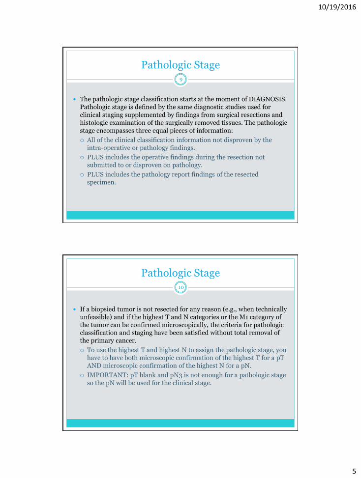
Case 2 – Case Vignette
HISTORY: 61 yr old white female, lifelong smoker, with multiple medical problems including recent adenoma on routine screening colonoscopy. Physical exam - negative.
CT CHEST: Negative
COLONOSCOPY : Transverse colon polyp @ 110cm – high grade dysplasia with focal well differentiated adenocarcinoma arising in an adenoma.
17
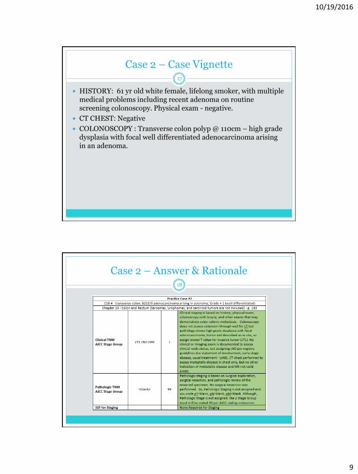
Case 2 – Answer & Rationale18
10/19/2016
10
Case 3 – Case Vignette
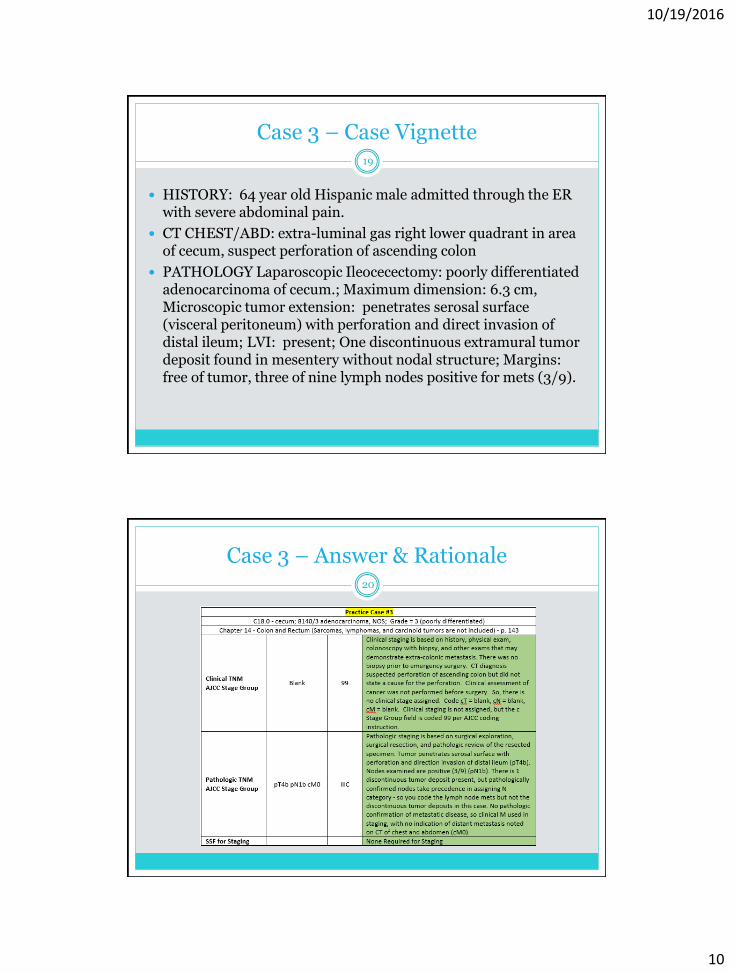
HISTORY: 64 year old Hispanic male admitted through the ER with severe abdominal pain.
CT CHEST/ABD: extra-luminal gas right lower quadrant in area of cecum, suspect perforation of ascending colon
PATHOLOGY Laparoscopic Ileocecectomy: poorly differentiated adenocarcinoma of cecum.; Maximum dimension: 6.3 cm, Microscopic tumor extension: penetrates serosal surface (visceral peritoneum) with perforation and direct invasion of distal ileum; LVI: present; One discontinuous extramural tumor deposit found in mesentery without nodal structure; Margins: free of tumor, three of nine lymph nodes positive for mets (3/9).
19
Case 3 – Answer & Rationale20

10/19/2016
11
Case 4 – Case Vignette
HISTORY: 49 year old white female admitted following recent colonoscopy showing malignant appearing mass in ascending colon. Family History: Father and brother had rectal cancer Physical Exam is essentially WNL.
CT CHEST/ABDOMEN: no abnormalities noted
COLONOSCOPY per history showed malignant appearing mass in proximal ascending colon – unknown if biopsy was taken to confirm malignancy.
CEA 0.6 – WNL
PATHOLOGY from Resection - Right hemicolectomy with appendix: Intermediate grade 2 neuroendocrine tumor (NET) of cecum (carcinoid tumor). Maximum dimension: 3.0 cm. Grossly the lesion invades through the muscularis propria into the underlying mesenteric adipose tissue. Microscopic tumor extension: invades through muscularis propria. Lymphovascular invasion: present (venous). Perineural invasion: not identified. Margins: free of tumor. One of twenty two lymph nodes positive for metastatic carcinoma (1/22).
21
Case 4 – Answer & Rationale22
10/19/2016
12
Case 5 – Case Vignette
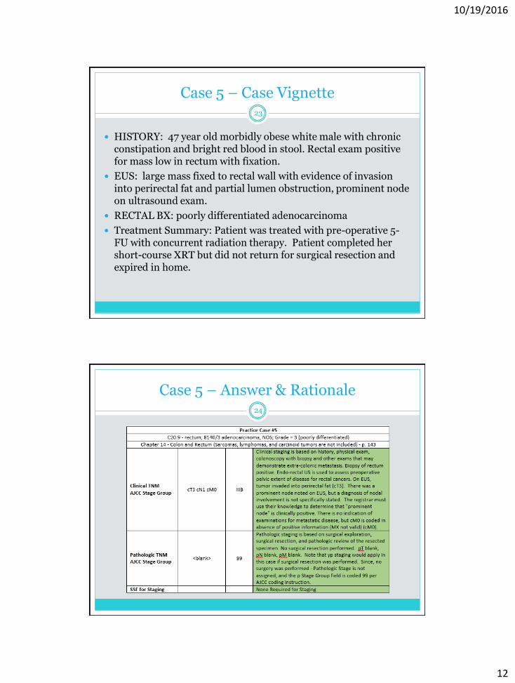
HISTORY: 47 year old morbidly obese white male with chronic constipation and bright red blood in stool. Rectal exam positive for mass low in rectum with fixation.
EUS: large mass fixed to rectal wall with evidence of invasion into perirectal fat and partial lumen obstruction, prominent node on ultrasound exam.
RECTAL BX: poorly differentiated adenocarcinoma
Treatment Summary: Patient was treated with pre-operative 5-FU with concurrent radiation therapy. Patient completed her short-course XRT but did not return for surgical resection and expired in home.
23
Case 5 – Answer & Rationale24

10/19/2016
13
Case 6 – Case Vignette25
HISTORY: 70-year-old female with right pleural effusion in January. Thoracentesis with bloody pleural fluid. Cytology showed no tumor cells. Patient admitted with right pleural effusion with a pleural-based mass for biopsy.
CT CHEST/ABD/PELVIS: nonspecific hilar and mediastinal lymph nodes. Soft tissue mass in RLL lung size 3.5 x 2.5cm. Extensive abnormal right pleural thickening with large right pleural effusion. Abdomen/Pelvis – neg
PROCEDURE: Mini Thoracotomy with VATS wedge resection RLL lung.
RLL LUNG WEDGE RESECTION: poorly differentiated adenocarcinoma typical of lung primary with extensive visceral pleural invasion. TTF1 and CK7 positive and CK20 negative. 3 hilar nodes negative.
FINAL DX: Adenocarcinoma of lung, right lower lobe.
Case 6 – Answer & Rationale26

10/19/2016
14
Case 7 – Case Vignette27
HISTORY: 58 yr old white male, smoker, with lung mass noted on CT. He has had repeated bouts of bronchopneumonia treated with antibiotics. He complains of shortness of breath, 15 pound weight loss, and mental status change. Admitted for workup and start of treatment.
CT CHEST/ABD/PELVIS: Large mass obstructing right upper lobe lung measuring at least 6cm with large mediastinal mass 5cm x 6cm in size. Large right-sided pleural effusion noted. Multiple cysts noted in liver.
MRI BRAIN: Diffuse 4th ventricle involvement with large cerebellar mass
BRONCHOSCOPY WITH BIOPSY: right upper lobe lung tumor, biopsy with small cell neuroendocrine carcinoma. CK7 +, Chromogranin + with SY38 positive consistent with small cell carcinoma of lung origin.
THORACENTESIS: pleural fluid + for malignant cells
Case 7 – Answer & Rationale28

10/19/2016
15
Case 8 – Case Vignette29
HISTORY: 65 year old male admitted with chest pain, cough, hoarseness and partial vocal cord paralysis. History of 1ppd smoker x 50yrs
CT CHEST: 7.5cm mass right main stem bronchus with supraclavicular node.
CT-GUIDED CORE BX RIGHT LUNG TUMOR MASS: Poorly differentiated squamous cell carcinoma. p63 and CK5 positive, Napsin and TTF1 neg - c/w squamous cell carcinoma of lung origin. (Positive IHC for p63 and CK5 supports the diagnosis of squamous cell carcinoma. Negative IHC for Napsinand TTF-1 argues against adenocarcinoma.)
ULTRASOUND-GUIDED CORE BX SUPRACLAVICULAR MASS: positive for metastatic squamous cell carcinoma of pulmonary origin.
FINAL DX: Biopsy-proven unresectable squamous cell carcinoma of right lung with vocal cord paralysis and positive supraclavicular lymph node on FNA.
Case 8 – Answer & Rationale30

10/19/2016
16
Case 9 – Case Vignette31
HISTORY: 55 yr old white female, non-smoker, with lung mass seen on routine chest x-ray. No clinical symptoms or complaints. Admitted for workup and surgical treatment for left upper lobe lung cancer.
CT CHEST: 3cm tumor in left upper lobe lung no lymphadenopathy.
FNA LEFT LUNG : non small cell carcinoma, favor adenocarcinoma
VATS WEDGE RESECTION LUL LUNG WITH NODE SAMPLING: moderately differentiated adenocarcinoma 2.5 x 2.8cm in size, wedge resection, with no involvement of surgical margins. 3 hilar lymph nodes sampled, 1 node with micrometastasis noted on IHC.
Case 9 – Answer & Rationale32

10/19/2016
17
Case 10 – Case Vignette
HISTORY: 47-year-old female presents for suspicious mole removal left forearm.
PUNCH BIOPSY SPECIMEN: Left dorsal forearm skin lesion - melanocytes invade beyond the papillary dermis to a maximal Breslow depth of 3.67 mm. Mild ulceration is present. One dermal mitosis is seen in one section. No microsatellitosis is identified.
FINAL DIAGNOSIS: - Malignant Melanoma - Breslow Depth: 3.67mm - Ulceration: Mild ulceration is present on the skin surface - Mitotic Index: 1 per square millimeter - The lesion extends to the peripheral edge of the biopsy. - Excision with appropriate margins is necessary. - Sentinel lymph node biopsy is warranted.
WIDE EXCISION SPECIMEN: Excision of malignant melanoma on left forearm. Skin, left forearm, excision: - Residual malignant melanoma - Surgical margins negative for melanoma. COMMENT: The residual malignant melanoma is all in-situ.
33
Case 10 - Answer & Rationale34
10/19/2016
18
Case 11 – Case Vignette
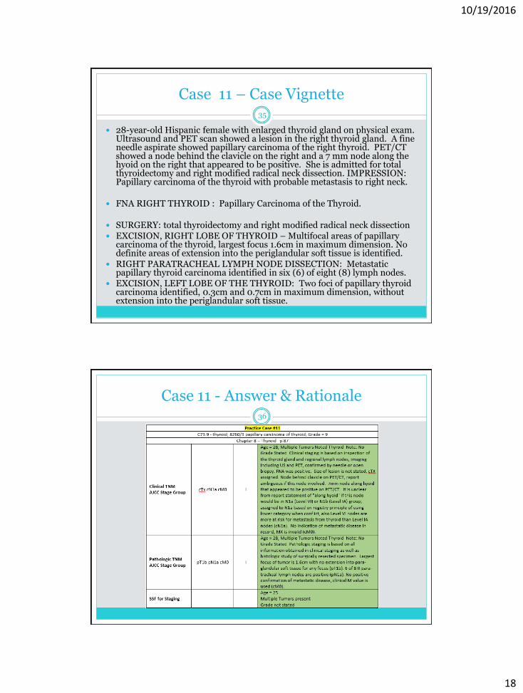
28-year-old Hispanic female with enlarged thyroid gland on physical exam. Ultrasound and PET scan showed a lesion in the right thyroid gland. A fine needle aspirate showed papillary carcinoma of the right thyroid. PET/CT showed a node behind the clavicle on the right and a 7 mm node along the hyoid on the right that appeared to be positive. She is admitted for total thyroidectomy and right modified radical neck dissection. IMPRESSION: Papillary carcinoma of the thyroid with probable metastasis to right neck.
FNA RIGHT THYROID : Papillary Carcinoma of the Thyroid.
SURGERY: total thyroidectomy and right modified radical neck dissection EXCISION, RIGHT LOBE OF THYROID – Multifocal areas of papillary
carcinoma of the thyroid, largest focus 1.6cm in maximum dimension. No definite areas of extension into the periglandular soft tissue is identified.
RIGHT PARATRACHEAL LYMPH NODE DISSECTION: Metastatic papillary thyroid carcinoma identified in six (6) of eight (8) lymph nodes.
EXCISION, LEFT LOBE OF THE THYROID: Two foci of papillary thyroid carcinoma identified, 0.3cm and 0.7cm in maximum dimension, without extension into the periglandular soft tissue.
35
Case 11 - Answer & Rationale36

10/19/2016
19
Case 12 – Case Vignette
65-year old female with right-sided dominant thyroid nodule. Recent PET/CT shows suspicious thyroid nodule as well as suspicious metastatic lesions in lung and bones.
PET/CT; intense focal increased FDG uptake in the right lung apex compatible with FDG Avid malignant process. Increased FDG uptake within the right lobe of the thyroid gland measuring 2.8cm suspicious for FDG AVID malignancy. T3 and T1 bone lesion suspicious for bony metastatic lesions
PATH: TOTAL THYROIDECTOMY: Anaplastic thyroid carcinoma, 4.0cm in general dimension, unifocal with extensive extrathyroidal extension, margin positive; LVI present, 0/5 lymph nodes with carcinoma. PAX-8 (+), TTF-1(+) AND P53(+)
66-year old female who was diagnosed with metastatic anaplastic carcinoma of the thyroid to the bone and lung. She is status post total thyroidectomy followed by chemotherapy and radiation to the H&N and bone. Latest images showing progression of disease in lungs.
IMRT to the thyroid and neck delivering 6600 CGY in 33 fractions/42 days IMRT to the T9 spine delivering 3500 CGY in 10 fractions/14 days. 05/25/16. weekly Carboplatin/Taxol X 7 weeks 04/19/16. Synthroid.112 MCG
37
Case 12 - Answer & Rationale38

10/19/2016
20
Case 13 – Case Vignette
75-year-old male with CT scan showing a mass centered on the right lobe of the thyroid extending into the superior mediastinum, multiple lung nodules, and mediastinal and left hilar adenopathy. Referred for FNA biopsy of the mass in the right thyroid.
RIGHT THYROID MASS, FNA: Non-Hodgkin large cell lymphoma
PERIPHERAL BLOOD SMEAR: Normal RBC and WBC morphology
BONE MARROW, ASPIRATION – Negative for malignant lymphoma
PET IMG W CT SKULL TO THIGH - IMPRESSION:
The right-sided neck mass is intensely hypermetabolic with SUV of greater than 13.
There is a solitary hypermetabolic node anterior to the left hilum with SUV of 4.
No hypermetabolism is seen in the lung nodules.
Two skeletal areas of hypermetabolism seen; one in right ilium and the other in the body of T11.
FNA vertebral T-11: Atypical lymphoid infiltrate consistent with large B-cell lymphoma
MEDICAL ONCOLOGY: Stage IV diffuse large B cell lymphoma involving bone and thyroid. Bulky thyroid mass 11 cm. IPI score 4. TREATMENT PLAN: R-CHOP.
39
Case 13 - Answer & Rationale40
10/19/2016
21
Case 14 - Case Vignette
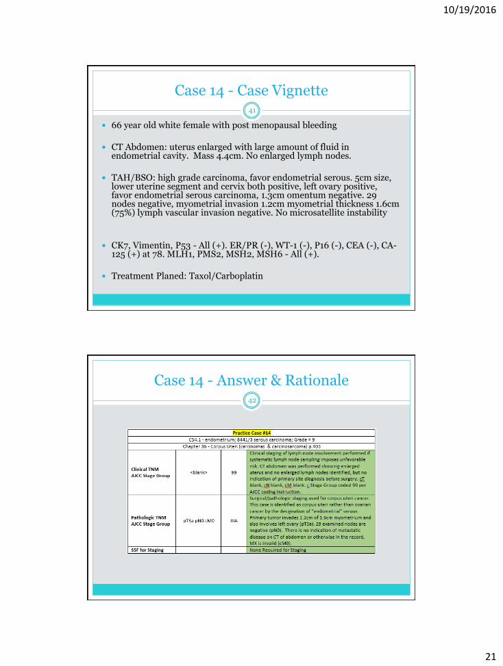
66 year old white female with post menopausal bleeding
CT Abdomen: uterus enlarged with large amount of fluid in endometrial cavity. Mass 4.4cm. No enlarged lymph nodes.
TAH/BSO: high grade carcinoma, favor endometrial serous. 5cm size, lower uterine segment and cervix both positive, left ovary positive, favor endometrial serous carcinoma, 1.3cm omentum negative. 29 nodes negative, myometrial invasion 1.2cm myometrial thickness 1.6cm (75%) lymph vascular invasion negative. No microsatellite instability
CK7, Vimentin, P53 - All (+). ER/PR (-), WT-1 (-), P16 (-), CEA (-), CA-125 (+) at 78. MLH1, PMS2, MSH2, MSH6 - All (+).
Treatment Planed: Taxol/Carboplatin
41
Case 14 - Answer & Rationale42
10/19/2016
22
Case 15 – Case Vignette
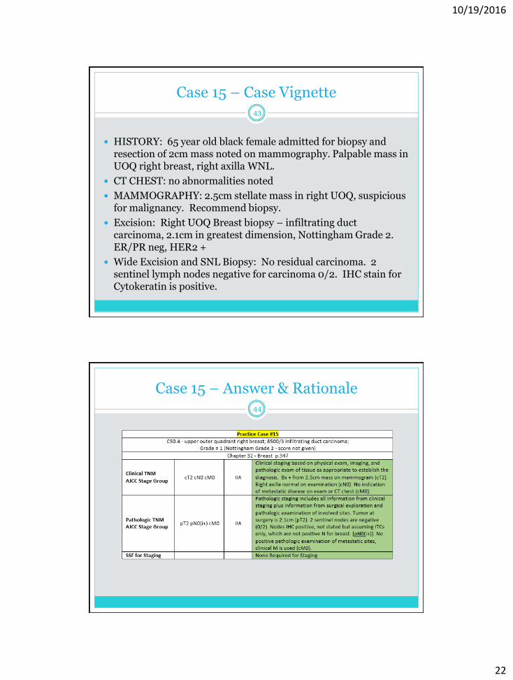
HISTORY: 65 year old black female admitted for biopsy and resection of 2cm mass noted on mammography. Palpable mass in UOQ right breast, right axilla WNL.
CT CHEST: no abnormalities noted
MAMMOGRAPHY: 2.5cm stellate mass in right UOQ, suspicious for malignancy. Recommend biopsy.
Excision: Right UOQ Breast biopsy – infiltrating duct carcinoma, 2.1cm in greatest dimension, Nottingham Grade 2. ER/PR neg, HER2 +
Wide Excision and SNL Biopsy: No residual carcinoma. 2 sentinel lymph nodes negative for carcinoma 0/2. IHC stain for Cytokeratin is positive.
43
Case 15 – Answer & Rationale44
10/19/2016
23
Case 16 – Case Vignette
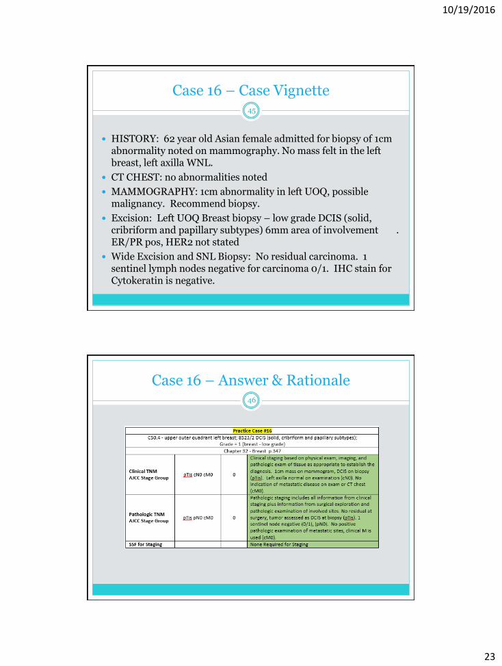
HISTORY: 62 year old Asian female admitted for biopsy of 1cm abnormality noted on mammography. No mass felt in the left breast, left axilla WNL.
CT CHEST: no abnormalities noted
MAMMOGRAPHY: 1cm abnormality in left UOQ, possible malignancy. Recommend biopsy.
Excision: Left UOQ Breast biopsy – low grade DCIS (solid, cribriform and papillary subtypes) 6mm area of involvement . ER/PR pos, HER2 not stated
Wide Excision and SNL Biopsy: No residual carcinoma. 1 sentinel lymph nodes negative for carcinoma 0/1. IHC stain for Cytokeratin is negative.
45
Case 16 – Answer & Rationale46

10/19/2016
24
Case 17 – Case Vignette
61 year old white female with mammo showing suspicious tumor in lateral aspect of right periareolar area. Physical Exam shows a palpable mass in periareolar region right breast @ 9:00 approximately 2cm in size, close to skin with extension to retroareolar area and overlying areola. Mass is not fixed to chest wall but may be contiguous to subcutaneous tissue. No palpable lymphadenopathy.
MAMMO – mass right breast @ 9:00, suspicious lymph node with thickening in right axilla
MRI Bilateral Breast – left breast neg. right breast in retroareolar area shows enhancing mass measuring 2.3cm. 1.4cm right axillary lymph node corresponds to recent biopsy of lymph node.
CT ABD/PELVIS neg and CXR neg
Right Breast @ 9:00, subareolar infiltrating ductal carcinoma Nottingham Grade 3/3. Core biopsy axillary lymph node – positive for metastatic ductal carcinoma.
ER POS. 40%/PR NEG. 0%/HER-2/NEU IHC NEG. 1+/KI-67 high proliferative index 95%
ONCOTYPE DX score 64/ER 5.5 NEG./<3.2 NEG./HER-2/NEU IHC <7.6 NEG.
Right Breast Wide Excision with right axillary sentinel node biopsy – No residual tumor after 5 cycles of Adria/Cytoxan + Taxol. 1 sentinel node negative after neoadjuvant chemotherapy
47
Case 17 – Answer & Rationale48
10/19/2016
25
Case 18 – Case Vignette
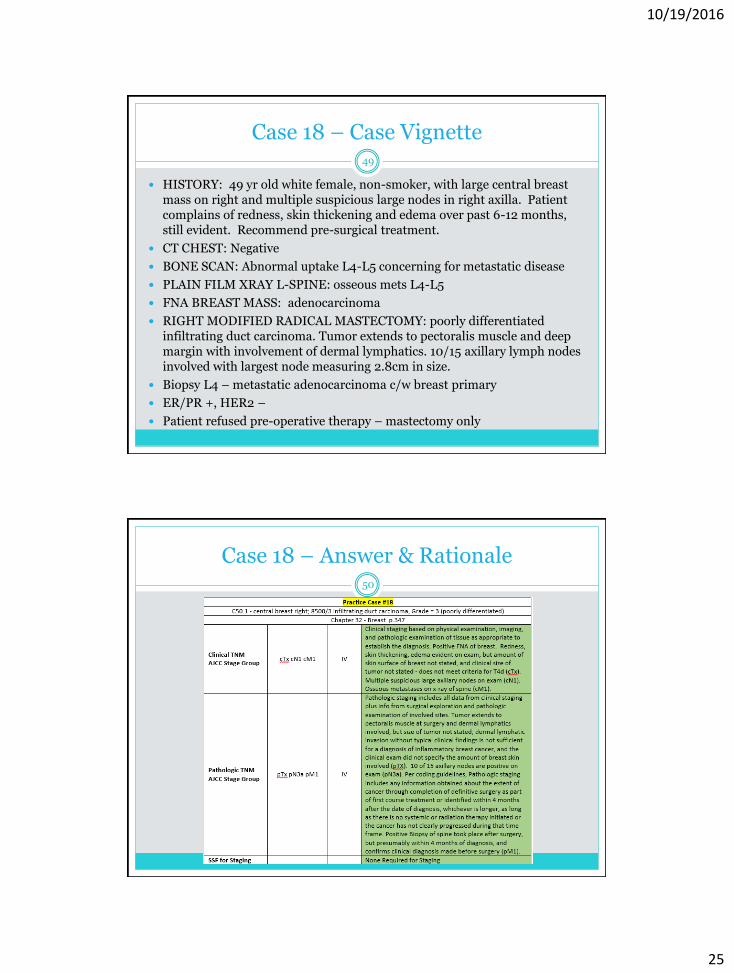
HISTORY: 49 yr old white female, non-smoker, with large central breast mass on right and multiple suspicious large nodes in right axilla. Patient complains of redness, skin thickening and edema over past 6-12 months, still evident. Recommend pre-surgical treatment.
CT CHEST: Negative
BONE SCAN: Abnormal uptake L4-L5 concerning for metastatic disease
PLAIN FILM XRAY L-SPINE: osseous mets L4-L5
FNA BREAST MASS: adenocarcinoma
RIGHT MODIFIED RADICAL MASTECTOMY: poorly differentiated infiltrating duct carcinoma. Tumor extends to pectoralis muscle and deep margin with involvement of dermal lymphatics. 10/15 axillary lymph nodes involved with largest node measuring 2.8cm in size.
Biopsy L4 – metastatic adenocarcinoma c/w breast primary
ER/PR +, HER2 –
Patient refused pre-operative therapy – mastectomy only
49
Case 18 – Answer & Rationale50
10/19/2016
26
Case 19 – Case Vignette
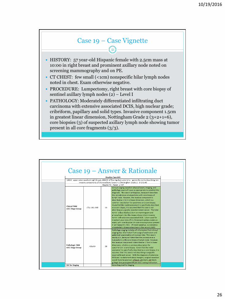
HISTORY: 57 year-old Hispanic female with 2.5cm mass at 10:00 in right breast and prominent axillary node noted on screening mammography and on PE.
CT CHEST: few small (<1cm) nonspecific hilar lymph nodes noted in chest. Exam otherwise negative.
PROCEDURE: Lumpectomy, right breast with core biopsy of sentinel axillary lymph nodes (2) – Level I
PATHOLOGY: Moderately differentiated infiltrating duct carcinoma with extensive associated DCIS, high nuclear grade; cribriform, papillary and solid types. Invasive component 1.5cm in greatest linear dimension, Nottingham Grade 2 (3+2+1=6), core biopsies (3) of suspected axillary lymph node showing tumor present in all core fragments (3/3).
51
Case 19 – Answer & Rationale52

10/19/2016
27
Case 20 – Case Vignette
59 year old white male with elevated PSA – biopsy-proven adenocarcinoma
MRI Prostate – 2cm area of tumor involving right mid gland and apex. Tumor abuts posterior wall without definitive extracapsular extension. No pelvic lymphadenopathy noted. Bone Scan is negative.
PSA=13.5
TRUS BX=Adenocarcinoma Gleason 3 + 4 = 7. No perineural invasion. Robot-Assisted Radical Prostatectomy with Bilateral Pelvic LN Dissection.
Radical Prostatectomy – 1.7cm dominant focus in right posterior peripheral zone from apex to mid gland. 20% of gland involved. Gleason 3 + 4 = 7. Tumor extends focally a fraction of a millimeter past the prostatic capsule resection margins. All final margins negative. Perineural invasion is identified in the specimen. 6 pelvic lymph nodes negative for metastatic adenocarcinoma.
53
Case 20 – Answer & Rationale54

10/19/2016
28
Case 21 – Case Vignette
HISTORY: 65 year old black male admitted with intermittent microscopic hematuria . History of prostate cancer. History of 1ppd smoker x 45yrs.
CT CHEST: no abnormalities noted
CT ABDOMEN: negative
CYSTOSCOPY: 2 papillary projections identified, one along the right lateral wall, the other in the trigone area of the bladder. TURBT was performed.
PATHOLOGY: Bladder biopsy (TURBT) – low grade papillary urothelial carcinoma (no mention of invasion)
FINAL DX: Papillary urothelial carcinoma of bladder, low grade. Repeat cystoscopy in 3 months.
55
Case 21 – Answer & Rationale56
10/19/2016
29
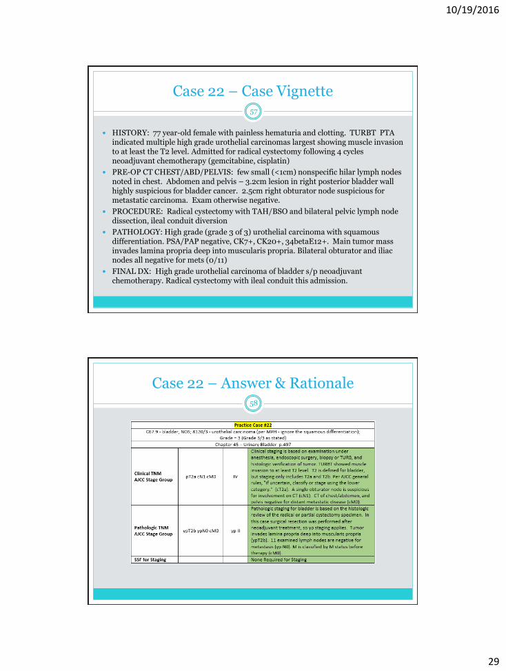
Case 22 – Case Vignette
HISTORY: 77 year-old female with painless hematuria and clotting. TURBT PTA indicated multiple high grade urothelial carcinomas largest showing muscle invasion to at least the T2 level. Admitted for radical cystectomy following 4 cycles neoadjuvant chemotherapy (gemcitabine, cisplatin)
PRE-OP CT CHEST/ABD/PELVIS: few small (<1cm) nonspecific hilar lymph nodes noted in chest. Abdomen and pelvis – 3.2cm lesion in right posterior bladder wall highly suspicious for bladder cancer. 2.5cm right obturator node suspicious for metastatic carcinoma. Exam otherwise negative.
PROCEDURE: Radical cystectomy with TAH/BSO and bilateral pelvic lymph node dissection, ileal conduit diversion
PATHOLOGY: High grade (grade 3 of 3) urothelial carcinoma with squamous differentiation. PSA/PAP negative, CK7+, CK20+, 34betaE12+. Main tumor mass invades lamina propria deep into muscularis propria. Bilateral obturator and iliac nodes all negative for mets (0/11)
FINAL DX: High grade urothelial carcinoma of bladder s/p neoadjuvant chemotherapy. Radical cystectomy with ileal conduit this admission.
57
Case 22 – Answer & Rationale58

10/19/2016
30
Case 23 – Case Vignette
HISTORY: 61 yr old man, lifelong smoker, with frequent and urgent urinary symptoms and microscopic hematuria noted on routine exam.
CT ABDOMEN: Negative
CT CHEST: Negative
CYSTOSCOPY: Flat urothelial carcinoma diffuse involvement of bladder - multiple biopsies with fulguration and administration Intravesical BCG
PATHOLOGY: flat urothelial carcinoma, high grade, diffuse - Tis
TREATMENT: TURBT with Intravesical BCG (now and for next 6 weeks)
59
Case 23 – Answer & Rationale60

10/19/2016
31
Case 24 – Case Vignette
HISTORY: 55 yr old white male, non-smoker, with elevated PSA and recurring prostatitis with minimal response to multiple course of antibiotics. DRE shows enlarged prostate with firm nodule in left lateral lobe of prostate. No other clinical symptoms or complaints. Admitted for treatment evaluation.
PSA: 10.3 ng/mL
CT CHEST: Negative
BONE SCAN: Abnormal uptake L4-L5 concerning for metastatic disease
PLAIN FILM XRAY L-SPINE: no evidence for osseous mets
TRUS-GUIDED BX PROSTATE: adenocarcinoma, Gleason 4+4=8, 9 of 12 cores positive
RADICAL RETROPUBIC PROSTATECTOMY WITH LYMPH NODE SAMPLING: moderately differentiated adenocarcinoma Gleason 4+4=8 with microscopic involvement of bladder neck. Negative surgical resection margins. 3 inguinal lymph nodes sampled, all negative
61
Case 24 – Answer & Rationale62

10/19/2016
32
Case 25 – Case Vignette
HISTORY: 2 year old white male child with abdominal distention, decreased bowel sounds and abdominal pain of several weeks duration.
Ultrasound Abdomen – large heterogeneous 21cm x 9.6cm space occupying lesion of uncertain origin.
CT Abdomen – large intra-abdominal space occupying lesion w/mass effect
CT Chest – no metastatic disease in the chest noted
Whole Body Bone Scan – negative for metastatic disease
Tumor Biopsy and Biopsy of Omental Implant – high-grade embryonal rhabdomyosarcoma
Plan: vincristine, dactinomycin, mesna, cyclophosphamide plus irinotecan
63
Case 25 – Answer & Rationale64

10/19/2016
33
8th ed. Purchase and Ordering Information65
http://www.springer.com/us/book/ 9783319406176
• AJCC Cancer Staging Manual – 8th edition, 2017
• COST: $119.99• ISBN: 978-3-319-40617-6
• 1429 pages• 512 illustrations• 187 color illustrations
• Required - Florida Mandate• FCDS will not purchase• Facility may purchase• Individual may purchase
• https://cancerstaging.org• http://springer.com• 1-800-SPRINGER
Questions66