Embed Size (px)
Citation preview

2019Course #4
Self-StudyCourse
Contact Us:Phone
614-292-6737
Toll Free1-888-476-7678
Fax614-292-8752
Webdentistry.osu.edu/sms
The Ohio State UniversityCollege of Dentistry305 W. 12th AvenueColumbus, OH 43210
The Ohio State University College of Dentistry is a recognized provider for ADA CERP credit. ADA CERP is a service of the American Dental Association to assist dental professionals in identifying quality providers of continuing dental education. ADA CERP does not approve or endorse individual courses or instructors, nor does it imply acceptance of credit hours by boards of dentistry. Concerns or complaints about a CE provider may be directed to the provider or to the Commission for Continuing Education Provider Recognition at www.ada.org/cerp.
The Ohio State University College of Dentistry is approved by the Ohio State Dental Board as a permanent sponsor of continuing dental education.This continuing education activity has been planned and implemented in accordance with the standards of the ADA Continuing Education Recognition Program (ADA CERP) through joint efforts between The Ohio State University College of Dentistry Office of Continuing Dental Education and the Sterilization Monitoring Service (SMS).
Course Instructions:
▪ Read and review the course materials.
▪ Complete the 12 question test. A total of 9 questions must be answered correctly for credit.
▪ Submit your answers online at:
http://dentistry.osu.edu/sms-continuing-education
▪ Check your email for your CE certification of completion (please check your junk/spam folder as well).
About SMS CE courses:
▪ TWO CREDIT HOURS are issued for successful completion of this self-study course for the OSDB 2019-2021 biennium totals.
▪ CERTIFICATE of COMPLETIONis used to document your CE credit and is emailed to each course participant.
▪ ALLOW 2 WEEKS for processing of your certificate.
Frequently Asked Questions:
Q: Who can earn FREE CE credits?
A: EVERYONE - All dental professionals in your office may earn free CE credits. Each person must read the course materials and submit an online answer form independently.
Q: Where can I find my SMS number?
A: Your SMS number can be found in the upper right hand corner of your monthly reports, or, imprinted on the back of your test envelopes. The SMS number is the account number for your office only, and is the same for everyone in the office.
Q: How often are these courses available?
A: Four times per year (8 CE credits).
2019 Course
#4
Written by:
Sonya Kalim, DMD, MDS, ABOMR Diplomate
Release Date:
November 11, 2019
8:30am EST
Last Day to Take Course
Free of Charge:
December 11, 2019
4:30pm EST
An Introduction to Cone-Beam Computed Tomography
This is an OSDB Category B: Supervised self-instruction course
2
About the Author
Sonya Kalim, DMD, MDS, ABOMR Diplomate
Dr. Kalim can be reached at [email protected]
Neither I nor my immediate family have any financial interests that
would create a conflict of interest or restrict my judgement with
regard to the content of this course.
IntroductionCone-beam computed tomography (CBCT) is a relatively
recent technology that has revolutionized the modern practice of
dentistry. In the United States (U.S.), CBCT machines became
available for purchase in the early 2000s1. Equipment size and
relative affordability have made it extremely accessible to dental
private practices and dental academic institutions across the
country. The U.S. Food and Drug Administration’s (FDA)
Nationwide Evaluation of X-Ray Trends (NEXT) Dental survey
found that between the years of 2014 and 2015, approximately
5,500 CBCT machines were being utilized to acquire an
approximated over 4 million pediatric and adult studies annually in
general dentistry, oral surgery, and orthodontic clinics.2 However,
with the ever-increasing popularity of CBCT equipment and the
diagnostic value they provide, these numbers are sure to increase
with time, and it has become increasingly essential to have an
introductory knowledge base regarding CBCT technology.
Dr. Sonya Kalim is an Assistant Professor of Oral and
Maxillofacial Radiology in the Section of Oral and Maxillofacial
Pathology and Radiology at The Ohio State University, College of
Dentistry. Dr. Kalim received her D.M.D. from the Maurice H.
Kornberg School of Dentistry at Temple University in 2014.
Afterwards, she entered the oral and maxillofacial radiology
residency program at the University of Connecticut School of
Dental Medicine, where she earned a certificate in oral and
maxillofacial radiology and a master's in dental science. Dr. Kalim
joined OSU as a faculty member in July of 2017. Her activities
include teaching responsibilities and interpretation of cone beam
CT scans. Dr. Kalim is certified by the American Board of Oral
and Maxillofacial Radiology.
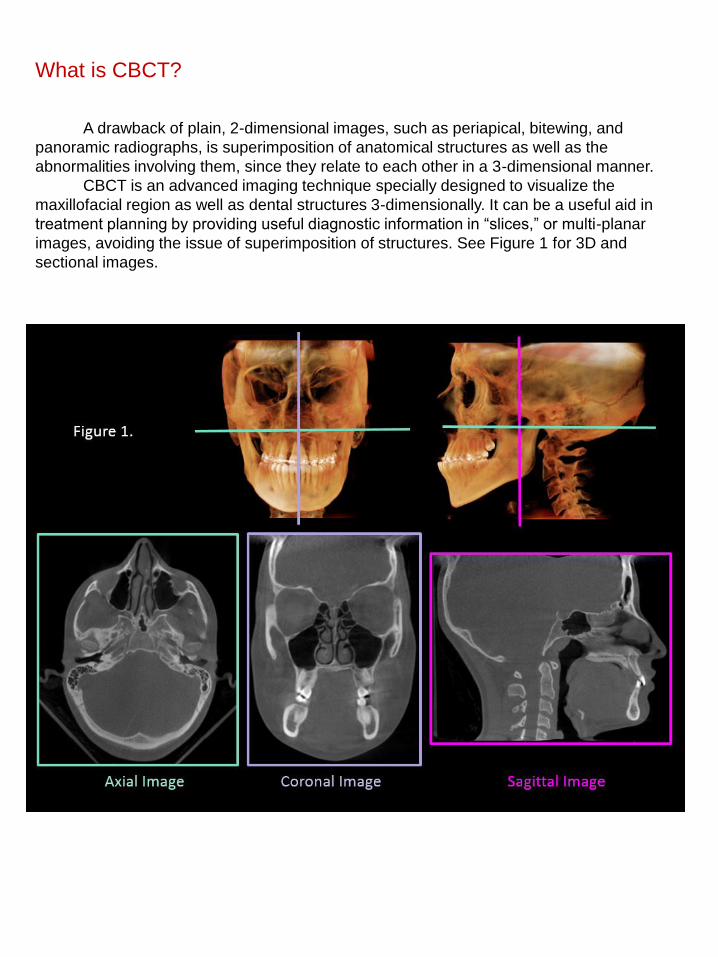
What is CBCT?
A drawback of plain, 2-dimensional images, such as periapical, bitewing, and
panoramic radiographs, is superimposition of anatomical structures as well as the
abnormalities involving them, since they relate to each other in a 3-dimensional manner.
CBCT is an advanced imaging technique specially designed to visualize the
maxillofacial region as well as dental structures 3-dimensionally. It can be a useful aid in
treatment planning by providing useful diagnostic information in “slices,” or multi-planar
images, avoiding the issue of superimposition of structures. See Figure 1 for 3D and
sectional images.
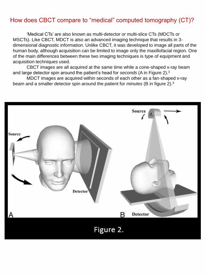
‘Medical CTs’ are also known as multi-detector or multi-slice CTs (MDCTs or
MSCTs). Like CBCT, MDCT is also an advanced imaging technique that results in 3-
dimensional diagnostic information. Unlike CBCT, it was developed to image all parts of the
human body, although acquisition can be limited to image only the maxillofacial region. One
of the main differences between these two imaging techniques is type of equipment and
acquisition techniques used.
CBCT images are all acquired at the same time while a cone-shaped x-ray beam
and large detector spin around the patient’s head for seconds (A in Figure 2).3
MDCT images are acquired within seconds of each other as a fan-shaped x-ray
beam and a smaller detector spin around the patient for minutes (B in figure 2).3
How does CBCT compare to “medical” computed tomography (CT)?
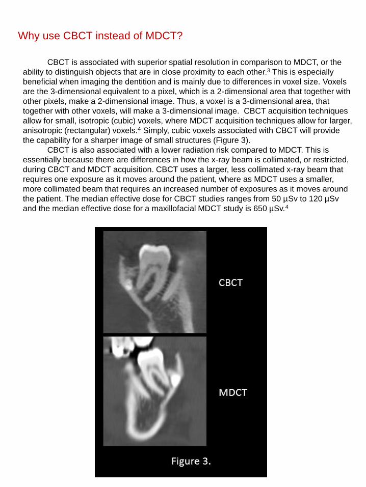
CBCT is associated with superior spatial resolution in comparison to MDCT, or the
ability to distinguish objects that are in close proximity to each other.3 This is especially
beneficial when imaging the dentition and is mainly due to differences in voxel size. Voxels
are the 3-dimensional equivalent to a pixel, which is a 2-dimensional area that together with
other pixels, make a 2-dimensional image. Thus, a voxel is a 3-dimensional area, that
together with other voxels, will make a 3-dimensional image. CBCT acquisition techniques
allow for small, isotropic (cubic) voxels, where MDCT acquisition techniques allow for larger,
anisotropic (rectangular) voxels.4 Simply, cubic voxels associated with CBCT will provide
the capability for a sharper image of small structures (Figure 3).
CBCT is also associated with a lower radiation risk compared to MDCT. This is
essentially because there are differences in how the x-ray beam is collimated, or restricted,
during CBCT and MDCT acquisition. CBCT uses a larger, less collimated x-ray beam that
requires one exposure as it moves around the patient, where as MDCT uses a smaller,
more collimated beam that requires an increased number of exposures as it moves around
the patient. The median effective dose for CBCT studies ranges from 50 µSv to 120 µSv
and the median effective dose for a maxillofacial MDCT study is 650 µSv.4
Why use CBCT instead of MDCT?
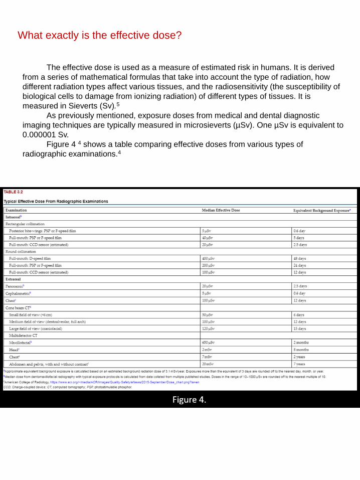
The effective dose is used as a measure of estimated risk in humans. It is derived
from a series of mathematical formulas that take into account the type of radiation, how
different radiation types affect various tissues, and the radiosensitivity (the susceptibility of
biological cells to damage from ionizing radiation) of different types of tissues. It is
measured in Sieverts (Sv).5
As previously mentioned, exposure doses from medical and dental diagnostic
imaging techniques are typically measured in microsieverts (µSv). One µSv is equivalent to
0.000001 Sv.
Figure 4 4 shows a table comparing effective doses from various types of
radiographic examinations.4
What exactly is the effective dose?
As the effective dose suggests, the probability of lethal damage to bodily tissues due to
radiation exposure heavily depends on the type of tissue being irradiated.5 Tissues that are
rapidly dividing (have a high turnover rate) and undifferentiated (do not complete a specifically
defined function) are the most radiosensitive.4 Most relevant to the profession of dentistry are
cells of the oral mucosa, salivary glands, thyroid gland, and bone marrow stem cells, due to
the regions that are imaged .
The radiation risk increases in children compared to adults due to the abundance of
rapidly dividing cells during developmental growth.4,6 Thus, additional caution should be
exercised when deciding to utilize advanced imaging in pediatric cases.6
For ALL patients, the decision to expose the patient to any kind of diagnostic radiation
should be properly justified and optimized with carefully chosen parameters according to the
As Low As Reasonably/Diagnostically Achievable (ALARA/ALADA) principle.4,6,7 The ALARA
or ALADA principle indicates that all practical and reasonable efforts should be made to keep
radiation exposure at a minimum while maintaining diagnostic efficacy.
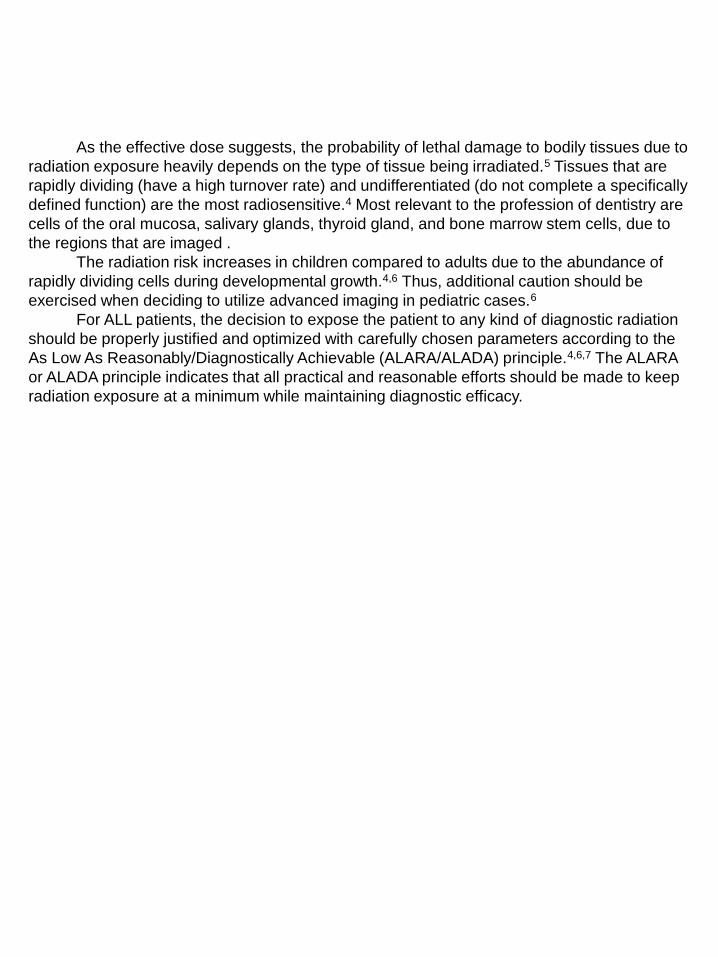
The radiation exposure resulting from CBCT acquisition significantly varies with
machinery (Figure 5 4), due to a lack of standardization between different brands of
equipment.8 Each CBCT machine offers various options for acquisition, most notably
differing in sizing of the field-of-view (FOV) (Figure 5 4).8 The FOV is the area imaged
during the scan, which is determined by the region(s) of interest. Generally, a smaller FOV
limits the area of exposure on the patient, but may not reduce the effective dose, depending
on the acquisition parameters used.
Acquisition parameters that modify the radiation dose include the scan time, the
number of raw images acquired during the scan, trajectory arc (whether the gantry spins
around the patient’s head for 180o or 360o), and exposure settings (mA and kVp).4,8
Reducing the values of the aforementioned factors will also lessen the radiation risk, but
may compromise the diagnostic quality of the scan. Additionally, utilizing a smaller voxel
size to increase spatial resolution will increase the effective dose.4,8 Therefore, a balance of
these factors must be achieved.
Why does the radiation risk estimated from CBCT range between
different values?
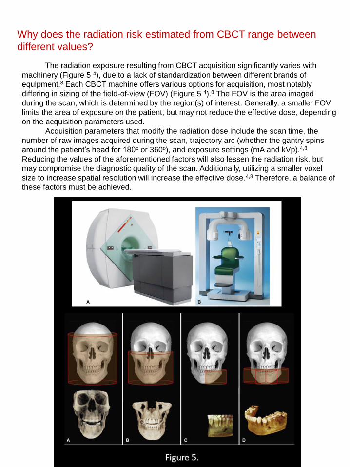
Due to the way CBCT images are acquired, there is an inherent amount of noise or
“graininess” captured in the images.4,9 This is due to a phenomenon known as scatter
radiation.4,9 When an image is acquired, some photons travel on a direct path through the
patient to the detector and some photons get absorbed by dense tissues; these photons
contribute to image formation. Other photons follow an altered trajectory to the receptor,
and thus, do not carry any diagnostic information, leading to image degradation (Figure 6 4).
Scatter radiation also results in poor soft tissue contrast, making CBCT less diagnostic for
assessment of soft tissue abnormalities.
What are the limitations of CBCT?
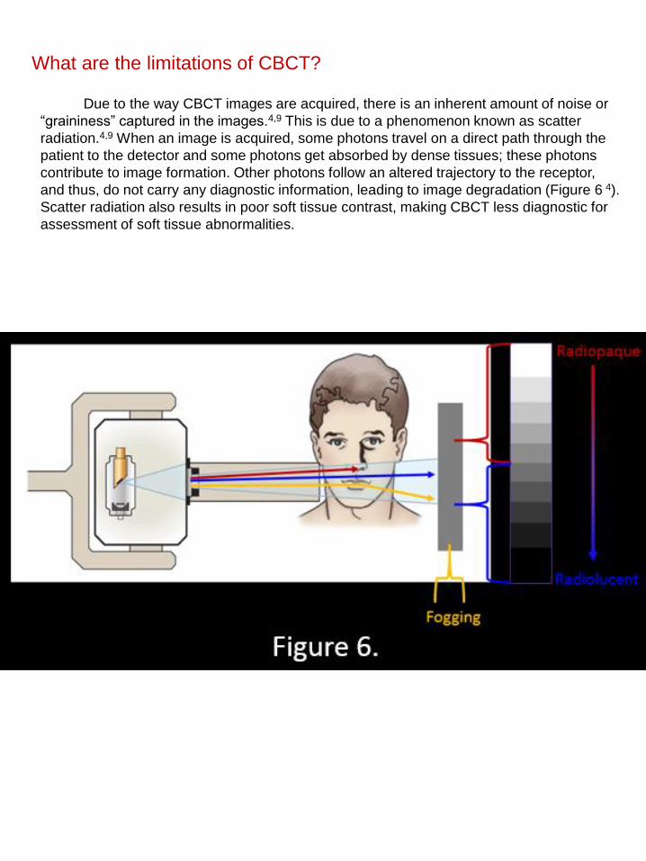
MDCT images are far superior to CBCT images regarding noise and soft tissue
contrast, due to less scatter radiation resulting from a more collimated x-ray beam, as
previously mentioned (Figure 7).
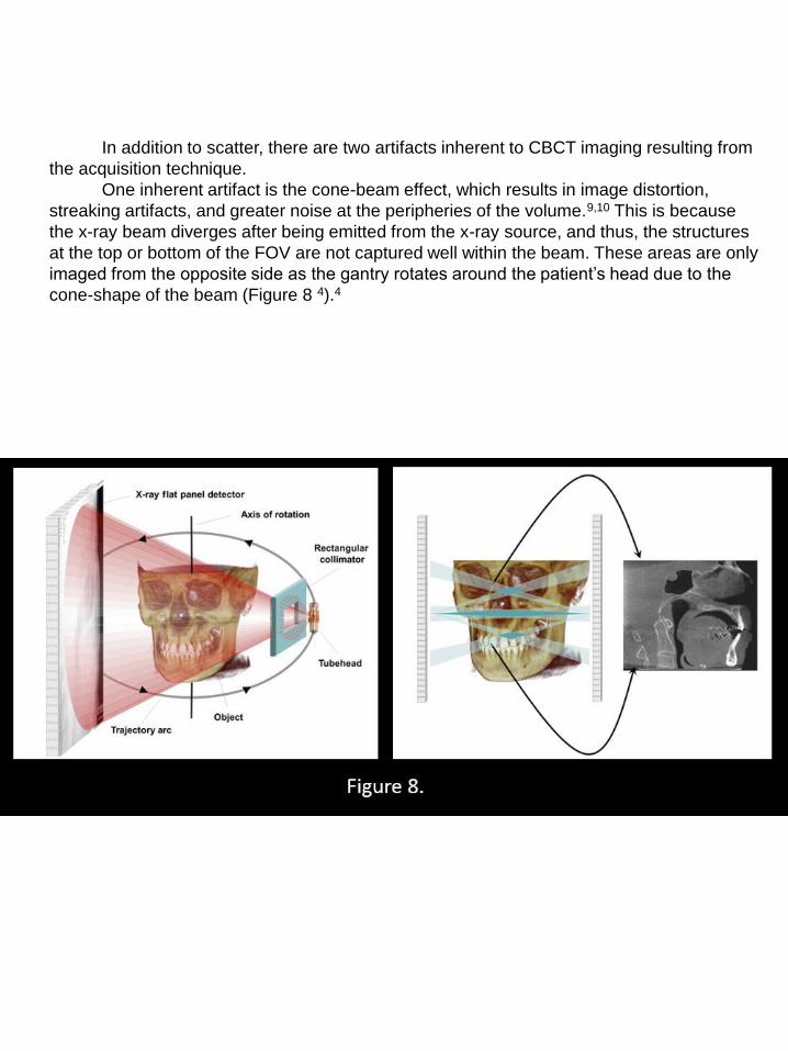
In addition to scatter, there are two artifacts inherent to CBCT imaging resulting from
the acquisition technique.
One inherent artifact is the cone-beam effect, which results in image distortion,
streaking artifacts, and greater noise at the peripheries of the volume.9,10 This is because
the x-ray beam diverges after being emitted from the x-ray source, and thus, the structures
at the top or bottom of the FOV are not captured well within the beam. These areas are only
imaged from the opposite side as the gantry rotates around the patient’s head due to the
cone-shape of the beam (Figure 8 4).4

Another inherent artifact is partial volume averaging, which results in obscuration of
small structures due to the difference in size of the structure(s) and the voxel used to image
it, where the resultant density of that voxel will be an average of all the densities of the
objects imaged.10
Aside from inherent artifacts, artifacts associated with the image acquisition
procedures can also undermine diagnostic value of the scan. One of these is patient
movement captured during scan acquisition, creating “double lines” across the images
(Figure 9 4).
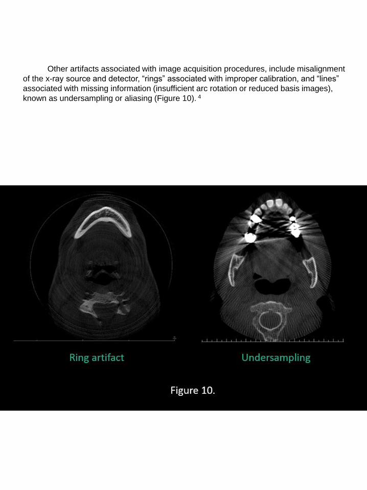
Other artifacts associated with image acquisition procedures, include misalignment
of the x-ray source and detector, “rings” associated with improper calibration, and “lines”
associated with missing information (insufficient arc rotation or reduced basis images),
known as undersampling or aliasing (Figure 10). 4
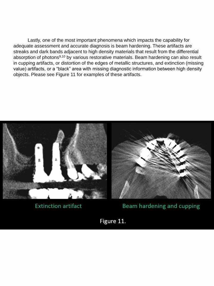
Lastly, one of the most important phenomena which impacts the capability for
adequate assessment and accurate diagnosis is beam hardening. These artifacts are
streaks and dark bands adjacent to high density materials that result from the differential
absorption of photons9,10 by various restorative materials. Beam hardening can also result
in cupping artifacts, or distortion of the edges of metallic structures, and extinction (missing
value) artifacts, or a “black” area with missing diagnostic information between high density
objects. Please see Figure 11 for examples of these artifacts.
Understanding the strengths and limitations of CBCT as an advanced imaging
modality and how it may be of use to discover valuable diagnostic information is essential.
However, due to the previously discussed risk posed to the patient from ionizing radiation
from radiographic examinations, it is important to follow proper selection criteria when
deciding to expose the patient. The potential benefits of gaining additional information that
may modify the patient’s treatment plan from the study should outweigh the radiation risk to
the patient.
CBCT studies are not a replacement for conventional plain images, especially in
diagnosis of common dental diseases. Artifacts from existing restorations may impede
accurate assessment of caries and periodontal disease. CBCTs are not recommended for
every patient and should only be taken when appropriately indicated after initial clinical and
radiographic examinations are completed.
What are common indications for CBCT studies?
CBCT studies are most commonly acquired for implant treatment planning and
applications in endodontics, orthodontics, and oral surgery. With regard to implant treatment
planning, CBCTs can be utilized to assess bone quality and bone quantity, location of
neighboring anatomical structures, integration of bone grafts, follow-up of symptomatic
implants after placement, and creation of surgical guides. Endodontic applications include
evaluation of resorptive defects (internal and external), suspected tooth fractures, iatrogenic
perforations, complex tooth morphology, and persistent periapical pathology. Other
indications include evaluation of impacted teeth, supernumerary teeth, pathologies, the
osseous components of the temporomandibular joints (TMJs), and paranasal sinuses.
CBCT images can also be utilized to manufacture 3-dimensional biomodels in preparation
for orthognathic surgery.
There are multiple published position statements available from the American
Academy of Oral and Maxillofacial Radiology (AAOMR) as well as other professional
organizations regarding adequate justification and optimization of the use of CBCT in these
fields of dentistry. The included links within the following summarizing text can be used to
view these specific recommendations in depth.
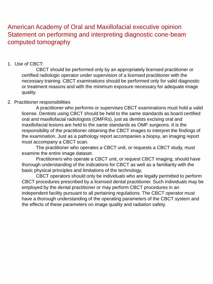
1. Use of CBCT:
CBCT should be performed only by an appropriately licensed practitioner or
certified radiologic operator under supervision of a licensed practitioner with the
necessary training. CBCT examinations should be performed only for valid diagnostic
or treatment reasons and with the minimum exposure necessary for adequate image
quality.
2. Practitioner responsibilities
A practitioner who performs or supervises CBCT examinations must hold a valid
license. Dentists using CBCT should be held to the same standards as board certified
oral and maxillofacial radiologists (OMFRs), just as dentists excising oral and
maxillofacial lesions are held to the same standards as OMF surgeons. It is the
responsibility of the practitioner obtaining the CBCT images to interpret the findings of
the examination. Just as a pathology report accompanies a biopsy, an imaging report
must accompany a CBCT scan.
The practitioner who operates a CBCT unit, or requests a CBCT study, must
examine the entire image dataset.
Practitioners who operate a CBCT unit, or request CBCT imaging, should have
thorough understanding of the indications for CBCT as well as a familiarity with the
basic physical principles and limitations of the technology.
CBCT operators should only be individuals who are legally permitted to perform
CBCT procedures prescribed by a licensed dental practitioner. Such individuals may be
employed by the dental practitioner or may perform CBCT procedures in an
independent facility pursuant to all pertaining regulations. The CBCT operator must
have a thorough understanding of the operating parameters of the CBCT system and
the effects of these parameters on image quality and radiation safety.
American Academy of Oral and Maxillofacial executive opinion
Statement on performing and interpreting diagnostic cone-beam
computed tomography
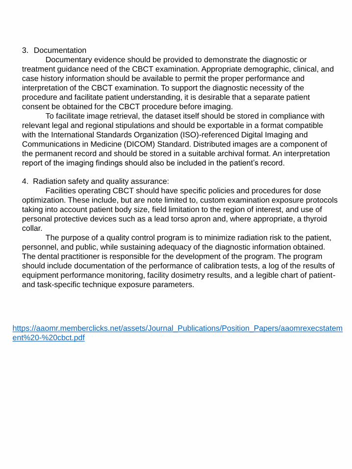
3. Documentation
Documentary evidence should be provided to demonstrate the diagnostic or
treatment guidance need of the CBCT examination. Appropriate demographic, clinical, and
case history information should be available to permit the proper performance and
interpretation of the CBCT examination. To support the diagnostic necessity of the
procedure and facilitate patient understanding, it is desirable that a separate patient
consent be obtained for the CBCT procedure before imaging.
To facilitate image retrieval, the dataset itself should be stored in compliance with
relevant legal and regional stipulations and should be exportable in a format compatible
with the International Standards Organization (ISO)-referenced Digital Imaging and
Communications in Medicine (DICOM) Standard. Distributed images are a component of
the permanent record and should be stored in a suitable archival format. An interpretation
report of the imaging findings should also be included in the patient’s record.
4. Radiation safety and quality assurance:
Facilities operating CBCT should have specific policies and procedures for dose
optimization. These include, but are note limited to, custom examination exposure protocols
taking into account patient body size, field limitation to the region of interest, and use of
personal protective devices such as a lead torso apron and, where appropriate, a thyroid
collar.
The purpose of a quality control program is to minimize radiation risk to the patient,
personnel, and public, while sustaining adequacy of the diagnostic information obtained.
The dental practitioner is responsible for the development of the program. The program
should include documentation of the performance of calibration tests, a log of the results of
equipment performance monitoring, facility dosimetry results, and a legible chart of patient-
and task-specific technique exposure parameters.
https://aaomr.memberclicks.net/assets/Journal_Publications/Position_Papers/aaomrexecstatem
ent%20-%20cbct.pdf
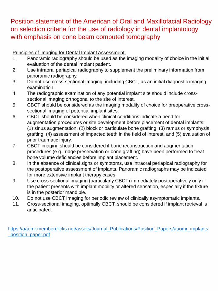
Principles of Imaging for Dental Implant Assessment:
1. Panoramic radiography should be used as the imaging modality of choice in the initial
evaluation of the dental implant patient.
2. Use intraoral periapical radiography to supplement the preliminary information from
panoramic radiography.
3. Do not use cross-sectional imaging, including CBCT, as an initial diagnostic imaging
examination.
4. The radiographic examination of any potential implant site should include cross-
sectional imaging orthogonal to the site of interest.
5. CBCT should be considered as the imaging modality of choice for preoperative cross-
sectional imaging of potential implant sites.
6. CBCT should be considered when clinical conditions indicate a need for
augmentation procedures or site development before placement of dental implants:
(1) sinus augmentation, (2) block or particulate bone grafting, (3) ramus or symphysis
grafting, (4) assessment of impacted teeth in the field of interest, and (5) evaluation of
prior traumatic injury.
7. CBCT imaging should be considered if bone reconstruction and augmentation
procedures (e.g., ridge preservation or bone grafting) have been performed to treat
bone volume deficiencies before implant placement.
8. In the absence of clinical signs or symptoms, use intraoral periapical radiography for
the postoperative assessment of implants. Panoramic radiographs may be indicated
for more extensive implant therapy cases.
9. Use cross-sectional imaging (particularly CBCT) immediately postoperatively only if
the patient presents with implant mobility or altered sensation, especially if the fixture
is in the posterior mandible.
10. Do not use CBCT imaging for periodic review of clinically asymptomatic implants.
11. Cross-sectional imaging, optimally CBCT, should be considered if implant retrieval is
anticipated.
Position statement of the American of Oral and Maxillofacial Radiology
on selection criteria for the use of radiology in dental implantology
with emphasis on cone beam computed tomography
https://aaomr.memberclicks.net/assets/Journal_Publications/Position_Papers/aaomr_implants
_position_paper.pdf
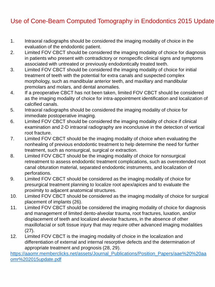
1. Intraoral radiographs should be considered the imaging modality of choice in the
evaluation of the endodontic patient.
2. Limited FOV CBCT should be considered the imaging modality of choice for diagnosis
in patients who present with contradictory or nonspecific clinical signs and symptoms
associated with untreated or previously endodontically treated teeth.
3. Limited FOV CBCT should be considered the imaging modality of choice for initial
treatment of teeth with the potential for extra canals and suspected complex
morphology, such as mandibular anterior teeth, and maxillary and mandibular
premolars and molars, and dental anomalies.
4. If a preoperative CBCT has not been taken, limited FOV CBCT should be considered
as the imaging modality of choice for intra-appointment identification and localization of
calcified canals.
5. Intraoral radiographs should be considered the imaging modality of choice for
immediate postoperative imaging.
6. Limited FOV CBCT should be considered the imaging modality of choice if clinical
examination and 2-D intraoral radiography are inconclusive in the detection of vertical
root fracture.
7. Limited FOV CBCT should be the imaging modality of choice when evaluating the
nonhealing of previous endodontic treatment to help determine the need for further
treatment, such as nonsurgical, surgical or extraction.
8. Limited FOV CBCT should be the imaging modality of choice for nonsurgical
retreatment to assess endodontic treatment complications, such as overextended root
canal obturation material, separated endodontic instruments, and localization of
perforations.
9. Limited FOV CBCT should be considered as the imaging modality of choice for
presurgical treatment planning to localize root apex/apices and to evaluate the
proximity to adjacent anatomical structures.
10. Limited FOV CBCT should be considered as the imaging modality of choice for surgical
placement of implants (26).
11. Limited FOV CBCT should be considered the imaging modality of choice for diagnosis
and management of limited dento-alveolar trauma, root fractures, luxation, and/or
displacement of teeth and localized alveolar fractures, in the absence of other
maxillofacial or soft tissue injury that may require other advanced imaging modalities
(27).
12. Limited FOV CBCT is the imaging modality of choice in the localization and
differentiation of external and internal resorptive defects and the determination of
appropriate treatment and prognosis (28, 29).
https://aaomr.memberclicks.net/assets/Journal_Publications/Position_Papers/aae%20%20aa
omr%202015update.pdf
Use of Cone-Beam Computed Tomography in Endodontics 2015 Update
1. Image appropriately according to clinical condition
1.1. The decision to perform a CBCT examination is based on the patient’s
history, clinical examination, available radiographic imaging, and the presence of a
clinical condition for which the benefits to the diagnosis and/or treatment plan outweigh
the potential risks of exposure to radiation, especially in the case of a child or young
adult.
1.2. Use CBCT when the clinical question for which imaging is required cannot
be answered adequately by lower-dose conventional dental radiography or alternate
non-ionizing imaging modalities.
1.3. Avoid using CBCT on patients to obtain data that can be provided by
alternate non-ionizing modalities (e.g., to produce virtual orthodontic study models).
1.4. Use a CBCT protocol that restricts the field of view (FOV), minimizes
exposure (mA and kVp), the number of basis images, and resolution yet permits
adequate visualization of the region of interest.
1.5. Avoid taking a CBCT scan solely to produce a lateral cephalogram and/or
panoramic view if the CBCT would result in higher radiation exposure than would
conventional imaging.
1.6. Avoid taking conventional 2D radiographs if the clinical examination
indicates that a CBCT study is indicated for proper diagnosis and/or treatment planning
or if a recent CBCT study is available.
2. Assess the radiation dose risk
2.1. Consider the RRL (relative radiation level) when assessing the imaging risk
for imaging procedures over a course of orthodontic treatment.
2.2. Because CBCT exposes patients to ionizing radiation that may pose
elevated risks to some patients (pregnant or younger patients), explain and disclosure
to patients radiation exposure risks, benefits and imaging modality alternatives and
document this in the patients’ records.
Clinical recommendations regarding use of cone-beam computed
tomography in orthodontics. Position statement by the American
Academy of Oral and Maxillofacial Radiology
3. Minimize patient radiation exposure
3.1. Perform CBCT imaging with acquisition parameters adjusted to the nominal
settings consistent with providing appropriate images of task specific diagnostic quality
for the desired diagnostic information required: 1) Use a pulsed exposure mode of
acquisition, 2) Optimize exposure settings (mA, kVp), 3) Reduce the number of basis
projection images, and 4) Employ dose reduction protocols (e.g., reduced resolution)
when possible.
3.2. When other factors remain the same, reduce the size of the FOV to match
the ROI; however, selection of FOV may result in automatic or default changes in other
technical factors (e.g., mAs) that should be considered because these concomitant
changes can result in an increase in dose.
3.3. Use patient protective shielding (such as, lead torso aprons and consider the
use of thyroid shields) when possible (e.g., maxillary only scan), to minimize exposure
to radiosensitive organs outside the FOV of the exposure.
3.4. Ensure that all CBCT equipment is properly installed, routinely calibrated and
updated, and meets all governmental requirements and regulations.
4. Maintain professional competency in performing and interpreting CBCT studies
4.1. Clinicians have an obligation to attain and improve their professional skills
through lifelong learning in regards to performing CBCT examinations as well as
interpreting the resultant images. Clinicians need to attend continuing education courses
(such as those offered by the American Dental Association Continuing Education
Recognition Program) to maintain familiarity with the technical and operational aspects
of CBCT and to maintain current knowledge of scientific advances and health risks
associated with the use of CBCT.
4.2. Clinicians have legal responsibilities when operating CBCT equipment and
interpreting images and are expected to comply with all governmental and third party
payer (e.g., Medicare) regulations.
4.3. It is important that patients/ guardians know about the limitations of CBCT
with regard to visualization of soft tissues, artifacts and noise.
Clinical recommendations regarding use of cone beam computed
tomography in orthodontics. Position statement by the American
Academy of Oral and Maxillofacial Radiology.
https://aaomr.memberclicks.net/assets/Journal_Publications/Position_Papers/1.%20clinical
%20recommendations%20%20regarding%20use%20of%20cbct%20in%20orthodontics.%2
0position%20statement%20by%20the%20american%20%20academy%20of%20oral%20an
d%20maxillofaci.pdf

Every CBCT scan should be thoroughly and systematically reviewed in its entirety,
REGARDLESS of the region(s) of interest and there should be an associated written report
that summarizes significant findings within the whole volume.
It is important to understand that the practitioner reviewing the CBCT study and
summarizing important findings is liable. Extensive knowledge of CBCT acquisition
techniques and artifacts, head and neck anatomy, anatomic variants, pathology, is essential
for adequate analysis and assessment of the study.
Oral and maxillofacial radiology (OMFR) is an American Dental Association (ADA)
recognized specialty with associated educational residencies and a board certification exam
(American Board of Oral and Maxillofacial Radiology or ABOMR). Oral and maxillofacial
radiologists offer reading services for any and all radiographs, in addition to CBCT studies.
What happens after the CBCT scan is taken?
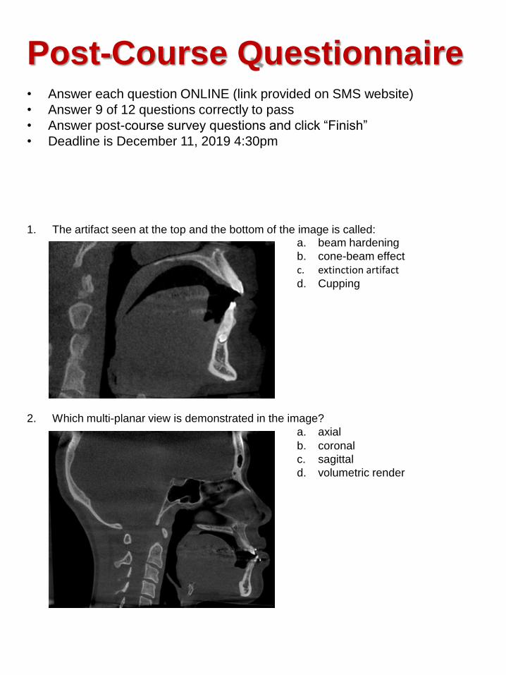
Post-Course Questionnaire• Answer each question ONLINE (link provided on SMS website)
• Answer 9 of 12 questions correctly to pass
• Answer post-course survey questions and click “Finish”
• Deadline is December 11, 2019 4:30pm
1. The artifact seen at the top and the bottom of the image is called:a. beam hardening
b. cone-beam effect
c. extinction artifactd. Cupping
2. Which multi-planar view is demonstrated in the image?
a. axial
b. coronal
c. sagittal
d. volumetric render
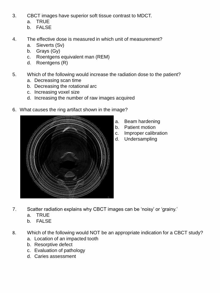
3. CBCT images have superior soft tissue contrast to MDCT.
a. TRUE
b. FALSE
4. The effective dose is measured in which unit of measurement?
a. Sieverts (Sv)
b. Grays (Gy)
c. Roentgens equivalent man (REM)
d. Roentgens (R)
5. Which of the following would increase the radiation dose to the patient?
a. Decreasing scan time
b. Decreasing the rotational arc
c. Increasing voxel size
d. Increasing the number of raw images acquired
6. What causes the ring artifact shown in the image?
a. Beam hardening
b. Patient motion
c. Improper calibration
d. Undersampling
7. Scatter radiation explains why CBCT images can be ‘noisy’ or ‘grainy.’
a. TRUE
b. FALSE
8. Which of the following would NOT be an appropriate indication for a CBCT study?
a. Location of an impacted tooth
b. Resorptive defect
c. Evaluation of pathology
d. Caries assessment
9. Which patient would be at the highest risk for adverse effects of radiation?
a. 60 year old male
b. 2 year old female
c. 8 year old maled. 30 year old female
10. CBCT images have superior spatial resolution to MDCT.
a. TRUE
b. FALSE
11. CBCT studies are recommended for all patients, regardless of treatment plan.
a. TRUEb. FALSE
12. The practitioner interpreting the scan would be liable for diagnosis of all significant
radiographic findings and is subject to the same standard as a trained oral and maxillofacial radiologist.
a. TRUE
b. FALSE
1. https://www.fda.gov/radiation-emitting-products/medical-x-ray-imaging/dental-cone-beam-computed-
tomography
2. https://cdn.ymaws.com/www.crcpd.org/resource/collection/BF80D39C-1287-4A8F-8617-0253743905D8/E-
17-6%20Technical%20White%20Paper%20-%20Cone%20Beam%20Compu.pdf
3. https://doi.org/10.3174/ajnr.A1653
4. Mallya, S. and Lam, E. W. N. (2019) White and Pharoah’s Oral Radiology: Principles and Interpretation. St.
Louis, Missouri: Elsevier.
5. https://journals.sagepub.com/doi/pdf/10.1177/ANIB_37_2-4
6. https://www.imagegently.org/Roles-What-can-I-do/Parent/Dentist
7. https://www.imagewisely.org/
8. https://www.birpublications.org/doi/10.1259/dmfr.20140197
9. http://www.ajnr.org/content/ajnr/30/6/1088.full.pdf
10. https://pubs.rsna.org/doi/10.1148/rg.246045065?url_ver=Z39.88-
2003&rfr_id=ori:rid:crossref.org&rfr_dat=cr_pub%3dpubmed
References