Embed Size (px)
Citation preview
5,6,9/99 Neuman Chapter 22
22: Peptides, Proteins, and -Amino Acids
Peptides
Protein Structure and Organization
Properties of -Amino Acids
Enzymes and Enzyme Catalysis
PreviewProteins are a major class of bioorganic molecules present in all organisms.They contain one or more polypeptide chains with the repeating generalstructure -(NH-CHR-C(=O))-. These repeating units come from 20 differentchiral -amino acids with the general structure H2N-CHR-CO2H. The R groups
play a major role in determining conformations of the peptide chains and theshapes of proteins. Free α-Amino acids are polyprotic acids because they have at
least two functional groups (CO2H and NH2) with acid and conjugate base forms.Some of these 20 α-amino acids also have an acid-base functional group in R.
Enzymes are proteins that catalyze biochemical reactions. These enzyme-catalyzed reactions take place in specific regions of the enzymes called active
sites.
22.1 PeptidesPeptides are polymers of -amino acids joined by amide or peptide bonds.
[graphic 22.1]
Peptide Structure (22.1A)Peptides with two -amino acid components are dipeptides, those with threeare tripeptides, and so on. Peptides with 3-10 α-amino acid components arecalled oligopeptides, while those with many α-amino acid components are
polypeptides. These terms are similar to the terms disaccharide,oligosaccharide, and polysaccharide that we learned when we studiedcarbohydrates (Chapter 20).
-Amino Acids in Peptides. Hydrolysis of peptides cleaves amide bonds(Chapters 15 and 16) releasing the individual α-amino acids. [graphic 22.2] α-
Amino acids in naturally occurring peptides generally have one R group and one
1
5,6,9/99 Neuman Chapter 22
H on Cα (H2N-CαHR-CO2H). We will see later in this chapter that the NH2 andCO2H groups of free α-amino acids exist as NH3+ and CO2- so the general
structure of these amino acids is actually +H3N-CHR-CO2-. [graphic 22.3]
-Amino Acids Can be D or L. When R is a group other than H, these α-
amino acids are chiral compounds with two enantiomeric forms because Cα ischiral (Chapter 4). We can identify the configuration of Cα as R or S if we knowthe structure of the Cα-R group, but we usually describe the two enantiomers asD or L. [graphic 22.4] Most α-Amino acids in naturally occurring peptides have
L-configurations.
The Definition of D and L for -Amino Acids. Definitions of D and L for
enantiomers of α-amino acids are based on those of D and L-glyceraldehyde (Chapter
20). [graphic 22.5] We define C(=O)O- of the amino acid as equivalent to C(=O)H of
glyceraldehyde (both have C=O) and place it at the top of the α-amino acid Fischer
projection. We define R of the amino acid as equivalent to CH2OH of glyceraldehyde and
place it at the bottom of the Fischer projection. The result is a pair of enantiomers with
NH3+ to the right (defined as D) or to the left (defined as L) on Cα. Although we will see
that one α-amino acid (proline, see Figure (graphic 22.6)) has an additional bond
between the aminium group and R (+H2N-R), its D and L assignments similarly depend
on the "right" or "left" orientation of its Cα-N bond.
The R Groups. While α-amino acids have many different R groups, we areinterested in the 20 R groups of the "standard" α-amino acids (Figure (graphic
22.6)) in naturally occurring peptides. [graphic 22.6] There are a few other Rgroups in naturally occurring peptides, but these "nonstandard" groups arisefrom biosynthetic modification of a "standard" R group already present in apeptide. [graphic 22.7] You can see that "standard" R groups are diverse. Theyinclude alkyl and aryl groups, heterocyclic rings, sulfur containing groups,alcohols, aminium ions, carboxylates, and amides. The R group of proline differsfrom the others because it chemically binds to its α-amino group. Isoleucine and
threonine each have an additional chiral C* (C3) in their R groups (Figure(graphic 22.6)). The naturally occurring stereoisomer of L-isoleucine is S at C3,while C3 of L-threonine is R. [graphic 22.9][graphic 22.8]
2
5,6,9/99 Neuman Chapter 22
Categories of "Standard" Amino Acids. In order to learn their names andR group structures, it is helpful for us to group the "standard" amino acids in thethree categories shown in Figure (graphic 22.6). R groups of the 5 "charged
polar" amino acids are electrically charged (- or +) at physiological pH values(about pH 7), while those of the remaining 15 amino acids are electrically neutralunder the same conditions. R groups of the 5 "uncharged polar" amino acidsform hydrogen bonds to each other and to water, while R groups of the"nonpolar" amino acids do not form hydrogen bonds.
Abbreviated Names. Each "standard" amino acid name has a three-letter
abbreviation as well as a one-letter designation (Figure (graphic 22.6)). The three-letter abbreviations are usually the first three letters of the full name, while one-letter designations correspond to the first letter of the name where possible. Youwill often see three-letter abbreviations used interchangeably with full names.
Glx and Asx. The abbreviation Glx refers to both Glu and Gln, and Asx refers to both
Asp and Asn. Amide groups of Gln and Asn sometimes hydrolyze to carboxylate groups
of Glu and Asp during amino acid analysis (described later in the chapter). [graphic
22.10] As a result, the relative amounts of carboxylate and amide side chains in a
naturally occurring peptide may be uncertain so they are grouped together as Glx or Asx.
Peptide Synthesis (22.1B)Laboratory syntheses of peptides make use of familiar reactions illustrated below.Biosynthesis of peptides involves nucleic acids so we defer this topic until Chapter23.
General Considerations. If we wish to make a dipeptide from two aminoacids (AAx and AAy), we must recognize that it can have two possible structures(AAx-AAy and AAy-AAx). [graphic 22.11] Chemists write peptide formulas sothat the first amino acid in the sequence is N-terminal (it has the unreactedNH2 group), while the last one is C-terminal (it has the terminal CO2H group).As a result, AAx is the N-terminal amino acid in AAx-AAy while AAy is the N-terminal amino acid in AAy-AAx. Each amino acid has an amino group and acarboxylic acid group, so we might expect that direct reactions of AAx and AAy (orof appropriate derivatives) may give not just AAx-AAy and AAy-AAx, but AAx-AAx and AAy-AAy as well [graphic 22.12] Since the number of possible
3
5,6,9/99 Neuman Chapter 22
combinations of amino acids increases rapidly as the size of the desired peptideincreases, chemists do not use direct reactions of amino acids to make peptides.
Automated Peptide Synthesis. We can avoid the problem of multipleproducts by using automated peptide synthesis. We illustrate its generalfeatures here for the synthesis of the tripeptide AAz-AAy-AAx: [graphic 22.13]
(1) Chemically bind AAx to a solid insoluble resin.(2) Couple AAy to AAx-Resin.(3) Couple AAz to AAy-AAx-Resin.(4) Remove AAz-AAy-AAx from the resin.
To accomplish these general steps we protect and deprotect NH2 groups andactivate CO2H groups. One way chemists protect NH2 groups is with tert-butyloxycarbonyl (t-Boc) groups from tert-butyloxycarbonyl chloride (t-
BocCl). [graphic 22.14] Trifluoroacetic acid in methylene chloride deprotects
NH2 groups by removing the t-Boc groups. We activate CO2H groups on N-protected amino acids (t-Boc-AA) by treating them withdicyclohexylcarbodiimide (DCCD). [graphic 22.15] The resulting carboxylicacid derivatives (C(=O)-O-DCCD) react readily with NH2 groups to form amidebonds (C(=O)-NH).
The detailed steps of automated peptide synthesis are:(1) Attach AAx to the Solid Support
Couple CO2H groups of N-protected AAx (t-Boc-AAx) with benzyl chloridegroups (Ar-CH2-Cl) on the resin solid support to form esters. [graphic 22.16]Deprotect t-Boc-AAx-Resin to give reactive NH2 group on each AAx.
(2) Couple AAy to AAx
Activate N-protected AAy (t-Boc-AAy) using DCCD and couple it with AAx-Resin. [graphic 22.17] An amide bond forms between AAy and AAx leading tot-Boc-AAy-AAx-Resin. Subsequent deprotection gives AAy-AAx-Resin with areactive NH2 group on each AAy.
4
5,6,9/99 Neuman Chapter 22
(3) Couple AAz to AAy
Activate N-protected AAz (t-Boc-AAz) using DCCD and couple it with AAy-AAx-Resin. [graphic 22.18] An amide bond forms between AAz and AAy
leading to t-Boc-AAz-AAy-AAx-Resin.
(4) Isolate AAz-AAy-AAx
Treat t-Boc-AAz-AAy-AAx-Resin with liquid HF cleaving t-Boc from AAz, aswell as the ester bond between AAx and the resin, giving free AAz-AAy-AAx.[graphic 22.19]
Protection of Functional Groups on R. We must also protect functional groups such
as NH2, OH, SH, and CO2H on amino acid side chains (R) during peptide synthesis by
converting them into derivatives such as benzyl groups. [graphic 22.20] These benzyl
groups cleave along with the t-Boc group and the ester linkage to the resin during
treatment with liquid HF in Step 4.
22.2 Protein Structure and OrganizationProteins are biological molecules made up of one or more polypeptides. Theamino acids of the polypeptides, and their relative order in the polypeptide, arethe protein's primary (1 ) structure. The 3-dimensional structure of localizedsegments of the protein's polypeptides is secondary (2 ) structure, while theoverall shape of a protein is its tertiary (3 ) structure. Quaternary (4 )
structure describes the way that the individual polypeptides interact with eachother in a protein with multiple polypeptides.
Primary (1 ) Structure (22.2A)
Amino acid content and amino acid sequence are our first concern inelucidation of the structure of a protein.
Content. We identify and quantify the amino acids in a polypeptide using anautomated amino acid analyzer that performs the following steps illustratedin Figure (graphic 22.21). [graphic 22.21]
(1) Hydrolysis of the polypeptide to its component amino acids by heating instrong acid (6 M HCl) or strong base (2 to 4 M NaOH).
5
5,6,9/99 Neuman Chapter 22
(2) Separation of the resulting mixture of amino acids into its individualamino acids by column chromatography.
(3) Derivatization of the amino acids so they are detected by spectroscopicmethods and displayed in a spectrum. [graphic 22.22] (Sometimesderivatization is done before chromatographic separation (step 2)).
(4) Identification of each spectral signal in the spectrum as that of a specificamino acid and determination of the amount of amino acid present from thesignal intensity.
Sequence. We determine the sequence of amino acids in a polypeptide byidentifying its component amino acids one at a time starting at its N-terminus orC-terminus. An important end-group analysis procedure for N-terminal aminoacids is Edman Degradation that first derivatizes and then cleaves the N-terminal amino acid of a polypeptide leaving the rest of the chain intact. [graphic22.23] Automated sequenators carry out N-terminal amino acid derivatization,cleavage, and analysis in a repetitive stepwise fashion allowing accuratedetermination of as many as 100 amino acid units in a peptide chain.
We can analyze C-terminal amino acids using carboxypeptidase enzymes.Such enzymes specifically hydrolyze (cleave) the peptide bond between the C-terminal amino acid and the rest of the chain. [graphic 22.24] They are usefulfor analyzing only the first few amino acids at the C-terminal end of a peptidebecause they cleave different C-terminal amino acids at different rates. As aresult, C-terminal amino acids of shortened chains formed during the analysisprocedure may cleave more rapidly than those of the original polypeptide. Thiscan quickly complicate the reaction mixture with a variety of individual aminoacids and shortened peptide chains. Aminopeptidase enzymes similarlyoperate on the N-terminal end of a peptide and have the same limitations ascarboxypeptidases.
When peptide chains are too long for accurate determination of their fullsequence by a method such as Edman degradation, we can cleave them intosmaller peptides using specific peptide cleavage reactions. We determine
6
5,6,9/99 Neuman Chapter 22
the sequences of these smaller peptides and use them to reconstruct the originalsequence of the complete peptide. One of the most specific peptide cleavagereactions uses the enzyme trypsin to cleave the peptide bond in CH(Rx)C(=O)—NHCH(Ry) where Rx is from Lys or Arg, while Ry is from any amino acid exceptPro. As a result, the C-terminus of each cleaved segment is Lys or Arg, with theexception of the segment with the original C-terminus. [graphic 22.25]
More about Enzymatic Peptide Cleavage. The carboxypeptidases and
aminopeptidases that we have just described are exopeptidases because they cleave
the terminal amide bonds connecting N-terminal or C-terminal amino acids to the rest of
the peptide chain. In contrast, trypsin is one of several enzymes that cleave amide
bonds "inside" the peptide chain (endopeptidases). Others are -chymotrypsin,
elastase, and pepsin. Enzymes are proteins that catalyze specific types of reactions.
We will discuss enzyme catalysis in the last section of this chapter.
Separation of Individual Peptide Chains. Polypeptide chains of proteinswith two or more peptide chains often covalently bind to each other with disulfidebonds (S-S) formed from neighboring SH groups of the amino acid Cys. [graphic22.26] In order to determine content and sequence of individual polypeptidechains of such proteins, we must break these disulfide bonds by reduction oroxidation. We then separate the chains using denaturing agents and isolatethem using chromatography. We describe denaturing agents later in this chapter.
Secondary (2 ) Structure (22.2B)Secondary (2 ) protein structure includes such localized structural features ofpeptide chains as the -helix, the -pleated sheet, and the planarity of amide
groups.
Planarity of Amide Groups. We learned in Chapter 15 that amide groupshave significant electron delocalization. [graphic 22.27] This causes them to beplanar since rotational barriers about their C-N bonds are 60 to 75 kJ/mol(rotational barriers about C-N single bonds are 12 to 20 kJ/mol). Whilesubstituents on amide bonds are cis or trans, the most favorable conformation inpeptides is that where Cα's of the chain are trans. As a result, we can representpeptide chains as a series of "planes", containing the amide groups, that connect
7
5,6,9/99 Neuman Chapter 22
at the Cα's. [graphic 22.28] Since these planes have a restricted range of relativeorientations, amide groups impart significant rigidity to a polypeptide.
Helical Structures. The -helix structure that Pauling and Corey proposed
in 1951 is a result of repeating planar trans amide groups in peptides. The C=Oof each amide group in the α-helical region hydrogen bonds to an N-H of another
amide group. Two intervening amide groups separate the hydrogen bondedamide groups leading to a favorable distance of 2.8 Å between O and N in theresulting C=O.....H-N hydrogen bonds. [graphic 22.29] Since Cα's have L-configurations, the α-helix adopts a right-handed twist so that the Cα-R groupspoint away from the chain. α-Helical regions contain on the order of 12 amino
acid residues.
-Pleated Sheets. Segments of peptide chains separated by manyintervening amino acids also hydrogen bond to each other to give -pleated sheets.[graphic 22.30] These -pleated sheets, that Pauling and Corey also proposed in
1951, are parallel or antiparallel and the hydrogen bonded segments can befrom the same or different peptide chains. A "sheet" includes 2 to 15 peptidechain segments that average about 6 amino acid residues in length. You can seefrom the figure that the C=O....H-N bonds in parallel β-pleated sheets are
distorted and they are less stable than those in antiparallel sheets where there isno distortion.
Other Structures. -Helical regions and -pleated sheets connect using otherpeptide structures called coils or loop conformations. -Helices and -pleated
sheets make up about 50% of peptide chains.
Tertiary (3 ) Structure (22.2C)We generally describe proteins at the 3 structural level as globular or fibrous.Proteins of both types contain all of the 2° structural elements we have just
described.
Fibrous Proteins. The fibrous protein of skin, bone, and muscle has anelongated "fiber-like" shape. An example is -keratin of hair, horns, and nails.[graphic 22.31] Two α-helical polypeptide chains wind together and group with
other polypeptide dimers to form protofilaments. Two protofilaments
8
5,6,9/99 Neuman Chapter 22
comprise a protofibril and four protofibrils wind together in a microfibril.Microfibrils cluster to form macrofibrils which come together in groups to formcells. A strand of hair includes many cells grouped together in a sheath.
Globular Proteins. The shapes of globular proteins also reflect their name.Most enzymes are globular proteins, and there are many others such as thefamiliar oxygen transport proteins myoglobin and hemoglobin. [graphic 22.32]Myoglobin has one peptide chain with 153 amino acid residues surrounding aheme group containing an Fe(II) atom. In muscle tissue, it accepts O2 fromoxygenated hemoglobin in blood. Hemoglobin has four polypeptides with hemegroups that have globular tertiary structures like myoglobin. Hemoglobin bindsoxygen in the lungs, transports it to myoglobin, and returns to the lungs with CO2
formed as a metabolic product in muscle tissue.
Factors that Determine Protein Shape (22.2D)The 3° structure of a protein depends on the interactions of the amino acid side
chains (the R groups) with each other and with the medium surrounding theprotein. These interactions involve electrostatic forces, hydrogen bonding,hydrophobic bonding, and disulfide bonds. [graphic 22.33] We will see belowthat the same forces also bind together the multiple peptide chains of a proteinin its quaternary (4 ) structure.
Hydrophobic Bonding. A major influence on the shape and stability of aprotein is the desire of nonpolar amino acid side chains to minimize theirexposure to H2O. This leads them to the interior of a protein where they interactwith each other by hydrophobic bonding. For example, nonpolar alkyl groupsinduce complementary dipoles in each other. While they are individually weak,these attractive induced dipolar interactions taken together have a major effecton the shape and stability of a protein (Figure (graphic 22.33)). Side chains withpermanent dipolar groups also interact attractively with each other and inducedipoles in nonpolar groups as well.
Thermodynamics of Hydrophobic Bonding. You may be surprised to learn that
the enthalpy (H) of a hydrocarbon in water is more favorable (lower) than the enthalpy of
the same hydrocarbon in a hydrocarbon medium. In spite of this, hydrocarbons prefer to
dissolve in hydrocarbons and not water because the entropy (S) of a hydrocarbon in
9
5,6,9/99 Neuman Chapter 22
water is less favorable (much lower) than it is in a hydrocarbon medium. Water
molecules form highly structured cages around hydrocarbon molecules leading to a
decrease in volume of the aqueous solution and a decrease in the entropy of the system.
The net result of these effects on H and S is that the free energy (G) for the hydrocarbon
in water is greater (less favorable) than in a hydrocarbon medium. The unfavorable
T∆S(water→hydrocarbon) term overwhelms the favorable ∆H(water→hydrocarbon) term in
the equation ∆G = ∆H - T∆S so that ∆G(water→hydrocarbon) is unfavorable.
Electrostatic Interactions and Hydrogen Bonding. In contrast toindividually weak hydrophobic interactions, ionic interactions between charged
polar side chains within a protein are individually strong (Figure (graphic22.33)). However, since charged polar groups readily hydrogen bond to H2O onthe exterior of a protein, these groups predominate on the exterior surface of aprotein. Uncharged polar groups such as OH and C(=O)NH2 form hydrogenbonds with each other in the interior of a protein, but they also hydrogen bond toH2O molecules on the surface of proteins and are present in both locations.
Disulfide Bonds. The SH groups of Cys stabilize and determine the shape ofproteins by forming disulfide bonds (S-S). Their effect on the shapes of proteinsis the basis of the "permanent wave" process that straightens or curls hair.[graphic 22.34] Application of a reducing agent to the hair breaks S-S bonds togive individual SH side chains. With disulfide bonds broken, curlers orstraighteners mechanically modify the shape of the hair. Treatment with anoxidizing agent reforms S-S bonds that hold the hair in a new shape. Disulfidebonds periodically cleave and reform over time allowing the strands of hair toslowly resume their natural configuration.
Quaternary (4 ) Structure. Most proteins have more than one polypeptide
chain and the ways that these multiple chains interact with each otherdetermine the quaternary (4 ) structure of the protein. The forces that hold these
individual polypeptide chains together are the same as those that determineprotein 3° structure. Hemoglobin (Figure (graphic 22.32)) has four peptidesubunits that come together to form its 4 structure.
Denaturation. We often refer to a protein with the same shape and activitythat it has in an organism as the native state or native form of the protein.
10
5,6,9/99 Neuman Chapter 22
Denaturation of a protein involves a change in the 4°, 3°, and/or 2° structure of
the native state. [graphic 22.35] While some proteins denature and then returnto their native state (reversible denaturation), 2°, 3°, and/or 4° structures of
many proteins are sufficiently fragile that denaturation is irreversible.Biochemists studying protein structure and function try to avoid proteindenaturation, except to separate individual peptide chains of a protein forcontent and sequence determination.
Native proteins are often just slightly more thermodynamically stable thandenatured proteins, so denaturation frequently occurs with relative ease.Denaturating agents, or conditions, include temperature increases, pH changes,detergents, water soluble organic compounds such as alcohols and urea, and ionssuch as ClO4-, SCN-, Ca+2, Ba+2, and guanidinium ion. [graphic 22.36] Atemperature increase provides energy to overcome attractive forces between Rgroups, while a change in pH alters the charge on acidic or basic side chains (nextsection) and the attractive interactions between R groups. The chemical agentsprovide intermolecular competition with the intramolecular attractive forcesbetween R groups in the native form.
22.3 Properties of -Amino AcidsWe have learned the structures of the 20 "standard" amino acids that organismsuse to make peptides and proteins, and have just seen how these R groupsinfluence the structures of peptides and proteins. In this section we examine theproperties of α-amino acids as acids and bases as well as their synthetic and
biosynthetic origins.
-Amino Acids Are Polyprotic Acids (22.3A)The "standard" α-amino acids are diprotic ("H2A") or triprotic acids ("H3A").
Diprotic -Amino Acids. Most "standard" α-amino acids are diprotic acids
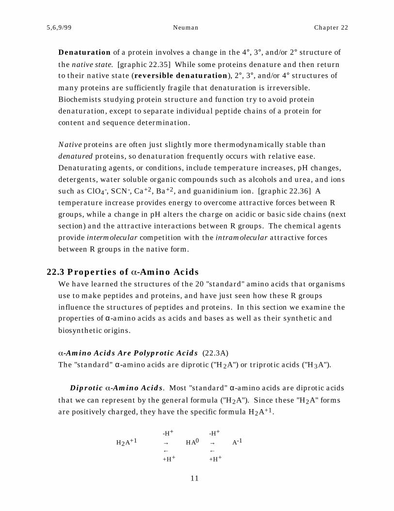
that we can represent by the general formula ("H2A"). Since these "H2A" formsare positively charged, they have the specific formula H2A+1.
-H+ -H+
H2A+1 → HA0 → A-1← ←+H+ +H+
11
5,6,9/99 Neuman Chapter 22
H2A+1 is in equilibrium with the uncharged monoprotonated form HA0 and it isin equilibrium with the unprotonated form A-1. An example is glycine whereH2A+1 is +H3N-CH2-CO2H, and A-1 is H2N-CH2-CO2- (NH3+ and CO2H haveeach lost a proton). [graphic 22.37] While these are the only possible structuresfor H2A+1 and A-1, HA0 could be either the zwitterion form +H3N-CH2-CO2-, orthe uncharged form H2N-CH2-CO2H. We stated at the beginning of the chapterthat the structure of HA0 is the zwitterion form. This seems reasonable since wewould expect a basic NH2 group and acidic CO2H group in the uncharged
structure to react with each other to form the NH3+ and CO2- groups of thezwitterion form. [graphic 22.38]
Quantitative support for this notion comes from pKa1 = 2.3 for the equilibriumbetween H2A+1 and HA0, and pKa2 = 9.6 for the equilibrium between HA0 and A-
1. [graphic 22.39] If HA0 is the zwitterion , pKa1 = 2.3 is that for ionization ofCO2H in H2A+1. However, if HA0 is the uncharged form, pKa1 = 2.3 is that forionization of the NH3+ group in H2A+1. Simple carboxylic acids (RCH2CO2H)have pKa's ≈ 5, while simple aminium ions (RCH2NH3+) have pKa's between 10
and 11. We can see that pKa1 = 2.3 is much closer to that for CO2H ionization(pKa ≈ 5) than for NH3+ ionization (pKa ≈10 to 11). This means that pKa2 = 9.6
corresponds to ionization of NH3+ in the zwitterion form of HA0 consistent withthe pKa ≅10 to 11 for ionization of NH3+ in a simple aminium ion (RCH2NH3+).
The lower pKa for ionization of CO2H in +H3N-CH2-CO2H, compared toionization of CO2H in RCH2CO2H, results from the electron withdrawing
inductive effect of the positively charged NH3+ group (Chapter 14) that stabilizes+H3N-CH2-CO2-. This effect causes the energy difference between +H3N-CH2-CO2H and +H3N-CH2-CO2- to be less than that between RCH2CO2H andRCH2CO2- resulting in a lower pKa for the amino acid. [graphic 22.40] BesidesGly, there are 12 other diprotic "standard" amino acids (Table 22.1). [Table 22.1,next page] Their values of pKa1 (Cα-CO2H) and of pKa2 (Cα-NH3+) areremarkably similar to those of Gly (an exception is pKa2 for Pro that isstructurally different from the others). The average value of pKa1 is about 2.2while that of pKa2 (excluding proline) is about 9.3. We will discuss the pI valueslater in this section.
12

5,6,9/99 Neuman Chapter 22
Diprotic Amino Acid Forms at Different pH Values. The pKa values forthe H2A+1, HA0, and A-1 forms of the 13 diprotic amino acids allow us to predictthe pH dependence of their concentrations. [graphic 22.41]
(1) H2A+1 (+H3N-CHR-CO2H) is the major form in solution at pH valuessmaller than pKa1 (below approximately 2.2).
(2) The concentrations of H2A+1 [+H3N-CHR-CO2H] and HA0 [+H3N-CHR-CO2-] are equal when the solution pH is equal to pKa1 (at approximately 2.2).
Table 22.1. Acid Dissociation Constants for Diprotic Amino Acids
Name pKa1 (Cα-CO2H) pKa2 (Cα-NH3+) pI
Nonpolar Ralanine 2.3 9.7 6.0glycine 2.3 9.6 6.0isoleucine 2.4 9.6 6.0leucine 2.4 9.6 6.0methionine 2.3 9.2 5.7phenylalanine 1.8 9.1 5.5proline 2.0 10.6 6.3tryptophan 2.8 9.4 5.9(?)valine 2.3 9.6 6.0
Uncharged Polar Rasparagine 2.0 8.8 5.4glutamine 2.2 9.1 5.7serine 2.2 9.2 5.7threonine 2.1 9.1 5.6
Average (2.2) (9.3)* (5.8)** Excluding proline
_______________________________________
(3) HA0 (+H3N-CHR-CO2-) is the major form when the solution pH is betweenpKa1 and pKa2 (between approximately 2.2 and 9.3)
(4) The concentrations of HA0 [+H3N-CHR-CO2-] and A-1 [H2N-CHR-CO2-]are equal when the solution pH is equal to pKa2 (approximately 9.3).
(5) A-1 (H2N-CHR-CO2-) is the major form when the solution pH is greaterthan pKa2 (above approximately 9.3).
13
5,6,9/99 Neuman Chapter 22
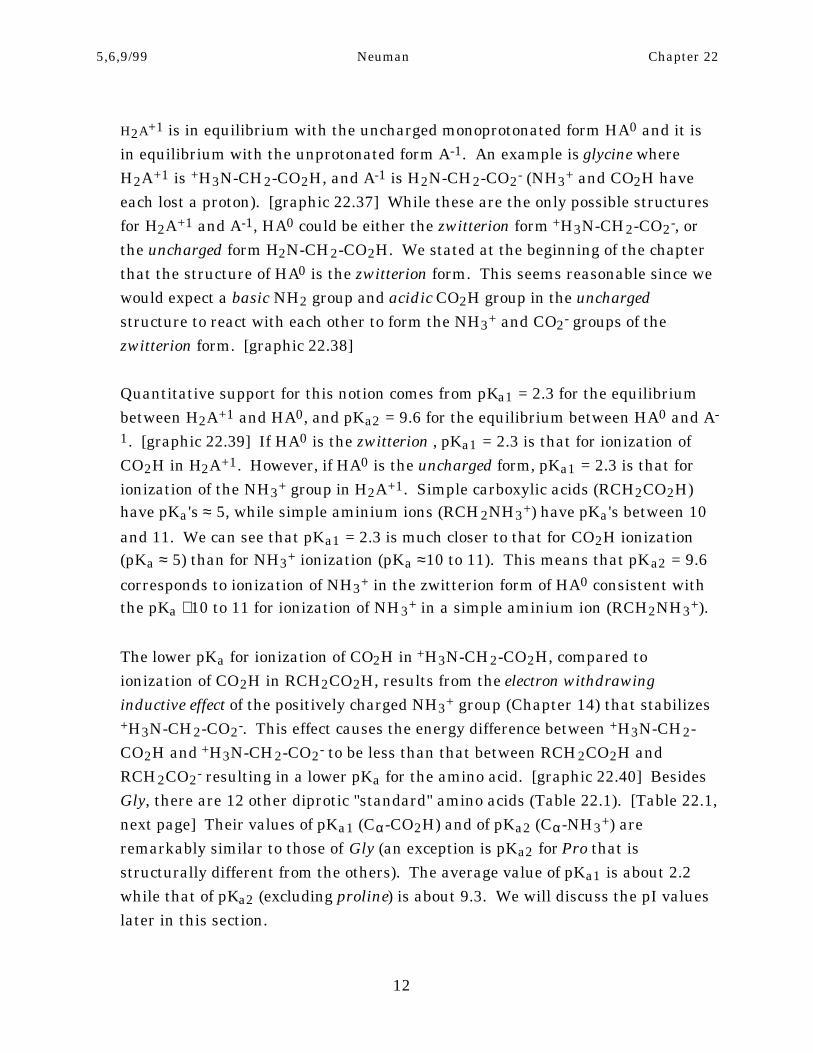
Triprotic -Amino Acids. The remaining 7 "standard" amino acids (Table
22.2) are triprotic acids ("H3A") because their R's have acidic or basic functionalgroups in addition to the Cα-CO2H and Cα-NH3+ groups. [Table 22.2] Thecharges on these forms depend on the specific R group.
-H+ -H+ -H+"H3A" → "H2A" → "HA" → "A"
← ← ←+H+ +H+ +H+
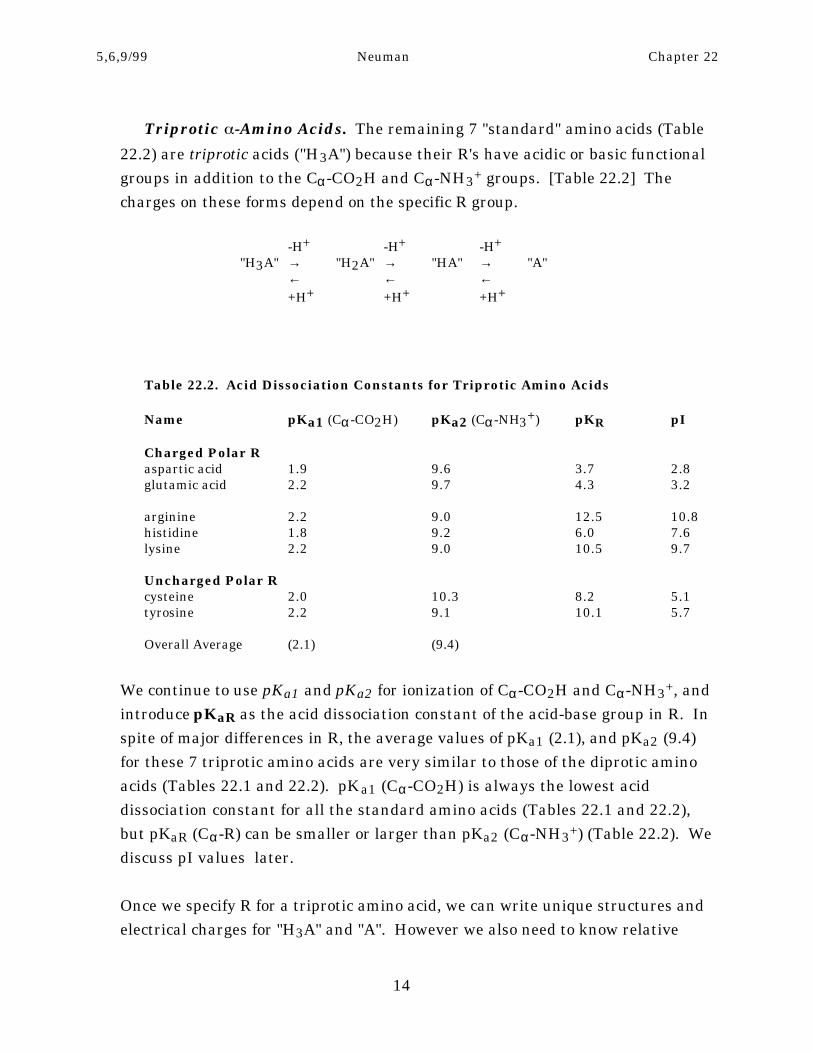
Table 22.2. Acid Dissociation Constants for Triprotic Amino Acids
Name pKa1 (Cα-CO2H) pKa2 (Cα-NH3+) pKR pI
Charged Polar Raspartic acid 1.9 9.6 3.7 2.8glutamic acid 2.2 9.7 4.3 3.2
arginine 2.2 9.0 12.5 10.8histidine 1.8 9.2 6.0 7.6lysine 2.2 9.0 10.5 9.7
Uncharged Polar Rcysteine 2.0 10.3 8.2 5.1tyrosine 2.2 9.1 10.1 5.7
Overall Average (2.1) (9.4)
We continue to use pKa1 and pKa2 for ionization of Cα-CO2H and Cα-NH3+, andintroduce pKaR as the acid dissociation constant of the acid-base group in R. Inspite of major differences in R, the average values of pKa1 (2.1), and pKa2 (9.4)for these 7 triprotic amino acids are very similar to those of the diprotic aminoacids (Tables 22.1 and 22.2). pKa1 (Cα-CO2H) is always the lowest aciddissociation constant for all the standard amino acids (Tables 22.1 and 22.2),but pKaR (Cα-R) can be smaller or larger than pKa2 (Cα-NH3+) (Table 22.2). Wediscuss pI values later.
Once we specify R for a triprotic amino acid, we can write unique structures andelectrical charges for "H3A" and "A". However we also need to know relative
14
5,6,9/99 Neuman Chapter 22
values of pKaR and pKa2 in order to write structures and electrical charges for"H2A" and "HA". Because their structures and charges depend on R, we willexamine the triprotic amino acids in three separate categories based on their Rgroups.
Aspartic and Glutamic Acid. Glu and Asp each have an acidic CO2H groupin R. Since they differ only in the number of CH2 groups connecting that CO2H toCα, their acid dissociation equilibria and associated pKa values are very similar.[graphic 22.42] Their pKaR values (≈ 4) are closer to pKa's of simple carboxylicacids (≈ 5) than pKa1 values (≈ 2) because one or two CH2 groups separate the
CO2H groups in R from NH3+. The relative concentrations of the four protonatedforms of Glu or Asp depend on pH as illustrated in Figure (graphic 22.43) andtheir specific electrical charges are H3A+1, H2A0, HA-1, andA-2. [graphic 22.43] At physiological pH (approximately pH 7), their majorforms are HA-1 where the R grtoups (CH2CO2- or CH2CH2CO2-) have anelectrical charge of -1.
Lysine, Arginine, and Histidine. In contrast to Asp and Glu, the threeamino acids Lys, Arg, and His all have basic functional groups in R. That group inLys is NH2, Arg has a guanidino group (NHC(=NH)NH2), while His has animidazole ring. [graphic 22.44] These groups are all positively charged whenthey are protonated (Figure (graphic 22.44)) so their four forms are H3A+2,H2A+1, HA0, and A-1. Although each R is basic, the pKaR values are significantlydifferent (Table 22.2). The pKa order for Lys and Arg is pKa1 < pKa2 < pKaR,while that for His is pKa1 < pKaR < pKa2. We illustrate the relativeconcentrations of their four forms as a function of pH in Figure (graphic 22.45).[graphic 22.45] At approximate physiological pH 7, the electrical charge on the Rgroups of Arg and Lys is +1. In contrast, the R of His is predominantly neutral atpH 7, but a significant amount of the protonated +1 form is also present.(Remember that R in Glu and Asp is -1 at pH 7.)
Cysteine and Tyrosine. The remaining triprotic amino acids are Cys andTyr. While their R groups are acidic, they are much weaker acids than the CO2Hgroups of Glu and Asp. [graphic 22.46] The SH group of Cys has an approximatepKaR of 8, while the phenol group of Tyr has an approximate pKaR of 10. As aresult, the pKa order for Cys is pKa1 < pKaR < pKa2 while that for Tyr is pKa1 <
15
5,6,9/99 Neuman Chapter 22
pKa2 < pKaR. In spite of the differences in pKa order for these two amino acids,the net electrical charges on the four forms of each of them are H3A+1, H2A0, HA-
1, and A-2. We show their relative concentrations as a function of pH in Figure(graphic 22.47). You can see that at pH 7 their R groups predominantly exist intheir uncharged protonated forms (CH2-SH or CH2-Ph-OH). [graphic 22.47]
Isoelectric Points (22.3B)The isoelectric point (pI) for any amino acid is the pH value where theelectrically neutral form has its highest concentration. pI values allow us topredict the behavior of amino acids in electrical fields. When we place a solutionof amino acids between positive and negative electrodes, amino acids with a net(+) charge migrate toward the negative electrode, those with a net (-) charge
migrate toward the positive electrode, while those that are uncharged do notmigrate. We can adjust the direction of migration of amino acids in an electricfield by raising or lowering pH with respect to their pI values in order to facilitatetheir separation in solution.
pI Values of Diprotic Amino Acids. We have seen that the electricallyneutral form of all diprotic amino acids is HA0. It is the major form at pH valuesbetween pKa1 and pKa2 (Figure (graphic 22.41)) and its maximum concentrationis at a pH (its pI value) midway between these two pKa values. For example, thepI for glycine (R = H) is 6.0 (pIGly = (pKa1 + pKa2)Gly/2 = (2.3 + 9.6)/2 = 6.0). pI'sof all the other diprotic amino acids are also about 6 since their pKa1 and theirpKa2 values are very similar (Table 22.1).
pI Values of Triprotic Amino Acids. In contrast to diprotic amino acids,the electrically neutral forms and pI values of triprotic amino acids depend on thestructure of R (Table 22.2). To a good approximation, the highest concentrationof the electrically neutral form (HnA0) of any triprotic acid occurs at a pH midwaybetween the two pKa's associated with that neutral form (pKa(n+1) and pKa(n)).[graphic 22.48] As a result, Asp and Glu have relatively low pI values of about 3(see Figure (graphic 22.43)), Arg, Lys, and His have relatively high pI valuesranging from 8 to 11 (see Figure (graphic 22.45)), and Cys and Tyr have pI valuesbetween 5 and 6 (see Figure (graphic 22.47)).
16
5,6,9/99 Neuman Chapter 22
pI Values of Proteins. A protein also has a pI value equal to the pH where the
number of its positively charged (+) R groups (from Arg, Lys, and His) is exactly equal to
the number of its negatively charged (-) side chains (from Asp, Glu, Cys and Tyr). In
contrast to amino acids, protein pI values cover the broad range from <1 to >12 because
pKa's of their R groups depend on whether R is folded inside or outside the protein and
how it interacts with other R's. Proteins often have their lowest solubility at pH = pI
and biochemists use this behavior to facilitate their isolation and purification.
Laboratory Synthesis of Amino Acids (22.3C)Chemists use the following reactions to synthesize α-amino acids in the
laboratory. They give racemic mixtures of the D and L enantiomers, so resolutionis required to obtain individual enantiomers.
Amination of -Bromo acids (Chapter xx).Br2 NH3
RCH2CO2H → RCH(Br)CO2H → RCH(NH2)CO2HPBr3
Strecker Synthesis (Chapter xx). KC≡N H3O+
RCH(=O) → RCH(NH2)C≡N → RCH(NH2)CO2HNH4+
Reductive Amination (Chapter xx). NH3
RC(=O)CO2H → RCH(NH2)CO2HNaBH4
Diethylacetamidomalonate Synthesis. (1) EtO- CH3C(=O)-HN-CH(CO2Et)2 → CH3C(=O)-HN-C(R)(CO2Et)2
(2) R-Br
H3O+
CH3C(=O)-HN-C(R)(CO2Et)2 → CH3C(=O)OH + +H3N-C(R)(CO2H)2
+H3N-C(R)(CO2H)2 → +H3N-CH(R)CO2H + CO2
17
5,6,9/99 Neuman Chapter 22
Biosynthesis of -Amino Acids (22.3D)
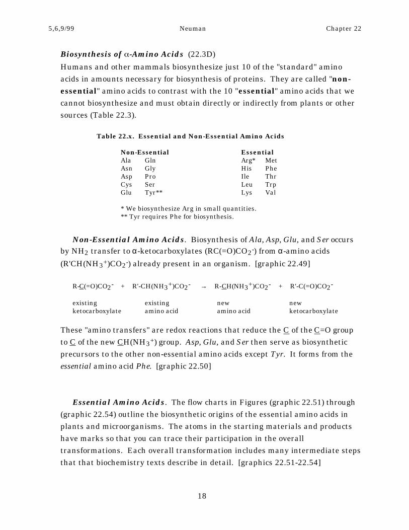
Humans and other mammals biosynthesize just 10 of the "standard" aminoacids in amounts necessary for biosynthesis of proteins. They are called "non-
essential" amino acids to contrast with the 10 "essential" amino acids that wecannot biosynthesize and must obtain directly or indirectly from plants or othersources (Table 22.3).
Table 22.x. Essential and Non-Essential Amino Acids
Non-Essential EssentialAla Gln Arg* MetAsn Gly His PheAsp Pro Ile ThrCys Ser Leu TrpGlu Tyr** Lys Val
* We biosynthesize Arg in small quantities.** Tyr requires Phe for biosynthesis.
Non-Essential Amino Acids. Biosynthesis of Ala, Asp, Glu, and Ser occursby NH2 transfer to α-ketocarboxylates (RC(=O)CO2-) from α-amino acids
(R'CH(NH3+)CO2-) already present in an organism. [graphic 22.49]
R-C(=O)CO2- + R'-CH(NH3+)CO2- → R-CH(NH3+)CO2- + R'-C(=O)CO2-
existing existing new newketocarboxylate amino acid amino acid ketocarboxylate
These "amino transfers" are redox reactions that reduce the C of the C=O groupto C of the new CH(NH3+) group. Asp, Glu, and Ser then serve as biosyntheticprecursors to the other non-essential amino acids except Tyr. It forms from theessential amino acid Phe. [graphic 22.50]
Essential Amino Acids. The flow charts in Figures (graphic 22.51) through(graphic 22.54) outline the biosynthetic origins of the essential amino acids inplants and microorganisms. The atoms in the starting materials and productshave marks so that you can trace their participation in the overalltransformations. Each overall transformation includes many intermediate stepsthat that biochemistry texts describe in detail. [graphics 22.51-22.54]
18
5,6,9/99 Neuman Chapter 22
22.4 Enzymes and Enzyme CatalysisEnzymes are proteins that catalyze biochemical reactions in organisms. We willfirst examine general aspects of enzymes and enzyme catalysis and then thespecific mechanistic details of catalysis by -chymotrypsin.
General Features (22.4A)All enzymes and enzyme catalyzed reactions share a number of general features.
Active Sites. In enzyme catalyzed reactions, the reactant biomolecule(substrate) binds to a region of the enzyme called its active site. The active site
is an indentation or cleft in the enzyme where R groups on amino acid residuesinteract with the substrate by noncovalent attractive forces. [graphic 22.55]These attractive forces include hydrophobic bonding, hydrogen bonding, andelectrostatic interactions. They are the same as those we described earlier forinteractions between R groups of polypeptides.
Enzyme Catalysis Mechanism. Once in the active site, a series of reactionstransforms the substrate into product. These reactions may use amino acid Rgroups in the active site as reagents, as well as other reactants that diffuse intothe active site. The general scheme involves reversible formation of an enzyme-substrate complex (ES) from enzyme (E) and substrate (S), followed by itsconversion into product (P) and regeneration of the enzyme.
k1 k2E + S ← ES → E + P
→k-1
The k2 step is generally not a single reaction, but includes a number ofsequential molecular transformations. We examine such a mechanism for α-
chymotrypsin catalyzed hydrolysis of peptides at the end of this section.
Substrate Specificity. Enzyme catalyzed reactions have stereochemical andgeometric specificity. Enzyme active sites have specific stereochemicalconfigurations because their peptide chains contain only L-amino acids. As aresult, active sites only interact with specific stereoisomers of chiral substrates,or they only catalyze stereospecific reactions on achiral substrates. As an
19
5,6,9/99 Neuman Chapter 22
example, the enzyme yeast alcohol dehydrogenase exclusively removes Ha
from the CH2 group of ethanol giving acetaldehyde containing only Hb. [graphic22.56] Besides their stereospecificity, enzymes often catalyze reactions on one oronly a few specific members of a general class of compounds. This geometric
specificity varies from enzyme to enzyme. Although yeast alcohol dehydrogenase
slowly dehydrogenates (oxidizes) a number of simple primary alcohols toaldehydes, it overwhelmingly favors ethanol as its substrate. In contrast, -
chymotrypsin effectively hydrolyzes amide bonds of peptides, amide bonds ofsimple amides, and ester bonds.
Types of Enzymes. Enzymes often have common names with the ending ase
added to the name of a substrate, or the name of the reaction, that they catalyze.In addition, systematic names classify them by the general type of process theycatalyze. Oxidoreductases oxidize or reduce substrates, transferases catalyzefunctional group transfers, hydrolases hydrolyze functional groups, lyases formdouble bonds, isomerases cause isomerization reactions, and ligases makechemical bonds.
-Chymotrypsin (22.4B)-Chymotrypsin is one of several hydrolase enzymes (commonly called
proteases) that catalyze hydrolysis of amide bonds of peptides to give smallerpeptide fragments. [graphic 22.57] It is a globular enzyme composed of 241amino acid residues.
-Chymotrypsin Active Site. The active site of -chymotrypsin contains the
R groups of its amino acids His 57, Asp 102, and Ser 195. Amino acid residues inpolypeptides have sequential numbers, so many other amino acids separate His
57, Asp 102, and Ser 195. In spite of this, their R groups are close neighbors inthe active site. They form hydrogen bonds with each other because of the folded3° structure of the protein. [graphic 22.58]
General Hydrolysis Mechanism. The OH group of Ser 195 adds to the C=Ogroup of a peptide bond initiating the series of reactions that leads to hydrolysisof the peptide (amide) bond. [graphic 22.59] We represent the enzymeschematically as "E-OH" where the OH is that of Ser 195 and R-C(=O)-NHR'represents the peptide. Hydrolysis cleaves the original peptide into a new N-
20
5,6,9/99 Neuman Chapter 22
terminal peptide fragment H2NR' and new C-terminal peptide fragment R-CO2H.
Detailed Hydrolysis Mechanism. The detailed mechanism in Figure(graphic 22.60) shows how the other two R groups in the active site participate.[graphic 22.60]
(1) The peptide (S) forms a complex (ES) with -chymotrypsin (E) in which an
amide bond is close to the OH of Ser 195.
(2) The OH of Ser 195 attacks C=O of the amide bond giving a tetrahedralintermediate.
(3) The C-N bond of the tetrahedral intermediate breaks giving the N-terminal peptide fragment (R'NH2) and an acylated enzyme. His 57 activatedby Asp 102 provides acid catalysis.
(4) R'NH2 diffuses from the active site and is replaced by H2O.
(5) H2O activated by hydrogen bonding to His 57, nucleophilically attacksC=O of the acyl-enzyme intermediate giving a new tetrahedral intermediate.
(6) The C-O-E bond of the tetrahedral intermediate breaks giving the C-terminal peptide fragment RCO2H that diffuses out of the active site of theenzyme.
Chapter Review
Peptides(1) Peptides contain α-amino acids (+H3N-CαHR-C(=O)O-) joined by amide bonds. (2) When R ≠
H, Cα 's are chiral and have L-configurations. (3) There are 20 "standard" α-amino acids
biosynthetically incorporated into naturally occurring polypeptides. (4) We can classify R groups
of the 20 "standard" α-amino acids as "nonpolar", "uncharged polar", and "charged polar". (5)
Names of amino acids have three-letter abbreviations and one-letter designations. (6) Peptides
are synthesized in the laboratory by automated peptide synthesis that starts with the C-
21
5,6,9/99 Neuman Chapter 22
terminal amino acid bound to a solid resin support and sequentially adds amino acids, using N-
protection and carbonyl activation.
Protein Structure and Organization
(1) Primary (1 ) protein structure includes amino acid content and sequence. (2) Content is
determined using automated amino acid analyzers that hydrolyze proteins, chromatographically
separate the individual amino acids, and provide a spectral display of their derivatives. (3)
Sequence is determined using Edman degradation (N-terminal), and carboxypeptidases (C-
terminal), in conjunction with peptide cleavage reactions catalyzed by endopeptidases. (4)
Before determining content or sequence, disulfide bonds are cleaved by reduction or oxidation.
(5) Secondary (2 ) protein structure includes planar electron delocalized amide groups, as well as
α-helices and β-pleated sheets resulting from hydrogen bonding between separated amide
groups. (6) The fibrous and globular tertiary (3 ) structures of proteins result from interactions of
the amino acid R groups with each other and with water. (7) R group interactions include
hydrophobic bonding, electrostatic interactions, hydrogen bonding, and disulfide bond formation.
(8) Protein quaternary (4°) structure is the result of interactions between individual polypeptides
in a protein with two or more peptide chains. (9) Native states of proteins are denatured by
heat, pH changes, certain organic compounds, and ions, because they disrupt favorable
interactions between R groups.
Properties of -Amino Acids
(1) Depending on their R group, α-amino acids are diprotic or triprotic acids. (2) For the 13
diprotic α-amino acids, pKa1 for Cα-NH3+ ≈ 2.2, while pKa2 for Cα-CO2H ≈ 9.3. (3) For
diprotic acids, H2A+ predominates below pH ≈ 2.2, HA predominates between pH ≈ 2.2 to 9.3,
while A- predominates above pH ≈ 9.3. (4) Values of pKa1 (Cα-NH3+) and pKa2 (Cα-CO2H) for
triprotic amino acids are about the same as those of diprotic acids, but pKaR values depend on
R. (5) Fully protonated forms of the triprotic acids Asp and Glu have the formula H3A+ and
their R groups are (-) at physiological pH, those of Lys, Arg, and His are H3A+2 and their R
groups are (+) at physiological pH, while those of Cys and Tyr are H3A+ and their R groups are
uncharged at physiological pH. (6) pI values for diprotic amino acids are approximately 6, those
of the triprotic acids Asp and Glu are about 3, those for Lys, Arg, and His are between 8 and 11,
while those of Cys and Tyr are between 5 and 6. (7) Racemic mixtures of α-amino acids arise
from (a) amination of α-bromocarboxylic acids, (b) the Strecker synthesis, (c) reductive amination
of α-ketocarboxylic acids, and (d) the diethylacetamidomalonate synthesis. (8) Humans and
other animals biosynthesize "non-essential" "standard" α-amino acids Ala, Asp, Glu, and Ser,
by amino transfer reactions to α-ketocarboxylates, and these in turn serve as biosynthetic
22
5,6,9/99 Neuman Chapter 22
precursors for Asp, Glu, and Ser. (9) Plants and microorganisms, but not humans or other
animals, biosynthesize "essential" "standard" α-amino acids.
Enzymes and Enzyme Catalysis
(1) Enzymes are proteins that catalyze biochemical reactions. (2) Substrates bind to the active
sites of enzymes and the resulting enzyme-substrate complexes undergo reactions leading to the
final product and regenerate active enzyme. (3) Enzyme-catalyzed reactions are stereochemically
and geometrically specific. (4) Enzymes are classified as oxidoreductases, transferases, hydrolases,
lyases, isomerases, and ligases. (5) -chymotrypsin is a hydrolase for peptide and amide bonds.
(6) In the active site of α-chymotrypsin, Ser 195 nucleophilically attacks the C=O of an amide
bond leading to formation of a tetrahedral intermediate, C-N cleavage is catalyzed by His 57
giving an N-terminal peptide fragment, H2O attacks the acyl-enzyme, and the resulting
tetrahedral intermediate decomposes to give a C-terminal peptide fragment and active enzyme.
23