Embed Size (px)
Citation preview
Int. J. Pharm. Sci. Rev. Res., 29(2), November – December 2014; Article No. 26, Pages: 143-156 ISSN 0976 – 044X
International Journal of Pharmaceutical Sciences Review and Research Available online at www.globalresearchonline.net
© Copyright protected. Unauthorised republication, reproduction, distribution, dissemination and copying of this document in whole or in part is strictly prohibited. © Copyright protected. Unauthorised republication, reproduction, distribution,
143
Farouk K El-Baz1, Hanan F Aly2*, Howida I Abd-Alla3, Safaa A Saad1 1Plant Biochemistry Department, National Research Centre, Dokki, Cairo, Egypt.
2Therapeutic Chemistry Department, National Research Centre, Dokki, Cairo, Egypt. 3Chemistry of Natural Compounds Department, National Research Centre, Dokki, Egypt.
*Corresponding author’s E-mail: [email protected]
Accepted on: 29-09-2014; Finalized on: 30-11-2014. ABSTRACT
The current research sheds light on the antidiabetic action of different extracts of Jatropha curcas leaves in the management of streptozotocin (STZ)-induced type 2 diabetes in rats. Active compounds isolated from the most bioactive extract were characterized and identified. Petroleum ether, ethyl acetate, successive and crude methanolic extracts were comparatively tested for their antidiabetic potential. Diabetes was induced in female Wistar rats using STZ (45 mg/kg body weight). Throughout the experimental period (30 days), diabetic rats showed significant elevation in glucose, liver function enzymes; aspartate and alanine transaminase (AST and ALT), alkaline phosphatase (ALP), triglycerides (TG), total cholesterol (TC), total lipids (TL), nitric oxide (NO), malondialdehyde (MDA) while, significant decrease was found in the levels of serum α-amylase, lactate dehydrogenase (LDH) and glutathione reduced (GSH) as compared to normal control rats. However, the level of total protein did not change. Oral administration of successive and crude extracts of J. curcas (250 mg/kg body weight) to diabetic rats returned the levels of AST, ALT, ALP, TL, NO, and MDA to the normal levels. While, different extracts of J. curcas fluctuated significant improvement in the levels of glucose, α-amylase, TG, TC, and GSH as compared to normal control rats. However, administration of J. curcas extracts did not reveal change in total protein levels and LDH activity. Moreover, diabetic rats-treated with antidiabetic Glibenclamide drug (10 mg/kg body weight) showed insignificant change for glucose, ALT, ALP, LDH, protein, TG, NO and MDA while, significant increase for α-amylase and TC was noticed. However, significant decrease for AST, TL and GSH was noticed as compared to normal control rats. Histopathological investigation of pancreas of diabetic rats showed perivascular inflammatory cells infiltration, necrosis of Langerhan’s islets and apoptosis of acinar epithelium. Morover, liver of diabetic rats revealed focal hepatic necrosis associated with inflammatory cells infiltration and portal infiltration with inflammatory cells. The current histopathological investigation revealed the regenerative and protective effect of extracts on β-cells and liver in diabetic rats and apparent normal. Active compounds isolated from ethyl acetate extract of J. curcas were characterized and identified. Three flavonoid glycosides; apigenin-7-O-β-rhamnoglucoside (rhiofolin), luteolin 6-C--D-glucopyranoside (isoorientin) and quercetin 3-O--D-glucopyranoside (isoquercetrin) were isolated from ethyl acetate extract for the first time from this specie. So, it could be concluded that, the antioxidant and anti-hyperglycemic properties of plant extracts may offer a potential therapeutic source for the treatment of diabetes. In addition, the present results proved that the ethyl acetate extract had bioactive compounds that protect against aberrations caused by diabetes in rats.
Keywords: Antidiabetic activity, Flavonoid glycosides, Glibenclamide, Jatropha curcas, STZ.
INTRODUCTION
iabetes mellitus (DM) is a chronic disease that is characterized by the deficiency in insulin production, insulin action or both of them leading
to hyperglycemia (increasing of glucose levels).1,2 Oxidative stress, inflammation, genetic, habitual, environmental and epigenetic are pathophysiological factors included in insulin resistance.3,4 Hyperglycemia exhibits the oxidative stress through several mechanisms including the activation of xanthine and nicotinamide adenine dinucleotide phosphate (NADPH) oxidases, cyclooxygenase, uncoupled nitric oxide synthase (NOS), glucose autoxidation, polyol pathway and formation of advanced glycation end products (AGE).5,6 Reactive Oxygen Species (ROS) may promote atherosclerosis by oxygenating and modifying low density lipoprotein cholesterol (LDL-C) to the oxidized form LDL-C, resulting in the accumulation of cholesterol in phagocytes and the formation of foam cells.2 Additionally, diabetic patients
with type 1 or type 2 diabetes fall under the risk of vascular disease greater with 2 to 4-fold than individuals without diabetes besides; they are exposed to the cardiovascular disease which is the precursor of death.7 In type 2 diabetes, the progression of β-cells loss may results from the increase of glucotoxicity, lipotoxicity, endoplasmic reticulum-induced stress and apoptosis.8,9 Moreover, diabetes induction with STZ leads to inflate and ultimately degenerate the Langerhans islets β-cells.10 In addition to hyperglycemia, STZ-induction results in atrial and ventricular stiffening accompanied by alterations in systolic and diastolic functions.11
Recently, utilization of plants as herbal therapy has been gaining popularity among clinicians and represents a good source of therapeutic agents due to their beneficial effects with minimum toxicity, natural origins, lower side effects and relatively lower costs as compared to synthetic drugs.2,5 Medicinal plants produce antihyperglycemic effects and are vital in new drugs
Bioactive Flavonoid Glycosides and Antidiabetic Activity of Jatropha curcas on Streptozotocin-Induced Diabetic Rats
D
Research Article
Int. J. Pharm. Sci. Rev. Res., 29(2), November – December 2014; Article No. 26, Pages: 143-156 ISSN 0976 – 044X
International Journal of Pharmaceutical Sciences Review and Research Available online at www.globalresearchonline.net
© Copyright protected. Unauthorised republication, reproduction, distribution, dissemination and copying of this document in whole or in part is strictly prohibited. © Copyright protected. Unauthorised republication, reproduction, distribution,
144
development due to their content of bioactive compounds (phytochemicals) that have plentiful of biological activities. Phytochemicals produce antidiabetic action throughout many modes of actions. For example, alkaloids inhibiting alpha-glucosidase resulting in reduce glucose transport through the intestinal epithelium.12 Also, phenolics have the ability to increase both of the serum insulin levels, the sensitivity of tissues to insulin action and stimulate the enzymes activity of glucose utilization while, flavonoids depress the high levels of glucose level, plasma cholesterol and triglycerides.1 Saponins stimulate the release of insulin and block the formation of glucose in the bloodstream.13
J. curcas (Euphorbiaceae family) is a semi-evergreen shrub or small tree, resistant to a high degree of aridity, allowing it to be grown in deserts. The genus name, Jatropha derived from the Greek word jatro´s (doctor) and trophe´ (food), which connotes its medicinal uses. Several biological activities were reported for the plant throughout the amelioration of neuropathic pain and treating inflammatory diseases such as gout.2 Thus, the present work aims to evaluate the hypoglycemic and antioxidant effects of J. curcas in STZ-induced diabetic rats through measuring the levels of glucose, α-amylase, liver function enzyme activities; (AST, ALT, and ALP), LDH, total protein, TG, TC, TL, NO, GSH and MDA beside histopathological examination of pancreas and liver, as well as the identification of bioactive compounds from the promising extract.
MATERIALS AND METHODS
Collection and preparation of plant material
J. curcas fresh leaves were collected from the farm of Aromatic and Medicinal Plant Department, Agriculture Research Centre, Egypt during July 2013. The plant was kindly authenticated by Mrs Treas Labib, Herbarium section, El-Orman Botanical Garden, Giza, Egypt. The leaves were washed with tap water then with distilled water to remove dust and dirt. Leaves were air dried under shade then grinded and homogenized to coarse powder finally stored in opaque screw tight jars until use.
Successive and crude extracts preparation
Successive extracts preparation
About 2.5 kg powdered leaves were extracted successively by cold maceration method with different solvents of increasing polarity on shaker (Heidolph) i.e. petroleum ether, ethyl acetate and methanol. The marc was dried each time before extraction with next solvent. After complete extraction the extracts were filtered by using Whatman grade No. 4 filter paper and Buchner. Filtrates were concentrated using Rotary evaporator (Heidolph) at 40°C under vacuum and stored in refrigerator (4°C) till biological assay and chemical analysis.
Crude methanolic extract preparation
About 300 g of powdered leaves of J. curcas were extracted by cold maceration in methanol on shaker. The extract was filtered by using Whatman grade No. 4 filter paper and Buchner. The filtrate was concentrated using Rotary evaporator at 40°C under vacuum and stored in refrigerator (4°C) till biological assay and chemical analysis.
Chemicals and reagents
All chemicals in the present study are of analytical grade, products of Sigma, Merck and Aldrich. All kits were the products of Biosystems (Alcobendas, Madrid, Spain), Sigma Chemical Company (St. Louis, MO, USA), Biodiagnostic Company (Cairo, Egypt).
Animals
110 female albino rats (130-150 g), were used for the evaluation of anti-diabetic effects of successive and crude extracts were provided by the Animal House of the National Research Center (NRC) and housed in a temperature-controlled environment (26-29°C) with a fixed light/dark cycle for one week as an adaptation period to acclimatize under normal combination with free access to water and food. The present study is approved by the Ethical Committee of the NRC, Egypt, provided that the animals will not suffer at any stage of the experiment.
Experimental design
110 female albino rats were selected for this study and divided into eleven groups of ten rats each as follows:
Group 1: Normal healthy control rats, Groups 2-5: Normal rats treated orally with 250 mg/kg body of petroleum ether, ethyl acetate, successive and crude methanolic extracts for 30 days.14 Group 6: Is considered as diabetic group; where type2 diabetes was induced by intraperitoneally injection of a single dose of STZ (45 mg/kg body weight dissolved in 0.01 M citrate buffer immediately before use.15,16 After injection, animals had free access for food, water and were given 5% glucose solution to drink overnight to encounter hypoglycaemic shock. Animals were checked daily for the presence of glycosuria.5 Animals were considered to be diabetic if glycosuria was present for 3 consecutive days. After 3 days of STZ injection fasting blood samples were obtained and blood sugar was determined (≥300 mg/dl). Hyperglycemic rats were used for the experiment and classified as follows:
Group 7-10: Diabetic rats oral administered 250 mg/kg body weight petroleum ether, ethyl acetate, successive and crude methanolic extracts for 30 days respectively, Group 11: Diabetic rats administered orally antidiabetic glibenclamide reference drug 10 mg/kg body weight daily for 30 days.17
Int. J. Pharm. Sci. Rev. Res., 29(2), November – December 2014; Article No. 26, Pages: 143-156 ISSN 0976 – 044X
International Journal of Pharmaceutical Sciences Review and Research Available online at www.globalresearchonline.net
© Copyright protected. Unauthorised republication, reproduction, distribution, dissemination and copying of this document in whole or in part is strictly prohibited. © Copyright protected. Unauthorised republication, reproduction, distribution,
145
Sample preparations
After 30 days of treatments, rats were fasted overnight (12-14 hours), anesthetized by diethyl ether and blood collected by puncture of the sublingual vein in clean and dry test tube, left 10 minutes to clot and centrifuged at 3000 rpm for serum separation. The separated serum was used for biochemical analysis of blood glucose and α-amylase levels, liver function enzymes (AST, ALT, ALP, and LDH), total protein and lipid profile (TG, TC and TL). After blood collection, rats of each group were sacrificed, liver and pancreas were removed immediately (a part was fixed in 10% formalin for histopathological examination). Liver in each exponential group weighed and homogenized in 5-10 volumes of appropriate medium using electrical homogenizer, centrifuged at 3000 rpm for 15 min, the supernatants (10%) were collected and placed in Eppendorff tubes then stored at -80°C and used for determination of oxidative stress markers (NO and MDA) as well as non-enzymatic antioxidant (GSH).
Biochemical examination
Glucose was determined in blood serum using colorimetric kits.18 Inhibitory activity of α-amylase was estimated.19 AST and ALT enzyme activities were assayed.20 ALP enzyme activity was determined.21 LDH enzyme activity was measured.22 Total protein (TP) content was assayed in serum.23 TG level was determined.24 TC level was estimated.25 TL level was measured.26 Liver nitrite (NO) level was estimated.27 GSH level was assayed in liver homogenate.22 Liver MDA level was estimated.28
Calculation:
Mean of control – mean of treated ×100 % change = Mean of control Mean of treated – mean of disease×100 % of improvement = Mean of control
Histopathological analysis
Pancreas and liver slices were fixed instantaneously in buffer neutral formalin (10%) for 24 hours for fixation then processed in automatic processors, embedded in paraffin wax (melting point 55-60°C) and paraffin blocks were obtained. Sections of 6 µm thicknesses were prepared and stained with Haematoxylin and Eosin (H&E) stain.29 The cytoplasm stained shades of pink, red and the nuclei gave blue colour. The slides were examined and photographed under a light microscope at a magnification power of x 400.
Phytochemical Investigation of J. curcas leaves for the main constituents
General
The NMR spectra were recorded at 300 and 500 (1H) and 75, 125 (13C) MHz, on a Varian Mercury 300 and JEOL GX-
500 NMR spectrometers and δ-values are reported as ppm relative to TMS in the convenient solvent. MS analyses were recorded on LCQ (Finnigan MAT, Bremen, Germany) UV analyses for pure samples were recorded, separately, as MeOH solutions and with different diagnostic UV shift reagents on a Shimadzu UV 240 (P/N 240 –58000). For column chromatography; Silica gel, Sephadex LH-20 (Pharmacia, Uppsala, Sweden), microcrystalline cellulose (E. Merck, Darmstadt, Germany) and polyamide S (Fluka, Steinheim, Switzerland) were used. For paper chromatography (PC); Whatmann no. 1 sheets (Whatmann ltd., Maidstone, Kent, England) were used.
Thin layer chromatographic technique (TLC)
It was performed on percolated silica gel plates using the following different solvent systems as: S1: n-BuOH-HOAc-H2O (4:1:5, upper layer); S2: 15 % aqueous HOAc; S3: CHCl3/MeOH (8:2); S4: C6H6/EtOAc (9.5:0.5); S5:
CH2Cl2/CH3OH (95:5); S6: C6H14/EtOAc (7:3); S7: CHCl3/MeOH (9:1); S8 (CHCl3 : EtOAc 9:1). The detection of spots was observed visually under UV and after spraying with different spraying reagent as I. naturstoff reagent (NA/PE) [a specific spray reagent for the detection of flavonoids.30 it was composed of a 1% methanolic solution of diphenylborinic acid ethanol amine complex and a 5% ethanolic solution of polyethylene glycol 400), heat the dry chromatogram at 120 C for 10 min. and showed under UV light (365 nm)]. II. AlCl3 (1% ethanol), and III. p-anisaldehyde-sulphuric acid.
Isolation and purification of the main constituents
On the basis of biological studies in the current study, the ethyl acetate extract was chosen for the isolation of its major components. This fraction was screened by PC using Whatman no. 1 sheets, and S1 and S2 as solvent systems. The chromatograms were examined under UV light (256 and 366 nm) before and after exposure to ammonia vapor, as well as, after spraying with reagent II. A sample of the ethyl acetate (20 g) was fractionated on a polyamide column (110 x 5, 60g). Elution was carried out using water, water-methanol mixtures with decreasing polarity till methanol, then acetone. Fractions, 100 ml each, were collected and monitored by PC, using Whatman no. 1 sheets and S3 as solvent system. Similar fractions of similar chromatographic profile were pooled together to yield four main fractions (FI – FIV). FII, FIII and FIV were the major and subjected to the isolation of their constituents. These fractions were chromatographed on different columns. A major compound 1 (120 mg) was obtained from FII and by purification through Sephadex LH-20 column and eluted with MeOH. Fraction FIII (150 mg) was subjected to repeated column chromatography on microcrystalline cellulose using n-BuOH-isopropanol-H2O (BIW, 4:1:5, top layer) as an eluent to give pure 2 (23 mg). Fraction FIV (285 mg) was chromatographed on Sephadex column and eluted with a gradient elution of MeOH-H2O (100:0-0:100 v/v) to give pure compounds of 3 (38 mg).
Int. J. Pharm. Sci. Rev. Res., 29(2), November – December 2014; Article No. 26, Pages: 143-156 ISSN 0976 – 044X
International Journal of Pharmaceutical Sciences Review and Research Available online at www.globalresearchonline.net
© Copyright protected. Unauthorised republication, reproduction, distribution, dissemination and copying of this document in whole or in part is strictly prohibited. © Copyright protected. Unauthorised republication, reproduction, distribution,
146
Statistical Analysis
Data presented as mean ± S.D, n=10. Statistical analysis was carried out by using one way analysis of variance (ANOVA) SPSS (version 7) computer program and Co-state computer program, where unshared letter is significant at P ≤ 0.05.
RESULTS
The current study was carried out to examine the antidiabetic activity of successive and crude extracts of J. curcas leaves beside the identification of bioactive compounds and histopathological examination.
Blood glucose level and α-amylase activity
Insignificant change was detected in glucose level in healthy negative control rats treated with successive (petroleum ether, ethyl acetate, methanol) and crude methanolic extracts of J. curcas as compared to normal control rats (Table 1). The activity of α-amylase was
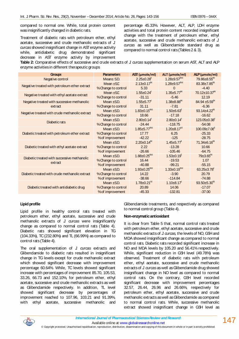
decreased significantly in the treatment of normal healthy rats with petroleum ether, ethyl acetate, successive and crude methanolic extracts with percentages decrease of 17.85, 19.62, 17.85 and 17.53%, respectively. Blood glucose level of diabetic rats showed significant increase (229.34%), while significant decrease in α-amylase activity was detected (35.71%) as compared to normal control rats. Treatment of diabetic rats with petroleum ether, ethyl acetate and crude methanolic extracts of J. curcas as well as Glibenclamide declared insignificant change in blood glucose level as compared to normal control rats. Although, successive methanolic extract recorded significant increase in glucose level with percentage of improvement 190.39%. While, α-amylase enzyme activity showed significant decrease with percentages of improvement 7.14, 9.31, 11.60, 10.71 and 14.33%, for petroleum ether, ethyl acetate, successive and crude methanolic extracts as well as Glibenclamide standard drug respectively (Table 1).
Table 1: Comparative effects of successive and crude extracts of J. curcas supplementation on blood glucose and α-amylase levels in different therapeutic groups
Groups Parameters Glucose (mg/dl) α-amylase (U/l) Negative control Mean± S.D. 95.72±4.66cd 1151.12±67.14a
Negative treated with petroleum ether extract Mean ±S.D
% Change to control 88.25±8.88cd
7.80 945.55±47.47b
17.85
Negative treated with ethyl acetate extract Mean ±S.D
% Change to control 94±3.74cd
1.79 925.25b
19.62
Negative treated with successive methanolic extract Mean ±S.D.
% Change to control 77.25±5.74d
19.29 945.55±47.47b
17.85
Negative treated with crude methanolic extract Mean ±S.D.
% Change to control 87.50±9.57cd
8.58 949.25±38.38b
17.53
Diabetic rats Mean ±S.D.
% Change to control 315.25±42.01a
-229.34 739.99±67.14d
35.71
Diabetic treated with petroleum ether extract Mean ±S.D.
% Change to control % of improvement
94±12.94cd
1.79 -231.14
822.22±67.13c 28.57 7.14
Diabetic treated with ethyl acetate extract Mean ±S.D.
% Change to control % of improvement
97.75±11.12cd
-2.12 -227.22
847.25±13.3bc 26.39 9.31
Diabetic treated with successive methanolic extract Mean ±S.D.
% Change to control % of improvement
133±15.89b
-38.94 -190.39
873.61±39.36bc 24.10 11.60
Diabetic treated with crude methanolic extract Mean ±S.D.
% Change to control % of improvement
118±12.06bc
-23.27 -206.06
863.33±47.47bc 25.00 10.71
Diabetic treated with antidiabetic drug Mean ±S.D.
% Change to control % of improvement
100.25±12.61cd
-4.73 -224.61
904.97±45.30bc 21.38 14.33
Liver function enzyme activities (AST, ALT and ALP), LDH and total protein content
Insignificant change in the enzyme activities of AST, ALT, ALP, LDH and total protein content were detected in healthy rats orally administrated successive and crude extracts of J. curcas (Table 2). However, ethyl acetate and successive methanolic extracts showed similarly
significant decrease in AST enzyme activity with percentage decrease 31.11%, as compared to normal control rats. While, diabetic rats exhibited significant increase in enzyme activities; AST, ALT and ALP (24.44, 118.75 and 54.08%, respectively) as compared to normal control rats. Although, LDH enzyme activity (Table 3) showed significant decrease in diabetic rats (59.61%) as
Int. J. Pharm. Sci. Rev. Res., 29(2), November – December 2014; Article No. 26, Pages: 143-156 ISSN 0976 – 044X
International Journal of Pharmaceutical Sciences Review and Research Available online at www.globalresearchonline.net
© Copyright protected. Unauthorised republication, reproduction, distribution, dissemination and copying of this document in whole or in part is strictly prohibited. © Copyright protected. Unauthorised republication, reproduction, distribution,
147
compared to normal one. While, total protein content was insignificantly changed in diabetic rats.
Treatment of diabetic rats with petroleum ether, ethyl acetate, successive and crude methanolic extracts of J. curcas showed insignificant change in AST enzyme activity while, antidiabetic drug demonstrated significant decrease in AST enzyme activity by improvement
percentage 45.33%. However, ALT, ALP, LDH enzyme activities and total protein content recorded insignificant change with the treatment of petroleum ether, ethyl acetate, successive and crude methanolic extracts of J. curcas as well as Glibenclamide standard drug as compared to normal control rats (Tables 2 & 3).
Table 2: Comparative effects of successive and crude extracts of J. curcas supplementation on serum AST, ALT and ALP enzyme activities in different therapeutic groups
Groups Parameters AST (µmole/ml) ALT (µmole/ml) ALP (µmole/ml) Negative control Mean± S.D. 2.25±0.28b 1.28±9.57bcd 79.86±8.55bc
Negative treated with petroleum ether extract Mean ±S.D % Change to control
2.13±0.17bc
5.33 1.28±9.57bcd
0 83.38±7.85bc
-4.40
Negative treated with ethyl acetate extract Mean ±S.D % Change to control
1.55±0.24d
-31.11 1.35±5.77bc
-5.46 70.12±10.37bc
12.19 Negative treated with successive methanolic
extract Mean ±S.D.
% Change to control 1.55±5.77d
31.11 1.38±8.88bc
-7.81 84.94 ±5.59bc
-6.36
Negative treated with crude methanolic extract Mean ±S.D. % Change to control
1.83±0.15bcd
18.66 1.50±6.63b
-17.18 93.14±4.22bc
-16.62
Diabetic rats Mean ±S.D. % Change to control
2.80±0.14a
-24.44 2.80±0.14a
-118.75 123.05±0.38a
-54.08
Diabetic treated with petroleum ether extract Mean ±S.D.
% Change to control % of improvement
1.85±5.77bcd
17.77 -42.22
1.20±8.17cd
6.25 -125
100.09±7.08b -25.33 -28.75
Diabetic treated with ethyl acetate extract Mean ±S.D.
% Change to control % of improvement
2.20±0.14bc
2.22 -26.66
1.45±5.77b
-13.28 -105.46
71.34±6.16bc 10.66 -64.75
Diabetic treated with successive methanolic extract
Mean ±S.D. % Change to control % of improvement
1.88±0.25bcd
16.44 -40.88
1.53±0.19b
-19.53 -99.21
79±3.65bc 1.07
-55.15
Diabetic treated with crude methanolic extract Mean ±S.D.
% Change to control % of improvement
1.93±0.25bcd
14.22 -38.66
1.33±0.19bcd
-3.90 -114.84
63.25±3.78c 20.79 -74.88
Diabetic treated with antidiabetic drug Mean ±S.D.
% Change to control % of improvement
1.78±0.21cd
20.89 -45.33
1.10±8.17d
14.06 -132.81
93.50±5.30bc -17.07 -37.00
Lipid profile
Lipid profile in healthy control rats treated with petroleum ether, ethyl acetate, successive and crude methanolic extracts of J. curcas were insignificantly change as compared to normal control rats (Table 4). Diabetic rats showed significant elevation in TG (104.33%), TC (232.80%) and TL (66.99%) as compared to control rats (Table 4).
The oral supplementation of J. curcas extracts and Glibenclamide to diabetic rats resulted in insignificant change in TG levels except for crude methanolic extract which showed significant decrease with improvement percentage 60.64%. While, TC levels showed significant increase with percentages of improvement 85.70, 105.53, 33.26, 66.73 and 152.10%, for petroleum ether, ethyl acetate, successive and crude methanolic extracts as well as Glibenclamide respectively. In addition, TL level showed significant decrease by percentages of improvement reached to 107.96, 103.21 and 91.39%, with ethyl acetate, successive methanolic and
Glibenclamide treatments, and respectively as compared to normal control group (Table 4).
Non-enzymatic antioxidant
It is clear from Table 5 that, normal control rats treated with petroleum ether, ethyl acetate, successive and crude methanolic extracts of J. curcas, the levels of NO, GSH and MDA showed insignificant change as compared to normal control rats. Diabetic rats recorded significant increase in NO and MDA levels by 105.20 and 56.41% respectively. While, significant reduction in GSH level (49.78%) was observed. Treatment of diabetic rats with petroleum ether, ethyl acetate, successive and crude methanolic extracts of J. curcas as well as Glibenclamide drug showed insignificant change in NO level as compared to normal control rats. On the contrary, GSH level recorded significant decrease with improvement percentages 32.57, 26.44, 26.96 and 26.66%, respectively for petroleum ether, ethyl acetate, successive and crude methanolic extracts as well as Glibenclamide as compared to normal control rats. While, successive methanolic extract showed insignificant change in GSH level as
Int. J. Pharm. Sci. Rev. Res., 29(2), November – December 2014; Article No. 26, Pages: 143-156 ISSN 0976 – 044X
International Journal of Pharmaceutical Sciences Review and Research Available online at www.globalresearchonline.net
© Copyright protected. Unauthorised republication, reproduction, distribution, dissemination and copying of this document in whole or in part is strictly prohibited. © Copyright protected. Unauthorised republication, reproduction, distribution,
148
compared to normal control rats (Table 5). Also, the MDA level demonstrated insignificant change post treatment of diabetic rats with petroleum ether, ethyl acetate,
successive and crude methanolic extracts as well as Glibenclamide, a standard anti-diabetic drug (Table 5).
Table 3: Comparative effects of successive and crude extracts of J. curcas supplementation on serum LDH enzyme activity and total protein content in different therapeutic groups.
Groups Parameters LDH (U/l) Protein (TP) (mg/ml) Negative control Mean± S.D. 26000±1.15a 87±2.94a
Negative treated with petroleum ether extract Mean ±S.D % Change to control
21250±0.46a
18.26 86.75±2.87a
0.28
Negative treated with ethyl acetate extract Mean ±S.D % Change to control
27000±0.20a
-3.84 85.50±1.29a
1.72
Negative treated with successive methanolic extract Mean ±S.D. % Change to control
23000±0.47a
11.53 87.25±3.86a
-0.28
Negative treated with crude methanolic extract Mean ±S.D. % Change to control
30500±0.24a
-17.30 84±1.83a
-3.44
Diabetic rats Mean ±S.D. % Change to control
10500±0.13b
59.61 86.25±4.84a
0.86
Diabetic treated with petroleum ether extract Mean ±S.D.
% Change to control % of improvement
24750±0.67a
4.80 54.80
87.25±2.87a -0.28 1.14
Diabetic treated with ethyl acetate extract Mean ±S.D.
% Change to control % of improvement
27500±0.21a
-5.76 65.38
86±0.82a 1.14 -0.28
Diabetic treated with successive methanolic extract Mean ±S.D.
% Change to control % of improvement
19225±0.37a
26.05 33.55
85.50±2.38a 1.72 -0.86
Diabetic treated with crude methanolic extract Mean ±S.D.
% Change to control % of improvement
24750±0.38a
4.80 54.80
89.50±3a -2.87 3.73
Diabetic treated with antidiabetic drug Mean ±S.D.
% Change to control % of improvement
23750±0.36a
8.65 50.96
89±3.56a -2.30 3.16
Table 4: Comparative effects of successive and crude extracts of J. curcas supplementation on lipid profile TG, TC and TL in different therapeutic groups.
Groups Parameters TG (mg/dl) TC (mg/dl) TL (mg/dl) Negative control Mean± S.D. 83.93±16.69b 24.20±7.01f 362.50±65.93b
Negative treated with petroleum ether extract Mean ±S.D % Change to control
53.47±15.04bc
36.29 25.20±9.11f
-4.13 352.69±56.47b
2.70
Negative treated with ethyl acetate extract Mean ±S.D % Change to control
63.73±11.10b
24.06 35.29±8.32ef
-45.83 365.37±24.53b
-0.79 Negative treated with successive methanolic
extract Mean ±S.D.
% Change to control 54.24±13.92bc
35.37 22.5±2.5f
7.02 313.99±27.10b
13.38
Negative treated with crude methanolic extract Mean ±S.D. % Change to control
60±16.33bc
28.51 33±2.16ef
-36.36 299.92±10.36b
17.26
Diabetic rats Mean ±S.D. % Change to control
171.50±30.61a
-104.33 80.54±18.54a
-232.80 605.34±199.37a
-66.99
Diabetic treated with petroleum ether extract Mean ±S.D.
% Change to control % Of improvement
73.82±7.62b
12.04 -116.38
59.80±3.22bcd
-147.10 -85.70
307.43±20.69b 15.19 -82.18
Diabetic treated with ethyl acetate extract Mean ±S.D.
% Change to control % Of improvement
76.61±10.93b
8.72 -113.05
55±4.08cd
-127.27 -105.53
213.98±14.04c 40.97
-107.96
Diabetic treated with successive methanolic extract
Mean ±S.D. % Change to control % Of improvement
76.61±10.93b
8.72 -113.05
72.49±12.76ab
-199.54 -33.26
231.17±30.07c 36.22
-103.21
Diabetic treated with crude methanolic extract Mean ±S.D.
% Change to control % Of improvement
33.03±6.75c
-60.64 -164.98
64.39±2.44bc
-166.07 -66.73
307.69±41.87b 15.12 -82.11
Diabetic treated with antidiabetic drug Mean ±S.D.
% Change to control % Of improvement
70.71±8.21b
15.75 -120.08
43.73±5.10de
-80.70 -152.10
274.04±12.13c 24.40 -91.39
Int. J. Pharm. Sci. Rev. Res., 29(2), November – December 2014; Article No. 26, Pages: 143-156 ISSN 0976 – 044X
International Journal of Pharmaceutical Sciences Review and Research Available online at www.globalresearchonline.net
© Copyright protected. Unauthorised republication, reproduction, distribution, dissemination and copying of this document in whole or in part is strictly prohibited. © Copyright protected. Unauthorised republication, reproduction, distribution,
149
Table 5: Comparative effect of successive and crude extracts of J. curcas supplementation on antioxidant scavenging activity in different therapeutic groups.
Groups Parameters NO (µmole/ml) GSH (mmole/l) MDA (ηmole/ml) Negative control Mean± S.D. 16.72±3.53b 101.07±6.07ab 0.39±2.99b
Negative treated with petroleum ether extract Mean ±S.D
% Change to control 18.56±1.98b
-11.00 102.53±9.30a
-1.44 0.40±2.87b
-2.56
Negative treated with ethyl acetate extract Mean ±S.D
% Change to control 15.47±4.45b
7.47 92±4.70abc
8.97 0.39±3.93b
0 Negative treated with successive methanolic
extract Mean ±S.D.
% Change to control 14.50±1.29b
13.27 90.23±4.95bc
10.72 0.41±2.08b
-5.12
Negative treated with crude methanolic extract Mean ±S.D.
% Change to control 17.22±4.53b
-2.99 90.73±4.30bc
10.23 0.38±4.06b
2.56
Diabetic rats Mean ±S.D.
% Change to control 34.31±3.33a
-105.20 50.75±7.18e
49.78 0.61±6.22a
56.41
Diabetic treated with petroleum ether extract Mean ±S.D.
% Change to control % Of improvement
11.63±1.38bc
30.44 -135.64
83.67±2.45cd
17.22 32.57
0.42±5.57b -7.69
-48.71
Diabetic treated with ethyl acetate extract Mean ±S.D.
% Change to control % Of improvement
14.41±1.15b
13.81 -119.01
77.48±6.85d
23.34 26.44
0.42±3.37b -7.69
-48.71
Diabetic treated with successive methanolic extract
Mean ±S.D. % Change to control % Of improvement
18.22±3.26b
-8.97 -96.23
99.23±1.75ab
1.82 47.96
0.41±1.25b -5.13
-51.28
Diabetic treated with crude methanolic extract Mean ±S.D.
% Change to control % Of improvement
14.65±1.39b
12.38 -117.58
78±4.32d
22.82 26.96
0.43±8.17b -10.25 -46.15
Diabetic treated with antidiabetic drug Mean ±S.D.
% Change to control % Of improvement
14.25±1.5b
14.77 -116.86
77.7±5.53d
23.12 26.66
0.43±2.36b -10.26 -46.15
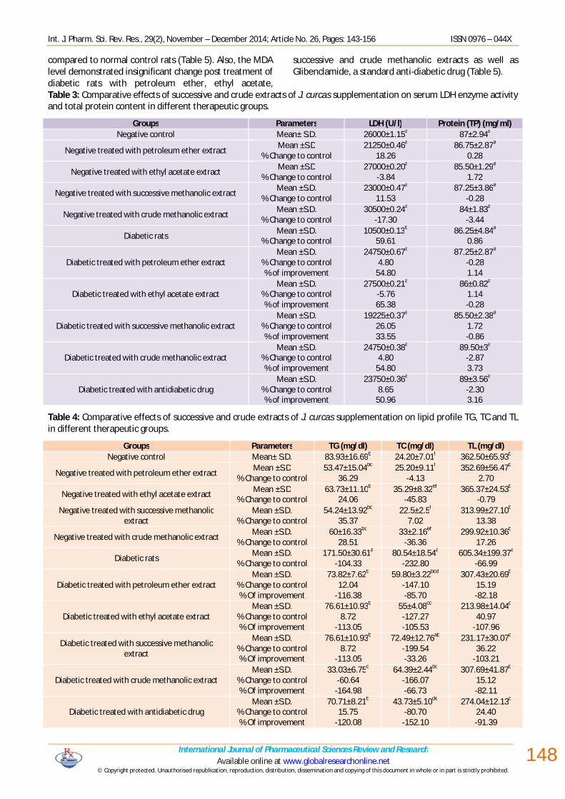
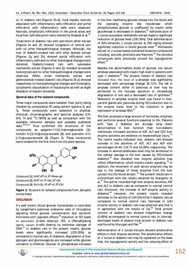
Figure (1a): Pancreas of normal control rats showing no histopathological changes (H & E X 400).
Figure (1b): Pancreas of diabetic rats showing perivascular inflammatory cells infiltration (H & E X 400).
Figure (1c): Pancreas of diabetic rats showing necrosis of islets of Langerhan’s (H & E X 400).
Figure (1d): Pancreas of diabetic rats showing apoptosis of acinar epithelium (H & E X 400).
Figure (1e): Pancreas of diabetic-treated rats with petroleum ether extract showing vacuolation of islets of Langerhan’s (H & E X 400).
Figure (1f): Pancreas of diabetic-treated rats with ethyl acetate extract showing vacuolation of islets of Langerhan's (H & E X 400).
Int. J. Pharm. Sci. Rev. Res., 29(2), November – December 2014; Article No. 26, Pages: 143-156 ISSN 0976 – 044X
International Journal of Pharmaceutical Sciences Review and Research Available online at www.globalresearchonline.net
© Copyright protected. Unauthorised republication, reproduction, distribution, dissemination and copying of this document in whole or in part is strictly prohibited. © Copyright protected. Unauthorised republication, reproduction, distribution,
150
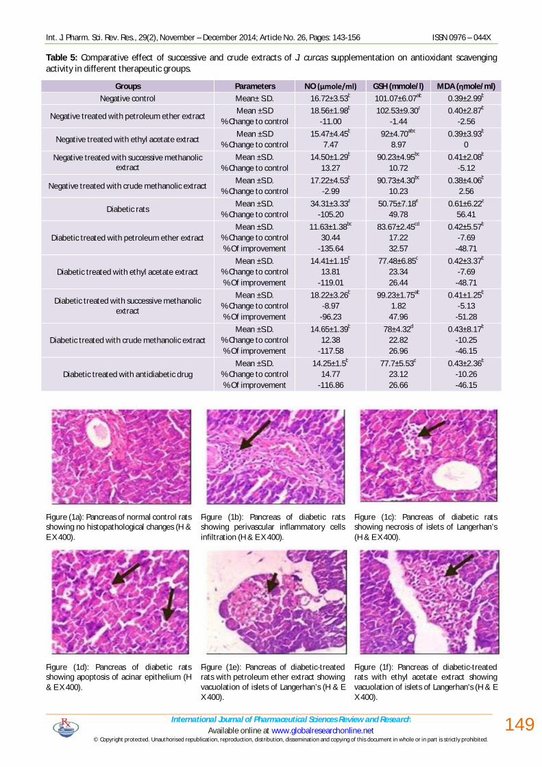
Figure (1g): Pancreas of diabetic-treated rats with successive methanolic extract showing dilatation of pancreatic duct (H & E X 400).
Figure (1h): Pancreas of of diabetic-treated rats with successive methanolic extract showing no histopathological changes (H & E X 400).
Figure (1i): Pancreas of diabetic-treated rats with crude methanolic extract showing dilatation of pancreatic duct (H & E X 400).
Figure (1j): Pancreas of diabetic-treated rats with crude methanolic extract showing no histopathological changes (H & E X 400).
Figure (1k): Pancreas of diabetic-treated rats with glibenclamide drug showing vacuolation of islets of Langerhan's (H & E X 400).
Figure (1l): Pancreas of diabetic-treated rats with glibenclamide drug showing no histopathological change (H & E X 400).
Figure 1: Histopathological examination of normal pancreas, diabetic and diabetic- treated rats.
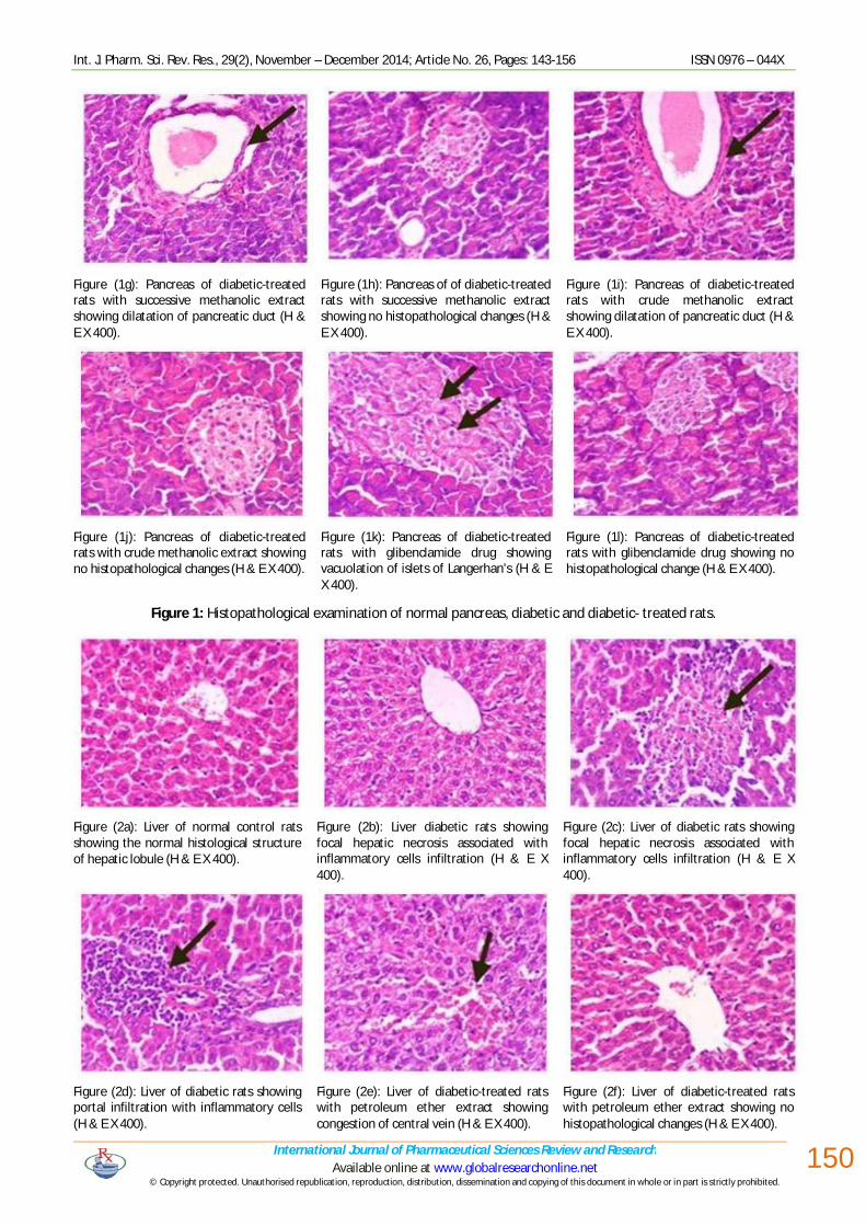
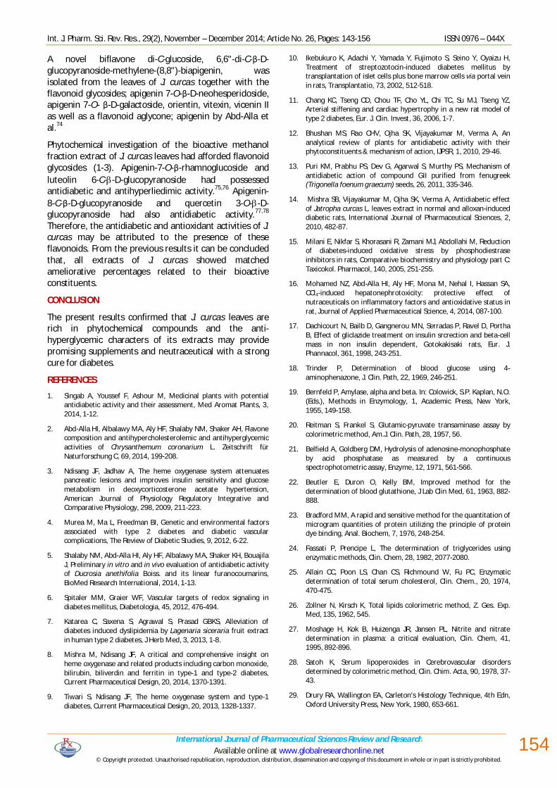
Figure (2a): Liver of normal control rats showing the normal histological structure of hepatic lobule (H & E X 400).
Figure (2b): Liver diabetic rats showing focal hepatic necrosis associated with inflammatory cells infiltration (H & E X 400).
Figure (2c): Liver of diabetic rats showing focal hepatic necrosis associated with inflammatory cells infiltration (H & E X 400).
Figure (2d): Liver of diabetic rats showing portal infiltration with inflammatory cells (H & E X 400).
Figure (2e): Liver of diabetic-treated rats with petroleum ether extract showing congestion of central vein (H & E X 400).
Figure (2f): Liver of diabetic-treated rats with petroleum ether extract showing no histopathological changes (H & E X 400).
Int. J. Pharm. Sci. Rev. Res., 29(2), November – December 2014; Article No. 26, Pages: 143-156 ISSN 0976 – 044X
International Journal of Pharmaceutical Sciences Review and Research Available online at www.globalresearchonline.net
© Copyright protected. Unauthorised republication, reproduction, distribution, dissemination and copying of this document in whole or in part is strictly prohibited. © Copyright protected. Unauthorised republication, reproduction, distribution,
151
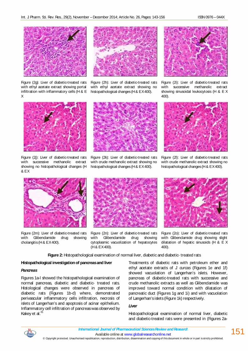
Figure (2g): Liver of diabetic-treated rats with ethyl acetate extract showing portal infiltration with inflammatory cells (H & E X
Figure (2h): Liver of diabetic-treated rats with ethyl acetate extract showing no histopathological changes (H & E X 400).
Figure (2i): Liver of diabetic-treated rats with successive methanolic extract showing sinusoidal leukocytosis (H & E X 400).
Figure (2j): Liver of diabetic-treated rats with successive methanolic extract showing no histopathological changes (H & E X
Figure (2k): Liver of diabetic-treated rats with crude methanolic extract showing no histopathological changes (H & E X 400).
Figure (2l): Liver of diabetic-treated rats with crude methanolic extract showing no histopathological changes (H & E X 400).
Figure (2m): Liver of diabetic-treated rats with Glibenclamide drug showing cholangitis (H & E X 400).
Figure (2n): Liver of diabetic-treated rats with Glibenclamide drug showing cytoplasmic vacuolization of hepatocytes (H & E X 400).
Figure (2o): Liver of diabetic-treated rats with Glibenclamide drug showing slight dilatation of hepatic sinusoids (H & E X 400).
Figure 2: Histopathological examination of normal liver, diabetic and diabetic- treated rats
Histopathological investigation of pancreas and liver
Pancreas
Figures 1a-l showed the histopathological examination of normal pancreas, diabetic and diabetic- treated rats. Histological changes were observed in pancreas of diabetic rats (Figures 1b-d) where, demonstrated perivascular inflammatory cells infiltration, necrosis of islets of Langerhan’s and apoptosis of acinar epithelium. Inflammatory cell infiltration of pancreas was observed by Kakey et al.31
Treatments of diabetic rats with petroleum ether and ethyl acetate extracts of J. curcas (Figures 1e and 1f) showed vacuolation of Langerhan’s islets. However, pancreas of diabetic-treated rats with successive and crude methanolic extracts as well as Glibenclamide was improved toward normal condition with dilatation of pancreatic duct (Figures 1g and 1i) and with vacuolation of Langerhan’s islets (Figure 1k) respectively.
Liver
Histopathological examination of normal liver, diabetic and diabetic-treated rats were presented in (Figures 2a-
Int. J. Pharm. Sci. Rev. Res., 29(2), November – December 2014; Article No. 26, Pages: 143-156 ISSN 0976 – 044X
International Journal of Pharmaceutical Sciences Review and Research Available online at www.globalresearchonline.net
© Copyright protected. Unauthorised republication, reproduction, distribution, dissemination and copying of this document in whole or in part is strictly prohibited. © Copyright protected. Unauthorised republication, reproduction, distribution,
152
o). In diabetic rats (Figures 2b-d), focal hepatic necrosis associated with inflammatory cells infiltration and portal infiltration with inflammatory cells were observed. Necrosis, lymphocytic infiltration in the portal areas and focal liver cells disruption were noticed by Masjedi et al.32
Treatments of diabetic rats with petroleum ether extract (Figures 2e and 2f) showed congestion of central vein with no other histopathological changes. Although, the liver of diabetic-treated rats with ethyl acetate extract (Figures 2g and 2h) showed portal infiltration with inflammatory cells and no other histological changes were detected. Diabetic-treated rats with successive methanolic extract (Figures 2i and 2j) revealed sinusoidal leukocytosis and no other histopathological changes were observed. While, crude methanolic extract and glibenclamide treated diabetic rats (Figures 2k-o) showed respectively no histopathological changes and cholangitis, cytoplasmic vacuolization of hepatocytes as well as slight dilatation of hepatic sinusoids.
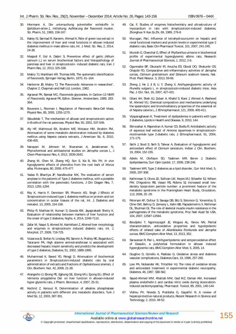
Spectral data of the isolated compounds
Three major compounds were isolated, their purity being checked by comparative PC using solvent systems S1 and S2. These compounds were subjected to physical, chemical, chromatographic and spectral analyses (UV, MS, 1H and 13C NMR) as well as comparison with the available reference samples and available published data.33,34,35 Thus confirmed the structures of the compounds as apigenin-7-O-β-rhamnoglucoside (1), luteolin 6-C--D-glucopyranoside (2), and quercetin 3-O--D-glucopyranoside (3) (Figure 3). These compounds were isolated for the first time from the plant species.
Compound (1): R=R1=R3=H, R2=Rham-glc Compound (2): R=R2=H, R1=Glc, R3=OH Compound (3): R=O-Glc, R1=R2=H, R3=OH
Figure 3: Structure of isolated compounds from Jatropha curcas leave
DISCUSSION
It's well known blood glucose homeostasis is controlled by Langerhan's pancreas endocrine cells of throughout adjusting blood glucose concentration and secretion hormones with opposite effects.36 Induction of STZ leads to pancreatic β-cells destroys. STZ, a DNA-alkylating agent, causes β-cells destroy by extensive damage of DNA.37 In diabetic rats in the present results, glucose levels were significantly increased (229.35%), as compared to normal rats. In diabetes, degradation of liver glycogen and gluconeogensis are increased while glucose utilization is inhibited. Glucose -6- phosphatase increases
in the liver, facilitating glucose release into the blood and the opposing enzyme like hexokinase which phosphorlyates glucose is unaffected by insulin while glucokinase is decreased in diabetes.38 Administration of J. curcas successive methanolic extract leads to significant reduction of glucose level (190.39%). Oral administration of 50% ethanolic J. curcas extract at 250 mg/kg showed significant reduction in blood glucose level.14 Methanolic extract of J. curcas leaves contained bioactive compounds including, steroids, phenolics and flavones.39 The penolics compounds were previously proved the hypoglycemic effect.40
Besides the abnormalities levels of glucose, low serum amylase associated with insulin high level in patients with type 2 diabetes.41 The present results of diabetic rats revealed that, the level of α-amylase was significantly decreased with percentage change 35.71%. The fall in amylase content either in pancreas or liver may be attributed to the increase secretion or intracellular degradation in vivo and a decreased rate of synthesis.38 Reduction in amylase enzyme level was found in liver, parotid glands and pancreas during STZ-induction due to the enzyme lower level to the reduction in gene expression of amylase RNA.42
The liver produces a large amount of hormones, enzymes, and performs several functions essential to life. Patients with Type 2 diabetes are strongly lying down abnormalities in liver function than non-diabetic individuals, increased activities of AST, ALT and GGT liver enzyme activities are evidence on hepatocellular injury.43 The recent results indicated that, diabetic rats showed increase in the activities of AST, ALT and ALP with percentages 24.44, 118.75 and 54.08%, respectively. The increase in aminotransferases level may be attributed to the cellular damage in the liver caused by STZ-induced diabetes.44 Also elevated liver enzyme activities may reflect inflammation, which impairs insulin signaling. 45 In addition, the increment of such serum enzymes may be due to the leakage of these enzymes from the liver cytosol into the blood stream. 46 The present results are in concomitant with the results obtained by Atangwho et al.47 The author recorded high liver enzyme activities; AST and ALT in diabetic rats as compared to normal control rats. Moreover, the increase of ALP enzyme activity in diabetes.48 However, the present results revealed decrease in the activity of LDH (59.61%) in diabetic rats as compared to normal control rats. Decrease in LDH enzyme activity in diabetic rats was observed and that is in agreement with the results of Cai.49 Total protein content of diabetic rats showed insignificant change (0.86%) as compared to normal control rats. In contrast, decreased levels of plasma total protein in diabetic rats was found by Murugan and Pari.50
Administration of J. curcas extracts showed ameliorative effects in liver enzyme activities. The ameliorative effects of J. curcas in diabetic rats may be explained on the base that, the hypoglycemic activity and the reducing effect of

Int. J. Pharm. Sci. Rev. Res., 29(2), November – December 2014; Article No. 26, Pages: 143-156 ISSN 0976 – 044X
International Journal of Pharmaceutical Sciences Review and Research Available online at www.globalresearchonline.net
© Copyright protected. Unauthorised republication, reproduction, distribution, dissemination and copying of this document in whole or in part is strictly prohibited. © Copyright protected. Unauthorised republication, reproduction, distribution,
153
petroleum ether, alcoholic and aqueous extracts of Phyllanthus Urinaria, plant (Euphorbiaceae family), on biochemical profile; AST, ALT and ALP in alloxan-induced diabetic rats may be attributed to the contents of tannins and flavonoids.51 In addition, the high activities of ALT and AST in formaldehyde-induced arthritic mice were significantly reduced with the treatment of the J. curcas methanolic leaves extract to normal or below normal value.52 Moreover, most of bioactive compounds (especially flavonoids and triterpenoids such as ursolic acid) showed a mechanism to improve liver and pancreas cells functions and hence normalized liver enzymes.53,54
The dyslipidemia associated with insulin resistance can be represented in increase of TG level carried in very-low-density lipoprotein (VLDL) particles, reduced high-density lipoprotein cholesterol (HDL-C) levels carried in small HDL particles and LDL-C levels that do not differ substantially from those of individuals without type 2 diabetes.55 It was observed that, the levels of TG, TC and TL in diabetic rats (Table 4) were increased in a significant way with percentages change reached to 104.33, 232.80 and 66.99%, respectively. Insulin activates lipoprotein lipase which hydrolyzes triglycerides. Insulin deficiency results in the failure of activate lipase enzyme consequently, causing hyper-triglyceridemia.56 Also, insulin deficiency leads to the elevated concentration in plasma free fatty acids as a result of increased free fatty acids outflow from fat depots, where the balance of the free fatty acids estrification, triglycerides lipolysis is displaced in favors of lipolysis. 56 Significant elevation in lipid profile in serum of diabetic rats was demonstrated by Sethi et al.57
In patients with type 2 diabetes, the ability of insulin to suppress hepatic production of large TG-rich VLDL (VLDL-TGs) leads to an elevation in plasma TG levels.58,59 Besides ,insulin resistance in skeletal muscle encourages energy conversion from carbohydrate ingestion into increased hepatic triglyceride synthesis, consequently leads to generate large numbers of triglyceride-rich lipoprotein particles, such as very-low-density lipoprotein (VLDL).60,61
Treatment of diabetic rats with J. curcas extracts revealed improvement in lipid profile. Where, crude methanolic extract for example showed significant decrease in the level of TG with percentage of improvement 164.98%. While, all extracts of J. curcas showed significant increase in TC with percentages of improvement in petroleum ether, ethyl acetate, successive and crude methanolic extracts reached to 85.70, 105.53, 33.26 and 66.73%, respectively. Ethyl acetate and successive methanolic extracts showed significant decrease in TL levels with percentages of improvement 107.96 and 103.21%, respectively. This may be explained on the basis of J. curcas oil seed cake possessed hypolipidaemic effect and these properties could be attributed to the presence of dietary fibers, phenolic compounds and storage proteins on oil seed cake.62 Also, the correction of insulin level induced by plants causing a regulation of carbohydrate and lipids metabolism by inhibition of lipolysis through
inhibition of hormone sensitive lipases activity in adipose tissue and suppresses the release of free fatty acids causing stimulation of lipogenesis.63
Oxygen-derived free radicals (ROS) are observed implicating in the pathophysiology of different disease, including diabetes mellitus.64 In DM, the oxidative stress can be resulted from the increased production of free radicals with/or a marked reduction of antioxidant defenses.65 The direct toxicity of NO is enhanced by its reacting with superoxide radical to give secondary toxic oxidizing species, such as peroxynitrite (ONOO) which is capable of oxidizing cellular structure and causes lipid peroxidation.66 In diabetic rats, NO and MDA levels were significantly increased with percentages 105.20 and 56.41%, respectively. Treatment of diabetic rats with different extracts of J. curcas as well as Glibenclamide standard drug showed normalization in GSH, NO and MDA levels as compared to normal control rats. However, successive methanolic extract showed the highest percentages of improvement in GSH and MDA levels reached to 47.96 and 51.28%, respectively.
Methanolic fraction of J. curcas (MFJC) treatment reversed the increase in lipid peroxide condition induced by Aflatoxin B1 (AFB1) near to normal levels.67 In accordance with these results; MFJC could protect liver against the AFB1-induced oxidative damage in rats.68
STZ selectively induces degenerative alterations and necrosis of pancreatic β-cells resulting in insulin deficiency and impairment in glucose oxidation.69 Treatment of J. curcas extracts showed enhancement in islet cell regeneration in diabetic rats and amelioration in both pancreas and hepatic architectures that apparent normal. The phytochemical screening of J. curcas leaves extracts possessed the presence of bioactive compounds including flavonoids, saponins, alkaloids, steroids and tannins.70 Rutin (flavonoid compound) has antioxidant and anti-inflammatory effects that lead to reduction of blood glucose level in the STZ-induced diabetic rats besides, functionally and formatively protection of pancreas, heart, liver, kidney, and retina tissues that attributed to diabetic complications.71 Thus, the protective effect of J. curcas leaves may be related to the presence of flavonoid compounds that lead to reducing the oxidative stress consequently, normalize hepatic and pancreas tissues structures and functions.
Flavonoids produce antidiabetic effect throughout many ways and have a wide range of biological activities, including antiallergic, antibacterial, antidiabetic, anti-inflammatory, antiviral, anti-proliferative, anti-mutagenic, antithrombotic, anticarcinogenic, hepatoprotective, estrogenic, insecticidal, and antioxidant activities.72 Moreover, the antidiabetic effect of flavonoids from Dracaena cochinchinensis (Asparagaceae) which possessed a potential hypoglycemic activity, in addition to, relieving dyslipidemia, tissue steatosis, and oxidative stress associated with T2DM.73
Int. J. Pharm. Sci. Rev. Res., 29(2), November – December 2014; Article No. 26, Pages: 143-156 ISSN 0976 – 044X
International Journal of Pharmaceutical Sciences Review and Research Available online at www.globalresearchonline.net
© Copyright protected. Unauthorised republication, reproduction, distribution, dissemination and copying of this document in whole or in part is strictly prohibited. © Copyright protected. Unauthorised republication, reproduction, distribution,
154
A novel biflavone di-C-glucoside, 6,6"-di-C-β-D-glucopyranoside-methylene-(8,8")-biapigenin, was isolated from the leaves of J. curcas together with the flavonoid glycosides; apigenin 7-O-β-D-neohesperidoside, apigenin 7-O- β-D-galactoside, orientin, vitexin, vicenin II as well as a flavonoid aglycone; apigenin by Abd-Alla et al.74
Phytochemical investigation of the bioactive methanol fraction extract of J. curcas leaves had afforded flavonoid glycosides (1-3). Apigenin-7-O-β-rhamnoglucoside and luteolin 6-C--D-glucopyranoside had possessed antidiabetic and antihyperliedimic activity.75,76 Apigenin-8-C-β-D-glucopyranoside and quercetin 3-O--D-glucopyranoside had also antidiabetic activity.77,78 Therefore, the antidiabetic and antioxidant activities of J. curcas may be attributed to the presence of these flavonoids. From the previous results it can be concluded that, all extracts of J. curcas showed matched ameliorative percentages related to their bioactive constituents.
CONCLUSION
The present results confirmed that J. curcas leaves are rich in phytochemical compounds and the anti-hyperglycemic characters of its extracts may provide promising supplements and neutraceutical with a strong cure for diabetes.
REFERENCES
1. Singab A, Youssef F, Ashour M, Medicinal plants with potential antidiabetic activity and their assessment, Med Aromat Plants, 3, 2014, 1-12.
2. Abd-Alla HI, Albalawy MA, Aly HF, Shalaby NM, Shaker AH, Flavone composition and antihypercholesterolemic and antihyperglycemic activities of Chrysanthemum coronarium L. Zeitschrift für Naturforschung C, 69, 2014, 199-208.
3. Ndisang JF, Jadhav A, The heme oxygenase system attenuates pancreatic lesions and improves insulin sensitivity and glucose metabolism in deoxycorticosterone acetate hypertension, American Journal of Physiology Regulatory Integrative and Comparative Physiology, 298, 2009, 211-223.
4. Murea M, Ma L, Freedman BI, Genetic and environmental factors associated with type 2 diabetes and diabetic vascular complications, The Review of Diabetic Studies, 9, 2012, 6-22.
5. Shalaby NM, Abd-Alla HI, Aly HF, Albalawy MA, Shaker KH, Bouajila J, Preliminary in vitro and in vivo evaluation of antidiabetic activity of Ducrosia anethifolia Boiss. and its linear furanocoumarins, BioMed Research International, 2014, 1-13.
6. Spitaler MM, Graier WF, Vascular targets of redox signaling in diabetes mellitus, Diabetologia, 45, 2012, 476-494.
7. Katarea C, Saxena S, Agrawal S, Prasad GBKS, Alleviation of diabetes induced dyslipidemia by Lagenaria siceraria fruit extract in human type 2 diabetes, J Herb Med, 3, 2013, 1-8.
8. Mishra M, Ndisang JF, A critical and comprehensive insight on heme oxygenase and related products including carbon monoxide, bilirubin, biliverdin and ferritin in type-1 and type-2 diabetes, Current Pharmaceutical Design, 20, 2014, 1370-1391.
9. Tiwari S, Ndisang JF, The heme oxygenase system and type-1 diabetes, Current Pharmaceutical Design, 20, 2013, 1328-1337.
10. Ikebukuro K, Adachi Y, Yamada Y, Fujimoto S, Seino Y, Oyaizu H, Treatment of streptozotocin-induced diabetes mellitus by transplantation of islet cells plus bone marrow cells via portal vein in rats, Transplantatio, 73, 2002, 512-518.
11. Chang KC, Tseng CD, Chou TF, Cho YL, Chi TC, Su MJ, Tseng YZ, Arterial stiffening and cardiac hypertrophy in a new rat model of type 2 diabetes, Eur. J. Clin. Invest, 36, 2006, 1-7.
12. Bhushan MS, Rao CHV, Ojha SK, Vijayakumar M, Verma A, An analytical review of plants for antidiabetic activity with their phytoconstituents & mechanism of action, IJPSR, 1, 2010, 29-46.
13. Puri KM, Prabhu PS, Dev G, Agarwal S, Murthy PS, Mechanism of antidiabetic action of compound GII purified from fenugreek (Trigonella foenum graecum) seeds, 26, 2011, 335-346.
14. Mishra SB, Vijayakumar M, Ojha SK, Verma A, Antidiabetic effect of Jatropha curcas L. leaves extract in normal and alloxan-induced diabetic rats, International Journal of Pharmaceutical Sciences, 2, 2010, 482-87.
15. Milani E, Nikfar S, Khorasani R, Zamani MJ, Abdollahi M, Reduction of diabetes-induced oxidative stress by phosphodiestrase inhibitors in rats, Comparative biochemistry and physiology part C: Taxicokol. Pharmacol, 140, 2005, 251-255.
16. Mohamed NZ, Abd-Alla HI, Aly HF, Mona M, Nehal I, Hassan SA, CCl4-induced hepatonephrotoxicity: protective effect of nutraceuticals on inflammatory factors and antioxidative status in rat, Journal of Applied Pharmaceutical Science, 4, 2014, 087-100.
17. Dachicourt N, Bailb D, Gangnerou MN, Serradas P, Ravel D, Portha B, Effect of gliclazide treatment on insulin srcrection and beta-cell mass in non insulin dependent, Gotokakisaki rats, Eur. J. Phannacol, 361, 1998, 243-251.
18. Trinder P, Determination of blood glucose using 4-aminophenazone, J. Clin. Path, 22, 1969, 246-251.
19. Bernfeld P, Amylase, alpha and beta. In: Colowick, S.P. Kaplan, N.O. (Eds.), Methods in Enzymology, 1, Academic Press, New York, 1955, 149-158.
20. Reitman S, Frankel S, Glutamic-pyruvate transaminase assay by colorimetric method, Am.J. Clin. Path, 28, 1957, 56.
21. Belfield A, Goldberg DM, Hydrolysis of adenosine-monophosphate by acid phosphatase as measured by a continuous spectrophotometric assay, Enzyme, 12, 1971, 561-566.
22. Beutler E, Duron O, Kelly BM, Improved method for the determination of blood glutathione, J Lab Clin Med, 61, 1963, 882-888.
23. Bradford MM, A rapid and sensitive method for the quantitation of microgram quantities of protein utilizing the principle of protein dye binding, Anal. Biochem, 7, 1976, 248-254.
24. Fassati P, Prencipe L, The determination of triglycerides using enzymatic methods, Clin. Chem, 28, 1982, 2077-2080.
25. Allain CC, Poon LS, Chan CS, Richmound W, Fu PC, Enzymatic determination of total serum cholesterol, Clin. Chem., 20, 1974, 470-475.
26. Zollner N, Kirsch K, Total lipids colorimetric method, Z. Ges. Exp. Med, 135, 1962, 545.
27. Moshage H, Kok B, Huizenga JR, Jansen PL, Nitrite and nitrate determination in plasma: a critical evaluation, Clin. Chem, 41, 1995, 892-896.
28. Satoh K, Serum lipoperoxides in Cerebrovascular disorders determined by colorimetric method, Clin. Chim. Acta, 90, 1978, 37-43.
29. Drury RA, Wallington EA, Carleton's Histology Technique, 4th Edn, Oxford University Press, New York, 1980, 653-661.
Int. J. Pharm. Sci. Rev. Res., 29(2), November – December 2014; Article No. 26, Pages: 143-156 ISSN 0976 – 044X
International Journal of Pharmaceutical Sciences Review and Research Available online at www.globalresearchonline.net
© Copyright protected. Unauthorised republication, reproduction, distribution, dissemination and copying of this document in whole or in part is strictly prohibited. © Copyright protected. Unauthorised republication, reproduction, distribution,
155
30. Hiermann A, Die untersuchung potentieller wirkstoffe in Epilobium-arten.1. mitteilung: Aufklarung der flavonoid muster, Sci. Pharm, 51, 1983, 158-167.
31. Kakey IS, Sarmad R, Kareem, Ahmad K, Role of green tea extract in the improvement of liver and renal functions in alloxan induced diabetes mellitus in male albino rats, Int. J. Med. Sci. Res, 2, 2014, 24-38.
32. Masjedi F, Gol A, Dabiri S, Preventive effect of garlic (Allium sativum L.) on serum biochemical factors and histopathology of pancreas and liver in streptozotocin- induced diabetic rats, Iran J Pharm Res, 12, 2013, 325-338.
33. Mabry TJ, Markham KR, Thomas MB, The systematic identification of flavonoids, Springer-Verlag, Berlin, 1970, 41–164.
34. Harborne JB, Mabry TJ, The Flavonoids: Advances in researches", Chapter 2, Chapman and Hall Ltd, London, 1982.
35. Agrawal PK, Bansal MC, Flavonoids glycosides, In Carbon-13 NMR of Flavonoids; Agrawal PK, Editor, Elsevier, Amsterdam, 1989, 283-364.
36. Bouwens L, Rooman I, Regulation of Pancreatic Beta-Cell Mass, Physiol Rev, 85, 2005, 1255-1270.
37. Szkudelski T, The mechanism of alloxan and streptozotocin action in B cells of the rat pancreas, Physiol Res, 50, 2001, 537-546.
38. Aly HF, Mahmoud EA, Ibrahim ME, Motawe HM, Ibrahim FM, Attenuation of some metabolic deterioration induced by diabetes mellitus using Nepeta cataria extracts, J American Sci, 6, 2010, 436-455.
39. Narayani M, Johnson M, Sivaraman A, Janakiraman N, Phytochemical and antibacterial studies on Jatropha curcas L, J Chem Pharmaceut Res, 4, 2012, 2639-2642.
40. Zhang M, Chen M, Zhang HQ, Sun S, Xia B, Wu FH, In vivo hypoglycemic effects of phenolics from the root bark of Morus alba, Fitoterapia, 80, 2009, 475-477.
41. Yadav R, Bhartiya JP, Nandkeoliar MK, The evaluation of serum amylase in the patients of Type 2 diabetes mellitus, with a possible correlation with the pancreatic functions, J Clin Diagen Res, 7, 2013, 1291-1294.
42. Roy K, Harris F, Dennison SR, Phoenix AD, Singh J Effects of Streptozotocin-induced type 1 diabetes mellitus on protein and ion concentration in ocular tissues of the rat, Int. J. Diabetes and metabol, 13, 2005, 154-158.
43. Philip R, Mathias M, Kumari S, Gowda KM, Jayaprakash Shetty K, Evaluation of relationship between markers of liver function and the onset of type 2 diabetes, Nujhs, 4, 2014, 2249-7110.
44. Zafar M, Naqvi S, Ahmed M, Kaimkhani Z, Altered liver morphology and enzymes in streptozotocin induced diabetic rats, Int. J. Morphol, 27, 2009, 719-725.
45. Vozarova B, Stefan N, Lindsay RS, Saremi A, Pratley RE, Bogardus C, Tataranni PA, High alanine aminotransferase is associated with decreased hepatic insulin sensitivity and predicts the development of type 2 diabetes, Diabetes, 51, 2002, 1889-1895.
46. Muhammad K, Saeed YD, Rongji D, Attenuation of biochemical parameters in Streptozotocin-induced diabetic rats by oral administration of extracts and fractions of Cephalotaxus sinensis, J. Clin. Biochem. Nut, 42, 2008, 21-28.
47. Atangwho IJ, Ebong PE, Egbung GE, Eteng MU, Eyong EU, Effect of Vernonia amygdalina Del. on liver function in alloxan-induced hyper glycemic rats, J. Pharm. Bioresour, 4, 2007, 25-31.
48. Kechrid Z, Kenouz R, Determination of alkaline phosphatase activity in patients with different zinc metabolic disorders, Turk J Med Sci, 12, 2003, 387-391.
49. Cai, F, Studies of enzymes histochemistry and ultrastructure of myocardium in rats with streptozotocin-induced diabetes, Zhonghua Yi Xue Za Zhi, 69, 1989, 276-8.
50. Murugan, Pari, Influence of tetrahydrocurcumin on hepatic and renal functional markers and protein levels in experimental type 2 diabetic rats, Basic Clin Pharmacol Toxicol, 101, 2007, 241-245.
51. Munish G, Chanchal G, Effect of Phyllanthus urinaria in biochemical profile of experimental hyperglycemic albino rats, Research Journal of Pharmaceutical Sciences, 1, 2012, 2-6.
52. Ogunnaike BF, Okutachi IR, Anucha ES, Gbodi OO, Shokunbi OS, Onajobi FD, Comparative anti-inflammatory activities of Jatropha curcas, Ocimum gratissimum and Solanum scabrum leaves, Nat. Prod. Plant Resour, 3, 2013, 59-66.
53. Zheng J, He J, Ji B, Li Y, Zhang X, Antihyperglycemic activity of Prunella vulgaris L. in streptozotocin-induced diabetic mice, Asia Pac. J. Clin. Nut, 16, 2007, 427-431.
54. Gilani AH, Shah AJ, Zubair A, Khalid S, Kiani J, Ahmed A, Rasheed M, Ahmed VU, Chemical composition and mechanisms underlying the spasmolytic and bronchodilatory properties of the essential oil of Nepeta cateria L, J. Ethnopharmacol, 121, 2009, 405-411.
55. Vijayaraghavan K, Treatment of dyslipidemia in patients with type 2 diabetes, Lipids in Health and Disease, 9, 2010, 144.
56. Shirwaikar A, Rajendran A, Kumar CD, Bodla R, Antidiabetic activity of aqueous leaf extract of Annona squamosa in streptozotocin-nicotinamide type 2-diabetic rats, J. Ethnopharmacol, 91, 2004, 171-175.
57. Sethi J, Sood S, Seth S, Talwar A, Evaluation of hypoglycemic and antioxidant effect of Ocimum sancatum, Indian J. Clin. Biochem, 19, 2004, 152-155.
58. Adiels M, Olofsson SO, Taskinen MR, Boren J, Diabetic dyslipidaemia, Curr Opin Lipidol, 17, 2006, 238-246.
59. Taskinen MR, Type 2 diabetes as a lipid disorder, Curr Mol Med, 5, 2005, 297-308.
60. Kathiresan S, Otvos JD, Sullivan LM, Keyes MJ, Schaefer EJ, Wilson PW, D'Agostino RB, Vasan RS, Robins SJ, Increased small low-density lipoprotein particle number: a prominent feature of the metabolic syndrome in the Framingham Heart Study, Circulation, 113, 2006, 20 -29.
61. Petersen KF, Dufour S, Savage DB, Bilz S, Solomon G, Yonemitsu S, Cline GW, Befroy D, Zemany L, Kahn BB, Papademetris X, Rothman DL, Shulman GI, The role of skeletal muscle insulin resistance in the pathogenesis of the metabolic syndrome, Proc Natl Acad Sci USA, 104, 2007, 12587-12594.
62. Boudjeko T, Ngomoyogoli JE, Woguia AL, Yanou NN, Partial characterization, antioxidative properties and hypolipidemic effects of oilseed cake of Allanblackia floribunda and Jatropha curcas, BMC Complem Alter Med, 13, 2013, 352.
63. Saravanan R, Pari L, Antihyperlipidemic and antiperoxidative effect of Diasulin, a polyherbal formulation in alloxan induced hyperglycemic rats, BMC Complem Alter Med, 5, 2005, 14.
64. Giuglino D, Ceriello A, Paslisso G, Oxidative stress and diabetes vascular complications, Diabetes Care, 19, 1996, 257-265.
65. Low PA, Nickander KK, Tritschler HJ, The roles of oxidative stress and antioxidant treatment in experimental diabetic neuropathy, Diabetes, 46, 1997, S38-S42.
66. Sayed-Ahmed MM, Khattab MM, Gad MZ, Osman AM, Increased plasma endothelin-1 and cardiac nitric oxide during doxorubicin-induced cardiomyopathay, Pharmacol. Toxicol, 89, 2001, 140-144.
67. Vishnu PV, Niveda S, Pratiksha G, Gayathri R, A review of hepatoprotective natural products, Recent Research in Science and Technology, 2, 2010, 49-52.
Int. J. Pharm. Sci. Rev. Res., 29(2), November – December 2014; Article No. 26, Pages: 143-156 ISSN 0976 – 044X
International Journal of Pharmaceutical Sciences Review and Research Available online at www.globalresearchonline.net
© Copyright protected. Unauthorised republication, reproduction, distribution, dissemination and copying of this document in whole or in part is strictly prohibited. © Copyright protected. Unauthorised republication, reproduction, distribution,
156
68. Balaji R, Suba V, Rekha N, Deecaraman M, Hepatoprotective activity of methanolic fraction of Jatropha curcas on aflatoxin B1 induced hepatic carcinoma, International Journal of Pharmaceutical Science, 1, 2009, 287-296.
69. De Carvalho EN, Ferreira LM, De Carvalho NAS, Abla LEF, Liebano RE, Viability of a random pattern dorsal skin flap, in diabetic rats, Acta Cir. Bras, 20, 2005, 225-228.
70. Nyembo K, Kikakedimau N, Mutambel H, Mbaya N, In vitro antibacterial activity and phytochemical screening of crude extracts from Jatropha curcas Linn, European J Med Plants, 2, 2012, 242-251.
71. Lee YJ, Jeune KH, The Effect of rutin on antioxidant and anti-inflammation in Streptozotocin-induced diabetic rats, Appl Microscopy, 43, 2012, 54-64.
72. Tringali C, Bioactive compounds from natural sources: isolation, characterization and biological properties, Taylor & Francis, London, 2001.
73. Chen F, Xiong H, Wang J, Ding X, Shu G, Mei Z, Antidiabetic effect of total flavonoids from Sanguis draxonis in type 2 diabetic rats, J Ethnopharmacol, 149, 2013, 729-36.
74. Abd-Alla HI, Moharram FA, Gaara AH, El-Safty MM, Phytoconstituents and immunomodulatory activity of Jatropha curcas L. leaves on humoral and cell-mediated immune response in chicks, Z Naturforsch, 64 C, 2009, 495-501.
75. Rao YK, Lee M, Chen K, Lee Y, Wu W, Tzeng Y, Insulin-mimetic action of rhoifolin and cosmosiin isolated from citrus grandis (L.) Osbeck Leaves: Enhanced adiponectin secretion and insulin receptor phosphorylation in 3T3-L1 Cells, Evidence-Based Complementary and Alternative Medicine, 2011, 2011, 1-9.
76. Sezik E, Aslan M, Yesilada E, Ito S, Hypoglycaemic activity of Gentiana olivieri and isolation of the active constituent through bioassay-directed fractionation techniques, Life Sci, 76, 2005, 1223-1238.
77. Choi JS, Islam MN, Ali MY, Kim YM, Park HJ, Sohn HS, Jung HA, The effects of C-glycosylation of luteolin on its antioxidant, anti-Alzheimer's disease, anti-diabetic, and anti-inflammatory activities, Arch Pharm Res, 2014.
78. Abdel-Sattar E, Abdel-Monem AR, Sleem AA, Biological and chemical study of Cleome paradoxa B.Br. Pharmacognosy Research, 1, 2009, 175-178.
Source of Support: Nil, Conflict of Interest: None.