Embed Size (px)
Citation preview

2nd AnnualGCC Single Cell
Omics Symposium
Oct. 7, 2021

The Gulf Coast Consortia (GCC), located in Houston, Texas, is a dynamic, multi- institutioncollaboration of basic and translational scientists, researchers, clinicians and students in thequantitative biomedical sciences, who benefit from joint training programs, topic-focusedresearch consortia, shared facilities and equipment, and exchange of scientific knowledge.GCCresearchconsortia gather interested faculty around research fociwithin thequantitativebiomedical sciences, and currently include AI in Healthcare, Antimicrobial Resistance,Cellular and Molecular Biophysics, Innovative Drug Discovery and Development,Immunology, Mental Health, Regenerative Medicine, Single Cell Omics, Theoretical andComputational Neuroscience, Translational Imaging and Translational Pain Research.Working together, GCC member institutions provide a cutting-edge collaborative trainingenvironment and research infrastructure beyond the capability of any single institution. GCCtraining programs currently focus onBiomedical Informatics, Computational Cancer Biology,MolecularBiophysics,Pharmacological Sciences,PrecisionEnvironmentalHealthSciencesand Antimicrobial Resistance. Current members include Baylor College of Medicine, RiceUniversity, University of Houston, The University of Texas Health Science Center atHouston, The University of TexasMedical Branch at Galveston, The University of TexasM.D.AndersonCancerCenter, and the Institute of Biosciences andTechnology of TexasA&MHealthScienceCenter.
Rui Chen, Baylor College of MedicineChair
GCCSCO Executive Steering Committee
Nicholas Navin, MD Anderson Cancer CenterCo-Chair
Noelle Anastsio, UT Medical Branch at GalvestonCaleb Bashor, Rice Univ.Cici Bauer, UT Health HoustonKen Chen, MD Anderson Cancer CenterCourtney Hodges, Baylor College of MedicineHongyu Miao, UT Health HoustonPeter McCaffrey, UT Medical Branch at GalvestonWeiyi Pang, Univ. of HoustonAndrew Roth, UT Medical Branch at GalvestonRosa Uribe, Rice Univ.Navin Varadarajan, Univ. of HoustonKurt Zhang, TAMHSC IBTYubin Zhou, TAMHSCH IBT

Thank you to our
sponsors!
Platinum Sponsors

Agenda
9:00am Welcome 9:05-10:00 Keynote Address Moderator: Kurt Zhang, Texas A&M Univ.
Generative Modeling for Single Cell Genomics: Tools and an Application for Studying How T Cells Develop in the Thymus
Nir Yosef, Univ. of California Berkeley Session 1: Single Cell in Cancer Moderator: H. Courtney Hodges, Baylor College of Medicine 10:00-10:25 Breast Cancer Evolution Through the Lens of Single Cell Genomics
Nicholas Navin, MD Anderson Cancer Center 10:25-10:50 Measuring Cytoskeletal Protein Complex Drug Response Variation by Single-Cell Fractionation Julea Vlassakis, Rice University 10:50-11:15 Resolved Spatial Transcriptomics of High-Grade Serous Ovarian Carcinoma
Elaine Stur, MD Anderson Cancer Center 11:15-12:00 Vendor Workshop Session
11:15-11:30 Harnessing the Power of full-length Transcriptome Analysis for Biomarker Discoveries Yanli Liu, Takara Bio
11:30-11:45 Enabling High-Dimensional Biology through Single-Cell Multiomics via the BD Rhapsody™ Platform
Wes Austin, BD Bioscience 11:45-12:00 Fueling the Century of Biology with Single Cell and Spatial Technologies Spence Fast, 10X Genomics 12:00-1:30 Lunch, Poster session, and vendor booths 12:00-12:20 Rapid Fire Presentations:
Time Resolved Single Cell Transcriptomic Atlas of Neural Crest Cells in Zebrafish Reveals Lineage-Specific Hox Transcriptional Code
Adam Howard, Rice Univ. Poster 5
Tumor and TME Metabolic Reaction Flux Framework from Bulk and Single Cell Gene
Expression Data Yuefan Huang, Univ. of Texas Health Science Center Houston Poster 6 Single-nucleus RNA Sequencing Reveals the Doxorubicin-Induced Cognitive Impairment
Reversal by HDAC6 Inhibition
Rajasekaran Mahalingam, MD Anderson Cancer Center Poster 12
Inquiries at Super-Resolution: Is Genome Organization a Platform for Gene Regulation? Guy Nir, Univ. of Texas Medical Branch at Galveston Poster 13

Agenda
12:00-12:50 Vendor booths/breakout rooms 12:20-12:50 Lunch 12:50-1:30 Poster Session 1:30-2:15 Keynote Address Moderator: Peter McCaffrey, Univ. of Texas Medical Branch at Galveston Decoding Decision Making in the Immune System with Single-cell Data Matt Spitzer, UCSF & Teiko Bio Session 2: Software Tools and Technologies Moderator: Ken Chen, MD Anderson Cancer Center 2:20-2:45 Single Cell Spatial Atlas of the Retina
Rui Chen, Baylor College of Medicine 2:45-3:10 Spatial Charting of Single Cell Transcriptomes in Tissues
Runmin Wei, MD Anderson Cancer Center 3:10-3:35 scTenifoldXct: Semi-supervised Manifold Alignment for Inference of Cellular Interactions
Through Construction of Intra- and Intercellular Gene Signaling Networks Yongjian Yang, Texas A&M Univ.
Session 3: Single Cell Genomics Applications Moderator: Peter McCaffrey, Univ. of Texas Medical Branch at Galveston 3:35-4:00 Single Cell RNA-seq and Mass Cytometry Reveals a Novel and a Targetable Population of
Macrophages in Idiopathic Pulmonary Fibrosis Ivan Rosas, Baylor College of Medicine
4:00-4:25 Single-cell Manifold-preserving Feature Selection for Detecting Rare Cell Populations
Shaoheng Liang, MD Anderson Cancer Center 4:25-4:50 Time-lapse Imaging Microscopy in Nanowell Grids (TIMING™): A Platform to Expand Multi-
Dimensional Profiling of Single Cells Mohsen Fathi, CellChorus
4:50 Closing Remarks H. Courtney Hodges, Baylor College of Medicine
Symposium Organizers: Kurt Zhang, Ken Chen, Peter McCaffrey, H. Courtney Hodges

Wes Austin is a single cell multiomics field applications specialist at BD Biosciences.He obtained his PhD in molecular biology from Emory University where he studied theepigenetic regulation of PD-1. Next, he moved to the NIH as a postdoctoral fellowwhere he focused on the transcriptional and immune repertoire profiling of tissue-likememory B cells following HIV infection. Wes joined BD Biosciences in January 2021,having most recently served as a subject matter expert for Sophia Genetics.
Wes Austin, PhD Application ScientistEnabling High-Dimensional Biology through Single-Cell Multiomics via the BD Rhapsody™ Platform
BD Biosciences
Presenters in alphabetical order

Dr. Chen is currently a professor at the Department of Molecular and Human Genetics at Baylor College of Medicine. Dr. Chen is the director of center of single cell omics and the ATC single cell genomics core. Dr. Chen’s research interests fall within the following interactive areas: 1) Identify and functional validate mutations and genes associated with inherited human retinal diseases; 2) Using single-cell multi-omics technology to generate the cell atlas of the human visual system and characterize diseases at the single-cell resolution; 3) Understand the molecular mechanisms of the retinal diseases through characterizing animal disease models and retinal organoids; 4) Develop gene and small molecule therapeutics for retinal degeneration diseases. With a combination of both experimental and computational approaches, Dr. Chen’s group focuses on understanding the genetics and genomics of the human visual system function and disease mechanisms.
Rui Chen, PhD ProfessorMolecular and Human GeneticsSingle Cell Spatial Atlas of the Retina
Baylor College of Medicine

Spence received his master’s degree in Forensic and Investigative Genetics from theUniversity of North Texas Health Science Center where he worked on mitochondrialgenetic markers in prostate cancer. Afterward, Spence joined the EmergingTechnologies Section of the Armed Forces DNA Identification Laboratory at DoverAirforce Base as a Research Scientist. His work focused on recovery and sequencingof highly degraded DNA samples as well as migrating Sanger sequencing methods toNGS platforms for forensic casework. Now Spence focuses his time on sciencecommunication and empowering customer research as a Science and TechnologyAdvisor at 10x Genomics.
Spence FastScience & Technology AdvisorFueling the Century of Biology with Single Cell and Spatial Technologies
10x Genomics

Mohsen Fathi is a Senior Scientist at CellChorus. He is responsible for managing thescience team, designing, and executing experiments. Prior to joining CellChorus, hewas a Research Assistant in the Single Cell Lab at the University of Houston. Mohsenhas six years of experience running TIMING assays including characterization andcytotoxicity screening of CAR T cells and CAR NK cells and characterization ofexosomes secretion. Mohsen holds a Ph.D. in Chemical and Biomolecular Engineeringfrom the University of Houston, and a BS from the Sharif University of Technology.
Mohsen Fathi, PhD Senior ScientistTime-lapse Imaging Microscopy in Nanowell Grids (TIMING™): A Platform to Expand Multi-Dimensional Profiling of Single Cells
CellChorus

Shaoheng Liang received his Bachelor's degree from Tsinghua University, Beijing,China, in 2016. He is now a Ph.D. candidate in the Department of Computer Scienceat Rice University, working in Dr. Ken Chen's computational biology lab at MDAnderson Cancer Center. His work focuses on using machine learning methods tointerpret single-cell data.
Shaoheng LiangPhD CandidateManifold-Preserving Feature Selection Helps Find Rare Cell Population Markers and Design Panels
Rice University/MD Anderson Cancer Center

Yanli Liu is a Senior R&D Group Leader at Takara Bio USA. Her group focuses ondeveloping single cell molecular biology assays for Takara Bio's automation platforms.She is an experienced technical leader in product innovation and development andhas led several lab studies to success.
Yanli Liu, PhDSenior R&D Group LeaderHarnessing the Power of Full-length Transcriptome Analysis for Biomarker Discoveries
Takara Bio USA

Dr. Nicholas Navin is a professor in the Department of Genetics and the Department ofBioinformatics and Computational Biology at the MD Anderson Cancer Center. He isthe director of the CPRIT 5M Single Cell Genomics Center and the co-director of theSequencing and Microarray Core Facility. Dr. Navin completed his Ph.D. andpostdoctoral studies at the Cold Spring Harbor Laboratory and Stony Brook University.Dr. Navin is internationally recognized for his seminal work on developing single cellDNA sequencing techniques. Dr. Navin developed the first single cell DNA sequencingmethod (Navin et al. 2011 Nature, citations: 1851) which played a pivotal role inestablishing the field of single cell genomics. His research work focuses on applyingsingle cell genomic technologies to understand the evolution of diseases such ascancer, where they have elucidated complex biological processes including invasion,metastasis and therapy resistance. In his previous work, he identified a punctuatedmodel of copy number evolution in breast cancer and discovered that multiple clonesco-invade surrounding tissues in premalignant breast cancer. His work has alsoshown transcriptional reprogramming and adaptive selection of clonal genotypesduring chemotherapy resistance in triple-negative disease. Dr. Navin’s laboratory isactively developing new genomic technologies for single cell sequencing, plasma DNAand spatial genomics, as well as computational approaches to analyze the resultinglarge-scale datasets. In recognition for his work, Dr. Navin has been the recipient ofmany prestigious awards, including the AAAS Wachtel Award, Damon RunyonInnovator Award, ACS Research Scholar Award, Andrew Sabin Fellowship, WilsonStone Award, Randall Innovator Award, Living Legend Basic Science Award and is afinalist for the Blavatnik Award in Life Sciences.
Nicholas Navin, PhD Professor, Genetics and BioinformaticsDirector, CPRIT Single Cell Genomics CenterCo-Director, Sequencing CoreBreast Cancer Evolution Through the Lens of Single Cell Genomics
MD Anderson Cancer Center

Dr. Rosas’ main area of research interest are the prevention, diagnosis and treatmentof pulmonary fibrosis, a condition that affects genetically susceptible individuals andthe elderly. Clinically his program has focused on defining populations at risk ofdeveloping pulmonary fibrosis, identifying disease biomarkers and the design andexecution of clinical trials testing novel treatments for pulmonary fibrosis. In thelaboratory, his research team employs cutting edge genomic technologies andtranslational models to determine how select molecular derangements contribute tothe development and progression of parenchymal lung disease. The long-term goal ofthis translational research program is to better understand mechanisms that contributeto disease progression and to inform the development of novel diagnostic andtherapeutic strategies for patients affected with fibrotic lung disease.
Ivan O. Rosas, MD Professor and Section ChiefPulmonary, Critical Care and Sleep MedicineLester and Sue Smith Chair in Lung HealthSingle Cell RNA-seq and Mass Cytometry Reveals a Novel and a Targetable Population of Macrophages in Idiopathic Pulmonary Fibrosis
Baylor College of Medicine
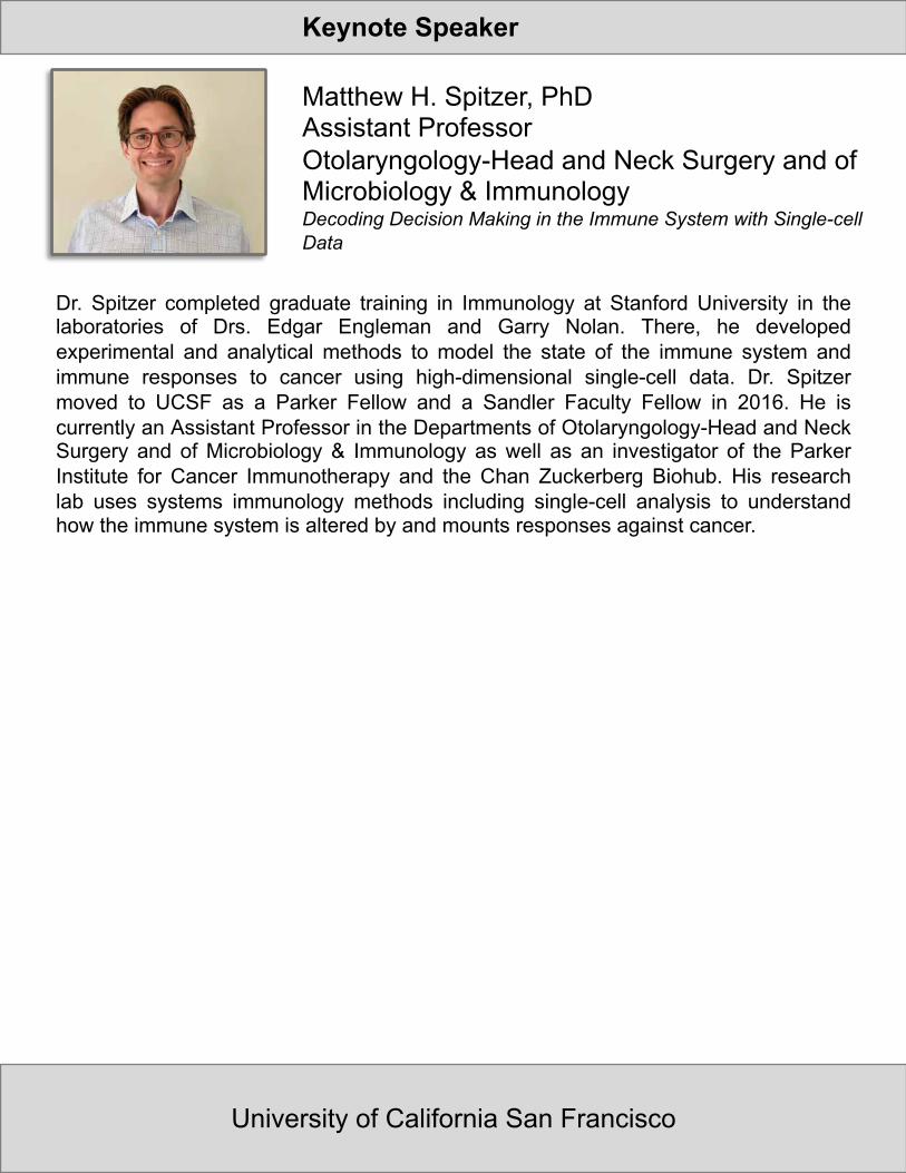
Dr. Spitzer completed graduate training in Immunology at Stanford University in thelaboratories of Drs. Edgar Engleman and Garry Nolan. There, he developedexperimental and analytical methods to model the state of the immune system andimmune responses to cancer using high-dimensional single-cell data. Dr. Spitzermoved to UCSF as a Parker Fellow and a Sandler Faculty Fellow in 2016. He iscurrently an Assistant Professor in the Departments of Otolaryngology-Head and NeckSurgery and of Microbiology & Immunology as well as an investigator of the ParkerInstitute for Cancer Immunotherapy and the Chan Zuckerberg Biohub. His researchlab uses systems immunology methods including single-cell analysis to understandhow the immune system is altered by and mounts responses against cancer.
Matthew H. Spitzer, PhDAssistant ProfessorOtolaryngology-Head and Neck Surgery and of Microbiology & Immunology Decoding Decision Making in the Immune System with Single-cell Data
University of California San Francisco
Keynote Speaker

Elaine Stur is a post-doctoral fellow at The University of Texas MD Anderson CancerCenter. She works in the lab of Dr. Anil Sood and her work has been focused on tumorheterogeneity, where she has been driving the single cell and spatial transcriptomicsunderstanding of ovarian tumors. Elaine also studies the immune suppressivemechanisms of ovarian cancer through the identification of possible new drug targets.
Elaine Stur, PhD PostdocResolved Spatial Transcriptomics of High-Grade Serous Ovarian Carcinoma
MD Anderson Cancer Center

Dr. Julea Vlassakis earned her Ph.D. in bioengineering and completed a postdoc in theHerr Lab at UC Berkeley. Her research investigated separation and thermodynamicpartitioning phenomena fundamentals to design integrated single-cell protein assays.She joined the Department of Bioengineering at Rice University as an AssistantProfessor in July 2021. She directs the Microtechnologies Laboratory for PediatricOncology with the goal of advancing targeted and precision therapies for pediatriccancers with a focus on Ewing sarcoma. To study the biochemistry and biophysics offusion oncoprotein interactions in proliferative and metastatic subpopulations, hergroup will develop single-molecule and single-cell technologies at the micro and nanoscales of cancer biology. Her research has garnered several honors including an NSFGraduate Research Fellowship, Burroughs Wellcome Fund Career Award at theScientific Interface, and the CPRIT Scholar Award.
Julea Vlassakis, PhD Assistant ProfessorBioengineeringMeasuring Cytoskeletal Protein Complex Drug Response Variation by Single-Cell Fractionation
Rice University

Dr. Runmin Wei is a Damon Runyon post-doctoral research fellow from Navin lab inMD Anderson Cancer Center. He earned his bachelor's degree in pharmaceuticalengineering and MS degree in pharmacology in China. He got his PhD degree fromUniversity of Hawaii Cancer Center with focusing on bioinformatics in metabolomicsand metagenomics. He is also interested in machine learning and deep learningbiomedical studies. He joined Navin Lab in MD Anderson in 2019. His study focuseson using single-cell and spatial sequencing technology to understand the intra-tumoralheterogeneity and tumor microenvironment in breast cancer.
Runmin Wei, PhD Postdoc Research FellowSpatial Charting of Single Cell Transcriptomes in Tissues
MD Anderson Cancer Center

Yongjian is a Ph.D. student from the Department of Electrical and ComputerEngineering, Texas A&M University. Currently, he is working at Dr. James Cai’s group,where they are studying the genetic basis of phenotypic variability and developingcomputational tools to identify genetic variants that dominate complex traits. Beforereturning to graduate school, Yongjian was employed by an IN-VITRO DIAGNOSTICScompany in Japan developing healthcare applications.
Yongjian YangPhD StudentElectrical and Computer EngineeringscTenifoldXct: Semi-supervised Manifold Alignment for Inference of Cellular Interactions Through Construction of Intra- and Intercellular Gene Signaling Networks
Texas A&M University

Nir Yosef received his Ph.D. in Computer Science from Tel Aviv University and thencompleted a postdoctoral training at the Broad Institute, where he worked ontranscriptional regulation of T cell differentiation. Nir joined the faculty at UC Berkeleyin 2014, where he is now an Associate Professor of Computer Science, a coremember at the Center of Computational Biology, and a Chan Zuckerberg Biohubinvestigator. The Yosef lab is developing data- centric methods for studying howchanges in transcription are associated with various phenotypes in the immunesystem. In that capacity, the lab is developing and applying computational tools thatleverage single cell genomics, with the goal of better understanding the factors thatcontribute to variability between cells, (e.g, metabolism, chromatin structure) and theirbroader implications (e.g., in autoimmunity). A second area of research is methoddevelopment for studying regulatory regions in the genome, based on chromatinprofiles and massively parallel reporter assays.
Nir Yosef, PhD Associate ProfessorComputer ScienceGenerative Modeling for Single Cell Genomics: Tools and an Application for Studying How T Cells Develop in the Thymus
University of California Berkeley
Keynote Speaker
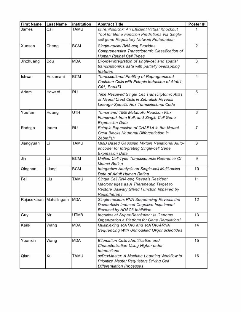
First Name Last Name institution Abstract Title Poster #James Cai TAMU scTenifoldKnk: An Efficient Virtual Knockout
Tool for Gene Function Predictions Via Single-cell gene Regulatory Network Perturbation
1
Xuesen Cheng BCM Single-nuclei RNA-seq Provides Comprehensive Transcriptomic Classification of Human Retinal Cell Types
2
Jinzhuang Dou MDA Bi-order integration of single-cell and spatial transcriptomics data with partially overlapping features
3
Ishwar Hosamani BCM Transcriptional Profiling of Reprogrammed Cochlear Cells with Ectopic Induction of Atoh1, Gfi1, Pou4f3
4
Adam Howard RU Time Resolved Single Cell Transcriptomic Atlas of Neural Crest Cells in Zebrafish Reveals Lineage-Specific Hox Transcriptional Code
5
Yuefan Huang UTH Tumor and TME Metabolic Reaction Flux Framework from Bulk and Single Cell Gene Expression Data
6
Rodrigo Ibarra RU Ectopic Expression of CHAF1A in the Neural Crest Blocks Neuronal Differentiation in Zebrafish
7
Jiangyuan Li TAMU MMD Based Gaussian Mixture Variational Auto-encoder for Integrating Single-cell Gene Expression Data
8
Jin Li BCM Unified Cell-Type Transcriptomic Reference Of Mouse Retina
9
Qingnan Liang BCM Integrative Analysis on Single-cell Multi-omics Data of Adult Human Retina
10
Fei Liu TAMU Single Cell RNA-seq Reveals Resident Macrophages as A Therapeutic Target to Restore Salivary Gland Function Impaired by Radiotherapy
11
Rajasekaran Mahalingam MDA Single-nucleus RNA Sequencing Reveals the Doxorubicin-Induced Cognitive Impairment Reversal by HDAC6 Inhibition
12
Guy Nir UTMB Inquiries at Super-Resolution: Is Genome Organization a Platform for Gene Regulation?
13
Kaile Wang MDA Multiplexing scATAC and scATAC&RNA Sequencing With Unmodified Oligonucleotides
14
Yuanxin Wang MDA Bifurcation Cells Identification and Characterization Using Higher-order Interactions
15
Qian Xu TAMU scDevMaster: A Machine Learning Workflow to Prioritize Master Regulators Driving Cell Differentiation Processes
16
Poster 1
scTenifoldKnk: An Efficient Virtual Knockout Tool for Gene Function Predictions Via
Single-cell gene Regulatory Network Perturbation
Osorio D1, Zhong Y2, Li G2, Xu Q1, Yang Y3, Tian Y4, Chapkin RS5, Huang JZ2, Cai JJ1,3,6,*
1Department of Veterinary Integrative Biosciences, 2Department of Statistics, 3Department of Electrical and Computer Engineering, 4Department of Veterinary Physiology and Pharmacology,
5Department of Nutrition, 6Interdisciplinary Program of Genetics, Texas A&M University, College Station, TX 77843, USA
*Corresponding authors: J.J.C. ([email protected])
Background: Gene knockout (KO) experiments are a proven approach for studying gene function. A
typical KO experiment usually involves the phenotypic characterization of KO organisms. The recent
advent of single-cell technology has greatly boosted the resolution of cellular phenotyping. Applications
of single-cell technology in KO experiments hold promises for providing unprecedented insights into
gene functions. However, the large-scale application of single-cell technology in systematic KO
experiments is prohibitive due to the vast resources required.
Hypothesis/Goals: We develop scTenifoldKnk, an efficient software tool that uses single-cell RNA
sequencing (scRNA-seq) data to perform virtual KO. It can be used to predict gene function, achieving
the goal of real-animal gene KO experiments.
Methods: In a scTenifoldKnk virtual KO analysis, a single-cell gene regulatory network (scGRN) is first
constructed from the scRNA-seq data of the wild-type (WT) samples. Then, a target gene is knocked out
from the adjacency matrix of constructed scGRN by setting weights of the gene’s outward edges to zeros.
This “pseudo-KO” scGRN is compared with the original WT scGRN to identify significantly
differentially regulated (DR) genes. We call these DR genes virtual-KO perturbed genes, which are used
to infer functions of the KO gene in analyzed cells.
Results: Using existing data sets, we demonstrate that the scTenifoldKnk-based virtual KO analysis
recapitulates the main findings of real-animal KO experiments and recovers the expected functions of
causal genes of Mendelian diseases in relevant cell types. Finally, we demonstrate the use of
scTenifoldKnk to perform systematic KO analysis, in which a large number of genes are individually
deleted, or a single gene is deleted in many tissues and cell types.
Conclusions: scTenifoldKnk is an efficient virtual KO tool for predicting gene function via scGRN
perturbation. It can be used to prioritize targets and predict outcomes prior to real-animal KO experiments,
as well as to conduct systematic KO to predict the functions of all genes.
Acknowledgments: This research was funded by Texas A&M University 2019 X-Grants and the DoD
grant GW200026 for J.J.C.
Poster 2
Single-nuclei RNA-seq Provides Comprehensive Transcriptomic Classification of Human Retinal Cell
Types
Cheng X1; Liang Q1, 2; Owen LA.3; Shakoor A3; Vitale AT3; Kim IK3; Morgan DJ3, 4; Li Y1; DeAngelis MM.3; Chen R1, 2
1.HGSC, Molecular and Human Genetics, Baylor College of Medicine, Houston, TX, United States.2.Verna and Marrs McLean Department of Biochemistry and Molecular Biology, Baylor College ofMedicine, Houston, TX, United States.3.Department of Ophthalmology and Visual Sciences, University of Utah, Salt Lake City, UT, UnitedStates.4.Department of Pharmacotherapy, the College of Pharmacy, University of Utah, Salt Lake City, UT,United States.
Corresponding author: Rui Chen, HGSC, Molecular and Human Genetics, One Baylor Plaza, Houston, TX, USA. E-mail: [email protected].
Background: The human retina is composed of many different neuronal and non-neuronal cell types, with their fraction in the tissue varying dramatically, ranging from 75% to less than 0.5%. Significant cell heterogeneity is observed within many retinal cell types. However, the number of cell subtypes and their molecular signature remain unknown.
Goals: Our study aims at generating the first version of human retinal cell atlas reference by characterizing the transcriptome and open chromatin profile for all cell types in the human retina.
Methods: Single-nuclei RNA-seq and single-nuclei ATAC-seq are carried out to profile healthy human retina from six individual donors using the 10x Genomics technologies. Each donor retina is dissected into three areas: the fovea, macula, and peripheral retina. A fractionation protocol was developed to enrich nuclei from rare neuron types, such as bipolar cells, amacrine cells, and retinal ganglion cells. Integrative data analysis is performed to identify cell subtypes, marker genes, chromatin signature, and transcription factors and modules. RNA in situ hybridization is performed to validate novel cell populations.
Results: A transcriptome profile is generated for over 300K nuclei, leading to the identification of over 70 cell types in the retina. Notably, the numbers of bipolar cell clusters (13) and amacrine cell clusters (39) both exceeded the previously reported primate and human studies. Through integration of the single-nuclei RNA-seq and single-nuclei ATAC-seq data, key transcription factors and transcription modules were identified at both major- and sub- cell-type level. Moreover, three-way comparison among mouse, monkey and human retina single-cell transcriptomic data revealed conserved and lineage specific cell types during evolution. Finally, it has been observed that genes associated with different human retina diseases show distinct cell type specific gene expression profiles, providing insight to potential disease mechanisms at cell subtype resolution.
Conclusions: The study represents the most comprehensive single-cell transcriptome and single-cell chromatin accessibility profiling for the human retina to date. Over 300K single nuclei are profiled and over 70 types of cells are identified, making it a high-quality dataset that can serve as the first version of a human retina cell atlas reference.
Acknowledgements: This work is supported by Cell Atlas of the Neural Retina Seed Networks, CZF2019-002425, Chan Zuckerberg Foundation.

Poster 3
Bi-order Integration of Single-Cell and Spatial Transcriptomics Data with Partially Overlapping
Features
Dou J1, Liang S1, Liang Q2, Choi J2, Li J2, Hu M3, Jiang X3, Chen R2, Chen K1
1. Department of Bioinformatics and Computational Biology, The University of Texas MD AndersonCancer Center
2. Department of Molecular and Human Genetics, Baylor College of Medicine, Houston, TX, 77030,USA
3. Department of Neuroscience, Baylor College of Medicine, Houston, TX, 77030, USACorresponding authors: Ken Chen. Email [email protected]
Background Charting a biological atlas of an organ, such as the brain, requires us to spatially resolve whole transcriptomes of single cells. Single-cell RNA-Seq (scRNA-seq) can measure transcriptomes comprehensively but lose spatial localization information of cells within organs. Image-based transcriptomic approaches, for example multiplexed error-robust fluorescence in situ hybridization (MERFISH), provide powerful means to measure both expression levels and locations of RNAs at single-cell resolution. However, they are limited in a subset of targeted genes, which impedes profiling of the whole transcriptome.
Goals Here, we devise bindSC, a tool that reconstructs a genome-wide spatial map at single-cell resolution by integrating scRNA-seq and image-based spatial data collected from the same region. The key advance of bindSC is that it achieves de novo alignment of both the rows and the columns of two different data matrices. This addresses the limitation of previous methods that are constrained to the subset of features shared by the two data matrices.
Methods
BindSC employs the bi-order canonical correlation analysis (bi-CCA) which introduces Z (transcriptomics matrix on targeted gene panel) to link X (spatial transcriptomics matrix on targeted gene panel) and Y (transcriptomics matrix on full gene panel). Bi-CCA iteratively updates Z to find an optimal solution which maximizes the correlation between X and Z and between Y and Z in the latent space simultaneously. Based on the integration, bindSC then transfers cell types annotated in scRNA-seq data onto matched spatial transcriptomics data.
Results/Conclusions We demonstrate that incorporating unshared genes significantly improves the integration of scRNA-seq and MERFISH data on mouse brain data. bindSC improves the percentage of annotated MERFISH cells from ~55% to 80% with maximal probability on cell type assignment being 0.9 (Fig.1 l). Our integration provides higher resolution on cell type annotation compared with that from MERFISH data only (Fig.1 a-h), especially for rare cell type annotation. The reconstructed genome-wide spatial map can thus reveal spatial gene expression patterning beyond the limitations of current spatial transcriptomics technologies (Fig. 1 i-k).
Acknowledgements This project has been made possible in part by the Human Cell Atlas Seeds Network Grant CZF2019-02425 to RC and KC.

Poster 3
Fig.1 Integration of single-cell and spatial transcriptomics using bindSC. We applied bindSC to integrate MERFISH (Zhang
et al., 2020) and scRNA-seq data (Yao et al., 2020) in the mouse primary motor cortex (MOp) region. a-h: Improvement of cell type annotation by projecting scRNA-seq cell types on spatial map. The panel a, c, e, g shows spatial distribution of four clusters (5,6,7,17) determined by MERFISH in one of the coronal slices. Each column denotes one cluster. The panel b, d, f, h show spatial distribution of scRNA-seq cell types based on bindSC label transfer. The cells are the same with the top but colored with scRNA-seq cell types. These MERFISH clusters are composed of multiple scRNA-seq cell types. i-k: Imputation of spatial gene expression for genes not included in MERFISH panel. Three
example genes are shown with Osr1 (i), Rnf152(j), and Otof (k). The darker color denotes higher expression level. l: Comparison of label transfer on three methods. We integrated two modalities using bindSC, CCA, and Harmony and then used Support Vector Machine (SVM) to perform label transfer for each method. The x-axis denotes the maximal prediction probability on assigning cell type for SVM. The y-axis denotes the proportion of MERFISH cells that are annotated with scRNA-seq cell types.
Zhang M, Eichhorn S W, Zingg B, et al. Molecular, spatial and projection diversity of neurons in primary motor cortex revealed by in situ single-cell transcriptomics[J]. Biorxiv, 2020.
Yao Z, Liu H, Xie F, et al. An integrated transcriptomic and epigenomic atlas of mouse primary motor cortex cell types[J]. Biorxiv, 2020.

Poster 4
Transcriptional Profiling of Reprogrammed Cochlear Cells with Ectopic Induction of Atoh1, Gfi1,
Pou4f3
Iyer A1, Hosamani I, 1Groves A
1. Dept. of Molecular and Human Genetics, Baylor College of Medicine
Corresponding author: Andrew K Groves, Molecular and Human Genetics, Baylor College of Medicine, 1 Baylor Plaza, Houston, Texas, Email: [email protected],
Background Sensory hair cells of the cochlea function as transducers of sound energy into electrical impulses which are then transmitted to the brain. Mammalian hair cells can be killed by exposure to loud sounds, certain antibiotics and chemotherapy drugs, as well as aging, which is one of the major causes of hearing loss. Once lost, mammalian cochlear hair cells do not regenerate. However, regeneration of hair cells has been observed in non-mammals, and research towards replicating this phenomenon in mammals has been actively pursued for over 20 years. Reprogramming of supporting cells of the cochlea into hair cells by inducing ectopic hair cell transcription factors has emerged as one of the top methods to achieve hair cell regeneration.
Hypothesis The ectopic expression of transcription factors Atoh1, Gfi1 and Pou4f3 can reprogram supporting cells in to hair cells.
Methods In order to reprogram supporting cells to hair cells, we used Sox9CreER mice, which drive recombination in supporting cells of the cochlea. We bred these mice with mice carrying a Cre-inducible tdTomato reporter gene targeted to one allele of the ROSA26 locus, and a second modified ROSA26 allele in which Atoh1 alone, or combination of Atoh1-Gfi1 or Atoh1-Gfi1-Pou4f3 transcription factors could be expressed in a Cre-inducible manner. Ectopic expression of these transcription factors was triggered by tamoxifen injection at P1 and tdTomato+ cells were FACS sorted at P8 and profiled by single cell RNA sequencing using the 10X Genomics Chromium V3 kit. The resulting data were aligned with the mm10 mouse reference genome and further analysis was performed with the Seurat R package. Briefly, after appropriate filtering of each of the datasets to remove low quality cells with low number of counts and any potential doublets with very high counts, the data were log normalized and the top 2000 variable genes in each dataset were identified. Next, an integrated analysis was performed to identify clusters that differed among the various genotypes and the clusters were classified into appropriate cell types based on their marker gene expression.
Results and Conclusions While we did not see any hair cell gene expressing clusters in the control (Sox9CreER x Ai9 reporter only) dataset, we were able to identify new clusters that expressed hair cell genes in the transcription factor induced datasets. We also saw a new population of supporting cells with similar transcriptional signature in all the induced datasets. The results of the single cell analysis reflected prior immunofluorescence experimental observations and serves as a good validation for the cellular reprogramming of the supporting cells in the inner ear.
Acknowledgements: This was supported by the grant R01DC014832.

Poster 5
Time Resolved Single Cell Transcriptomic Atlas of Neural Crest Cells in Zebrafish Reveals Lineage-
Specific Hox Transcriptional Code
Howard AGA1, Uribe RA1
1. BioSciences Department, Rice University
Corresponding author: Rosa Uribe, Department of BioSciences, Rice University, 6100 Main Street-MS140, Houston, TX, E-mail: [email protected]
Background: Neural crest cells (NCCs) are a highly migratory, multipotent, and transient stem cell population essential to vertebrate development. Born along the neural tube at early developmental stages, NCCs are regionally specified along the anterior-posterior axis to contribute to a vast number of cell lineages, such as craniofacial cartilage, pigment cells, and large tracts of the peripheral nervous system. In on our recently published analysis of posterior zebrafish NCC, we discovered an emerging combinatorial expression code of transcripts that encode for Hox transcription factors, which was specific to NCC-derived neural lineages. A Hox expression code has been characterized within the cranial NCC, but prior unto had not been observed within the context of the posterior neural NCC lineages. Importantly, the combinatorial while code persisted throughout the embryonic-to-larval transition, it remained unclear if this code persisted in NCC at other developmental stages. A comprehensive description of the combinatorial changes of Hox expression in NCC will greatly contribute to our understanding of posterior NCC specification, fate acquisition, and differentiation.
Goals: The goal of this study is to map the expression in combination of Hox transcription factors across several posterior NCC lineages over time.
Methods: Several published single cell RNA transcriptomic (scRNA-seq) studies characterizing FACS-sorted NCC and NCC-derived cells were computationally merged using the Seurat (v3.1.1) integration pipeline in Rstudio (v3.6.3). Data was sourced from NCBI GEO database to include the following datasets: 24 hours post fertilization) (GSE163907), 48-50 hpf (GSE152906), 68-70 hpf (GSE152906), 5 days post fertilization (GSE131136), Juvanile (GSE131136), and adult tissues (GSE131136). All datasets were reprocessed to standardize cell thresholds and labeled with slight modification according to the original publications for consistency.
Results: Integration of datasets produced a cohesive and expected distribution of cell identities, as noted by cells from separate temporal stages constellating proximally to presumptively more differentiated cells from older stages. Throughout this comprehensive transcriptomic atlas, Hox Transcriptional codes were observed in several NCC-lineages in every dataset analyzed, but was particularly robust in datasets from 48 hpf through adult lineages. Discrete lineages were detected for neural tissues, particularly the enteric nervous system.
Conclusions: The generation of a multi-stage comprehensive transcriptional atlas from multiple scRNA-seq studies will serve as a valuable tool for the NCC zebrafish community. Additionally, this atlas of cell lineages demonstrates the complexity and robustness of combinatorial Hox expression codes in posterior NCC throughout development.
Acknowledgements: A special thanks goes to the members of the Uribe Lab who have supported this project. Additional thanks goes to Dr. Ezra Lencer for providing digital resources from their recent publication. Funding for this project was provided by Rice University, Cancer Prevention & Research Institute of Texas (CPRIT) Recruitment of First-Time Tenure Track Faculty Members (CPRIT-RR170062) and the NSF CAREER Award (1942019) awarded to R.A.U.

Poster 6
Tumor and TME Metabolic Reaction Flux Framework from Bulk and Single Cell Gene Expression
Data
Huang Y 1, Mohanty V 1, Dede M 1, Daher M 2, Li L 2, Rezvani K2, Chen K 1
1. Department of Bioinformatics and Computational Biology, The University of Texas MD Anderson
Cancer Center, Houston, TX 77030
2. Department of Stem Cell Transplantation and Cellular Therapy, University of Texas MD Anderson
Cancer Center, Houston, TX 77030
Corresponding author: Ken Chen, Department of Bioinformatics and Computational Biology, The
University of Texas MD Anderson Cancer Center, Houston, TX, 77030
Background: The metabolic reprogramming in both TME (Tumor Microenvironment) and tumor has been
considered a hallmark of cancer because tumor cells often alter metabolic strategies under nutrient-deprived
conditions to support their growth and survival. The TME, including immune cells, cancer-associated
fibroblast, stromal cells, is acidic, hypoxic, and nutrient-deficient. As a result, tumor cells either compete
or cooperate for nutrients with other cell types to sustain tumor proliferation. A fundamental interest in
cancer research is to understand the underlying mechanisms of those altered activities in tumors and TME
and explore potential rational metabolic therapeutical targeting.
Goals: However, our understanding of metabolic competition and cooperation in TME is still limited.
RNA-seq and scRNA-seq datasets provide unprecedented resolution of tumor inter- and intra-heterogeneity,
but its use in understanding metabolic activities is still lacking.
Methods: To fill this gap, we developed a computational framework to infer the global metabolic reaction
network from RNA-seq and scRNA-seq data. Our method utilized FBA (Flux Balance Analysis) coupled
with quadratic programming to estimate 13,082 metabolic reaction fluxes. We then employed a graph-based
approach to summarize the biochemical reaction fluxes into individual pathways activity scores.
Results: We validated our predicted fluxes using NCI-60 experimental exchange flux data. Through further
application on TCGA data, we accurately validated the key metabolic difference in oxidative and glycolytic
tumors. In addition, we discovered a novel metabolic reaction associated with hypoxia. We also applied
our method on public single-cell data and observed a dynamic, metabolic interaction between tumor and
immune cells under different TME compositions. Our high-resolution metabolic networks can significantly
facilitate downstream analysis and generate dynamic metabolic interaction TME profiles beyond what the
current methods can provide.
Acknowledgement: This project has been made possible by MD Anderson Moon shots program.

Poster 7
Ectopic Expression of CHAF1A in the Neural Crest Blocks Neuronal Differentiation in Zebrafish
Ibarra-Garcia-Padilla R 1, Tao L2,3, Barbieri E2,3, Uribe RA1
1. Department of Biosciences, Rice University, Houston, TX 77005, USA.2. Department of Pediatrics, Section of Hematology-Oncology, Texas Children’s Cancer and
Hematology Centers, Baylor College of Medicine, Houston, TX 77030, USA.3. Dan L Duncan Comprehensive Cancer Center, Baylor College of Medicine, Houston TX 77030
Corresponding author: Rosa A. Uribe, Department of Biosciences, Rice University, 6100 Main St, Houston, TX 77005, E-mail: [email protected]
Background: Neural Crest Cells (NCC) are a transitory multipotent stem cell population, present during vertebrate embryogenesis, that migrate to distinct sites within the developing embryo and give rise to a variety of cell types, including neurons and glia of the sympathetic nervous system. CHAF1A, a member of the chromatin assembly factor (CAF-1), is required for development in the early embryo, however its roles during vertebrate neuronal development are not well known. In humans, CHAF1A is highly expressed in neuroblastoma (NB), a neural crest-derived pediatric cancer, suggesting it may be involved in the neural crest to neuroblastoma transformation.
Hypothesis/Goals: In an effort to gain insight into CHAF1A’s role in NB progression, our goal was to test the hypothesis that CHAF1A blocks NCC differentiation towards a neuronal fate in vivo.
Methods: By leveraging recent single cell RNA-seq datasets from NCC and NCC-derived cells in the vertebrate model zebrafish, as well Hybridization Chain Reaction (HCR), we examined the spatiotemporal expression of chaf1a during NCC development and during early and late neuronal differentiation. Additionally, we generated constructs for overexpression of human CHAF1A in the NCC lineage and in vivo performed clonal analysis, after injection into zebrafish embryos, to quantify changes in neuronal differentiation.
Results: We discovered that zebrafish chaf1a is strongly expressed in the NCC pre-migratory population and during early NCC migration phases. During later phases of development, we found that chaf1a drastically decreased in NCC-derived cells undergoing neuronal differentiation, suggesting that its exclusion is necessary for neuron differentiation. Indeed, ectopic expression of human CHAF1A in zebrafish NCC lineage was sufficient to prevent neuronal differentiation in vivo.
Conclusions: These data show that chaf1a is expressed during early neural crest development, but its expression is rapidly lost in cells undergoing neuronal differentiation, and increased CHAF1A in NCC blocks neuronal differentiation. Overall, this project increases our understanding of CHAF1A and sheds light on its potential role during neural crest differentiation and NB oncogenesis.
Acknowledgements: Funding for this project provided by Cancer Prevention & Research Institute of Texas grant RR170062 and the American Cancer Society RSG-19-107-01.

Poster 8
MMD Based Gaussian Mixture Variational Auto-encoder for Integrating Single-cell Gene Expression
Data
Li J1,2, King R1,3, Huang JZ2, Zhang KK1,4*
1. Center for Epigenetics & Disease Prevention, Rigor and Reproducibility Core, Institute of Biosciences
& Technology, College of Medicine, Texas A&M University, Houston, TX
2. Department of Statistics, Texas A&M University, College Station, TX
3. Department of Computer Science and Engineering, Texas A&M University, College Station, TX
4. Department of Nutrition and Food Sciences, Texas A&M University, College Station, TX
*Corresponding author: Ke K. Zhang, Center for Epigenetics & Disease Prevention, Institute of
Biosciences & Technology, College of Medicine, Texas A&M University, 2121 W Holcombe Blvd,
Houston, TX, [email protected]
Background: Single-cell RNA sequencing is revolutionizing biomedical research by revealing molecular
variations at a cellular resolution. A number of statistical and computational methods have been proposed
to process and analyze data from a single experiment. Deep learning method, particular variational auto-
encoder (VAE) has been proven a powerful tool in learning latent representations of gene expression
patterns, which facilitates downstream analysis, such as visualization, clustering, and differential
expression. However, it remains a major challenge to perform integrative analysis of single cell data across
different datasets.
Methods: We developed a neural network based Gaussian mixture VAE for single-cell analysis, which
learned latent representation of each cell and achieved optimal clustering of cells by their physiological
types. Furthermore, with a robust incorporation of maximum mean discrepancy (MMD) regularization, the
model automatically minimized the batch effects across multiple datasets and regenerated normalized gene
expression data for subsequent statistical analysis.
Results: We demonstrated the effectiveness of our model for dimension reduction and clustering on various
settings of simulated data as well as real data (PBMC). The effectiveness of our model on batch correction
is observed on a diverse set of real data with emphasis on liver data.
Acknowledgements: This study was supported by NIH grant 1R01DK112368-01 and AHA grant
15GRANT25700195 for K.K.Z.

Poster 9
Unified Cell-Type Transcriptomic Reference of Mouse Retina
Li J, Choi J, Ferdous S, Chen R
HGSC, Department of Molecular and Human Genetics, Baylor College of Medicine, Houston, TX,
77030, USA
Corresponding author: Rui Chen, email: [email protected]
Background Single cell RNA sequencing (scRNA-Seq) has advanced the assessment of cellular heterogeneity level at the single cell resolution by transcriptional similarities and differences. Data resources of scRNA-Seq have been largely produced and extensively studied for mouse retina. These data resources form a powerful tool to study cellular components, transcriptome relationships, and regulatory mechanisms underlying various retinal diseases and biological processes.
Goals The large volume of mouse retinal scRNA-Seq data have been released in separate repositories, restricting them from wide-use in mouse retina communities. In this work, we are presenting a unified cell-type reference for wild-type mouse retina using our generated single-cell/nucleus RNA-Seq experiments complementing publicly available samples. This unified reference serves an easy-to-use data resource of mouse retina for mouse retina communities.
Methods To achieve the most comprehensive mouse retina cells, we produced extensive single-cell/neucli RNA-Seq cells from our in-house wild-type mouse samples. By incorporating publicly available yet separate hosted samples, we have collected the most comprehensive single cells for mouse retina. These collected single cells undergo integration after removing sample effects using scVI 1. The low-dimensional representations of integrated cells are used to measure cell similarities and cell clustering by Leiden algorithm. Major cell types are annotated by matching publicly annotated cells and major cell marker genes. Subtypes of amacrine cells (ACs), bipolar cells (BPs), and retinal ganglion cells (RGCs) are generated by sub-clustering and annotation on major type specific cells. Annotated major cell types and subtypes form a unified reference of mouse retina. To facilitate the public use of the reference, we deposit the reference at cellxgene 2 for visualization and gene signature inspection. Pretrained classifiers using scPred 3 are also released to annotate new mouse retinal cells.
Results and conclusions We have collected over 220,000 mouse retinal cells from in-house experiments and public samples. These a quarter million cells formed 11 major cell types and over 100 subtypes (Figure 1A). To facilitate the public use, our reference has been deposited at cellxgene for visualization and inspection of gene signatures (Figure 1B). Pretrained classifiers are shared in public URL for annotating incoming new mouse retina cells. This universal reference facilitates the single-cell studies of mouse retina.
Acknowledgements

Poster 9
Figure 1. Unified reference of mouse retina. (A) Annotated cell types of mouse retina. (B) Visualization of the reference at cellxgene.
Reference:
1 Lopez, R., Regier, J., Cole, M. B., Jordan, M. I. & Yosef, N. Deep generative modeling for single-cell transcriptomics. Nature Methods 15, 1053-1058, doi:10.1038/s41592-018-0229-2 (2018).
2 Megill, C. et al. cellxgene: a performant, scalable exploration platform for high dimensional sparse matrices. bioRxiv, 2021.2004.2005.438318, doi:10.1101/2021.04.05.438318 (2021).
3 Alquicira-Hernandez, J., Sathe, A., Ji, H. P., Nguyen, Q. & Powell, J. E. scPred: accurate supervised method for cell-type classification from single-cell RNA-seq data. Genome Biol 20, 264, doi:10.1186/s13059-019-1862-5 (2019).

Poster 10
Integrative Analysis on Single-cell Multi-omics Data of Adult Human Retina
Liang Q1, 2, Cheng X1, Wang J1, Owen L3,4, Li Y1, DeAngelis M3,4,5, Chen R1, 2
1. Department of Molecular and Human Genetics, Baylor College of Medicine, Houston, TX, 77030,United States.
2. Verna and Marrs McLean Department of Biochemistry and Molecular Biology, Baylor College ofMedicine, Houston, TX, 77030, United States.
3. Department of Ophthalmology and Visual Sciences, University of Utah, Salt Lake City, UT, 84132,United States.
4. Department of Population Health Sciences, University of Utah, Salt Lake City, UT, 84132, United States.
5. Department of Ophthalmology, Jacobs School of Medicine and Biomedical Engineering, University at Buffalo SUNY, and the VA Western New York Healthcare System, Buffalo, NY 14215, USA, Buffalo, NY 14215, United States.
Corresponding author: Rui Chen, Department of Molecular and Human Genetics, Baylor College of Medicine, Houston, TX, 77030, United States. E-mail: [email protected]
Abstract
Background. Cell types in the human retina are highly heterogeneous with their abundance varies by several orders of magnitude and is functionally required for the human visual system. However, a complete map of the human retinal cell types is lacking and moreover, the gene regulation network behind it is largely unrevealed.
Hypothesis. We hypothesize that a large portion of the heterogeneity of the transcriptome of retinal cells are originated from their diverse chromatin landscape.
Methods. To decipher the complexity, we generated a multi-omics single-cell atlas of the adult human retina including over 250K nuclei for single-nuclei RNA-seq and 150K nuclei for single-nuclei ATAC-seq. To balance the number of cells from abundant and rare cell types, we performed antibody-based nuclei sorting to enrich rare cells, mainly amacrine cells and retinal ganglion cells.
Results. We were able to integrate the snATAC-seq data to snRNA-seq data, generating a chromatin accessibility atlas for over 50 retinal cell types. We identified 70k distal cis-element-gene pairs for over 11k genes, with a majority of them being cell type specific. In addition, our distal cis-element list had a significant enrichment of previously discovered elements from bulk sequencing studies and showed improvement in the interpretability of the element.
Conclusion. Taken together, this new dataset represents the most comprehensive single-cell multi-omics profiling for the human retina that enables in-depth molecular characterization of most cell subtypes.
Acknowledgements. We thank the Human Cell Atlas Seed Network Grant CZF2019-02425 to RC for supporting this study.

Poster 11
Single Cell RNA-seq Reveals Resident Macrophages as a Therapeutic Target to Restore Salivary Gland
Function Impaired by Radiotherapy
Zhao Q1, Zhang L1, Hai B1, Wang J2, Baetge L3, Deveau M3, Kapler G1, Feng J2, and Liu F1
1. Molecular and Cellular Medicine Department, College of Medicine, Texas A&M University.2. Department of Biomedical Sciences, College of Dentistry, Texas A&M University, Dallas, TX.3. Department of Small Animal Clinical Sciences, College of Veterinary Medicine, Texas A&M
University.
Corresponding Author: Fei Liu, Texas A&M University, 206 Olsen Blvd, 242BReynolds Medical Bldg., College Station, TX 77843. E-mail: [email protected]
Background. Salivary gland (SG) destruction is a major adverse effect of radiotherapy for head and neck cancer. Consequent dry mouth severely compromises quality of life and is difficult to remedy. My laboratory found that the transient activation of Hedgehog signaling pathway after radiation rescued salivary function (Hai et al., 2014; 2016; 2018), but the exact mechanisms remain unclear.
Goals. Determine SG cells directly responsive to Hedgehog activation and their roles in the rescue of SG function damaged by radiation.
Methods. Mouse SGs were not treated or transferred with control GFP gene or Sonic Hedgehog (Shh) gene, and then collected at 7 days later for single cell RNA sequencing with 10x Chromium platform (10x Genomics). Data were analyzed with Cell Ranger, R package scater, and Seurat, and visualized with the Uniform Approximation and Projection (UMAP) method. Findings were verified by lineage tracing in mouse models and qRT-PCR and ELISA of SG cells isolated with FACS.
Results. In non-treated SGs, resident macrophages (rMφs) are abundant and interact with epithelial progenitors (EP) and endothelial cells (Endo) through paracrine homeostatic factors such as C1q, Hgf and Csf1/2. SG rMφs are the major type of cells expressing key membrane mediators of Hedgehog signaling, while lineage tracing confirmed that Mφs are major Hedgehog-responsive cells in SGs. Intra-SG transfer of Shh but not GFP gene greatly enhanced the paracrine homeostatic interaction between rMφs with other SG cells. SG rMφs are also the major type of cells expressing the oxidative stress sensor Trpm2 that causes cell death in response to radiation. Consistently, SG rMφs are severely damaged by radiation, restored by Hedgehog activation, and required for the restoration of salivary function by Hedgehog activation following radiation.
Conclusions. SG rMφs are a promising therapeutic target to restore salivary function impaired by radiotherapy, while single cell RNA sequencing is a powerful approach to determine roles of cell subsets such as rMφs during tissue regeneration.
Acknowledgements. This study is funded by NIH 1R01DE022975. We sincerely thank Drs. Andrew Hillhouse and Kranti Konganti in TAMU Genomics Core for scRNA-seq.

Poster 11
A: Cell clusters identified in NT SGs with scRNA-seq. iMφs: infiltrating macrophages, ILCs: innate lymphoid cells, Basal: basal/myo-epithelial cells. B: Scheme of paracrine homeostatic interaction between SG rMφs and other cells and the potential of modulating rMφs for restoring salivary function impaired by radiotherapy.

Poster 12
Single-nucleus RNA Sequencing Reveals the Doxorubicin-Induced Cognitive Impairment Reversal by
HDAC6 Inhibition
Mahalingam R1*, McAlpin B1*, Singh A1, Dharmaraj S1, Boukelmoune N1, Kavelaars A1, Heijnen CJ1
1. Department of Symptom Research, Laboratory of Neuroimmunology, The University of Texas MDAnderson Cancer Center, Houston, TX 77030*These authors contributed equally
Corresponding author: Dr. Cobi J Heijnen, Chair, Department of Symptom Research, The University of Texas MD Anderson Cancer Center, Houston, TX 77030, E-mail: [email protected]
Background: Doxorubicin (DOX) is a highly effective chemotherapy used to treat breast cancer. Unfortunately, up to 60% of survivors report chemotherapy-induced cognitive impairment (CICI) characterized by deficits in working memory, processing speed and executive functioning. Recent reports show the promising effect of histone deacetylase 6 (HDAC6) inhibition on treating cognitive impairment in neurodegenerative disorders. We recently demonstrated that the ACY-1083, an HDAC6 inhibitor, successfully reversed cognitive impairment in a cisplatin-induced model of CICI.
Hypothesis/Goals: In this study, we hypothesize that HDAC6 inhibition by ACY-1083 can reverse DOX-induced cognitive impairment. We explored transcriptomics changes using single-nucleus RNA sequencing (snRNA-seq).
Methods: Mice were treated with doxorubicin HCL (5 mg/kg/week, Pfizer, New York, NY) or PBS intraperitoneally for 4 weeks, followed by 1 week of rest. Mice were then treated with the blood-brain barrier permeable HDAC6 inhibitor ACY-1083 (10 mg/kg/day, Regenacy Pharmaceuticals, Waltham, MA) or vehicle intraperitoneally daily for 2 weeks. The nuclei from the mouse hippocampus were isolated. The 10x Genomics Chromium Next GEM 3’ Single Cell Reagent kits v3.1 was used for the library preparation and sequencing was done with Illumina NovaSeq 6000. The sequencing data were processed with CellRanger (version 6.0). All the downstream analysis was performed with the Seurat package.
Results: The integrated analysis of control, DOX, and DOX+ACY treated samples revealed 11 cell types. The pathway analysis indicated that DOX treatment affected genes related to mitochondrial function. The analysis of the microglia population showed an increase in neurodegeneration-associated genes and loss of microglia homeostasis genes that suggest a neurodegenerative microglia phenotype closely resembling stage 1 disease-associated microglia (DAMs). In the dentate gyrus (DG) neuron population, HDAC6 inhibition enriched a sub-cluster that express genes related to neuronal development and synaptogenesis. Similarly, HDAC6 inhibition increased the mature oligodendrocyte population. Behavioral testing confirmed that ACY-1083 reversed CICI. Overall, transcriptomics changes in the multiple cell types and the enrichment of a subpopulation expressing genes related to neuronal development and synaptogenesis could be the mechanism for reversing the cognitive impairment in our CICI mouse model.
Conclusions: The snRNA-seq reveals changes in gene expression in specific cell populations including microglia, oligodendrocytes, and DG neurons. These novel findings advance our understanding of the effects of DOX and ACY-1083 treatment on different cell types which has potential implications in treating CICI.
Acknowledgments: This study was funded by NIH RO1CA208371, NS073939, and NIH RO1CA227064 to CJH and AK, and the MD Anderson Cancer Center Core Grant NIH P30CA016672.

Inquiries at Super-Resolution: Is Genome Organization a Platform for Gene Regulation?
Nir G1,7, Farabella I2, Perez-Estrada C3, Ebeling CG4, Beliveau BJ5, Sasaki HM6, Lee DS7, Stuckey JA8, Yin P9, Aiden EL3, Marti-Renom MA2, Wu C7
1. Department of Biochemistry and Molecular Biology, UTMB, Galveston, TX, USA2. CNAG-CRG, Centre for Genomic Regulation (CRG), Barcelona Institute of Science and Technology
(BIST), Barcelona, Spain3. Center for Genome Architecture, Department of Molecular and Human Genetics, Baylor College of
Medicine, Houston, Texas, USA4. Bruker Nano Inc., Salt Lake City, Utah, USA5. Department of Genome Sciences, University of Washington, Seattle, Washington, United States of
America6. 10X Genomics, CA, USA7. Department of Genetics, Harvard Medical School, Boston, Massachusetts, USA8. Bruker Nano Inc., Middleton, Wisconsin, United States of America9. Wyss Institute for Biologically Inspired Engineering, Harvard University, Boston, Massachusetts,
USA
Corresponding author: Guy Nir, Department of Biochemistry and Molecular Biology, UTMB, 14th street, Galveston, TX, USA, email: [email protected]
Background. The way that the genome is folded and packed in the nucleus is of major importance. A multitude of studies have shown that the organization of genomes can influence gene expression. However, other studies contrasted this view by showing that genome wide disruption of chromosomal loop domains does not lead to significant changes in gene expression. These contrasting views are likely to be affected by these ensemble-level studies averaging over millions of structures and transcriptomes.
Goal. Here we introduce a new multiplexed imaging technology for visualizing chromosomal DNA at super-resolution and its integration with Hi-C data to produce three-dimensional models of chromosome organization. The method we develop bridges the gap between imaging, fixed-cell methods such as FISH, and molecular methods such as Hi-C.
Results. Using the super-resolution microscopy methods of OligoSTORM and OligoDNA-PAINT, we trace eight Mbp of human chromosome 19, which is the longest stretch of genomic DNA investigated thus far with super-resolution microscopy. Leveraging this technology, we discover different packaging levels of genomic elements that range in size from a few kilobases to over a megabase.
Conclusions. We obtain evidence that maternal and paternal homologous regions are organized differently. And, focusing on chromosomal regions that contribute to compartments, we discover distinct structures that can predict whether such regions correspond to compartments of active or inactive genes. Funding: Cancer Prevention Research Institute of Texas (R210018), UT STAR award.
Poster 13

Poster 14
Multiplexing scATAC and scATAC&RNA Sequencing with Unmodified Oligonucleotides
Wang K1,7, Xiao Z1,7, Yan Y1,2,7, Ye R2,3, Hu M1, Bai S1, Sei E1, Qiao Y3, Chen H4, Lim B5, Lin SH2,3, Navin
NE1,2,6,8*
1. Department of Genetics, UT MD Anderson Cancer Center, Houston, TX 77030, USA
2. Graduate School of Biological Sciences, UT MD Anderson Cancer Center, Houston, TX 77030, USA
3. Department of Radiation Oncology, UT MD Anderson Cancer Center, Houston, TX 77030, USA
4. Department of Pathology, UT MD Anderson Cancer Center, Houston, TX 77030, USA
5. Department of Breast Medical Oncology, UT MD Anderson Cancer Center, Houston, TX 77030, USA
6. Department of Bioinformatics and Computational Biology, UT MD Anderson Cancer Center, Houston, TX 77030,
USA
7. These authors contributed equally
8. Lead contact
*Correspondence: [email protected] (N.N.)
Background
Single-cell ATAC-seq (scATAC-seq) methods have emerged as powerful tools to measure the chromatin
accessibility landscape and investigate the role of transcription factors at promoters, enhancers, activators and
insulators at single cell genomic resolution. While microdroplet-based methods can generate data on thousands
of cells from a single sample, it remains difficult and expensive to run these assays on large sample collections,
since each sample must be run in a single microfluidics channel.
Goals
Develop a simple, highly scalable and accuracy multiplexing method to realize multiplexing large-scale
scATAC-seq and single cell ATAC&RNA co-assays (10X Genomics) on microdroplet platforms. If possible,
the method will avoid any additional experimental steps to barcode and label the cells in advance.
Methods
The SNuBar-ATAC approach utilizes a simple workflow by adding a single oligonucleotide barcode during
the existing tagmentation step in the scATAC assays to label each sample with a unique identifier that is
demultiplexed in the data post-processing steps.
Results
We validated the performance, efficiency and scalability of SNuBar-ATAC in cell line experiments and applied
it to two different biological applications, including profiling chromatin accessibility changes induced by drug
treatment combinations and studying macro-spatial areas of breast tissue regions. Additionally, we applied
SNuBar for multiplexing single cell ATAC&RNA co-assays of combined samples from cell lines and human
breast tissues.
Conclusions
Our data show that SNuBar is a highly accurate, easy-to-use and scalable system for multiplexing scATAC-
seq and scATAC&RNA-seq experiments.
Funding sources
This study was supported by grants to N.E.N. from the American Cancer Society (129098-RSG-16-092-01-
TBG), the NIH National Cancer Institute (RO1CA240526, RO1CA236864, UO1 CA216468), the Emerson
Collective Cancer Research Fund and the CPRIT Single Cell Genomics Center (RP180684). N.E.N. is an
AAAS Fellow, AAAS Wachtel Scholar, Andrew Sabin Family Fellow and Jack & Beverly Randall Innovator.

Poster 14
This study was supported by the MD Anderson Sequencing Core Facility Grant (CA016672). We thank Hongli
Tang, Louis Ramagli, and Erika Thompson for their help with next-generation sequencing.

Poster 15
Bifurcation Cells Identification and Characterization Using Higher-order Interactions
Wang Y1, Mohanty V1, Liang S1, Tan Y1, Li J5, Navin N2, Li Z3, Issa GC4, Andreeff M4, Chen R5, Chen K1
1. Department of Bioinformatics and Computational Biology2. Department of Genetics3. Department of Biostatistics4. Department of Leukemia, The University of Texas MD Anderson Cancer Center5. Molecular and Human Genetics, Baylor College of Medicine
Corresponding author: Ken Chen, Department of Bioinformatics and Computational Biology, The University of Texas MD Anderson Cancer Center, Houston, TX, [email protected]
Background Multi-cell organism development involves a lot of transitions between different cell types and cell states. If the behaviors of the cell change qualitatively after the transitions, it’s called bifurcation. The bifurcation process is heterogeneous and contains important gene regulatory networks changes. These changes play an important role in cell fate determination for both normal and disease.
Hypothesis/Goals Tumor cells genesis and metastasis can be described using the dynamic process. By using the math equations, one can identify bifurcation cells and gain more in-depth information about tumor dynamics from the quantitative aspect. This information can potentially help with prognosis and finding the right targets for the therapy.
Methods Single-cell RNA-seq data were first pre-processed for QC. The cell type of each cell were manually identified using marker genes. Within each cell type, cells were ordered using pseudotime and formed meta-cells. Top 100 most variable genes were extracted for Pearson’s correlation coefficient calculation. The absolute value of correlations were used to identify bifurcation cells. Gene pairs with highest correlation coefficients were selected for further analysis and interpretation.
Results We used single-cell RNA-seq data of normal fetal developmental foveal retina RGCs at week 10 to calculate the Pearson’s correlation coefficients of top 100 most variable genes. We found three groups of bifurcation cells in RGCs and some adhesion related genes. We also applied this method to AML patients data. We found the percentage of bifurcation tumor cells were higher in both two non-responder patients after the treatment than before the treatment. Gene pairs which have a high Pearson’s correlation within these bifurcation cells are stemness related, including CD34 and SPINK2. This indicates the regulatory relations of these stemness genes are stronger after the treatment comparing to those before the treatment.
Conclusions and Discussion Bifurcation is an important and dynamic process during cell development for both normal and disease. We proposed a quantitative bottom-up model to describe this process and mathematically proved bifurcation cells tend to have higher absolute values of gene-pair Pearson’s correlation coefficients. However, further analysis such as regulatory network inference are needed to get the whole picture of cell fate determination.
Acknowledgements We thank all the members in Dr. Ken Chen’s lab for comments and suggestions; all the members in Dr. Rui Chen’s lab for retina samples preparation, sequencing and annotation; all the people in AML Moonshot MRD project for sample preparation, sequencing and clinical information collection.

Poster 16
scDevMaster: A Machine Learning Workflow to Prioritize Master Regulators Driving Cell
Differentiation Processes
Xu Q1, Li G2, Osorio D3, Zhong Y4, Yang Y5, Cai JJ1, 5
1. Department of Veterinary Integrative Biosciences, Texas A&M University2. Department of Statistics, Texas A&M University3. Norwegian Center for Molecular Medicine (NCMM), University of Oslo4. Key Laboratory of Advanced Theory and Application in Statistics and Data Science-MOE, School of
Statistics, East China Normal University5. Department of Electrical and Computer Engineering, Texas A&M University
Corresponding author: James J. Cai, 660 Raymond Stotzer Pkwy, College Station, TX 77843, E-mail: [email protected]
Background: Understanding cell differentiation processes and underlying gene regulatory programs are extremely important in fields such as disease molecular biology and regenerative medicine. Current experimental methods to unravel master regulators driving cellular differentiation processes are typically laborious, low-throughput and often require extensive prior knowledge. However, prior knowledge is sometimes inaccessible. These constraints have limited the identification of master regulators for many given specific cellular processes.
Goals: To build an unsupervised framework using single cell RNA-seq (scRNA-seq) datasets, which can more efficiently unravel (1) master regulators for a given cellular differentiation process and (2) the underlying gene regulatory network associated.
Methods:We developed scDevMaster, a machine learning workflow, to solve the above issues in master regulators identification. scDevMaster includes three steps. The first step is to construct several gene regulatory networks (GRNs) in chronological order. We use the pseudotime determined by Monocle 3 and the differentiation potency calculated by CCAT to infer the cell stage. Next, we use principal-component (PC) regression method on cells from the same stage to build a GRN based on their scRNA-seq data. The second step is to regress the stage index on the interaction level for each pair of genes with GRNs at different stages. The beta coefficient of the linear regression illustrates the trend of their interactions across the stages. By collecting the beta coefficient of all pairs of genes, we obtain a beta matrix, which reflects the global profile of gene regulations as a function of pseudotime. In the last step, we use manifold learning method to compress the information of the beta matrix into a low dimensional matrix and identify genes that regulate differentially across the pseudotime.
Results:We validated our framework through analyzing three published time-resolved datasets, including 1) differentiating neurons from zebrafish hindbrain, 2) human SCC6 cell line treated with cetuximab, and3) developing mouse cardiomyocytes. The top-ranking candidates and associated signaling pathways foundby scDevMaster aligned well with previous studies, suggesting their role as master regulators controllingcell differentiation during the corresponding cellular processes.
Conclusions:We demonstrated that scDevMaster was able to prioritize high confidence candidate master regulators driving complex biological phenomena such as cell differentiation process and cellular response to drug treatments.
Acknowledgements:This research was funded by Texas A&M University 2019 X-Grants and the DoD grant GW200026 for J.J.C.


Explore what’s possiblewith innovative research toolsOvercome experimental limitations and gain the freedom to pursue your next discovery with our complete research solution. From leading-edge cell analyzers, sorters, multiomics instrumentation and informatics to advanced reagents, we’re committed to providing the critical tools you need to propel your research forward.
So, go beyond your research limitations and explore with confidence. Discover the difference.
bdbiosciences.com/explore
For Research Use Only. Not for use in diagnostic or therapeutic procedures. BD and the BD Logo are trademarks of Becton, Dickinson and Company. © 2021 BD. All rights reserved. BD-26644 (v1.0) 0221

BEST-IN-CLASS PRODUCTSEXPERT SUPPORTSUPERIOR VALUE
takarabio.com
Discover novel biomarkers.Understand unique single-cell characteristics.
Reveal interconnections between samples.
Takara Bio USA, Inc. United States/Canada: +1.800.662.2566 • Asia Pacific: +1.650.919.7300 • Europe: +33.(0)1.3904.6880 • Japan: +81.(0)77.565.6999
For Research Use Only. Not for use in diagnostic procedures. © 2021 Takara Bio Inc. All Rights Reserved. All trademarks are the property of Takara Bio Inc. or its affiliate(s) in the U.S. and/or other countries or their respective owners.
Certain trademarks may not be registered in all jurisdictions. Additional product, intellectual property, and restricted use information is available at takarabio.com.
Learn more: takarabio.com/icell8cx