Embed Size (px)
Citation preview

Epidemiology and molecular genetics of congenitalcataracts
窑Review窑
1Department of Vascular Endocrine Surgery, Xijing Hospital,Fourth Military Medical University, Xi'an 710032, ShaanxiProvince, China2Department of Respiratory, Xijing Hospital, Fourth MilitaryMedical University, Xi'an 710032, Shaanxi Province, China3Department of Anatomy and Editorial Office of Chinese Journal ofNeuroanatomy, Fourth Military Medical University, Xi'an 710032,Shaanxi Province, China4Outpatient Department of Oncology, Institute of Tumour, FourthMilitary Medical University, Xi'an 710032, Shaanxi Province,ChinaCorrespondence to: Zhi-Kui Li. Department of Respiratory,Xijing Hospital, Fourth Military Medical University, Xi'an 710032,Shaanxi Province, China; Chang-Tai Xu. Editorial Office ChineseJournal of Neuroanatomy, Fourth Military Medical University,Xi'an 710032, Shaanxi Province, China. [email protected]:2011-06-25 Accepted:2011-07-10
Abstract·Congenital cataract is a crystallin severe blinding disease
and genetic factors in disease development are important.Crystallin growth is under a combination of genes and theirproducts in time and space to complete the coordination roleof the guidance. Congenital cataract-related genes, includedcrystallin protein gene (CRYAA, CRYAB, CRYBA1/A3, CRYBA4,CRYBB1, CRYBB2, CRYBB3, CRYGC, CRYGD, CRYGS), gapjunction channel protein gene (GJA1, GJA3, GJA8),membrane protein gene (GJA3, GJA8, MIP, LIM2),cytoskeletal protein gene (BF-SP2), transcription factor genes(HSF4, MAF, PITX3, PAX6), ferritin light chain gene (FTL),fibroblast growth factor (FGF) and so on. Currently, there areabout 39 genetic loci isolated to which primary cataracts havebeen mapped, although the number is constantly increasingand depends to some extent on definition. We summarizedthe recent advances on epidemiology and genetic locations ofcongenital cataract in this review.
·KEYWORDS: congenital cataract; crystallin protein gene;
gap junction channel protein gene; membrane protein gene;cytoskeleton protein; transcription factor genes; ferritin lightchain gene; growth factor gene
DOI:10.3980/j.issn.2222-3959.2011.04.20
Yi J, Yun J, Li ZK, Xu CT, Pan BR. Epidemiology and molecular
genetics of congenital cataracts. 2011;4(4):422-432
INTRODUCTION
P aediatric cataract is a major cause of childhoodblindness. Several genes have been identified in
association with congenital and paediatric cataracts. Theaim was to determine the incidence of cataract in apopulation, the proportion of hereditary cataracts, the modeof inheritance, and the clinical presentation. Congenitalcataract is common with visual disability in children, due toabnormal metabolism of embryonic lens transparency resultsa severe blinding disease. Pathogenesis of congenitalcataract is considered complex with 1/4-1/3 family heredity.Congenital cataract can occur in isolation, or as eyesyndrome and developmental defects. A multi-system per-formance can be secondary to systemic metabolic disease[1-3].Lens development depends on a combination of genes andtheir products in time and space to complete thecoordination role of the guidance.The lens transmits light of wavelengths from 390nm to1200nm efficiently, extending well above the limit of visualperception (about 720nm). Lens transparency results fromappropriate architecture of lens cells and tight packing oftheir proteins, resulting in a constant refractive index overdistances approximating the wavelength of light [1].Ultrastructurally, the lens comprises an anterior layer oforganelle rich cuboidal epithelial cells covering a large fibercell mass making up the bulk of the lens (Figure 1). Layers
Epidemiology and molecular genetics of congenital cataracts
Figure 1 Structure of the mature human lens Cell divisionoccurs in the 10 and 2 O'clock positions of the anterior epithelia,and cells move laterally until they invert in the bow region of thelens and begin loosing their organelles to form cortical fiber cells.Nuclear fiber cells are laid down relatively early in development.The ends of the more peripheral fiber cells meet an the sutures,shown here as vertical lines but seen clinically as anterior andposterior Y structures
422
陨灶贼 允 韵责澡贼澡葬造皂燥造熏 灾燥造援 4熏 晕燥援 4熏 Aug.18, 圆园11 www. IJO. cn栽藻造押8629原愿圆圆源缘员苑圆 8629-83085628 耘皂葬蚤造押陨允韵援 圆园园园岳员远猿援糟燥皂
of nucleated cortical fiber cells form highly orderedconcentric shells around the non-nucleated and essentiallyorganelle-free central fiber cells which make up the lensnucleus. The ends of the more peripheral fiber cells abut onbranched anterior and posterior sutures. The cellulararchitecture and arrangement of the fiber cells andparticularly their sutures are critical for light transmissionand lens transparency [2]. In addition, the stability and closeordering of lens crystallins, which make up 80-90% of thesoluble proteins in the lens, are critical for lenstransparency. The high protein content of the lens andespecially the lens nucleus, approximately 60% of the wetweight, the highest of any tissue, is particularly importantfor refraction and focusing of light. Solutions of lenscrystallins are highly transparent, and as they areconcentrated to levels above 450mg/mL, light scatteringactually decreases[1-3].Cataracts can be defined by the age at onset: a congenital orinfantile cataract presents within the first year of life; ajuvenile cataract presents within the first decade of life; apresenile cataract presents before the age of about 45 years,and senile or age-related cataract after that. Between 8.3and 25 percent of congenital cataracts are believed to beinherited. The lens alone may be involved, accounting for
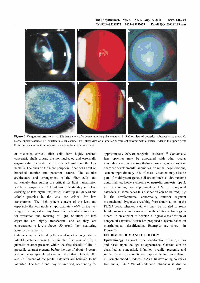
approximately 70% of congenital cataracts [3]. Conversely,lens opacities may be associated with other ocularanomalies such as microphthalmia, aniridia, other anteriorchamber developmental anomalies, or retinal degenerations,seen in approximately 15% of cases. Cataracts may also bepart of multisystem genetic disorders such as chromosomeabnormalities, Lowe syndrome or neurofibromatosis type 2,also accounting for approximately 15% of congenitalcataracts. In some cases this distinction can be blurred,in the developmental abnormality anterior segmentmesenchymal dysgenesis resulting from abnormalities in thePITX3 gene, inherited cataracts may be isolated in somefamily members and associated with additional findings inothers. In an attempt to develop a logical classification ofcongenital cataracts, Merin has proposed a system based onmorphological classification. Examples are shown inFigure 2 [1].EPIDEMIOLOGY AND ETIOLOGYEpidemiology Cataract is the opacification of the eye lensand based upon the age at appearance. Cataract can beclassified as congenital, infantile, juvenile, presenile andsenile. Pediatric cataracts are responsible for more than 1million childhood blindness in Asia. In developing countrieslike India, 7.4-15.3% of childhood blindness is due to
Figure 2 Congenital cataracts A: Slit lamp view of a dense anterior polar cataract; B: Reflex view of posterior subcapsular cataract; C:Dense nuclear cataract; D: Punctate nuclear cataract; E: Reflex view of a lamellar pulverulent cataract with a cortical rider in the upper right;F: Sutural cataract with a pulverulent nuclear lamellar component
423

cataract. The prevalence of cataract in children has beenestimated between 1-15/10,000 children. Hereditary,metabolic and other ocular or systemic disorders and traumaare known factors responsible for cataract in children. InIndia, half of all childhood cataracts are Idiopathic. Out of172 children, 88.4% had non-traumatic cataract and 11.6%had traumatic cataracts. Among non-traumatic cataracts,7.2% were hereditary, 4.6% were due to congenital rubellasyndrome, 15.1% were secondary and 73.0% wereundetermined. In the group of undetermined cases, duringpregnancy 67% of the mother had history of illness, and22% had taken medications during pregnancy. Inadequateawareness of the causative factors of cataracts within thesociety has lead to increase of cataracts in children[4-6].Considerable progress has been made in characterizingphenotypes, determining the prevalence and incidence invarious population groups, and understanding risk factorsfor cataract. Cataract surgery research has documentedfunctional improvements following surgery[5-7]. Cataract is anindependent marker of early mortality, providing a possiblesystem to study the aging process. Promising future work incataract epidemiology is highlighted. Despite the availabilityof cataract surgery, cataract is still the leading cause ofblindness worldwide. From a public health standpoint,research that can identify ways to delay onset orprogression, or achieve the holy grail of prevention ofcataract, should remain a leading priority[7].Foster [8] reported that cataract is the most importantcause of treatable childhood blindness. There are 200 000children blind from cataract worldwide, and 20 000 to 40 000children with developmental bilateral cataract are born eachyear. Rubella is still an important cause of preventabledisease in many countries. In the developing world, there isa need to improve early case detection and referral servicesand to establish centers with expertise in the assessment,surgical treatment, and long-term management of the childwith cataract. Blindness due to cataract presents anenormous problem in India not only in terms of humanmorbidity but also in terms of economic loss and socialburden. The WHO/NPCB (National Programme for Controlof Blindness) survey has shown that there is a backlog ofover 22 million blind eyes (12 million blind people) inIndia, and 80.1% of these are blind due to cataract. Theannual incidence of cataract blindness is about 3.8 million.The present annual level of performance is in the order ofabout 1.6-1.9 million cataract operations. To clear thebacklog of cataract cases by the year 2000 and to tackle therising incidence, 5-6 million cataract operations annuallywill have to be performed as against the present rate of 1.7million per year[9].
To describe the prevalence and risk factors for cataract in anAustralian population aged 40 years and older, McCarty
[10] have shown that cataract is a public health problem inAustralia, which, like other developed countries, isexperiencing a demographic shift toward more elderlypeople in the population. The first step in the design of anappropriate response to the expected increase in the numberof cases of cataract is to describe the age-specificprevalence and risk factors of cataract. Many of the riskfactors that we identified in the population are potentiallymodifiable through public health campaigns[11-14].Etiology Many etiological studies on childhood cataracthave been carried out in developed as well as developingcountries to determine causative factors. Studies performedin various parts of India show variation in etiological factorsaffecting childhood cataract. In south India amongnon-traumatic cataracts, 25% were due to hereditary, 15%were due to congenital rubella syndrome and 51% wereidiopathic. Nearly half of non-traumatic cataracts in thispopulation are due to potentially preventable causes likecongenital rubella syndrome and autosomal dominantdisease.In the paediatric cataract population examined,approximately half of the patients were diagnosed in thefirst year of life. More than 18% had a positive familyhistory of cataracts. Of patients with hereditary cataracts 8%presented with unilateral involvement. Identification of thegenes that cause paediatric and congenital cataract shouldhelp clarify the aetiology of some sporadic and unilateralcataracts. The results showed by Wirth [15] that 421patients with paediatric cataract were identified, which givesan estimated incidence of 2.2 per 10,000 births. Of the 342affected individuals with a negative family history, 50%were diagnosed during the first year of life, and 56/342(16%) were associated with a recognised systemic diseaseor syndrome. Unilateral cataract was identified in 178/342(52% ) of sporadic cases. Seventy-nine children (from 54nuclear families) had a positive family history. Of these 54families, 45 were recruited for clinical examination andDNA collection. Ten nuclear families were subsequentlyfound to be related, resulting in four larger pedigrees. Thus,39 families have been studied. The mode of inheritance wasautosomal dominant in 30 families, X linked in four,autosomal recessive in two, and uncertain in three. In total,178 affected family members were examined; of these 8%presented with unilateral cataracts and 43% were diagnosedwithin the first year of life[12-14].Cataract is responsible for about 10% blindness amongchildren in India. Etiology of cataract is not well definedespecially for childhood cataracts and epidemiological data
Epidemiology and molecular genetics of congenital cataracts
424

陨灶贼 允 韵责澡贼澡葬造皂燥造熏 灾燥造援 4熏 晕燥援 4熏 Aug.18, 圆园11 www. IJO. cn栽藻造押8629原愿圆圆源缘员苑圆 8629-83085628 耘皂葬蚤造押陨允韵援 圆园园园岳员远猿援糟燥皂
for Indian population is not available in details. Out of 172children, 88.4% had non-traumatic cataract and 11.6% hadtraumatic cataracts. Among non-traumatic cataracts, 7.2%were hereditary, 4.6% were due to congenital rubellasyndrome, 15.1% were secondary and 73.0% wereundetermined. In the group of undetermined cases, duringpregnancy 67% of the mother had history of illness, and22% had taken medications during pregnancy. Healtheducation of women to childbearing age and school childrencan decrease incidence of pediatric cataracts[16].MOLECULAR GENETICSCataracts can be isolated or can occur in association with alarge number of metabolic diseases and genetic syndromes[17].Isolated congenital cataracts tend to be highly penetrantMendelian traits, with autosomal dominant more commonthan autosomal recessive cataracts. Currently, there areabout 39 genetic loci to which isolated or primary cataractshave been mapped, although the number is constantlyincreasing and depends to some extent on definition (Table 1).Of these, several are associated with additional abnormalities,mostly as part of developmental syndromes. These tend toresult from mutations in genes encoding transcriptionalactivators, and most of these have been identified bysequencing candidate genes in patients with developmentalanomalies. A notable exception is the 琢B-crystallin gene,CRYAB, which is widely expressed in various tissues,especially muscle. Mutations in CRYAB can cause aspectrum of abnormalities ranging from isolated cataracts tomild cataracts associated with myopathy. A secondcounterexample is the ferritin gene, which causes thehyperferritinemia-cataract syndrome (Table 1)[1,16,17].Crystallin proteins Three major classes of ubiquitouscrystallins are found in the vertebrate eye lens. In themolecular structure of crystallin proteins, there are琢-crystallin protein (40%), 茁-crystallin protein (35%) and酌-crystallin protein (25%). The ratio of the crystalin proteincomposition and its spatial sequence in the maintenance ofcrystallin transparency is very important[18-22]. When the geneis mutated, the crystallin proteins are not only the abnormalprotein structure and affect it's closely packed, but also toreduce the solubility of crystallin proteins to form opacities.Alpha-crystallin proteins Alpha-crystallin protein familyhas two members: 琢A-crystallin (CRYAA) and 琢B-crystallin(CRYAB) gene located in the 21q22.3 gene and located inthe CRYAA 1lq22.3-q23.1 CRYAB coding, respectively [18].琢A-crystallin, a small heat shock protein with chaperone-like activity, forms dynamic multimeric complexes. Recentlywe described the spontaneous generation of a mutant protein(super 琢A-crystallin) by exon duplication arisen via exonshuffling confirming a classic hypothesis[16-18]. Comparison of
super 琢A-crystallin, which is viable in a mouse skeletalmuscle cell line, with normal 琢A-crystallin shows that it hasdiminished thermostability, increased exposure ofhydrophobic patches, a larger complex size and lost itschaperone activity. However, super 琢A-crystallin subunitsexchange as readily between complexes as does normal琢A-crystallin. These data indicate that chaperone-likeactivity may vanish independent of subunit hydrophobicityand exchangeability [19,20]. Crystallins are separated into twoclasses: taxon-specific, or enzyme, and ubiquitous. Thelatter class constitutes the major proteins of vertebrate eyelens and maintains the transparency and refractive index ofthe lens. Since lens central fiber cells lose their nucleiduring development, these crystallins are made and thenretained throughout life, making them extremely stableproteins. Mammalian lens crystallins are divided into 琢, 茁,and 酌 families; Beta and 酌 crystallins are also considered asa super family. Alpha and 茁 families are further divided intoacidic and basic groups. Seven protein regions exist incrystallins: four homologous motifs, a connecting peptide,and N- and C-terminal extensions. Alpha crystallins arecomposed of two gene products: alpha-A and alpha-B, foracidic and basic, respectively. Alpha crystallins can beinduced by heat shock and are members of the small heatshock protein (sHSP also known as the HSP20) family.They act as molecular chaperones although they do notrenature proteins and release them in the fashion of a truechaperone; instead they hold them in large solubleaggregates. Post-translational modifications decrease theability to chaperone. These heterogeneous aggregatesconsist of 30-40 subunits; 琢-A and 琢-B subunits have a 3:1ratio, respectively. Two additional functions of alphacrystallins are an autokinase activity and participation in theintracellular architecture. Alpha-A and 琢-B gene productsare differentially expressed; 琢-A is preferentially restrictedto the lens and 琢-B is expressed widely in many tissues andorgans. Elevated expression of 琢-B crystallin occurs inmany neurological diseases; a missense mutation cosegregatedin a family with a desmin-related myopathy [21,22].CRYAA (crystallin, 琢A)Chromosome: 21;Location: 21q22.3
CRYAB (crystallin, 琢B)Chromosome: 11;Location: 11q22.3-q23.1
Beta -crystallin proteins Beta-crystallins, the mostheterogeneous, differ by the presence of the C-terminalextension (present in the basic group, none in the acidic
425

Table 1 Mapped human cataracts Locus Chrom Inh MIM CCV (Volkmann) 1p36 AD 115665 CTPP 1p34-p36 AD 116600 FOXE3 NM_012186 1p32 AD 107250, 601094 GJA8 NM_005267 1q21-q25, 2p24 AD,AR 116200 CCNP 2p12 AD 607304 CRYGC NM_020989 2q33-q35 AD 123660, 123680, 601286 CRYGD NM_006891 2q33-q35 AD,AR 115700, 123690 BFSP2 NM_003571 3q22.1 AD 603212 CRYGS NM_017541 3q26.3-qter AD 123730 GCNT2 NM_001491 6p24-p23 AR 110800 EYA1 NM_172060 8q13.3 AD 601653 CAAR 9q13-q22 AR 212500 PITX3 NM_005029 10q25 AD 602669 CRYAB NM_001885 11q23.3-24.2 AD 123590 AQP0 NM_012064 12q12-14.1 AD 601286 GJA3 NM_021954 13q11-13 AD 601885 CHX10 NM_182894 14q24.3 AR 142993 CCSSO 15q21-q22 AD 605728 HSF4 NM_001538 16q21 AD,AR,S 602438
MAF NM_001031804 16q22-q23 AD 177074
CTAA2 17p13 AD 601202
CRYBA3 NM_005208 17q11-q12 AD 600881
CCA1 (Cerulean - blue dot) 17q24, 19q13, 19q13.4 AD,AR 115660
FTL NM_000146 19q13.33 AD
LIM2 NM_002316 19q13.4 AR 154045
BFSP1 NM_001195 20p11.23-p12.1 AR 603307
CPP3 20p12-q12 AD 605387
CHMP4B NM_176812 20q11.22 AD 610897
CRYAA NM_000394 21q22.3 AD, AR, Spo radi c 123580
CRYBB2 NM_00496 22q11.2 AD 123620
CRYBB1 NM_001887 22q11.2-q12.1 AD, AR 600929
CRYBB3 NM_004076 22q11.23-q12.1 AR 123630
CRYBA4 NM_001886 22q11.2 AD 123631
CXN Xp22 XL 300457
NHS NM_198270 Xp22.13 XL 300457 Specific mutations are described above the entry for the gene or locus. The cDNA sequence changes are given in reference to the NCBI sequence identifier in the Locus column. Chrom: chromosomal location, Inh: inheritance pattern, cDNA: changes in the NCBI DNA sequence listed in the Locus column, AA: changes in the protein sequence, MIM: Mendelian Inheritance in Man reference. Specific mutations identified are listed above the gene. AD: autosomal dominant, AR: autosomal recessive, XL: X-linked, S: sporadic. Genes and loci are shown in bold, while individual mutations and their descriptions are shown in small lettering above
group). Beta-crystallins form aggregates of different sizesand are able to self-associate to form dimers or to formheterodimers with other 茁-crystallins. This gene, a 茁 acidicgroup member, encodes two proteins (crystallin, 茁A3 andcrystallin, 茁A1) from a single mRNA, the latter protein is17 a shorter than crystallin, 茁A3 and is generated by use ofan alternate translation initiation site. Deletion of exons 3and 4 causes the autosomal dominant disease zonularcataract with sutural opacities. This gene, a 茁 basic group
member, is part of a gene cluster with 茁-A4, 茁-B1, and茁-B3. A chain-terminating mutation was found to causetype 2 cerulean cataracts. The major 茁-crystallin structuresare as follows.CRYBA1 (crystallin, 茁A1)Chromosome: 17;Location: 17q11.2
Epidemiology and molecular genetics of congenital cataracts
426

陨灶贼 允 韵责澡贼澡葬造皂燥造熏 灾燥造援 4熏 晕燥援 4熏 Aug.18, 圆园11 www. IJO. cn栽藻造押8629原愿圆圆源缘员苑圆 8629-83085628 耘皂葬蚤造押陨允韵援 圆园园园岳员远猿援糟燥皂
CRYBA2 (crystallin, 茁A2)Chromosome: 2; Location: 2q34-q36
CRYBB1 (crystallin, 茁B1)Chromosome: 22; Location: 22q11.2; 22q12.1
CRYBB3 (crystallin, 茁B3)Chromosome: 22; Location: 22q11.23-q12.1
Gama -crystallin proteins Alpha and 茁 families arefurther divided into acidic and basic groups. Seven proteinregions exist in crystallins: four homologous motifs, aconnecting peptide, and N- and C-terminal extensions [21,22].酌-crystallins are a homogeneous group of highlysymmetrical, monomeric proteins typically lackingconnecting peptides and terminal extensions. They aredifferentially regulated after early development. Four酌-crystallin genes (酌-A through 酌-D) and three pseudogenes(酌-E, 酌-F, 酌-G) are tandemly organized in a genomicsegment as a gene cluster. Whether due to aging ormutations in specific genes, 酌-crystallins have been involvedin cataract formation.CRYGA (crystallin, 酌A)Chromosome: 2; Location: 2q33-q35
CRYGB (crystallin, 酌B)Chromosome: 2; Location: 2q33-q35
CRYGC (crystallin, 酌C)Chromosome: 2; Location: 2q33-q35
CRYGD (crystallin, 酌D)Chromosome: 2; Location: 2q33-q35
CRYGE (crystallin, 酌E) [Mus musculus]Chromosome: 1; Location: 1 C2; 1 32.0 cM
CRYGF (crystallin, 酌G)Chromosome: 1; Location: 1 C3; 1 32.0 cM
Gap junction proteins Gap junctions were firstcharacterized by electron microscopy as regionallyspecialized structures on plasma membranes of contactingadherent cells. These structures were shown to consist ofcell-to-cell channels. Proteins, called connexins, purifiedfrom fractions of enriched gap junctions from differenttissues differ. The connexins are designated by theirmolecular mass. Another system of nomenclature dividesgap junction proteins into 2 categories, 琢 and 茁, accordingto sequence similarities at the nucleotide and amino acidlevels. For example, CX43 (121014) is designatedα -1 gapjunction protein, whereas CX32 (GJB1; 304040) and CX26are called 茁-1 and 茁-2 gap junction proteins, respectively.This nomenclature emphasizes that CX32 and CX26 aremore homologous to each other than either of them is toCX43. Gap junction proteins include the following threetypes: 1)GJB2 (Gap junction 茁2), 26kDa, Connexin 26(CX26) Gene map locus: 13q11-q12; 2) GJB1(Gap junction茁1) 32kDa, Connexin 32 (CX32) Gene map locus: Xq13.1;3)GJA1 (Gap junction 琢1), 43kDa, connexin 43 (CX43)gene map locus: 20q11.Using the paired xenopus oocyte assay, Mese [23]
functionally analyzed 5 CX26 mutations associated withautosomal recessive neurosensory deafness (DFNB1A;220290). Three of the mutants were unable to formfunctional channels; the other 2 did electrically couple cells,but their voltage gating properties were different from wildtype CX26 channels. The deafness associated with CX26mutations is caused not only by reduced potassiumrecirculation in the inner ear, but also by abnormalities inthe exchange of other metabolites through the cochlear gap[23].Elias [24] showed that the gap junction subunits CX26and CX43 (121014) are expressed at the contact pointsbetween radial fibers and migrating neurons, and that acutedown regulation of CX26 or CX43 impairs the migration ofneurons to the cortical plate. Unexpectedly, gap junctions donot mediate neuronal migration by acting in the classicalmanner to provide an aqueous channel for cell-cellcommunication. Instead, gap junctions provide dynamicadhesive contacts that interact with the internal cytoskeletonto enable leading process stabilization along radial fibers aswell as the subsequent translocation of the nucleus. The gapjunction adhesions are necessary for glial-guided neuronalmigration[24].Membrane proteins Lens is a non-vascular tissue; the vastmajority of lens cells rely on glycolysis to meet energysupply. Located in the center of the lens fiber rely on
427

passive diffusion to accept less than adequate nutrition, andby special transport to provide the nutrients and themetabolic products shipped to the lens surface.A membrane protein is a protein molecule that is attachedto, or associated with the membrane of a cell or anorganelle. More than half of all proteins interact withmembranes. Biological membranes consist of aphospholipid bilayer and a variety of proteins thataccomplish vital biological functions. 1) Structural proteinsare attached to microfilaments in the cytoskeleton whichensures stability of the cell; 2) Cell adhesion moleculesallow cells to identify each other and interact. Such proteinsare involved in immune response, for example; 3)Membrane enzymes produce a variety of substancesessential for cell function; 4) Membrane receptor proteinsserve as connection between the cell's internal and externalenvironments; 5) Transport proteins play an important rolein the maintenance of concentrations of ions. Thesetransport proteins come in two forms: carrier proteins andchannel proteins.The structures of membrane proteins are stabilized by weakinteractions and influenced by additional interactions withthe solubilizing environment. The influence of theenvironment on membrane protein structures is especiallysignificant. Despite the significant functional importance ofmembrane proteins, the structural biology has beenparticularly challenging as shown by the low number ofmembrane protein structures determined. Integral membraneproteins are present in a heterogeneous environment thatposes major obstacles for existing structural methodologies.Aquaporin proteins Aquaporins are proteins embedded inthe cell membrane that regulate the flow of water.Aquaporins are integral membrane proteins from a largerfamily of major intrinsic proteins (MIP) that form pores inthe membrane of biological cells [25]. Aquaporin proteins aremade up of six transmembrane 琢-helices arranged in aright-handed bundle, with the amino and the carboxyltermini located on the cytoplasmic surface of the membrane.The amino and carboxyl halves of the sequence showsimilarity to each other, in what appears to be a tandemrepeat. Some researchers believe that this results from anearly evolution event that saw the duplication of thehalf-size gene. There are also five interhelical loop regions(A-E) that form the extracellular and cytoplasmic vestibules.Loops B and E are hydrophobic loops that contain thehighly, although not completely conserved, asparagine-proline-alanine (NPA) motif, which overlap the middle ofthe lipid bilayer of the membrane forming a 3-D 'hourglass'structure where the water flows through. This overlap formsone of the two well-known channel constriction sites in the
peptide, the NPA motif and a second and usually narrowerconstriction known as 'selectivity filter' or ar/R selectivityfilter. Aquaporins form tetramers in the cell membrane, witheach monomer acting as a water channel. The differentaquaporins contain differences in their peptide sequence,which allows for the size of the pore in the protein to differbetween aquaporins. The resultant size of the pore directlyaffects what molecules are able to pass through the pore,with small pore sizes only allowing small molecules likewater to pass through the pore [26,27]. Mice homozygous forinactivating mutations in the aquaporin-0 gene developcongenital cataracts[28].Major intrinsic protein (MIP) is a member of thewater-transporting aquaporins as well as the originalmember of the MIP family of channel proteins. The functionof the fiber cell membrane protein encoded by this gene isundetermined, yet this protein is speculated to play a role inintracellular communication. The MIP protein is expressedin the ocular lens and is required for correct lens function.MIP (Major intrinsic protein)Chromosome: 12; Location: 12q13
Lens intrinsic membrane protein 2 This gene encodesan eye lens-specific protein (19kDa) found at the junctionsof lens fiber cells, where it may contribute to cell junctionalorganization. It acts as a receptor for calmodulin, and mayplay an important role in both lens development andcataractogenesis. Mutations in this gene have beenassociated with cataract formation. Alternatively splicedtranscript variants encoding different isoforms have beenfound for this gene.LIM2 (Lens intrinsic membrane protein 2)Chromosome: 19; Location: 19q13.4
Beaded filament structural protein 2 Frame of the lenscells and the cytoplasm by the cytoskeleton in the lensprotein interactions determined. Fibrin-like beads (beadedfilament structural protein 2, BFSP2) is the only kind ofexpression only in the eyes of cytoskeletal proteins(cytoskeletal protein) structure.More than 99% of the vertebrate ocular lens is comprised ofterminally differentiated lens fiber cells. Two lens-specificintermediate filament-like proteins, the protein product ofthis gene (phakinin), and filensin, are expressed only afterfiber cell differentiation has begun. Both proteins are foundin a structurally unique cytoskeletal element that is referredto as the beaded filament (BF). Mutations in this gene havebeen associated with juvenile-onset, progressive cataracts
Epidemiology and molecular genetics of congenital cataracts
428

陨灶贼 允 韵责澡贼澡葬造皂燥造熏 灾燥造援 4熏 晕燥援 4熏 Aug.18, 圆园11 www. IJO. cn栽藻造押8629原愿圆圆源缘员苑圆 8629-83085628 耘皂葬蚤造押陨允韵援 圆园园园岳员远猿援糟燥皂
and Dowling-Meara epidermolysis bullosa simplex[29].BFSP2 (Beaded filament structural protein 2)Chromosome: 3; Location: 3q22.1
Transcription factors Transcription factor has animportant role in embryonic development of eye, mutationsin these genes often because eye hypoplasia syndrome, thesegenes is to study the importance of eye hypoplasia syndromecandidate genes. Antibody-dependent cell-mediatedcytotoxicity (ADCC) associated with the occurrence oftranscription factor genes, including heat shock transcriptionfactor 4 gene (HSF4), MAF (v-maf musculoaponeuroticfibrosarcoma oncogene homolog), pituitary homeobox gene3 (PITX3), paired homeobox containing gene 6 (PAX6) andso on.Heat shock transcription factor 4 Heat shocktranscription factors (HSFs) activate heat-shock responsegenes under conditions of heat or other stresses. HSF4 lacksthe carboxyl-terminal hydrophobic repeat which is sharedamong all vertebrate HSFs and has been suggested to beinvolved in the negative regulation of DNA binding activity.Two alternatively spliced transcripts encoding distinctisoforms and possessing different transcriptional activityhave been described.HSF4 (Heat shock transcription factor 4)Chromosome: 16; Location: 16q21
v -maf musculoaponeurotic fibrosarcoma Much of ourknowledge about the underlying mechanisms ofcataractogenesis has come from the genetic analysis ofaffected families: there are contributions from genes codingfor transcription factors (such as MAF, PITX3, FoxE3) andstructural proteins such as crystallins or connexins [30]. Inaddition, there are contributions from enzymes affectingsugar pathways (particularly the galactose pathway) andfrom a quite unexpected area: axon guidance molecules likeephrins and their receptors. Cataractous mouse lenses can beidentified easily by visual inspection, and a remarkablenumber of mutant lines have now been characterized.Generally, most of the mouse mutants show a similarphenotype to their human counterparts; however, there aresome remarkable differences. It should be noted that manymutations affect genes that are expressed not only in thelens, but also in tissues and organs outside the eye[31-33].The protein encoded by this gene is a DNA-binding, leucinezipper-containing transcription factor that acts as ahomodimer or as a heterodimer. Depending on the binding
site and binding partner, the encoded protein can be atranscriptional activator or repressor. This protein plays arole in the regulation of several cellular processes, includingembryonic lens fiber cell development, increased T-cellsusceptibility to apoptosis, and chondrocyte terminaldifferentiation. Defects in this gene are a cause ofjuvenile-onset pulverulent cataract as well as congenitalcerulean cataract 4 (CCA4). Two transcript variants encodingdifferent isoforms have been found for this gene[31-33].MAF (v-maf musculoaponeurotic fibrosarcoma)Chromosome: 16; Location: 16q22-q23
Paired -like homeodomain 3 This gene encodes amember of the RIEG/PITX homeobox family, which is inthe bicoid class of homeodomain proteins. Members of thisfamily act as transcription factors. This protein is involvedin lens formation during eye development. Mutations of thisgene have been associated with anterior segmentmesenchymal dysgenesis and congenital cataracts[32,33].PITX3 (Paired-like homeodomain 3)Chromosome: 10;Location: 10q25
Paired box gene 6 This gene encodes paired box gene 6,one of many human homologs of the Drosophilamelanogaster gene prd. In addition to the hallmark featureof this gene family, a conserved paired box domain, theencoded protein also contains a homeo box domain. Bothdomains are known to bind DNA, and function as regulatorsof gene transcription. This gene is expressed in thedeveloping nervous system, and in developing eyes.Mutations in this gene are known to cause ocular disorderssuch as aniridia and Peter's anomaly. Alternatively splicedtranscript variants encoding either the same or differentisoform have been found for this gene. Mutations associatedwith congenital cataracts form 40, of which only 1 species iscaused by mutations in the pathogenesis of the independentoccurrence of ADCC (G18W), the mutation interfering withthe target gene PAX6 binding, and reduce its transcriptionalactivation function located in the 1lp13. A novel PAX6 genemutation was identified in a Chinese aniridia family. Thismutation may also contribute to congenital cataracts in theseaniridia patients[33-35].PAX6 (Paired box gene 6)Chromosome: 11;Location: 11p13
429
Ferritin light chains Why ferritin concentration can leadto cataracts, it is not clear. Studies suggest that ferritin forcataract may be two factors: first, ferritin light chain andheavy chain in the balance of the population makes the freeiron ions and active oxygen content increases, damage tolens function. Second, the cells increase in ferritin levelsmay lead to the aggregation of lens proteins. This geneencodes the light subunit of the ferritin protein. Ferritin isthe major intracellular iron storage protein in prokaryotesand eukaryotes. It is composed of 24 subunits of the heavyand light ferritin chains. Variation in ferritin subunitcomposition may affect the rates of iron uptake and releasein different tissues. A major function of ferritin is thestorage of iron in a soluble and nontoxic state. Defects inthis light chain ferritin gene are associated with severalneurodegenerative diseases and hyperferritinemia-cataractsyndrome. This gene has multiple pseudogenes[36].The mutation observed in FTL in this family highlights thephenotypic heterogeneity of the disorder in relation to thegenotype as the identical mutation (32 G>A) has previouslybeen reported in two Italian families with entirely differentphenotypes. It is also the first report of hereditaryhyperferritinemia-cataract syndrome in a family of Indianorigin[37].FTL (Ferritin light chain)Chromosome: 19; Location: 19q13.33
Fibroblast growth factors The protein encoded by thisgene is a member of the fibroblast growth factor (FGF)family. FGF family members possess broad mitogenic andcell survival activities, and are involved in a variety ofbiological processes, including embryonic development, cellgrowth, morphogenesis, tissue repair, tumor growth andinvasion. This protein is a potent epithelial cell-specificgrowth factor, whose mitogenic activity is predominantlyexhibited in keratinocytes but not in fibroblasts andendothelial cells. Studies of mouse and rat homologs of thisgene implicated roles in morphogenesis of epithelium,reepithelialization of wounds, hair development and earlylung organogenesis [ 38]. Using linkage analysis, Swaroop
[39] believed that it may be fibroblast growth factor 7(FGF7) gene mutation associated with congenitalcataract-related.FGF7 (fibroblast growth factor 7)Chromosome: 15; Location: 15q21.2
Other proteins The new genes that cause congenital
cataracts are constantly discovered. Studies suggest thatthere is no more than 12 disease genes identifiedchromosomal regions associated with congenital cataracts,which include lp32, lp36, 1pter, 2p12, 2p24-pter, 6p24,8q13.3, 15q21-22, 15q22.3 -q23.1, 17p12-13, 17q24,20p12-q12 and so on [1,17,39-49].SUMMARYThe hereditary congenital cataracts provides some insightinto those biological systems most important for developingand maintaining lens transparency, or at least those whichare most easily disrupted in above overview. Although thenormal lens and cataract in clinical molecular biology morecomplete description is beyond the scope of the review,more detailed reviews are available [48-53]. Other importantfunctional systems include cytoskeletal and membraneproteins, especially those limited to or favored in the lens.As suggested by their high expression levels in the lens, thecrystallins are the most common group of proteins mutatedin inherited congenital cataracts. Growth and differentiationfactors also frequently are seen causing congenital cataracts,often in association with other findings in theirdevelopmental spectra [54]. Finally, a varied group of proteinscan also cause congenital cataracts. Together these studiesprovide insights into lens biology easily accessible in noother way. They can also be of direct clinical benefit insome families [55]. In addition, while the pathophysiology ofcongenital and hereditary cataracts differs in fundamentalways from that of age related cataracts, the study ofcongenital cataracts can provide insights into themechanisms of lens transparency and to some of the ways inwhich it can be lost as the lens ages[53-56].Congenital cataract is particularly serious because it has thepotential for inhibiting visual development, resulting inpermanent blindness. Inherited cataracts represent a majorcontribution to congenital cataracts, especially in developedcountries. While cataract represents a common end stage ofmutations in a potentially large number of genes actingthrough varied mechanisms in practice most inheritedcataracts have been associated with a subgroup of genesencoding proteins of particular importance for themaintenance of lens transparency and homeostasis [57]. Theincreasing availability of more detailed information aboutthese proteins and their functions and is making it possibleto understand the pathophysiology of cataracts and thebiology of the lens in general.REFERENCES1 Hejtmancik JF. Congenital cataracts and their molecular genetics.
2008;19(2):134-149
2 Kuszak JR, Zoltoski RK, Sivertson C. Fibre cell organization in crystalline
lenses. 2004;78(3):673-687
3 Haargaard B, Wohlfahrt J, Fledelius HC, Rosenberg T, Melbye M. A nationwide
Epidemiology and molecular genetics of congenital cataracts
430

陨灶贼 允 韵责澡贼澡葬造皂燥造熏 灾燥造援 4熏 晕燥援 4熏 Aug.18, 圆园11 www. IJO. cn栽藻造押8629原愿圆圆源缘员苑圆 8629-83085628 耘皂葬蚤造押陨允韵援 圆园园园岳员远猿援糟燥皂
Danish study of 1027 cases of congenital/infantile cataracts: etiological and clinical
classifications. 2004;111(12):2292-2298
4 Johar SR, Savalia NK, Vasavada AR, Gupta PD. Epidemiology based etiological
study of pediatric cataract in western India. 2004;58(3):115-121
5 Heijl A, Leske MC. Cataract epidemiology. 2007;114(1):201
6 Chandrasekaran S, Cumming RG, Rochtchina E, Mitchell P. Associations
between elevated intraocular pressure and glaucoma, use of glaucoma medications,
and 5-year incident cataract: the Blue Mountains Eye Study.
2006;113(3):417-424
7 West S. Epidemiology of cataract: accomplishments over 25 years and future
directions. 2007;14(4):173-178
8 Foster A, Gilbert C, Rahi J. Epidemiology of cataract in childhood: a global
perspective. 1997;23(Suppl 1):601-604
9 Vajpayee RB, Joshi S, Saxena R, Gupta SK. Epidemiology of cataract in India:
combating plans and strategies. 1999;31(2):86-92
10 McCarty CA, Mukesh BN, Fu CL, Taylor HR. The epidemiology of cataract in
Australia. 1999;128(4):446-465
11 Agarwal PK, Bowman R, Courtright P. Child eye health tertiary facilities in
Africa. 2010;14(3):263-266
12 Prakalapakorn SG, Rasmussen SA, Lambert SR, Honein MA. National Birth
Defects Prevention Study. Assessment of risk factors for infantile cataracts using a
case-control study: National Birth Defects Prevention Study, 2000-2004.
2010;117(8):1500-1505
13 You C, Wu X, Zhang Y, Dai Y, Huang Y, Xie L. Visual impairment and delay
in presentation for surgery in chinese pediatric patients with cataract.
2011;118(1):17-23
14 Wang KJ, Li SS, Yun B, Ma WX, Jiang TG, Zhu SQ. A novel mutation in MIP
associated with congenital nuclear cataract in a Chinese family. 2011;17:
70-77
15 Wirth MG, Russell-Eggitt IM, Craig JE, Elder JE, Mackey DA. Aetiology of
congenital and paediatric cataract in an Australian population.
2002;86(7):782-786
16 Johar SR, Savalia NK, Vasavada AR, Gupta PD. Epidemiology based etiological
study of pediatric cataract in western India. 2004;58(3):115-121
17 Hejtmancik JF, Kaiser-Kupfer MI, Piatigorsky J. Molecular biology and
inherited disorders of the eye lens. In: Scriver CR, Beaudet AL, Valle D, Sly WS,
Childs B, Kinzler KW, Vogelstein B, editors. The Metabolic and Molecular Basis of
Inherited Disease. 8th ed, McGraw Hill; New York 2001:6033-6062
18 Yang G, Xiong C, Li S, Wang Y, Zhao J. A recurrent mutation in CRYGD is
associated with autosomal dominant congenital coralliform cataract in two
unrelated Chinese families. 2011;17:1085-1089
19 Kumar M, Agarwal T, Khokhar S, Kumar M, Kaur P, Roy TS, Dada R. Mutation
screening and genotype phenotype correlation of α-crystallin, γ-crystallin and
GJA8 gene in congenital cataract. 2011;17:693-707
20 Mothobi ME, Guo S, Liu Y, Chen Q, Yussuf AS, Zhu X, Fang Z. Mutation
analysis of congenital cataract in a Basotho family identified a new missense allele
in CRYBB2. 2009;15:1470-1475
21 Chen Q, Ma J, Yan M, Mothobi ME, Liu Y, Zheng F. A novel mutation in
CRYAB associated with autosomal dominant congenital nuclear cataract in a
Chinese family. 2009;15:1359-1365
22 Hansen L, Yao W, Eiberg H, Kjaer KW, Baggesen K, Hejtmancik JF,
Rosenberg T. Genetic heterogeneity in microcornea-cataract: five novel mutations
in CRYAA, CRYGD, and GJA8. 2007;48 (9):
3937-3944
23 Mese G, Londin E, Mui R, Brink PR, White TW. Altered gating properties of
functional Cx26 mutants associated with recessive non-syndromic hearing loss.
2004;115:191-199
24 Elias LAB, Wang DD, Kriegstein AR. Gap junction adhesion is necessary for
radial migration in the neocortex. 2007;448(3):901-907
25 Agre P. The aquaporin water channels. 2006;3(1):5-13
26 Gonen T, Walz T.The structure of aquaporins. 2006;39 (4):
361-396
27 Chepelinsky AB. Structural function of MIP/aquaporin 0 in the eye lens; genetic
defects lead to congenital inherited cataracts.
265-297
28 Okamura T, Miyoshi I, Takahashi K, Mototani Y, Ishigaki S, Kon Y, Kasai N.
Bilateral congenital cataracts result from a gain-of-function mutation in the gene
for aquaporin-0 in mice.
29 Ma X, Li FF, Wang SZ, Gao C, Zhang M, Zhu SQ. A new mutation in BFSP2
(G1091A) causes autosomal dominant congenital lamellar cataracts.
2008;14:1906-1911
30 Churchill A, Graw J. Clinical and experimental advances in congenital and
paediatric cataracts. 2011;366 (1568):
1234-1249
31 Berry V, Francis PJ, Prescott Q, Waseem NH, Moore AT, Bhattacharya SS. A
novel 1-bp deletion in PITX3 causing congenital posterior polar cataract.
2011;17:1249-1253
32 Churchill A, Graw J. Clinical and experimental advances in congenital and
paediatric cataracts. 2011;366 (1568):
1234-1249
33 Wang KJ, Zhu SQ, Cheng J. Progress in pathogenic genes and their functions of
congenital cataract. 2010;46(3):280-284
34 Cai F, Zhu J, Chen W, Ke T, Wang F, Tu X, Zhang Y, Jin R, Wu X. A novel
PAX6 mutation in a large Chinese family with aniridia and congenital cataract.
2010;16:1141-1145
35 Song S, Liu Y, Guo S, Zhang L, Zhang X, Wang S, Lu A, Li L. A novel PAX6
gene mutation in a Chinese family with aniridia. 2005;11:335-337
36 Wussuki-Lior O, Abu-Horowitz A, Netzer I, Almer Z, Morad Y, Goldich Y,
Yahalom V, Pras E, Pras E. Hematologic biomarkers in childhood cataracts.
2011;17:1011-1015
37 Vanita V, Hejtmancik JF, Hennies HC, Guleria K, Nürnberg P, Singh D,
Sperling K, Singh JR. Sutural cataract associated with a mutation in the ferritin
light chain gene (FTL) in a family of Indian origin. 2006;12:93-99
38 Xiao Y, Zhao B, Gao Z, Pan Q. Overaccumulation of transforming growth
factor-β1 and basic fibroblast growth factor in lens epithelial cells of congenital
cataract. 2009;44(2):189-192
39 Swaroop A, Xu JZ, Pawar H, Jackson A, Skolnick C, Agarwal N. A conserved
retina-specific gene encodes a basic motif/leucine zipper domain.
1992;89(1):266-2670
40 Vanita V, Singh JR, Singh D, Varon R, Sperling K. Novel mutation in the
gamma-S crystallin gene causing autosomal dominant cataract. 2009;15:
476-481
41 Gao L, Qin W, Cui H, Feng G, Liu P, Gao W, Ma L, Li P, He L, Fu S. A novel
locus of coralliform cataract mapped to chromosome 2p24-pter.
2005;50(6):305-310
42 Pras E, Raz J, Yahalom V, Frydman M, Garzozi HJ, Pras E, Hejtmancik JF. A
nonsense mutation in the glucosaminyl (N-acetyl) transferase 2 gene (GCNT2):
association with autosomal recessive congenital cataracts.
2004;45(6):1940-1945
43 Hilal L, Nandrot E, Belmekki M, Chefchaouni M, El Bacha S, Benazzouz B,
Hajaji Y, Gribouval O, Dufier J, Abitbol M, Berraho A. Evidence of clinical and
genetic heterogeneity in autosomal dominant congenital cerulean cataracts.
2002;23(4):199-208
44 Khaliq S, Hameed A, Ismail M, Anwar K, Mehdi SQ. A novel locus for
autosomal dominant nuclear cataract mapped to chromosome 2p12 in a Pakistani
family. 2002;43(7):2083-2087
45 Vanita, Singh JR, Sarhadi VK, Singh D, Reis A, Rueschendorf F,
Becker-Follmann J, Jung M, Sperling K. A novel form of "central pouchlike"
cataract, with sutural opacities, maps to chromosome 15q21-22.
2001;68(2):509-514
431
2009;190 (IV):
2003;81(4):361-368

46 Yamada K, Tomita HA, Kanazawa S, Mera A, Amemiya T, Niikawa N.
Genetically distinct autosomal dominant posterior polar cataract in a
four-generation Japanese family. 2000;129(2):159-165
47 Ionides AC, Berry V, Mackay DS, Moore AT, Bhattacharya SS, Shiels A. A
locus for autosomal dominant posterior polar cataract on chromosome 1p.
1997;6(1):47-51
48 Kramer P, Yount J, Mitchell T, LaMorticella D, Carrero-Valenzuela R, Lovrien
E, Maumenee I, Litt M. A second gene for cerulean cataracts maps to the beta
crystallin region on chromosome 22. 1996;35(3):539-542
49 Padma T, Ayyagari R, Murty JS, Basti S, Fletcher T, Rao GN, Kaiser-Kupfer
M, Hejtmancik JF. Autosomal dominant zonular cataract with sutural opacities
localized to chromosome 17q11-12. 1995;57(4):840-845
50 Stambolian D, Ai Y, Sidjanin D, Nesburn K, Sathe G, Rosenberg M, Bergsma
DJ. Cloning of the galactokinase cDNA and identification of mutations in two
families with cataracts. 1995;10(3):307-312
51 Eiberg H, Lund AM, Warburg M, Rosenberg T. Assignment of congenital
cataract Volkmann type (CCV) to chromosome 1p36. 1995;96 (1):
33-38
52 McKay JD, Patterson B, Craig JE, Russell-Eggitt IM, Wirth MG, Burdon KP,
Hewitt AW, Cohn AC, Kerdraon Y, Mackey DA. The telomere of human
chromosome 1p contains at least two independent autosomal dominant congenital
cataract genes. 2005;89(7):831-834
53 Hejtmancik JF, Piatigorsky J. Lens Proteins and their Molecular Biology. In:
Alpert DM, Jakobiec FA, Azar DT, Gragoudas ES, editors.
. W.B.Saunders Co.; Philadelphia, 2000:1409-1428
54 Hejtmancik JF, Datiles M. Congenital and Inherited Cataracts. In: Tasman W,
Jaeger EA, editors. Lippincott Willliams and
Wilkins; Philadelphia, 2001:1-22
55 Reches A, Yaron Y, Burdon K, Crystal-Shalit O, Kidron D, Malcov M, Tepper
R. Prenatal detection of congenital bilateral cataract leading to the diagnosis of
Nance-Horan syndrome in the extended family. 2007;27:662-664
56 Finzi S, Li Y, Mitchell TN, Farr A, Maumenee IH, Sallum JM, Sundin O.
Posterior polar cataract: genetic analysis of a large family.
2005;26(3):125-130
57 Medina-Martinez O, Jamrich M. Foxe view of lens development and disease.
2007;134(8):1455-1463
Epidemiology and molecular genetics of congenital cataracts
432


![Pdf pdf projet_educatif-2[1]](https://img.pdfslide.net/doc/110x75/5499da0bac7959092e8b5a10/pdf-pdf-projeteducatif-21.jpg)