Embed Size (px)
Citation preview

3 Photosynthesis in Attacked Plantsand Crops
3.1 INTRODUCTION
In the previous chapter, we saw that attack by pathogens, herbivores or parasitic plants canlead to substantial reductions in plant growth and reproductive output. This, in turn, can resultin serious crop losses in crop production systems and altered community structure in naturalsystems. The mechanisms underlying altered plant growth, development and yield can varydepending on the plant and its attacker, including the plant organ attacked, and whether theattacker is a biotroph or necrotroph, whether it is a root or shoot parasite and whether it is achewing insect or a sapsucker. Attacked plants can lose leaf area and root surface, or moreinsidiously, their vascular tissues can be disrupted. It is important to remember, however, thatquite often, only part of the plant is attacked, and when considering plant responses, bothattacked and non-attacked tissues need to be examined. Furthermore, as discussed in Chapter 2,plants can compensate for damage.
Rates of photosynthesis can be altered considerably after attack, although the nature ofthe change (i.e. decreased or increased photosynthetic rates) and the underlying mechanismswill depend on the plant–attacker interaction. When writing this chapter, I have assumed thatthe readers will have an understanding of photosynthesis. If some re-familiarisation with thisprocess is necessary, I recommend the excellent accounts provided by Smith et al. (2010) andScott (2008).
3.2 PHOTOSYNTHESIS IN DISEASED PLANTS
A common response to pathogen infection is a reduction in the rate of photosynthesis inthe infected leaves. How this change is brought about can vary, depending, for example, onwhether the pathogen is a biotroph or necrotroph, whether it is a virus or a bacterium, whether itproduces a toxin and also which tissues are attacked. As indicated previously, another impor-tant consideration is the heterogeneity of infection, which can apply not only to the wholeplant, but also to single leaves.
Physiological Responses of Plants to Attack, First Edition. Dale R. Walters.© 2015 Dale R. Walters. Published 2015 by John Wiley & Sons, Ltd.

42 Physiological Responses of Plants to Attack
3.2.1 Photosynthesis in plants infected with biotrophicfungal pathogens
3.2.1.1 Changes in whole leaves
Infection by biotrophic fungal pathogens, such as downymildews, powdery mildews and rusts,commonly leads to reduced rates of net photosynthesis (Fig. 3.1). For example, So & Thrower(1976) examined the effects of light and heavy infection with the rust fungus, Uromycesappendiculatus, on rates of photosynthesis in the second trifoliate leaves of the legume Vignasesquipedalis. In the lightly infected plants, rates of photosynthesis were reduced by 14%7 days after inoculation, and by 17 days after inoculation, photosynthetic rates were reducedby 45% compared to uninfected leaves (Fig. 3.1a; So & Thrower, 1976). In contrast, photo-synthesis was reduced earlier, more rapidly, and more substantially, in heavily infected plantscompared to uninfected plants. Thus, rates of photosynthesis in heavily infected leaves werereduced by 18% by 4 days after inoculation, while by 17 days after inoculation, photosynthe-sis in infected leaves was reduced by 73% compared to the uninfected controls (Fig. 3.1a;So & Thrower, 1976). Similar steady and substantial reductions in rates of photosynthesiswere observed in oak leaves infected with the powdery mildew fungus, Microsphaera alphi-toides (Hewitt & Ayres, 1975; Fig. 3.1b) and Arabidopsis thaliana infected with the white
0
10
20
30
40
50
60
70
80
90
100
0 4 7 10 14 17 20
Rate of
photosynthesis
(mm3
CO2 cm−2
h−1)
(a)
Healthy
Heavily infected
Lightly infected
Days after inoculation (d)
Days after inoculation (d)
0
0.5
1
1.5
2
2.5
3
3.5
4
1 2 3 4 5 6 7
Net
photosynthetic
rate (mg CO2
dm−2 h−1)
Days after inoculation (d)
(b)Healthy
Infected
0
1
2
3
4
5
6
7
8
4 8 14
Photosynthesis
(μmol CO2 m−2
s−1)
Uninfected(c)
Infected
Fig. 3.1 Photosynthesis in leaves infected with biotrophic fungal pathogens. (a) effects of the rust,Uromyces appendiculatus, on photosynthesis in the second trifoliate leaves of Vigna sesquipedalis. Leaveswere either not infected (Healthy), lightly infected or heavily infected. Data from So and Thrower (1976).(b) effects of infection by the powdery mildew fungus, Microsphaera alphitoides, on photosynthetic rates inoak leaves. Hewitt and Ayres (1975). Reproduced with permission of Elsevier. (c) Rates of photosynthesis inleaves of Arabidopsis thaliana infected with the white blister rust pathogen, Albugo candida. Tang et al.(2006). Reproduced with permission of John Wiley & Sons.

Photosynthesis in Attacked Plants and Crops 43
blister rust, Albugo candida (Fig. 3.1c; Tang et al., 1996). However, steady and substantialreductions in rates of net photosynthesis are not always observed, and indeed, exactly whathappens to photosynthesis is dependent not only on the host–pathogen interaction, but also onthe host plant variety. This is nicely illustrated by the work of Haigh et al. (1991), who exam-ined changes in rates of net photosynthesis in four genotypes of oat (Avena sativa) differingin partial resistance to the powdery mildew fungus, Erysiphe graminis f.sp. avenae. Powderymildew infection reduced rates of net photosynthesis in second leaves of the susceptible geno-type Mostyn, with significant reductions evident from 5 days after inoculation (Fig. 3.2; Haighet al., 1991). In a more susceptible genotype (1674), no such reduction was observed, while inthe resistant genotypes Maldwyn and 1621, rates of net photosynthesis were reduced, but onlylater in the experiment, 9 days after inoculation (Fig. 3.2). Interestingly, powdery mildew hadno effect on photosynthesis in fifth leaves on these plants. Thus, the decline in photosynthesis,which is usually associated with infection with powdery mildews, appeared to be delayed orcompensated for in oat (Haigh et al., 1991).
Reductions in rates of photosynthesis could result from alterations in one or more of severalmechanisms, including altered stomatal behaviour, decreased chlorophyll concentrations,perturbations in the light reactions of photosynthesis and reductions in the activities andamounts of Calvin cycle enzymes. Powdery mildew and rust infections can lead to alteredstomatal behaviour (see Chapter 6), and such changes can influence rates of photosynthesis.For example, in pea plants infected with the powdery mildew fungus Erysiphe pisi, stomataopened more widely in the light in infected leaves than in healthy leaves 24 hours after inoc-ulation. Thereafter, stomatal opening was progressively reduced by infection, and stomatafailed to close completely in the dark until, 7 days after inoculation, all movements ceasedand stomata remained partly open (Ayres, 1976). In this case, reduced stomatal opening wasthought to be partly responsible for reduced rates of net photosynthesis during the later stagesof infection (Ayres, 1976).
In leaves infected with biotrophic fungal pathogens, chlorosis is a common symptom,and indeed, there is often a progressive decline in chlorophyll content in such leaves. Scholesand Farrar (1987) observed a steady decline in total chlorophyll in barley leaves infectedwith brown rust, while the decline in chlorophyll concentration in leaves of A. thalianainfected with A. candida correlated well with the decline in photosynthetic rate in infectedleaves (Tang et al., 1996) (Fig. 3.3). A significant correlation between reduced rates ofphotosynthesis and chlorophyll levels was also found in leaves of V. sesquipedalis infectedwith rust (So & Thrower, 1976). However, such correlations are not always observed, asdemonstrated in wheat infected with the rust Puccinia striiformis (Doodson et al., 1964).Indeed, in oak infected with powdery mildew, photosynthesis started to decline before totalchlorophyll levels (Hewitt, 1976).
What about changes in the light reactions of photosynthesis? After all, chloroplasts inleaves infected with rusts and powdery mildews undergo marked changes in ultrastructure,particularly in the later stages of infection. Powdery mildew infection of sugar beet and rustinfection of broad bean were found to effect a preferential inhibition of non-cyclic photophos-phorylation in isolated chloroplasts (Montalbini & Buchanan, 1974; Magyarosy et al., 1976).Chloroplasts isolated from infected leaves showed a substantial (∼45%) decrease in the rateof non-cyclic electron transport (water as the electron donor and NADP or ferricyanide as theelectron acceptor) and attendant phosphorylation. Infection had no effect on the coupling ofphosphorylation to photosynthetic electron transport (photophosphorylation), as determined

44 Physiological Responses of Plants to Attack
0
5
10
15
20
25
Rate of net
photosynthesis
(μmol CO2
m−2 s−1)
Rate of net
photosynthesis
(μmol CO2
m−2 s−1)
Rate of net
photosynthesis
(μmol CO2
m−2 s−1)
Rate of net
photosynthesis
(μmol CO2
m−2 s−1)
Rate of net
photosynthesis
(μmol CO2
m−2 s−1)
Rate of net
photosynthesis
(μmol CO2
m−2 s−1)
Rate of net
photosynthesis
(μmol CO2
m−2 s−1)
Rate of net
photosynthesis
(μmol CO2
m−2 s−1)
Infected
Control
1 3 5 7 9
Days after inoculation (d)
0
2
4
6
8
10
12
14
16
18
20 MaldwynSecond leaf
1 3 5 7 9
Days after inoculation (d)
0
5
10
15
20
25MostynSecond leaf
1 3 5 7 9
Days after inoculation (d)
0
2
4
6
8
10
12
14
16
18
201647Second leaf
1 3 5 7 9
Days after inoculation (d)
0
2
4
6
8
10
12
14
16
18
1 3 5 7 9
Days after inoculation (d)
(a) (b)
1674Fifth leaf
Fifth leaf
0
2
4
6
8
10
12
14
16
18
20Mostyn
1 3 5 7 9
Days after inoculation (d)
0
2
4
6
8
10
12
14
16
18MaldwynFifth leaf
1 3 5 7 9Days after inoculation (d)
0
2
4
6
8
10
12
14
16
18Infected
Control
1 3 5 7 9Days after inoculation (d)
(i)
(iii)
(v)
(vii) (viii)
(vi)
(iv)
(ii)
Fig. 3.2 Changes in the rate of net photosynthesis in infected and control oat leaves, exhibiting differentlevels of partial resistance to powdery mildew. Second leaf (a) and fifth leaf (b). Haigh et al. (1991).Reproduced with permission of John Wiley & Sons.

Photosynthesis in Attacked Plants and Crops 45
0
1
2
3
4
5
6
7
8
4 8 14
Photosynthesis
(μmol CO2
m−2
s−1
)
Uninfected(b)
Infected
0
50
100
150
200
250
300
350
400
4 8 14
Chlorophyll
(mg m−2
)
Days after inoculation (d) Days after inoculation (d)
(a)
Uninfected
Infected
Fig. 3.3 Chlorophyll concentration (a) and rates of net photosynthesis (b) in leaves of Arabidopsisthaliana infected with the white blister rust pathogen, Albugo candida. Tang et al. (2006). Reproduced withpermission of John Wiley & Sons.
by the ratio of ATP formed to NADP reduced (P:2e) (Magyarosy et al., 1976). This suggeststhat the pathogen causes a block in the non-cyclic electron transport chain. Initial thoughtsthat this effect might have been the result of a pathogen-produced compound were dispelledwhen extensive washing of chloroplast membranes from rust-infected leaves produced norelief from the pathogen-induced reduction in the activity of the non-cyclic electron transportchain (Montalbini et al., 1981). In fact, it is more likely that infection leads to alterations in thecomponents of the non-cyclic electron transport chain, because the cytochrome content of theelectron transport chain was decreased by approximately 33% in chloroplasts isolated fromsugar beet leaves infected with powdery mildew (Magyarosy & Malkin, 1978). Because thephotosystem I (PS I) and photosystem II (PS II) reaction centres and the bound iron-sulphurproteins were unaffected by infection, it appears that infection by these obligately biotrophicpathogens specifically altered the content of certain carriers involved in the electron transportchain, thereby reducing the rate of non-cyclic electron transport. Later work using chlorophyllfluorescence kinetics demonstrated a progressive decline in the rate of photosynthetic electrontransfer in leaves of a susceptible barley variety infected with the powdery mildew fungus,Blumeria graminis f. sp. hordei (Swarbrick et al., 2006).
Substantial changes in the activities of Calvin cycle enzymes have also been found tooccur in leaves infected with biotrophic fungal pathogens. Infection of barley leaves withthe powdery mildew fungus led to a significant reduction in activity of the CO2 fixing
0
20
40
60
80
100
120
140
3 6 12 24
Rubisco
activity
(μmoles CO2
cm−2
h−1
)
Days after inoculation (d) Days after inoculation (d)
(a)
Control
Infected
0
2
4
6
8
10
12
14
16
18
20
3 6 12 24
Rubisco
protein
(μg cm−2
)
(b)
Control
Infected
Fig. 3.4 Effects of powdery mildew infection on (a) Rubisco activity and (b) Rubisco protein in barleyleaves. Walters and Ayres (1984). Reproduced with permission of John Wiley & Sons.

46 Physiological Responses of Plants to Attack
0
5
10
15
20
25
30
Rubisco
activity
(μmol CO2
m−2
s−1
)
(a)
Infected
Uninfected
0
0.2
0.4
0.6
0.8
1
1.2
1.4
1.6
1.8
4 8 14 4 8 14
Rubisco
protein
(g equivalents
m−2
)
(b)
Uninfected
Infected
Days after inoculation (d) Days after inoculation (d)
Fig. 3.5 Rubisco activity (a) and Rubisco protein (b) in leaves of Arabidopsis thaliana infected with thewhite blister rust pathogen, Albugo candida. Tang et al. (2006). Reproduced with permission of John Wiley& Sons.
enzyme, ribulose-1,5-bisphosphate carboxylase (Rubisco), brought about by a reduction inthe amount of Rubisco protein after infection (Fig. 3.4; Walters & Ayres, 1984). A similarsituation was observed in A. thaliana infected with the white blister rust A. candida, withreductions in Rubisco protein and activity of about 50% by 2weeks after inoculation (Fig. 3.5;Tang et al., 1996). Interestingly, work on powdery-mildew-infected sugar beet leaves founda decrease in the amount of Rubisco protein, but not its specific activity (activity per unitprotein) after infection (Gordon & Duniway, 1982). However, as the latter authors pointed out,Rubisco activity might not be responsible for limiting the flux of carbon through the Calvincycle, as the activities of other enzymes in that pathway might also be affected by infection.This was examined in barley leaves infected with powdery mildew, and activities of threeenzymes of the pathway (3-Phosphoglycerate kinase, NAD+ glyceraldehyde-3-phosphatedehydrogenase and NADP+ glyceraldehyde-3-phosphate dehydrogenase) were found tobe reduced substantially after infection, with possible implications for the regeneration ofribulose-1,5-bisphosphate (Walters & Ayres, 1984).
Perhaps, reductions in Rubisco protein and activity in leaves infected with biotrophic fungalpathogens should not be surprising, as it was well established that powdery mildew infec-tion reduced ribosomes and rRNA in chloroplasts (e.g. Bennett & Scott, 1971; Dyer & Scott,1972). In fact, subsequent work showed that mRNA coding for the small and large subunits ofRubisco was reduced substantially in barley leaves infected with powdery mildew, with reduc-tions already evident just 1 day after inoculation (Higgins et al., 1985). This was confirmedby later work, which showed that the expression of genes encoding Rubisco and chlorophylla/b-binding protein was reduced substantially in a compatible interaction between barley andpowdery mildew (Swarbrick et al., 2006).
3.2.1.2 Changes in localised regions of infected leaves
From the previous section, it is clear that infection by biotrophic fungal pathogens such aspowdery mildews and rusts can reduce rates of net photosynthesis in whole leaves. What isless clear, perhaps, is the relative importance of individual partial processes in accountingfor reduced photosynthetic rates in different host–pathogen interactions. In leaves of manyhost plants, infection by a rust or powdery mildew does not lead to uniform coverage of theleaf surface. Indeed, fungal pustules can be separated by areas of the leaf that appear to beuninfected. Is it possible that photosynthesis and its partial processes might be differentially

Photosynthesis in Attacked Plants and Crops 47
affected in these different regions of the leaf? Well, a sizeable body of work over the past25 years or so shows that different regions of an infected leaf can behave quite differently.
As hinted previously, leaves infected with biotrophic fungal pathogens are often hetero-geneous, consisting of cells invaded by the fungus, as well as cells that are not invaded butnevertheless are modified by the presence of the pathogen. In addition, although the area of leafsurrounding a rust pustule might appear, at least to the naked eye, to be free of fungal growth,there will be hyphae radiating out from the pustule into the surrounding mesophyll tissue.There will also be changes within the pustule, as tissues at the centre will have been interact-ing with the fungus for longer than tissues towards the edge of the pustule. As infected leavessenesce and become increasingly chlorotic, green islands appear. These are a characteristicfeature ofmany biotrophic infections and become apparent only during the later stages of infec-tion, when infection sites remain green, while the remainder of the leaf senesces (Scott, 1972).
Various studies have examined the effect of biotrophic fungal pathogens on photosynthesisin localised areas of an infected leaf, usually by excising small areas of the leaf. Despite thedisadvantages of using excised leaf tissue (e.g. damage to tissue and consequent physiologi-cal responses), interesting and useful data have been obtained. In the studies of Scholes andFarrar (1985) and Roberts and Walters (1988) described in the following sections, photosyn-thesis was measured by following oxygen evolution. Because these measurements were madeunder conditions of saturating CO2 concentration, any changes observed were likely to be dueto changes in chloroplasts, rather than to altered diffusion of CO2 to chloroplasts. Scholesand Farrar (1985) carried out their experiments on pustules of the rust Uromyces muscari atthree different developmental stages: pre-sporulation pustules, mature sporulating pustules andpustules surrounded by green islands (Fig. 3.6). Rates of photosynthesis were greatly reducedin all diseased tissues, but the largest reductions were in sporulating pustules (Fig. 3.6). Inorder to determine how rust infection affected the photochemical reactions within chloroplasts,Scholes and Farrar (1985) used chlorophyll fluorescence kinetics and found changes in twomajor parameters of chlorophyll fluorescence, Fvar (variable fluorescence) and Fq (fluores-cence quenching). Fvar is a measure of the oxidation–reduction status of the electron acceptorsbetween PS II and PS I and is a direct indicator of PS II activity (Baker, 2008). Fq dependson a number of factors, including the rate of re-oxidation of the primary electron acceptor Qand the plastoquinone pool, the proton and other cation electrochemical gradients across thethylakoid membranes, the ATP concentration in the external environment of the thylakoid. Fqis therefore a direct indicator of the ability of the thylakoids to generate electrochemical gra-dients across the membranes and to stimulate ATP production (Scholes & Farrar, 1985; Baker,2008). Fvar and Fq were progressively reduced in rust pustules on bluebell leaves (Fig. 3.6),suggesting that non-cyclic electron transport and general chloroplast integrity were impairedduring disease development (Scholes & Farrar, 1985).
Scholes and Farrar (1985) also examined changes in photosynthesis in green island tissuesfrom infected leaves. Rates of photosynthesis in green islands were roughly half of rates mea-sured in control tissues but double the rates obtained in pustules. Similar results were obtainedusing green islands from powdery-mildew-infected barley leaves, where the apparent quantumyield of photosynthesis, thought to be a sensitive indicator of damage to the electron transportsystem, was reduced by 47% compared to control tissues (Coghlan & Walters, 1992). Usingquantitative imaging of chlorophyll fluorescence in oat leaves infected with the crown rustfungus, Puccinia coronata, Scholes and Rolfe (1996) found that green island tissue was stillphotosynthetically active, albeit at greatly reduced rates.

48 Physiological Responses of Plants to Attack
Pre-sporulationpustule
Mature sporulationpustule
Green island surrounding sporulating pustuleControl
FPhotosynthesisTissue var Fq
Control
Pre-sporulation pustule
Mature sporulation pustule
Green island surrounding
sporulating pustule
Pustule
55 26.7 32.0
28.2 16.2 14.4
13.2 1.0 1.6
27.8 23.3 13.9
3.4 4.3
Fig. 3.6 Photosynthesis and chlorophyll fluorescence (Fvar and Fq) in localised regions of bluebell leavesinfected with the rust, Uromyces muscari. Photosynthesis was measured as the rate of oxygen evolution(μmol g chlorophyll−1 s−1). Scholes and Farrar (1985). Reproduced with permission of Elsevier.
0
0.5
1
1.5
2
2.5
3
3.5
4
4.5
Uninfected leaf
Infected
region of
rusted leaf
Uninfected
region of
rusted leaf
Photosynthesis
(μmol O2
m−2
s−1
)
(a)
0
10
20
30
40
50
60
70
80
90
Uninfected leaf
Rubisco
actvity
(μmol CO2 g−1
fresh weight
h−1
)
(b)
Infected
region of
rusted leaf
Uninfected
region of
rusted leaf
Fig. 3.7 Rates of photosynthesis (a) and Rubisco activity (b) in localised regions of leek leaves infectedwith rust, Puccinia allii. Data from Roberts and Walters (1988).
Rates of photosynthesis were also found to be greatly reduced within fungal pustules inrusted-infected leek leaves but much less so in regions between pustules (Fig. 3.7a; Roberts &Walters, 1988). These changes were accompanied by reduced activity of Rubisco in pustuleregions but not in regions between pustules (Fig. 3.7b; Roberts & Walters, 1988).
In the host–pathogen systems examined in the previous paragraphs, the photosyntheticdecline in infected leaves appeared to be due, in large part, to reductions in photosynthesis

Photosynthesis in Attacked Plants and Crops 49
0
2
4
6
8
10
12
Uninfectedleaf
Infectedleaf-
pustule
Infectedleaf -
betweenpustules
Rate of
photosynthesis
(μmol O2
m−2 s−1)
Fig. 3.8 Rates of photosynthesis in uninfected leaves of barley and in localised regions of barley leavesinfected with rust, Puccinia hordei. Data from Scholes and Farrar (1986).
in fungal pustules, with less pronounced reductions occurring in regions between pustules.However, this situation does not hold for all host–pathogen systems. Thus, in barleyleaves infected with brown rust (P. hordei), the decline in net photosynthesis in infectedleaves was attributable largely to a reduction in photosynthetic rate in the regions betweenpustules (Fig. 3.8; Scholes & Farrar, 1986). In fact, the photosynthetic rate in pustules wasconsiderably greater than rates measured either in uninfected leaves or in tissues betweenpustules (Fig. 3.8). The mechanism(s) underlying these changes remain unknown. However,an important lesson to take away from these studies is that it is not wise to generalise regardingdifferent host–pathogen systems.
3.2.1.3 Changes in photosynthesis in uninfected leaves on otherwise infected plants
Most work on photosynthesis in plants infected with biotrophic fungal pathogens has beencarried out on infected leaves. However, under field conditions, it is unlikely that all leaveson a plant will be infected. If this is so, then it appears reasonable to ask what happens tophotosynthesis in uninfected leaves on otherwise infected plants. After all, the photosyntheticoutput of the whole plant will comprise rates of photosynthesis in all leaves.
Enhanced rates of net photosynthesis in uninfected leaves on otherwise infected plantshave been reported from various systems, including rusted French bean (Livne & Daly, 1966),powdery-mildew-infected barley (Williams & Ayres, 1981; Walters & Ayres, 1983a), rustedleek (Roberts &Walters, 1986) and rusted broad bean (Murray &Walters, 1992). In mildewedbarley, increased photosynthesis in upper, uninfected third leaves on plants with the lower twoleaves infected (Fig. 3.9) was associated with an increase in the amount and activity of Rubisco(Walters & Ayres, 1983a). Interestingly, in broad bean, the increased photosynthesis in upper,uninfected leaves on plants with the two lower leaves infected with rust was accompanied by asignificantly enhanced resistance of those leaves to rust infection (Fig. 3.10;Murray&Walters,1992). Moreover, shading the upper leaves in an attempt to abolish the increase in photosyn-thesis in the upper, uninfected leaves reduced but did not prevent the enhanced resistance torust infection occurring in those leaves (Table 3.1). These data suggest that, certainly in thishost–pathogen system, the increased photosynthetic rates in the upper uninfected leaves on

50 Physiological Responses of Plants to Attack
0
5
10
15
20
25
30
35
3 6 9 12
Rate of net
photosynthesis
(mg CO2
dm−2 h−1)
Days after inoculation (d)
Uninfected leaf on infected
plant
Control
Fig. 3.9 Effects of powdery mildew infection of the lower two leaves of barley on rates of netphotosynthesis in uninfected third leaves. Controls were leaves from uninfected plants. Walters and Ayres(1983). Reproduced with permission of Elsevier.
0
2
4
6
8
10
12
Rate of net
photosynthesis
(mg CO2
m−2 h−1)
Days after inoculation (d)
(a)
Control
Uninfected leaf on
infected plant
0
5
10
15
20
25
30
35
3 6 9 12 15 14 18 21 29
Percentageof
leaf areainfected
(b)
Infected
Control
Days after inoculation of lower leaves (d)
Fig. 3.10 (a) Rates of net photosynthesis in upper leaves of broad beans. In controls, the lower twoleaves were not infected, while in infected plants, the lower two leaves were rust infected. In both cases,upper leaves were not infected. (b) Percentage of leaf area infected of upper leaves of broad bean afterprior inoculation of the lower two leaves with rust (infected). Upper leaves were challenge inoculated withrust 1 day after inoculation of the lower leaves. In controls, the lower two leaves were not inoculated withrust. Murray and Walters (1992). Reproduced with permission of John Wiley & Sons.
otherwise infected plants is important in providing energy to finance defence reactions in thoseleaves (Murray & Walters, 1992).
3.2.1.4 Photosynthesis in plants infected with the clubroot pathogen
Plasmodiophora brassicae is a soil-borne, obligate parasite within the class Phytomyxea(plasmodiophorids) of the protist supergroup Rhizaria. It causes clubroot, a major disease ofthe family Brassicaceae. It is characterized by the development of large, club-shaped gallson the roots of susceptible plants, which give the disease its name. The formation of gallshinders the capacity of the roots to take up water and nutrients from the soil. This results in thedevelopment of above-ground symptoms in affected plants, including wilting and stunting, aswell as yellowing of the leaves and premature senescence.

Photosynthesis in Attacked Plants and Crops 51
Table 3.1 Effect of shading the upper leaves of broad bean on the induction ofsystemic resistance to rust infection in those leaves. In infected plants, upper leaveswere challenge inoculated 2 days after inoculation of the lower leaves. In controls, thelower leaves were not inoculated with rust.
Treatment Irradiance(𝛍mol m−2 s−1)
Rate of netphotosynthesis(mg CO2 m−2 h−1)
Leaf areainfected (%)
Control 690 6.5±0.8 47.8±3.1Infected (−shade) 690 11.7±1.4 3.4±0.4Infected (shade +) 500 7.6±0.5 16.2±2.2Infected (shade ++) 20 0.8±0.2 40.1±3.9
Source: Murray and Walters (1992). Reproduced with permission of John Wiley & Sons.
Table 3.2 Photosynthetic rate (μmol CO2 m−2 s−1) and stomatalconductance (mol m−2 s−1) of Arabidopsis thaliana and Brassica campestrisplants infected with clubroot.
Plant species Control Infected
A. thaliana Photosynthesis 12.12±1.2 6.73±0.61Stomatal conductance 0.37±0.05 0.10±0.01
B. campestris Photosynthesis 7.86±0.14 9.46±0.97Stomatal conductance 0.21±0.03 0.58±0.13
Source: Evans and Scholes (1995). Reproduced with permission of Association of AppliedBiologists and J. Scholes.
In a study of the effects of clubroot on carbon assimilation and metabolism, Evansand Scholes (1995) found different responses in A. thaliana and Brassica campestris. InA. thaliana, photosynthetic rate, measured at atmospheric CO2 concentration, was reducedby 50% compared to control plants 5weeks after inoculation (Table 3.2). This appeared tobe the result of a substantial reduction in stomatal conductance. These results contrastedwith those obtained with B. campestris, where photosynthetic rate and stomatal conductancewere increased after clubroot infection (Table 3.2). Interestingly, when photosynthesis wasmeasured at saturating CO2 concentration, clubroot had no effect on A. thaliana, showing thatinfected plants had the same maximum capacity for photosynthesis as uninfected plants. Thissuggests that P. brassicae was not exerting a biochemical limitation on host photosynthesisin A. thaliana (Evans & Scholes, 1995). But what of the different photosynthetic responsesshown by A. thaliana and B. campestris? According to Evans and Scholes (1995), thismight have been the result of differences in architecture and position of the galls on thetwo plant species. In A. thaliana, following gall formation in hypocotyl tissue, roots ofinfected plants started to rot at the hypocotyl base and became detached from the plant. Suchplants exhibited visible signs of wilting towards the end of the infection cycle. This was notobserved in B. campestris, where galls formed in both root and hypocotyl tissue. It appearspossible therefore that there might have been a reduction in the number of functional roots inA. thaliana infected with clubroot, thereby reducing water uptake and leading eventually toreduced stomatal conductance and reduced rates of photosynthesis (Evans & Scholes, 1995).

52 Physiological Responses of Plants to Attack
3.2.2 Photosynthesis in plants infected with hemibiotrophicand necrotrophic fungal pathogens
Many foliar pathogens reduce photosynthetic rates by destroying leaf tissue. For example,epidemics of late blight of potato, caused by the hemibiotrophic Oomycete pathogenPhytophthora infestans, can result in substantial defoliation, resulting in reduced rates ofphotosynthesis. However, loss of photosynthetic leaf area does not always lead to reductionsin photosynthesis, because as we have seen in the previous sections, uninfected tissues mightcompensate for such losses.
Septoria tritici blotch (STB), caused by the ascomycete fungus Mycosphaerella gramini-cola (asexual stage: Septoria tritici), is one of the most important foliar diseases of wheat.M. graminicola is a hemibiotrophic pathogen, which means it is biotrophic early in the infec-tion process, deriving its nutrition from the apoplast around living cells. During this biotrophicphase, there are no apparent symptoms. Subsequently, host tissue becomes chlorotic and thennecrotic, as the pathogen kills the surrounding host cells and becomes necrotrophic (utilisingdead host tissue). Infection of wheat by M. graminicola reduces rates of net photosynthesis,although the reduction in photosynthetic activity was greater than could be accounted for byvisible STB symptoms (Shtienberg, 1992). Later work revealed that as long as no symptomswere visible, STB did not significantly affect rates of net photosynthesis. However, with theexpression of symptoms, photosynthetic rates were reduced, with the reductions in photosyn-thesis becomingmore significant as symptoms progressed from chlorotic to necrotic (Fig. 3.11;Robert et al., 2006). In an attempt to quantify the effect of disease in asymptomatic areas ofinfected leaves, Bastiaans (1991) introduced the concept of the virtual lesion. A virtual lesioncomprises a visible lesion and an adjacent area in which photosynthetic activity is negligible.The relationship between disease severity and photosynthesis is described by a single parame-ter, β, which is the ratio of virtual to visible lesions. The value of β indicates whether the effectof disease on photosynthesis is higher (β> 1), lower (β< 1) or equal (β= 1) to that accountedfor by the area of visible symptoms on the leaf. Thus, a value of β greater than 1 is interpretedas an indication that, in addition to reducing the leaf area capable of carrying out photosyn-thesis, the disease also reduced photosynthesis in the green leaf tissue surrounding the visiblelesion. For diseases caused by some necrotrophic pathogens, the virtual lesion could resultfrom the production and diffusion of toxins into the area surrounding the lesion. With STB,although chlorotic symptoms were associated with a significant reduction in net photosynthe-sis, the effect was less than could be accounted for by the symptom area (β< 1). This suggeststhat chlorotic areas of these leaves are still photosynthetically active and agrees with work onpowdery mildew and rust (see Section 3.2.1.2; Coghlan & Walters, 1992; Scholes & Rolfe,1996). In contrast, photosynthesis in necrotic areas was reduced to a greater extent than couldbe accounted for by the symptom area (β= 1.35) (Robert et al., 2006). A similar situation wasfound in bean (Phaseolus vulgaris) infectedwith the anthracnose pathogen, the hemibiotrophicfungusColletotrichum lindemuthianum (Lopes&Berger, 2001). The values of β obtainedwerehigh (β> 8), indicating that photosynthesis in the green area beyond the necrotic symptomswas severely impaired. Rates of net photosynthesis were also strongly and negatively corre-lated with disease severity in two poplar (Populus spp.) hybrids infected with the Marssoninaleaf spot fungus, Marssonina brunnea, with large differences in response by the two hybrids(Fig. 3.12; Erickson et al., 2003). The high values of β obtained in this work indicated thatimpairment of photosynthetic activity extended beyond the visibly damaged leaf tissue. The

Photosynthesis in Attacked Plants and Crops 53
Date 1 Date 2
0.2 0.4 0.6 0.8 10.2 0.4 0.6 0.8 10.2
Proportion of STB
chlorotic areaProportion of STB necrotic area
0
Experiment 2Experiment 1
N0N1
1.2
1.0
0.8
0.6
0.4
Net photo
synth
esis
in d
iseased leaf
rela
tive
to c
ontr
ol le
af
0.2
0.0
−0.2N1 N2 0.4 0.6 0.8 1
(a)
Latency
(b)
Chlorosis
(c)
Necroses
Fig. 3.11 Net photosynthetic rate in diseased leaf relative to control leaf during the development of STB.(a) 7 dai (days after inoculation), green latent tissue, measurements on flag leaves. Data are from twoexperiments: experiment 1 with standard fertilization level (diamonds) and experiment 2 on plants with lowfertilization treatment N0 (circles), standard fertilization treatment N1 (triangles) and high fertilizationtreatment N2 (squares). No symptoms were visible, and the x-axis represents the different treatments. (b) 13dai, chlorotic symptoms. Data are from experiment 1: measurements on flag leaves, standard fertilizationlevel. Line indicates y=1− x. (c) Necrotic symptoms and two assessment dates: date 1 is 20 dai and 19dai for experiments 1 and 2, respectively; date 2 is 27 dai and 28 dai for experiments 1 (solid symbols)and 2 (open symbols), respectively. STB necrotic symptoms were assessed as the total necrotic area(including sporulating and non-sporulating necrosis). Data are from experiment 1: flag leaves (triangles)and second leaves (diamonds) and from experiment 2 for the three fertilization treatments: low fertilizationtreatment N0 (circles), standard fertilization treatment N1 (triangles) and high fertilization treatment N2(squares). Robert et al. (2006). Reproduced with permission of Oxford University Press.
reductions in photosynthesis appeared to result from disruption of the photosynthetic machin-ery by the pathogen (Erickson et al., 2003).
It appears reasonable to expect that foliar infection will alter rates of photosynthesis.However, what should we expect following infection of roots or stems by necrotrophicpathogens? Pythium aphanidermatum is a major contributor to root rot of bell pepper(Capsicum annuum). Infected roots develop necrosis of the tips, followed by expansivebrowning and decay. Symptoms on aerial plant parts often include stunted shoots and fewer,smaller fruits. Inoculation of pepper plants with P. aphanidermatum led to reduced rates ofwhole plant net photosynthesis (Johnstone et al., 2005). This reduction in photosynthesiswas translated into a 28% reduction in cumulative carbon gain 7 days after inoculation andoccurred before the appearance of visible symptoms on the shoot. The data suggested thatphotosynthesis was reduced as a result of reduced leaf area and was not caused by inefficientwater transport from roots to the shoot (Johnstone et al., 2005). Photosynthesis was alsoreduced in Rhododendron macrophyllum infected with Phytophthora ramorum, the causeof sudden oak death. In this case, photosynthetic capacity was reduced by 21% 3weeksafter inoculation of stems with the pathogen (Fig. 3.13; Manter et al., 2007). At this stage,there were no symptoms on the leaves. One week later, at 4weeks after inoculation, stemlesions had developed. This was accompanied by a loss in water transport capacity, leading tostomatal closure and to a further decline in photosynthetic activity (Fig. 3.13). This suggests

54 Physiological Responses of Plants to Attack
1.4
1.2
1.0
0.8R
ela
tive
photo
synth
esis
0.6
0.4
0.2
0.0
0.80.60.4
Disease severity
0.20.0
Fig. 3.12 Relative net photosynthesis (Y) of leaves in relation to disease severity (x, fraction of leafsurface with visible lesions) for two hybrid poplar clones, NM-6 (unfilled symbols) and DN-34 (filledsymbols), infected with Marssonina brunnea. Trends in the data were described by the model Y= (1 – x)β,which gave significantly different β-values for NM-6 (β=1.49) and DN-34 (β=6.14). Erickson et al.(2003). Reproduced with permission of John Wiley & Sons.
0
10
20
30
40
50
60
1 2 3 4
Maximum
carboxylation
rate
(μmol m–2
s–1
)
Time (weeks)
(a)
control
inoculated
0
20
40
60
80
100
120
140
Stomatal
conductance
(mmol m–2
s–1
)
(b)
control
inoculated
0
10
20
30
40
50
60
70
80
90
1 2 3 4
Stem specific
hydraulic
conductivity
(g m–1
s–1
MPa–1
)
Time (weeks)
(c)
control
inoculated
1 2 3 4
Time (weeks)
Fig. 3.13 (a) Maximum rate of carboxylation, (b) stomatal conductance and (c) stem-specific hydraulicconductivity of Rhododendron macrophyllum artificially inoculated with Phytophthora ramorum. Manteret al. (2007). Reproduced with permission of American Phytopathological Society.

Photosynthesis in Attacked Plants and Crops 55
that the reduction in photosynthesis observed in this host–pathogen system occurs in twodistinct phases, an initial stage before symptom appearance and a second stage after symptomappearance. Reductions in photosynthesis in the second stage are attributable to the lossof water supply capacity associated with stem necrosis, while photosynthetic reductions inthe first stage are suggestive of a toxin or a host-derived signal. Interestingly, Manter et al.(2007) isolated two elicitins (molecules secreted by the pathogen that manipulate host cellstructure and function) from P. ramorum, both of which reduced photosynthetic activity inleaves of three compatible host plants. However, whether these elicitins are responsible forthe reductions in photosynthesis in planta remains to be established.
Vascular wilt pathogens such as Verticillium dahliae cause water stress in host plants byreducing the hydraulic conductance of the xylem (Beckman, 1987). V. dahliae is the majorcause of the early dying syndrome affecting potatoes in Wisconsin, USA. The syndrome ischaracterised by gradual leaf chlorosis, necrosis and defoliation, beginning at the base of theplant, and yield losses can be as great as 50% (Rowe et al., 1987). An early symptom ofthis disease is a reduction in the rate of photosynthesis, which appears to be the result ofwater-stress-induced stomatal closure, which limits the supply of CO2 (Fig. 3.14; Bowdenet al., 1990; Bowden & Rouse, 1991). V. dahliae also infects sunflower. However, in thisplant, although photosynthesis is reduced, the effects are small and occur late (Sadras et al.,2000). Indeed, the effects of V. dahliae on plant leaf area were first detected 31 days after inoc-ulation, whereas effects on photosynthesis were detected 66 days after inoculation (Fig. 3.15).Moreover, the effects of infection on leaf area preceded any reduction in shoot growth, whilethe reduction in photosynthetic rate occurred when substantial growth reductions were alreadypresent (Fig. 3.15; Sadras et al., 2000). The authors concluded that the effects of V. dahliaeon sunflower resembled the response of the plant to water deficit, with reduced leaf expansionearly in the season and accelerated leaf senescence in older plants accounting for decreasedplant leaf area, and this reduced plant leaf area, rather than leaf photosynthesis, accounting forthe reduced plant growth (Sadras et al., 2000).
3.2.3 Photosynthesis in incompatible interactions betweenplants and fungal pathogens
In the previous sections, we have dealt with, for the most part, compatible interactions, that isinteractions between a susceptible host and a virulent pathogen, which give rise to disease.We have only briefly mentioned incompatible interactions. In an incompatible interactionbetween a plant and a pathogen, resistance is generated by the rapid activation of a range ofdefences, including cell wall reinforcement, generation of reactive oxygen species, accumu-lation of pathogenesis-related proteins and phytoalexin biosynthesis (Walters, 2010). In someincompatible interactions, there is rapid localised death of host cells at the site of attemptedinfection, a phenomenon known as the hypersensitive response (HR). The deployment ofdefences requires energy and carbon skeletons. As we shall see in chapter 4, rates of darkrespiration tend to increase in plants resisting pathogen challenge. However, although respi-ratory increases are important for defence, such changes might not be compatible with themetabolic requirements for photosynthetic carbon assimilation (Scheibe, 1991). If this is so,what happens to photosynthesis in incompatible plant–pathogen interactions?
Back in the mid-1960s, Scott and Smillie (1966) used manometric techniques to measureevolution and uptake of oxygen by leaf discs but could find no change in photosynthesis inleaves of a resistant barley cultivar inoculated with powdery mildew. Later work by Walters

56 Physiological Responses of Plants to Attack
0
2
4
6
8
10
12
14
16
Uninoculated Inoculated
Assim
ilatio
n r
ate
(μ
mo
l m–
2 s
–1)
(a)
0
0.05
0.1
0.15
0.2
0.25
0.3
0.35
0.4
0.45
Uninoculated Inoculated
Sto
mata
l co
nd
uct
an
ce (
mo
l m–
2 s
–1)
(b)
0
50
100
150
200
250
Uninoculated Inoculated
Inte
rce
llula
r C
O2 (
pp
m)
(c)
Fig. 3.14 Effects of infection by Verticillium dahliae on (a) assimilation rate, (b) stomatal conductance,and (c) intercellular CO2 concentration in young potato leaves from a field experiment in 1988. Bowdenand Rouse (1991). Reproduced with permission of American Phytopathological Society.
and Ayres (1983b), using whole plants, found that rates of net photosynthesis were reducedsignificantly in two incompatible interactions between barley and the powdery mildew fungus.Although these reductions were not long-lived, lasting just a few days, under field conditionswhere plants are continually challenged by pathogens, reductions in photosynthesis might beconsiderably more prolonged. Reductions in grain yield are known to occur in incompatiblebarley–powdery mildew interactions and have been associated with increased rates of darkrespiration (Smedegaard-Petersen & Stolen, 1981). However, decreased photosynthetic ratescould also contribute to such yield reductions.
In the work of Walters and Ayres (1983b), reductions in photosynthesis were apparent by2 days after inoculation with powdery mildew. In tobacco reacting hypersensitively to thehemibiotrophic pathogen Phytophthora nicotianiae, photosynthetic activity was reduced by6 hours after inoculation, largely as a result of stomatal closure (Fig. 3.16; Scharte et al., 2005).Subsequently (>6 hours after inoculation), the photosynthetic electron chain was interrupted,and photosynthesis collapsed completely (Fig. 3.16). In this system, hypersensitive cell death

Photosynthesis in Attacked Plants and Crops 57
0
5
10
15
20
25
30
35
40
45
50
Leaf
photosynthetic
rate
(μmol CO2
m−2 s−1)
Control
Inoculated
(a)
0
200
400
600
800
1000
1200
1400
1600
Leaf area
(cm2 per plant)
Control(b)
Inoculated
0
50
100
150
200
250
37 45 52 58 66 74 86
31 37 50 64 79 94
37 50 64 79 94
Shoot dry
matter
(g per plant)
Days after inoculation (d)
Days after inoculation (d)
Days after inoculation (d)
(c)
Control
Inoculated
Fig. 3.15 Effects of inoculation of the sunflower hybrid Sankol with Verticillium dahliae on photosynthesisand growth parameters. Photosynthetic rate (a), leaf area (b), and shoot dry matter (c) of control (notinoculated) and inoculated plants. Sadras et al. (2000). Reproduced with permission of Oxford UniversityPress.

58 Physiological Responses of Plants to Attack
0
10
20
30
40
50
60
70
80
90
100
Open stomata
(Percentage
of counted)
Hours post infection (h)
(a)
Control
Infected
0
20
40
60
80
100
120
1 3 6 9 12
Hours post infection (h)
1 3 6 9 12
Photosynthetic
electron
transport
(μmol electrons
m−2
s−1
)
(b)
Control
Infected
Fig. 3.16 Stomatal aperture (a) and photosynthesis (b) in an incompatible interaction between tobaccoand Phytophthora nicotianae. The percentage of open stomata relative to total stomata was counted at theinfection site. Photosynthesis was measured as changes in the capacity of photosynthetic electron transport(PET) at 2% oxygen, under which conditions, photorespiration is largely suppressed. Scharte et al. (2005).Reproduced with permission of John Wiley & Sons.
did not occur until photosynthesis declined completely. The authors proposed that in anincompatible host–pathogen interaction, photosynthesis and assimilatory metabolism mustbe switched off to initiate the increased respiration and other processes required for defence(Scharte et al., 2005). In the tobacco – P. nicotianae interaction, reductions in photosynthesiswere accompanied by changes in intercellular sugar transport and invertase activity. Howalterations in carbohydrate metabolism relate to changes in photosynthetic activity anddefence reactions will be dealt with fully in Chapter 5.
3.2.4 Photosynthesis in plants infectedwith bacterial pathogens
Infection with bacterial pathogens such as Pseudomonas and Xanthomonas spp. can lead toprofound changes in photosynthetic metabolism. Over the past few years, a number of stud-ies have examined the effects of infection by various pathovars of Pseudomonas syringaeon photosynthesis in different hosts. P. syringae is a gram-negative bacterium that infectsa wide variety of plants and causes necrotic symptoms in leaves, stems and fruit. It can alsobe found growing epiphytically and endophytically on plant foliage without causing diseasesymptoms (Hirano & Upper, 2000). P. syringae can enter the plant through natural open-ings such as stomata and hydathodes or through mechanical wounds. It is considered to be ahemibiotrophic pathogen because it is able to obtain nutrients from living host cells in orderto multiply in the apoplast and infect neighbouring tissues. P. syringae pv. tomato DC3000infects A. thaliana, and this host–pathogen system was used by Bonfig et al. (2006) to studythe effect of virulent and avirulent strains of the bacterium on host photosynthesis. By mea-suring chlorophyll fluorescence parameters (maximum quantum yield of PS II and effectivequantum yield of PS II), it was shown that infection with either strain of P. syringae reducedphotosynthetic activity in A. thaliana (Fig. 3.17). These reductions in photosynthesis occurredbefore the development of visible symptoms, with changes detectable at 3 hours after inocula-tion with the virulent strain and 48 hours after challenge with the avirulent strain (Fig. 3.17).More detailed analysis of chlorophyll fluorescence suggested that infection by P. syringaeexerts a direct effect on the reaction centres of PS II (Bonfig et al., 2006). When photosynthetic

Photosynthesis in Attacked Plants and Crops 59
∗∗ ∗
∗∗ ∗∗ ∗
∗
∗∗
0.90.80.70.60.50.40.30.20.10.0
MgCI2vir
106 cfu ml−1 107 cfu ml−1 108 cfu ml−1
avr vir
(a) (b)
(c) (d)
avr vir avr
Fv/F
m
0.7
0.6
0.5
0.4
0.3
0.2
0.1
0.0MgCI2
vir
106 cfu ml−1 107 cfu ml−1 108 cfu ml−1
avr vir avr vir avr
Y/(
II)
0.90.80.70.60.50.40.30.20.10.0
MgCI2vir
106 cfu ml−1 107 cfu ml−1 108 cfu ml−1
avr vir avr vir avr
Fv/F
m
0.7
0.6
0.5
0.4
0.3
0.2
0.1
0.0MgCI2
vir
106 cfu ml−1 107 cfu ml−1 108 cfu ml−1
avr vir avr vir avr
Y/(
II)
Fig. 3.17 Effects of P. syringae infection on the maximum quantum yield of photosystem II (Fv/Fm) and effective quantum yield of photosystem II Y(II) ofArabidopsis thaliana. Leaves were infiltrated with the virulent (vir) or the avirulent (avr) strain of P. syringae or with MgCl2 (control) and measured at (a, c) 3 hand (b, d) 24 h after infiltration. Stars indicate significant differences (P<0.001) compared to the MgCl2 treated control. Bonfig et al. (2006). Reproduced withpermission of Springer Science + Business Media.

60 Physiological Responses of Plants to Attack
gene expression was examined, qualitative differences were observed between the two strains.Thus, expression of RbcS (encoding the small subunit of ribulose bisphosphate carboxylase)and Cab2 (encoding the chlorophyll a/b binding protein) was reduced after inoculation withthe virulent strain but not the avirulent strain (Bonfig et al., 2006). These findings are instruc-tive, because although both strains reduce photosynthetic activity, only the virulent strainrepressed activity of these two photosynthetic marker genes. Subsequent work by Bergeret al. (2007) found that the plant-derived lipid signalling molecule 12-oxo-phytodienoic acid(OPDA) exerted similar effects on chlorophyll fluorescence to those resulting from P. syringaeinfection. Infiltration of A. thaliana leaves with OPDA led to a rapid reduction in the maxi-mum quantum yield of PS II and, coupled with the fact that OPDA accumulates in A. thalianaleaves 24 hours after inoculation with P. syringae, suggests the involvement of OPDA in thedown-regulation of photosynthesis infected leaves (Berger et al., 2007).
Other pathovars of P. syringae also exert profound effects on photosynthesis. For example,P. syringae pv. tagetis produces a phytotoxin, tagetitoxin, during infection of many plants(Mitchell & Durbin, 1981). Infection of sunflower with this bacterial pathogen leads to a largereduction in rates of photosynthesis, accompanied by reduced stomatal conductance and largereductions in photosynthetic leaf area (Robinson et al., 2004). As pointed out by Robinsonet al. (2004), such large reductions in photosynthetic activity should not be surprising, asRubisco activity was found to be greatly reduced in wheat leaves treated with purified tageti-toxin (Lukens & Durbin, 1985).
Xanthomonas citri pv. citri (Xcc) is responsible for citrus canker, one of the most devastat-ing diseases of citrus worldwide. The bacterium produces a biologically active plant natriureticpeptide (PNP)-like protein (XacPNP), which is not present in other bacteria. PNPs are a class ofextracellular, systemically mobile peptides capable of eliciting a range of plant responses thatis important in homeostasis and growth. Infection of orange plants (Citrus sinensis cv. Valen-cia) with wild type Xcc and a Xcc mutant lacking XacPNP resulted in a down-regulation ofphotosynthesis, although the reduction in photosynthesis in plants inoculated with the XacPNPdeletion mutant was markedly more dramatic (Garavaglia et al., 2010; Fig. 3.18). Since theXacPNP deletion mutant died earlier in orange leaves than the wild-type bacterium, it wassuggested that XacPNP is responsible for maintaining host tissue in better condition, therebyfacilitating enhanced survival of the pathogen in the plant. Garavaglia et al. (2010) proposedthat Xcc acquired and adapted a plant protein, mimicking its function to maintain host tissue ina condition better suited to its biotrophic lifestyle, for example, by directly or indirectly mod-ulating and/or sustaining chloroplast function. Thus, when the wild type Xcc infects orangeleaves, the XacPNP overcomes host necrosis earlier by counteracting the shutting down ofphotosynthesis, allowing the bacterium to survive for longer in the host tissue.
3.2.5 Photosynthesis in plants infected with viruses
In most plant–virus interactions, rates of photosynthesis decline as infection progresses(Balachandran et al., 1997). For example, in grapevine (Vitis vinifera cv. Malvasia) infectedwith grapevine fan leaf virus (GFLV), photosynthesis was reduced by about 50% (Sampolet al., 2003). Although stomatal conductance was reduced by infection, it was not responsiblefor the decreased photosynthesis. Furthermore, chlorophyll fluorescence analysis indicatedthat damage to PS II was not the main factor limiting photosynthesis in virus-infected plants.Instead, the main factor limiting photosynthesis in GFLV-infected plants was decreasedcarboxylation capacity, resulting from reduced activity and activation state of Rubisco(Sampol et al., 2003).

Photosynthesis in Attacked Plants and Crops 61
0
0.2
0.4
0.6
0.8
1
1.2
1.4
1.6
Control XccWT ΔXacPNP
CO2
assimilation,
(mmol m−2 s−1)
Fig. 3.18 Effect of infiltrating leaves of orange plants with the pathogenic bacterium Xanthomonas citripv. citri (XccWT) or the bacterium lacking the PNP protein (ΔXacPNP) on photosynthesis. Measurements ofCO2 assimilation were taken 48 hours after infiltration. Garavaglia et al. (2010). © 2010 Garavagliaet al. CC BY 4.0.
Changes in photosynthesis in virus-infected plants can occur very quickly. For example, ina study of the responses of two potato cultivars, the resistant Santé and the susceptible Igor,to an aggressive isolate of Potato Virus Y (PVYNTN), photosynthetic genes were up-regulatedjust 0.5 hours after inoculation (Baebler et al., 2009). In the resistant Santé at this time, theup-regulation of photosynthetic genes was accompanied by an up-regulation of genes involvedin chlorophyll synthesis. The authors suggested that the up-regulation might be a consequenceof a general stress response triggering an increase in energy consumption. In many plant–virusinteractions, reductions in photosynthesis and down-regulation of photosynthetic genes areassociated with the appearance of symptoms. However, in cultivars Santé and Igor inoculatedwith PVYNTN, there was a substantial down-regulation of photosynthetic genes (predomi-nantly genes involved in regulating PS II) in both cultivars as soon as 12 hours after inoculation(Baebler et al., 2009).
A suppression of photosynthetic activity (down-regulation of photosynthetic genes andreduction in photosynthetic rate) was also found in tomato seedlings inoculated with a mild oran aggressive isolate of the potexvirus Pepino mosaic virus (PepMV) (Hanssen et al., 2011).It was suggested that, in addition to prioritising resources towards defence rather than primarymetabolism, reduced photosynthetic activity might also protect the photosynthetic apparatusagainst oxidative damage or, indeed, might be a consequence of oxidative damage.
3.3 PHOTOSYNTHESIS IN PLANTS INFECTEDWITH NEMATODES
Plant parasitic nematodes are agricultural pests responsible for global agricultural losses ona considerable scale. Root-knot nematodes (RKN) of the genus Meloidogyne include highlyadapted obligate plant pathogens in temperate and tropical regions. Anatomical changes in
62 Physiological Responses of Plants to Attack
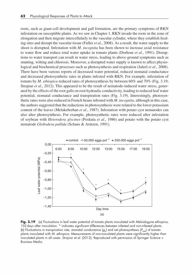
roots, such as giant-cell development and gall formation, are the primary symptoms of RKNinfestation on susceptible plants. As we saw in Chapter 1, RKN invade the roots in the zone ofelongation and then migrate intercellularly to the vascular cylinder, where they establish feed-ing sites and disrupt the vascular tissue (Fuller et al., 2008). As a result, the water supply to theshoot is disrupted. Infestation with M. incognita has been shown to increase axial resistanceto water flow and reduce total water uptake in tomato plants (Dorhout et al., 1991). Disrup-tions to water transport can result in water stress, leading to above-ground symptoms such asstunting, wilting and chlorosis. Moreover, a disrupted water supply is known to affect physio-logical and biochemical processes such as photosynthesis and respiration (Jaleel et al., 2008).There have been various reports of decreased water potential, reduced stomatal conductanceand decreased photosynthetic rates in plants infested with RKN. For example, infestation oftomato byM. ethiopica reduced rates of photosynthesis by between 60% and 70% (Fig. 3.19;Strajnar et al., 2012). This appeared to be the result of nematode-induced water stress, gener-ated by the effects of the root galls on root hydraulic conductivity, leading to reduced leaf waterpotential, stomatal conductance and transpiration rates (Fig. 3.19). Interestingly, photosyn-thetic rates were also reduced in French beans infested withM. incognita, although in this case,the authors suggested that the reductions in photosynthesis were related to the lower potassiumcontent of the leaves (Melakeberhan et al., 1987). Infestation with potato cyst nematodes canalso alter photosynthesis. For example, photosynthetic rates were reduced after infestationof soybean with Heterodera glycines (Poskuta et al., 1986) and potato with the potato cystnematode Globodera pallida (Schans & Arntzen, 1991).
6:00
0,00
Day time
Leaf w
ate
r pote
ntial (M
Pa)
−0,20
−0,40
−0,60
−0,80
−1,00
−1,20
−1,40
8:00
*
*
*
*
*
*
10:00
control 50.000 eggs pot−1 250.000 eggs pot−1
12:00 13:00 15:00 17:00 19:00
(a)
Fig. 3.19 (a) Fluctuations in leaf water potential of tomato plants inoculated with Meloidogyne ethiopica,102 days after inoculation. * indicates significant differences between infested and non-infested plants.(b) Fluctuations in transpiration rate, stomatal conductance (gs) and net photosynthesis (Pnet) of tomatoplants inoculated with M. ethiopica. Measurements of non-inoculated plants were significantly higher thaninoculated plants in all cases. Strajnar et al. (2012). Reproduced with permission of Springer Science +Business Media.

Photosynthesis in Attacked Plants and Crops 63
control
9:40
20
16
12
8
4
0
0
0
2
4
6
8
10
0.1
0.2
0.3
0.4
10:30 12:05 13:05
Time of day
15:20 17:20 18:40
50.000 eggs pot−1
Tra
nspiration (
mm
ol H
2O
m−2
s−1
)g
s (m
mol H
2O
m−2
s−1
)P
net (μ
mol C
O2 m
−2 s
−1)
250.000 eggs pot−1
(b)
Fig. 3.19 (continued).

64 Physiological Responses of Plants to Attack
Root-lesion nematode disease is caused by members of the genus Pratylenchus. Lesionnematodes are migratory endoparasites that enter the host root to feed and reproduce and movefreely through or out of the root tissue. They do not become sedentary in the roots, as dothe cyst or root-knot nematodes, and feeding is restricted almost entirely to the root cortex.The root lesion nematode P. coffeae is a major pest of coffee in several countries, causingpoor root growth, leaf loss and reductions in yield (Campos et al., 1990). Infestation of coffeeseedlings with these nematodes led to reductions in root and shoot fresh weight and in ratesof photosynthesis (Mazzafera et al., 2004; Fig. 3.20). Since these nematodes do not establishfeeding sites similarly to root knot nematodes, the authors suggest that the rapid reductionsin photosynthesis observed after infestation of coffee seedlings with P. coffeae is the result ofdirect damage to the root.
As mentioned in Section 3.2.2, potato early dying disease is caused primarily by the fungalpathogen V. dahliae. However, the root lesion nematode P. penetrans interacts synergisticallywith V. dahliae, resulting in enhancement of the visual symptoms of the disease and reducing
0
100
200
300
400
500
600
700
800
900
1000
Fresh mass (mg)
Inoculum level
(a)
Leaves
Roots
0
100
200
300
400
500
600
700
Total
radioactivity
(cpm)
Inoculum level
(b)
8000 nematodes1000 nematodes0 nematodes
8000 nematodes1000 nematodes0 nematodes
Fig. 3.20 Effect of Pratylenchus coffeae on growth and photosynthesis in coffee seedlings. (a) Fresh massof leaves and roots and (b) photosynthesis (total radioactivity assimilated, expressed as counts per minute).Adapted from Mazzafera et al. (2004). Reproduced with permission of Springer Science + Business Media.

Photosynthesis in Attacked Plants and Crops 65
yield (MacGuidwin & Rouse, 1990). When potato plants grown under controlled conditionswere infected with both V. dahliae and P. penetrans, photosynthesis was reduced significantly,despite the fact that there was little effect on photosynthesis when plants were inoculated withthe pathogen or nematode singly (Saeed et al., 1997). These reductions in photosynthesis wereaccompanied by reduced stomatal conductance and transpiration rates but not by a reductionin intercellular CO2 concentration. This suggested that the reduced rates of photosynthesis inthe jointly infected plants were not due simply to local plugging of the xylem vessels but morelikely to a combination of stomatal and non-stomatal factors (Saeed et al., 1997). Subsequentwork by Saeed et al. (2007) found similar effects on photosynthesis over 3 years of experimentsunder field conditions.
3.4 PHOTOSYNTHESIS IN PLANTS INFESTEDWITH INSECTS
Plant responses to insect herbivory tend to be assessed from the guild perspective, where differ-ent insect guilds are defined on the basis of their feeding mechanisms, such as chewing insects,piercing/sucking insects and so on (see Chapter 1). Welter (1989) used the guild approach toexamine a substantial number of articles dealing with plant responses to insect herbivory andfound that in more than half of all interactions, rates of photosynthesis were reduced. Thisstudy found that insect defoliation generally increased photosynthesis in remaining leaves,while insects feeding on cell contents tended to decrease photosynthesis (Welter, 1989). How-ever, since that study was conducted, it has become clear that plant photosynthetic responsesto insect herbivory are not as straightforward as what the analysis by Welter (1989) suggests,as we will see in the following sections.
3.4.1 Photosynthesis in plants attacked by chewing insectsVarious studies have demonstrated reduced rates of photosynthesis in the remaining leaf tis-sue following herbivory by chewing insects. For example, in a study of photosynthesis in wildparsnip infested with caterpillars of the cabbage looper, Trichoplusia ni, Zangerl et al. (2002)found that a single caterpillar feeding for 24 hours significantly decreased the photosyntheticactivity of the remaining leaf tissue. Moreover, using fluorescence imaging, they found largepatches of cells where photosynthesis was depressed, well beyond the area of leaf consumedby the caterpillars. Incredibly, the indirectly affected area on the leaves was six times greaterthan that directly affected by tissue removal. However, because the indirectly affected areaof the leaf remained photosynthetically active, albeit at a reduced rate, its contribution tothe overall reductions in photosynthetic activity was only three times that of the direct effect(Zangerl et al., 2002; Fig. 3.21). Later work examining the effects of T. ni on photosynthesisin A. thaliana found that the magnitude of the effects observed depended on the developmen-tal stage of the attacking herbivore (Tang et al., 2006). First instar larvae of T. ni feed on theunderside of leaves, making small holes, avoiding leaf veins and leaving the upper epidermisintact. In contrast, fourth instar larvae make large holes, consuming minor and major veins andthe leaf epidermis. Herbivory by first instar larvae reduced photosynthesis more strongly in theremaining leaf tissue than did herbivory by fourth instar larvae (Fig. 3.22). Feeding by bothfirst and fourth instars increased transpiration in the dark substantially, and because stomatawere closed, most of the water would have been lost from the cut edges of the leaf. A. thaliana

66 Physiological Responses of Plants to Attack
50
45
40
35
30
25
20
15
10
Perc
enta
ge r
eduction in
photo
synth
etic c
apacity
5
0
Direct Indirect
(image)
Indirect
(gas exchange)
Combined
Source Effects
−5
Fig. 3.21 Magnitudes of direct, indirect and total effects of caterpillar damage on suppression ofphotosynthesis in wild parsnip foliage. The black bars depict data obtained by fluorescence imaging, andthe white bar depicts data from gas exchange. The area of leaflet measured in all cases was 6 cm2. Theline inside each bar is the mean, the ends of the bars show the 10th and 90th percentiles, and the whiskersshow the 5th and 95th percentiles. (n=10.) Zangerl et al. (2002). Reproduced with permission of NationalAcademy of Sciences, USA.
00.0
0.2
0.3
Phosto
synth
etic e
ffic
iency
0.4
0.5
10 20 30
Area removed (%)
40
First instar
Fourth instar
50 60
Fig. 3.22 Correlation between the proportion of Arabidopsis leaf tissue removed (relative to areameasured) and photosystem II operating efficiency (ΦPSII) of the areas of the leaf characterized asphotosynthetically depressed. Leaf area with ΦPSII within or below the lower 10% of the averagedistribution for control leaves was considered photosynthetically depressed. Each point represents a leaf onone plant, 4 days after it was exposed to herbivory. Tang et al. (2006). Reproduced with permission ofOxford University Press.

Photosynthesis in Attacked Plants and Crops 67
has reticulate vasculature, allowing water to move around sites of damage to supply nearbytissue. However, although such vasculature might be able to compensate for water loss froma large hole, it appeared to be less effective in compensating for water loss from many smallholes, such as the damage resulting from first instar larvae (Tang et al., 2006).
In the work of Tang et al. (2006), the reductions in photosynthesis appeared to be the resultof localised water stress caused by damage to leaves. These workers found no change in theexpression of a gene encoding the small subunit of Rubisco. However, in other plant–insectinteractions, there can be substantial changes in gene expression. In the interaction betweenthe specialist herbivore Manduca sexta and its natural host Nicotiana attenuata, there wasa strong down-regulation of photosynthetic genes, accompanied by substantial up-regulationof defence-related genes (Hermsmeier et al., 2001; Hui et al., 2003). The latter authors sug-gested that the down-regulation of genes related to photosynthesis might allow attacked plantsto reinvest resources into other processes, such as those involved in defence (see Box 3.1).Interestingly, a down-regulation of photosynthetic genes and an up-regulation of genes relatedto secondary metabolism were also found in potato leaves treated with regurgitant of the Col-orado potato beetle, Leptinotarsa decemlineata (Lawrence et al., 2008).
As indicated at the start of this section, plant photosynthetic responses to insect herbivoryare not straightforward. Although there are many reports of reduced photosynthesis in remain-ing leaf tissue following herbivory, some of whichwe have just dealt with, there are also reportsof increased photosynthetic activity following herbivory (e.g. Holman & Oosterhuis, 1999) orartificial defoliation (Turnbull et al., 2007). Some studies have also found no effect of defo-liation on photosynthesis. Thus, Peterson et al. (2004) studied the photosynthetic responsesof several legume species to injury caused by mass consumption of leaf tissue. Photosyn-thesis was not significantly affected by either insect-induced or artificial defoliation. Similarresults were obtained with a number of varieties of soybean and alfalfa (e.g. Peterson et al.,1992; Peterson & Higley, 1996), suggesting that there is a common modality of response bylegumes to mass consumption of leaf tissue by insects (Peterson et al., 2004). However, sincechanges in photosynthesis were not detected in apple, crab apple, cucumber and tomato aftermass leaf consumption by insects (Welter, 1989; Peterson et al., 1996; Burkness et al., 1999),the common modality of response appears not to be limited to legumes.
The studies described previously examined photosynthesis in individual plants and leaves.What happens if we move from single plants to large plant populations, such as stands ofconifers or hardwood trees? Such studies are conducted to examine the impact of disturbance,such as forest fires or insect attacks on carbon cycling in forests. Insect attacks can influence netecosystem productivity, the net uptake of CO2 by the forest, via their impact on gross ecosys-tem photosynthesis (Pg) and ecosystem respiration. For example, measurements made in ahardwood forest in Wisconsin that had suffered 37% defoliation as a result of attack by foresttent caterpillars revealed a 24% reduction in Pg compared to non-outbreak years (Cook et al.,2008). In British Columbia, outbreaks of the mountain pine beetle (Dendroctonus ponderosae)resulted in tree mortality on such a scale that effects on the carbon balance of forests in BritishColumbia were possible. A modelling study predicted the cumulative impact of the mountainpine beetle outbreak between 2000 and 2020 would be a net loss of 270 million tonnes ofcarbon (Kurz et al., 2008), while estimates of Pg over the infestation area from 2002 to 2005revealed a reduction of 10–20% compared to pre-outbreak levels (Coops & Wulder, 2010).However, a subsequent study of two lodgepole pine-dominated stands in British Columbia, an85-year-old stand first attacked by mountain pine beetle in 2006 and a 110-year-old stand first

68 Physiological Responses of Plants to Attack
attacked in 2003, revealed that although net ecosystem production was negative after the initialattack, it increased substantially in the following year, as a result of increased productivity ofthe remaining trees and vegetation (Brown et al., 2010). Indeed, subsequent measurementsshowed that the recovery of net ecosystem production increased rapidly in both stands, due toan increase in Pg and photosynthetic capacity (Brown et al., 2012).
3.4.2 Photosynthesis in plants attackedby piercing-sucking insects
Damage to plants caused by insects with piercing-sucking mouthparts is often less evidentthan injury caused by insects with chewing mouthparts. Piercing-sucking insects may feed onsap of xylem, phloem or other plant cells, and their feeding site and the amount of damagethey cause to plant tissues can vary greatly. For example, although some workers found nosignificant effect of aphid feeding on photosynthesis (e.g. on cotton; Gomez et al., 2006),there are many reports of reductions in photosynthesis following feeding by piercing-suckinginsects, including aphids. Thus, reductions in photosynthetic rates of up to 50% were obtainedafter infestation of soybean with the phloem-feeding soybean aphid, Aphis glycines (Macedoet al., 2003). The magnitude of the reductions was surprising given the relatively low aphiddensities used (e.g. ∼50% reduction at aphid densities> 20/leaflet) and the fact that many ofthe leaves exhibited no visible symptoms of aphid damage (Macedo et al., 2003).
Photosynthesis can also be affected by spider mite infestation. The feeding apparatus ofspider mites consists of paired and partially fused cheliceral stylets, which they use to piercethe leaf surface and epidermis and disrupt the underlying mesophyll. Damage at the feedingsite includes punctured and collapsed epidermal cells and a disrupted cuticle. The twospottedspider mite, Tetranychus urticae, has a stylet long enough to reach the photosynthetically activemesophyll tissue. Indeed, infestation of soybean with T. urticae resulted in reduced rates ofphotosynthesis (de Freitas Bueno et al., 2009). The reduction in photosynthesis was due toreduced stomatal conductance, as no significant changes were observed in chlorophyll contentor chlorophyll fluorescence parameters.
Reduced photosynthetic activity was also found in interactions between two host plants,savoy cabbage (Brassica oleracea) and French bean (Phaseolus vulgaris), and the phy-tophagous stink bugs, Murgantia histrionica and Nezara viridula (Velikova et al., 2010).M. histrionica feeds using a lacerate and flush approach, which involves heavy damage tothe mesophyll tissue as a result of mechanical laceration of the cells and extra-oral digestionby salivary enzymes (Miles, 1972). In contrast, N. viridula adults feed on leaf veins with astylet-sheath feeding mode, destroying only a few cells and causing little mechanical damage(Miles, 1972). Rates of photosynthesis decreased rapidly in both cabbage infested withM. histrionica and bean infested with N. viridula (Fig. 3.23A and B). Although transpirationrate and stomatal conductance were reduced by 8 hours of insect feeding, the reduction inphotosynthesis was greater and more rapid than the changes in these parameters (Fig. 3.23Aand B). In bean infested with N. viridula, the substantial reduction in photosynthesis wasconfirmed by a large, transient inhibition in the photochemical efficiency of PS II, whiledamage to the maximal quantum yield of PS II was limited and transient, indicating that therewas no permanent damage to the photochemistry of the infested leaves. In this interaction,there was a more complete recovery of chlorophyll fluorescence than photosynthetic rate,and so photosynthesis was not permanently impaired. On cabbage, M. histrionica causedvisible and permanent damage to the leaf lamina, and in this case, a permanent impairment
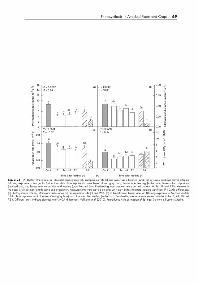
Photosynthesis in Attacked Plants and Crops 69
a
(a)
(A)
(b)
(c) (d)
P = 0.0002
F = 6.84
P = 0.0008
F = 5.42P < 0.0001
F = 16.92
P < 0.0001
F = 16.03
a
a
a
Cont
16 0.20
0.15
0.10
0.05
0.00
12
10
8
6
4
2
0
14
12
10
8
Photo
synth
esis
rate
(μm
ol m
−2 s
−1)
Sto
mata
l conducta
nce (
mol m
−2 s
−1)
WU
E (μm
ol C
O2 m
mol−
1 H
2O
)
Tra
nspiration r
ate
(m
mol m
−2 s
−1)
6
4
2
0
2.0
1.5
1.0
0.5
0.00 24 48
Time after feeding (h)
72 24 Cont 0 24 48
Time after feeding (h)
72 24
ab
a
ab
bc bc
bcbc
b
b
bbb
ab
b
c cc
cc
bc bcb
dd
Fig. 3.23 (A) Photosynthesis rate (a), stomatal conductance (b), transpiration rate (c) and water use efficiency (WUE) (d) of savoy cabbage leaves after an8-h long exposure to Murgantia histrionica adults. Bars represent control leaves (Cont, grey bars), leaves after feeding (white bars), leaves after oviposition(hatched bar), and leaves after oviposition and feeding (cross-hatched bar). Post-feeding measurements were carried out after 0, 24, 48 and 72 h, whereas inthe cases of oviposition, and feeding and oviposition, measurements were carried out after 24 h only. Different letters indicate significant (P<0.05) differences.(B) Photosynthesis rate (a), stomatal conductance (b), transpiration rate (c) and WUE (d) of French bean leaves after an 8-h long exposure to Nezara viridulaadults. Bars represent control leaves (Cont, grey bars) and of leaves after feeding (white bars). Post-feeding measurements were carried out after 0, 24, 48 and72 h. Different letters indicate significant (P<0.05) differences. Velikova et al. (2010). Reproduced with permission of Springer Science + Business Media.

70 Physiological Responses of Plants to Attack
a
(a)
(B)
(b)
(c) (d)
P = 0.0033
F = 5.66
P = 0.0032
F = 5.68P = 0.1112
F = 2.16
P = 0.0953
F = 2.29
a
a
a
Cont
16 0.20
0.15
0.10
0.05
0.00
12
10
8
6
4
2
0
14
12
10
8
Photo
synth
esis
rate
(μm
ol m
−2 s
−1)
Sto
mata
l conducta
nce (
mol m
−2 s
−1)
WU
E (μm
ol C
O2 m
mol−
1 H
2O
)
Tra
nspiration r
ate
(m
mol m
−2 s
−1)
6
4
2
0
2.0
1.5
1.0
0.5
0.00 24 48
Time after feeding (h)
72 Cont 0 24 48
Time after feeding (h)
72
ab
a
ab
ab ab
bc
bb
b
bb
ab
cc
c
bcb
Fig. 3.23 (continued).

Photosynthesis in Attacked Plants and Crops 71
of photosynthetic photochemistry was found in all damaged areas of leaves. In this system,photosynthesis did recover, although the recovery was incomplete and was driven by improvedphotosynthetic performance in unaffected areas of leaves (Velikova et al., 2010).
Compensatory increases in photosynthesis have also been obtained in plants attacked bypiercing-sucking insects. Thus, photosynthetic capacity of field-grown Pinus radiata trees wassignificantly greater after artificial defoliation or infestation with the phloem-feeding aphid,Essigella californica (Eyles et al., 2011), while compensatory increases in photosynthesisoccurred in beech trees (Northofagus solandri var. solandri) infested with the scale insect,Ultracoelostoma assimile (Dungan et al., 2007).
3.4.3 Photosynthetic changes following oviposition
Insect eggs represent a future threat to plants and the ability to detect oviposition, therebyallowing activation of appropriate defences, could provide an advantage to the host. Indeed,direct and indirect defences to oviposition have been detected, including the development ofnecrotic, a necrotic zone at the site of oviposition in Brassica nigra, leading to desiccationof the egg and mortality and the release of volatiles, attracting parasitoids of eggs (Shapiro &Devay, 1987; Meiners & Hilker, 2000; Hilker et al., 2002). If oviposition can affect defences,is it possible that the process affects plant primary metabolism? This was studied by Schröderet al., 2005, who examined the effects of egg deposition by the sawfly Diprion pini onphotosynthesis in Scots pine (Pinus sylvestris). Sawfly adults were allowed to lay eggs on thelower section of P. sylvestris twigs and rates of photosynthesis measured in the upper sectionof the twigs and in untreated twigs. Rates of net photosynthesis in oviposition-induced pinetwigs were significantly lower than in untreated twigs. Might this reduction in photosynthesisrepresent a trade-off between defence and photosynthetic activity, or might it simply be aconsequence of oviposition and the associated wounding of tissue, resulting in localisedwater stress and stomatal closure (Schröder et al., 2005)? Equally, given that some insectshave been shown to be sensitive to CO2 gradients (e.g. Stange et al., 1995), is it possiblethat the egg parasitoid or the herbivorous sawfly uses the changes in photosynthesis for
Box 3.1 Photosynthesis or defence: priorities for plants under attackPlants live in a hostile environment. They face attack from pathogens, pests and evenother plants, and because they are sessile, getting up and running away is not an option.As a result, plants have evolved a formidable array of defences to deal with attackers(Walters, 2010). But defence does not come cheaply, requiring metabolic resources,including carbon, nitrogen and supplies of energy and reducing power. The problemis that plants require not only to defend themselves, but also to grow and reproduce,and the latter processes also require metabolic resources. So plants under attack face adilemma – to grow or to reproduce. In reality, the choice might not be quite as starkas that, but there is still the need to prioritise the use of metabolic resources. Materialsused to synthesise defensive compounds or build defence structures are not available forgrowth and reproduction unless there is turnover (Gómez & Zamora, 2002). As a result,plants mounting a robust defence might incur costs in the form of reduced growth andreproductive fitness (Zavala et al., 2004).

72 Physiological Responses of Plants to Attack
Many of the metabolic resources for growth, reproduction and defence come fromphotosynthesis. As a result, there might also be a trade-off between photosynthesis anddefence. For example, the production of defensive furanocoumarins in wild parsnipis associated with reduced photosynthetic rates (Zangerl et al., 1997, 2002), whilein soybean, infection by an avirulent isolate of the bacterial pathogen Pseudomonassyringae pv. glycinea led to a down-regulation of photosynthesis (Zou et al., 2005).Tang et al. (2009) compared the spatial pattern of photosynthesis to that of inductionof the defence-related cinnamate-4-hydroxylase (C4H) gene in A. thaliana attackedby T. ni. They found that in areas of leaves where C4H expression was up-regulated,photosynthesis was reduced. However, the reduction in photosynthesis spread furtherinto surrounding areas of the leaf than did the expression of C4H. The authors suggestedthat the decrease in photosynthesis in areas of the leaf where C4H was induced mightrepresent a trade-off between defence and photosynthesis. They also suggested that thespread of photosynthetic damage beyond areas of C4H expression might reflect damageto the photosynthetic apparatus by other defences (e.g. glucosinolates) or non-defenceprocesses such as water stress (Tang et al., 2009).
As we have seen so far in this chapter, there are a great many examples where insectattack (and indeed pathogen attack) is associated with reductions in photosynthesis. How-ever, photosynthesis and defence are not always inversely related. For example, photo-synthesis and nicotine production are positively correlated in N. sylvestris (Baldwin &Ohnmeiss, 1994), while in transgenic N. tabacum with decreased expression of transketo-lase, an enzyme involved in photosynthesis as well as contributing to the phenylpropanoidpathway, photosynthesis and accumulation of defensive compounds are reduced (Henkeset al., 2001). Interestingly, it has been observed that plant genotypes that are able to main-tain photosynthetic activity under insect challenge exhibit greater tolerance to attack. Thus,tolerant lines of barley and wheat attacked by the Russian wheat aphid were able to main-tain photosynthetic rates, or recover photosynthetic capacity more rapidly, compared tosusceptible lines (Haile et al., 1999; Franzen et al., 2007; Gutsche et al., 2009).
Nevertheless, as we have already seen in this chapter, the expression of photosyntheti-cally related genes is commonly down-regulated after insect attack (e.g. Hui et al., 2003;Ralph et al., 2006; Tang et al., 2006), and compensatory increases in photosynthesis arerare (Kerchev et al., 2012). Down-regulation of photosynthesis might free up resources,making them available for production of defences. It has been suggested that the rarityof compensatory increases in photosynthesis in plant–insect interactions could reflect thepossibility that synthesis of secondary metabolites for defence is not costly to the plant interms of carbon and energy, because under most conditions, plants operate to ensure a sur-plus of carbon and energy, stored as starch and other carbohydrates (Kerchev et al., 2012).
There is a cost to everything in life. Of course, defence is costly. It requires metabolicresources after all. Whether the costs incurred by mounting defences become large enoughto be measured is likely to depend on the growth conditions plants find themselves in.Under optimal conditions, such costs might be too small to detect, whereas under limitingconditions, defence costs might well be more apparent. Plants have a prodigious anabolicpotential that allows them to throw just about everything at attackers in order to defendthemselves (Schwachtje & Baldwin, 2008). Moreover, plants are very plastic, a trait thatmight well be part of its defensive armoury (Karban et al., 1997).

Photosynthesis in Attacked Plants and Crops 73
orientation or to locate a host (Schröder et al., 2005)? Irrespective of the possible adaptivesignificance of this oviposition-induced reduction in photosynthesis, it is interesting to notethat in a study of oviposition by adults of Pieris brassicae on A. thaliana, gene expressionprofiling revealed a transcript signature that was remarkably similar to that obtained dur-ing a hypersensitive response (Little et al., 2007). The changes in gene expression includedthe up-regulation of defence-related genes and the down-regulation of genes associated withgrowth and photosynthesis.
3.5 PHOTOSYNTHESIS IN PLANTS INFECTEDWITH PARASITIC PLANTS
As we saw in Chapter 2, parasitic plants can lower growth and reproductive output of theirhosts. They can also affect host photosynthesis in a variety of ways and at a range of scales,from the leaf through to the whole plant. The impact of parasitic plants on host photosynthesisdepends on the species of parasite and host and, importantly, cannot always be correlatedwith the degree of dependence of the parasite on the host. Broadly, the effects of parasiticplants on host photosynthesis can be classified in terms of direct effects on removal of hostresources (source–sink interactions) and indirect or non-source–sink interactions (Watling& Press, 2001). In this section, we look at the effects of a range of different parasitic plantson photosynthesis in their hosts and examine the mechanisms responsible for the changesobserved.
3.5.1 Photosynthesis in plants infected with hemiparasites
Striga hermonthica is a root hemiparasite of C3 and C4 cereals in the semi-arid tropics andtypically causes stunting of host plants, with associated reductions in growth and grain yield(Stewart & Press, 1990). The effects of Striga on host growth and yield may be due, in part, toits role as an additional sink for host carbon, inorganic solutes and water (Press et al., 1990).However, although S. hermonthica is chlorophyllous, rates of photosynthesis tend to be low,while respiration rates are high. This leads to little, if any, carbon gain for the parasite, withthe result that it is dependent on the host for supplies of carbon (Graves et al., 1989). Variousworkers have reported reduced rates of photosynthesis in Striga-infected plants compared touninfected controls. For example, Press et al. (1987) found that infection of sorghum witheither S. hermonthica or S. asiatica markedly reduced photosynthetic capacity in the host.The very high rates of transpiration of the parasite predisposed the sorghum to water stress,although the resulting reduction in stomatal conductance was not sufficient to account for theStriga-induced reductions in photosynthesis. Later work by Frost et al. (1997) examined gasexchange in two varieties of sorghum infected with S. hermonthica, the susceptible CSH-1and Ochuti, a variety with some tolerance to the parasite under field conditions. Growth ofboth varieties was reduced substantially by infection. In the susceptible CSH-1, photosynthe-sis was reduced in infected plants and declined over time, whereas in Ochuti, photosynthesiswas not greatly affected by infection (Fig. 3.24). An examination of the kinetics of photo-synthetic induction in these plants is instructive (Fig. 3.25). In both CSH-1 and Ochuti, therate of photosynthesis, measured by CO2 assimilation or using the chlorophyll fluorescenceparameter ΦII, increased rapidly in leaves of uninfected plants, reaching a steady state after10–15minutes. This was accompanied by a rapid increase in stomatal conductance in both

74 Physiological Responses of Plants to Attack
0
2
4
6
8
10
12
14
16
30 35 40 45
Photosynthesis
(μmol CO2
m−2 s−1)
Host age (days)
(a)
CSH-1
uninfected
infected
0
2
4
6
8
10
12
14
16
30 35 40 45
Host age (days)
(b)
Ochuti
uninfected
infected
Photosynthesis
(μmol CO2
m−2 s−1)
Fig. 3.24 Rate of photosynthesis of the youngest fully emerged leaf of the two sorghum cultivars andOchuti, either uninfected or infected with Striga hermonthica. Frost et al. (1997). Reproduced withpermission of John Wiley & Sons.
varieties. In leaves of CSH-1 infected with S. hermonthica, both photosynthesis and stomatalconductance increased slowly on irradiation and 15minutes later had nearly reached steadystate but were lower than in leaves from uninfected controls (Fig. 3.25). In Striga-infectedOchuti, photosynthesis and stomatal conductance also increased slowly after illumination butafter some 30minutes, reached the same values as the uninfected controls. Overall, the dataprovided by Frost et al. (1997) suggest that lower values of stomatal conductance were the pri-mary cause of the reduced rates of photosynthesis in the Striga-infected sorghum. What couldcause such reductions in stomatal conductance? Well, transpiration rates in infected CSH-1were lower than in uninfected controls, and this was accompanied by an increased concen-tration of the plant growth regulator abscisic acid (ABA) in the xylem sap and leaf tissue ofinfected CSH-1 plants. Since ABA is known to be involved in controlling stomatal responsesto various environmental stresses, including drought (Wilkinson & Davies, 2002), it is possi-ble that the increased ABA found in the xylem sap and leaves of CSH-1 was responsible forthe lower stomatal conductance.

Photosynthesis in Attacked Plants and Crops 75
0
0.1
0.2
0.3
0.4
0.5
0.6
0.7
2.5 5 7.5 10 12.5 15
ΦII
(i)
(a) (b)
Uninfected
Infected
CSH-1
0
0.1
0.2
0.3
0.4
0.5
0.6
0.7
5 10 15 20 25
(i)
Infected
OCHUTI
Uninfected
0
2
4
6
8
10
12
Photosynthesis
(μmol CO2
m−2
s−1
)
(ii)Uninfected
Infected
CSH-1
0
2
4
6
8
10
12(ii)
Uninfected
Infected
OCHUTI
0
0.01
0.02
0.03
0.04
0.05
0.06
0.07
0.08
Time after start of illumination (minutes)
Time after start of illumination (minutes)
(iii)
Uninfected
Infected
CSH-1
0
0.01
0.02
0.03
0.04
0.05
0.06
0.07
2.5 5 7.5 10 12.5 15 5 10 15 20 25
2.5 5 7.5 10 12.5 15 5 10 15 20 25
Stomatal
conductance
(mol m−2
s−1
)
Stomatal
conductance
(mol m−2
s−1
)
(iii)
Uninfected
Infected
OCHUTI
Photosynthesis
(μmol CO2
m−2
s−1
)
Time after start of illumination (minutes)
Time after start of illumination (minutes) Time after start of illumination (minutes)
Time after illumination (minutes)
ΦII
Fig. 3.25 Photochemical quenching of chlorophyll fluorescence (ΦII), rate of photosynthesis and stomatalconductance of the youngest fully emerged leaf of the two sorghum cultivars CSH-1 and Ochuti, irradiatedat 900 μmol m−2 s−1 following a period of dark adaptation. Plants were 40 days old and were eitheruninfected or infected with Striga hermonthica. Note the difference in scale of the x-axis between the twocultivars. Frost et al. (1997). Reproduced with permission of John Wiley & Sons.
There is also evidence for increased photoinhibition in plants infected with Striga. This isimportant because if photoprotective mechanisms are inadequate, sustained exposure to highlight could damage the photosynthetic machinery, leading to a chronic reduction in quantumyield and increased costs for repairing thylakoid proteins associated with electron transport(Demmig-Adams & Adams, 1992). Ramlan and Graves (1996) studied photoinhibition insorghum infected with S. hermonthica and found that once the parasite had emerged aboveground, photosynthesis declined in infected plants, which also became severely photoinhibitedafter exposure to high light. These plants took longer to recover from photoinhibition thanuninfected plants and after 24 hours, still had lower quantum yields than uninfected plants,suggesting that they had sustained damage to their photosynthetic machinery (Ramlan &Graves, 1996).

76 Physiological Responses of Plants to Attack
Facultative root hemiparasites, such as Rhinanthus minor, often have a wide host rangeand may form haustorial connections with a number of different host plant species. Indeed,R.minor can grow on more than 20 host plants (Gibson &Watkinson, 1989), although grassesand legumes tend to be the best hosts for the parasite in terms of its growth and reproduction(Cameron et al., 2006). In general, these hosts are significantly damaged by the parasite, whileforbs (herbaceous species that are not graminoids) tend to remain undamaged. In the grassPhleum bertolinii, R. minor reduced biomass production significantly, while no significanteffects on growth were found with the forb, Plantago lanceolata (Cameron et al., 2008). Theseeffects on host biomass production were mirrored by changes in photosynthetic activity. Thus,infection of Phleum by R.minor led to significant reductions in the quantum efficiency of PS II(ΦII) and chlorophyll concentration, while no such effects were found in Plantago (Figs 3.26and 3.27). Interestingly, when photosynthesis was measured in R. minor growing on the twohosts, although the two photosynthetic parameters (Fv/Fm and ΦII) of the parasite growingon Phleum were similar to those of a healthy plant, both parameters were greatly reduced inR. minor growing on Plantago (Fig. 3.26 – Fv/Fm not shown in this figure) (Cameron et al.,2008). From these data, it appears as though Plantago is able to suppress growth of R. minorby suppressing the electron transport rate (ΦII).
Cassytha pubescens is a stem hemiparasite with an identical habit and morphology toplants in the holoparasitic genus Cuscuta. However, unlike Cuscuta, which is heterotrophicand phloem tapping, C. pubescens is autotrophic and xylem tapping. Infection with this par-asite can kill the invasive weeds Cytisus scoparius and Ulex europaeus in the Mount LoftyRanges of South Australia. Infection of C. scoparius with this parasite leads to significantreductions in rates of photosynthesis, and because Rubisco contents and chlorophyll concen-trations were not altered by infection, the photosynthetic reductions were probably the result oflower stomatal conductance (Shen et al., 2010; Fig. 3.28). Infected plants also exhibited lowerefficiency of PS II across the diurnal cycle and were also more susceptible to photoinhibition.Indeed, the authors suggested that the lower photosynthetic rates, combined with increasedsusceptibility to photoinhibition, were likely to be responsible for the poor performance andeven death of infected plants.
3.5.2 Photosynthesis in plants infected with holoparasites
As we saw in Chapter 1, holoparasites are achlorophyllous (or almost so) and as such aredependent on their hosts for their supply of carbon. Orobanche species are holoparasitic andnot only have they lost any photosynthetic ability, but also their root system is vestigial, therebymaking them reliant on their host for nitrogen and inorganic nutrients too (Parker & Riches,1993). Moreover, Orobanche species can be important parasites on various dicotyledenousplants, and infection can lead to reductions in growth and yield (e.g. Barker et al., 1996).
A study of tobacco infected with O. cernua showed that infected plants reduced growthsubstantially and that this growth reduction could be accounted for directly by diversion ofdry matter towards the parasite (Hibberd et al., 1998). This contrasts markedly with the sit-uation in Striga-infected plants described in the previous section, where the parasitic plantusually accounts for only a small proportion of the difference in biomass between the hostand the parasite. In tobacco infected with O. cernua, the effect of the parasite on host growthwas directly attributable to the sink activity of the parasite, with reductions in host growthdetermined by the size of the parasite sink. In this system, infected plants maintained the sameleaf area as uninfected plants, but leaf senescence was delayed. Indeed, although the rate of

Photosynthesis in Attacked Plants and Crops 77
0.8(a)
Infected hostUninfected host
(b)
(c)
0.6
0.4Φ
PS
II
0.2
0.0
0.8
0.6
0.4
ΦP
SII
0.2
0.0
0.8
0.6
0.4
ΦP
SII
0.2
0.00 500 1000
Actinic light intensity (μmol m−2 s−1)
1500
RhinanthusParasitizing Phleum
Phleum
Parasitizing Plantago
Plantago
2000 2500
Fig. 3.26 Quantum efficiency of photosystem II (ΦPSII) for host plants either infected or uninfected by theparasitic plant Rhinanthus minor: (a) Phleum bertolonii and (b) Plantago lanceolata; and (c) ΦPSII for theparasite growing with these species. Cameron et al. (2008). Reproduced with permission of OxfordUniversity Press.

78 Physiological Responses of Plants to Attack
Parasite
5
2.39
(±0.08)
2.41
(±0.26)
2.75
(±1.25)
1.84
(±0.26)
2.98
(±4.20)
1.92
(±0.07)
4
3
Tota
l am
ount of chlo
rophyll
(mg g
−1)
2
Phleum Plantago
1
0
Infected host
Uninfected host
Fig. 3.27 Chlorophyll concentration in infected and uninfected host plants Phleum bertolonii andPlantago lanceolata parasitized by Rhinanthus minor and for the parasite growing with these species.Chlorophyll a:b ratios are given above the corresponding bar. Cameron et al. (2008). Reproduced withpermission of Oxford University Press.
0
2
4
6
8
10
12
14
16
Uninfected host
Assimilation
rate (μmol
CO2
m−2
s−1
)
(a)
0
0.02
0.04
0.06
0.08
0.1
0.12
Stomatal
conductance
(mol m−2
s−1
)
(b)
0.00
0.10
0.20
0.30
0.40
0.50
0.60
0.70
Chlorophyll
concentration
(g m−2
)
(c)
0
20
40
60
80
100
120
Rubisco
(Percentage
of control)
(d)
Infected host Uninfected host Infected host
Uninfected host Infected host Uninfected host Infected host
Fig. 3.28 (a) Stem photosynthesis, (b) stomatal conductance, (c) concentration of chlorophylla+ chlorophyll b, and (d) Rubisco content, for Cytisus scoparius plants either uninfected or infected withCassytha pubescens. Shen et al. (2010). Reproduced with permission from CSIRO Publishing.

Photosynthesis in Attacked Plants and Crops 79
(a) (b)16
14
12
10
8
6
2
00 20 30 40 50 60 70 1 2 3 4
Young OldLeaf positionDays after planting
4
Rate
of net photo
synth
esis
(μm
ol m
−2 s
−1)
16
14
12
10
8
6
2
0
4
Rate
of n
et p
hoto
synth
esis
(μm
ol m
−2 s
−1)
Fig. 3.29 (a) The rate of net photosynthesis (μmol m–2 s–1) of the youngest fully expanded leaf between30 and 65 days after planting. Uninfected tobacco (○) or tobacco infected with Orobanche cernua (◾).(b) The rate of net photosynthesis (μmol m–2 s–1) in leaves from the top to the bottom of the canopy. Leafpositions are as follows: 1, the youngest fully expanded leaf at 65 days after planting; 2, the fourthyoungest fully expanded leaf; 3, the eighth youngest fully expanded leaf; 4, the twelfth youngest fullyexpanded leaf. Hibberd et al. (1998). Reproduced with permission of John Wiley & Sons.
net photosynthesis of the youngest fully expanded leaf was not altered in infected plants, theage-dependent decline in net photosynthesis found in leaves from uninfected plants through-out the canopy was retarded in infected plants, thus maintaining their rates of photosynthesisfor longer (Fig. 3.29; Hibberd et al., 1998).
The parasitic angiospermCuscuta can be considered a holoparasite or an intermediate para-site, as it contains some chlorophyll, although its photosynthesis makes little contribution to itstotal carbon requirement (0.6%; Jeschke et al., 1994).C. reflexa parasitizes herbaceous speciessuch as various legumes and indeed, on one of these hosts, Lupinus angustifolius, it can attractas much as 82% of the current photosynthate (Jeschke et al., 1994). Infection by Cuscutacan reduce fruit set in L. angustifolius substantially and can eventually kill its host. Neverthe-less, infection of L. angustifolius with C. reflexa resulted in a sink-induced stimulation of hostphotosynthesis (Jeschke et al., 1994). On the non-leguminous host Ricinus communis, infec-tion by this parasite reduced host growth but did not cause irreversible damage. In this case,infection also led to a substantial, sink-dependent increase in host photosynthesis (Jeschke& Hilpert, 1997), as it did on Coleus blumei (Jeschke et al., 1997). On the latter host, theincreased photosynthesis was attributed to increased chlorophyll concentration, greater stom-atal conductance, an apparent delay in leaf senescence and relief of feedback inhibition ofphotosynthesis (Jeschke et al., 1997). Why should the effects of infection by C. reflexa lead tosuch different effects on growth of L. angustifolius and Ricinus communis? It was suggestedthat the severity of the effects on L. angustifoliusmight reflect the competition between the hostplant, its nitrogen-fixing symbiont,Rhizobium, andC. reflexa; of course, in the non-leguminousR. communis, the extra competitive factor of the symbiont was not present.
Cuscuta campestris is also holoparasitic, but in contrast to O. cernua and C. reflexa, itseffects on host photosynthetic activity are very different. Infection ofMikania micrantha withC. campestris can reduce host biomass substantially and can inhibit host flowering, but inthis system, host photosynthesis is severely compromised. Thus, infection by C. campestris

80 Physiological Responses of Plants to Attack
(a) (b)
(c)
00
5
10
20
Pn (μm
ol C
O2 m
−2 s
−1)
15
0
5
10
20
15
10 20 30 40 50 60 0
Time (DAP)
10 20 30 40 50 60
(d)
Fig. 3.30 Mean of the net photosynthetic rates (Pn) of leaves from different leaf order positions ofMikania micrantha, uninfected (open circles) and infected (closed circles) by Cuscuta campestris, between 7and 65 days after parasitization (DAP). (a), (b), (c), and (d) are for the 1st, 4th, 8th and 12th fully expandedmature leaves, respectively. Shen et al. (2007). Reproduced with permission from Oxford University Press.
significantly reduced rates of net photosynthesis immediately after infection, with theinhibitory effect becoming greater with time (Shen et al., 2007; Fig. 3.30). This decrease innet photosynthesis appeared to be due, in part, to lower light use efficiency and to a lowercarboxylation efficiency compared to uninfected plants.
3.6 THE CARING ROBBER? HARDLY!
Parasitism is very much a one-sided affair. The parasite takes and the attacked plant has to putup with it. In most cases, this taking by the parasite has deleterious effects on the host plant.However, as Watling and Press (2001) ponder, there is surely an advantage to the parasite inkeeping the host in a fit state for as long as the association lasts. Why then, do parasites tend toimpair host photosynthesis and growth? Watling and Press (2001) were referring to hemipar-asitic plants when they asked this question, but it also pertains to pathogens and pests. Theysuggested that there is no short-term disadvantage to, for example, annual hemiparasites, inreducing photosynthesis and weakening the host, because they complete their life cycle beforethe host dies. There can, however, be long-term implications of reducing host photosynthesisand vigour, as a negative effect on host reproductive output could put hemiparasites with high

Photosynthesis in Attacked Plants and Crops 81
host specificity at a disadvantage and furthermore, could exert a significant effect on commu-nity structure (Marvier & Smith, 1997; Press, 1998).
3.7 CONCLUSIONS
As we have seen in this chapter, a common reaction to parasite attack is a reduction in hostphotosynthesis. It has been argued that this represents a host response aimed at ensuring thatresources are available for defensive actions. Whether this is true or not, it has become clearover the past few years that changes in host photosynthesis are only part of a network ofinterconnected metabolic events, linked closely to changes in carbohydrate metabolism andsignalling. These aspects will be covered in detail in Chapter 5. In the meantime, Chapter 4will look at changes in respiratory processes in attacked plants. Such changes are important insusceptible and resistant plants, with consequences for host growth.
RECOMMENDED READING
Baker NR, 2008. Chlorophyll fluorescence: a probe of photosynthesis in vivo. Annual Review of Plant Biology59, 89–113.
Bonfig KB, Schreiber U, Gabler A, Roitsch T, Berger S, 2006. Infection with virulent and avirulent P. syringaestrains differentially affects photosynthesis and sink metabolism in Arabidopsis leaves. Planta 225, 1–12.
Cameron DD, Geniez J-M, Seel WE, Irving LJ, 2008. Suppression of host photosynthesis by the parasitic plantRhinanthus minor. Annals of Botany 101, 573–578.
Zangerl AR, Hamilton JG, Miller TJ, Crofts AR, Oxborough K, Berenbaum MR, De Lucia EH, 2002. Impactof herbivory on photosynthesis is greater than the sum of its holes. Proceedings of the National Academyof Sciences of the United States of America 99, 1088–1091.
REFERENCES
Ayres PG, 1976. Patterns of stomatal behaviour, transpiration and CO2 exchange in pea following infection bypowdery mildew (Erysiphe pisi). Journal of Experimental Botany 27, 354–363.
Baebler S, Krecic H, Rotter A, Kogovšek P, Cankar K, Kok EJ, Gruden K, Kovac M, Žel J, Pompe-Novak M,Ravnikar M, 2009. PVYNTN elicits a diverse gene expression response in different potato genotypes in thefirst 12 h after inoculation. Molecular Plant Pathology 10, 263–275.
Baker NR, 2008. Chlorophyll fluorescence: a probe of photosynthesis in vivo. Annual Review of Plant Biology59, 89–113.
Balachandran S, Hurry VM, Kelley SE, Osmond CB, Robinson SA, Rohozinski GGR, Sims DA, 1997. Con-cepts of plant biotic stress. Some insights into the stress physiology of virus-infected plants, from theperspective of photosynthesis. Physiologia Plantarum 100, 203–213.
Baldwin IT, Ohnmeiss TE, 1994. Coordination of photosynthetic and alkaloidal responses to damage in unin-ducible and inducible Nicotiana sylvestris. Ecology 75, 1003–1014.
Barker ER, Press MC, Scholes JD, QuickWP, 1996. Interactions between the parasitic angiospermOrobancheaegyptica and its tomato host: growth and biomass allocation. New Phytologist 133, 637–642.
Bastiaans L, 1991. Ratio between virtual and visual lesion size as a measure to describe reduction in leafphotosynthesis in rice due to leaf blast. Phytopathology 81, 611–615.
Beckman CH, 1987. The nature of wilt diseases in plants. St. Paul, MN, USA: APS Press.Berger S, Benediktyová Z, Matouš K, Bonfig K, Mueller MJ, Nedbal L, Roitsch T, 2007.Visualisation of
dynamics of plant-pathogen interaction by novel combination of chlorophyll fluorescence imaging andstatistical analysis: differential effects of virulent and avirulent strains of P. syringae and of oxylipins onA. thaliana. Journal of Experimental Botany 58, 797–806.

82 Physiological Responses of Plants to Attack
Bennett J, Scott KJ, 1971. Inorganic polyphosphates in the wheat stem rust fungus and in rust-infected wheatleaves. Physiological Plant Pathology 1, 185–198.
Bonfig KB, Schreiber U, Gabler A, Roitsch T, Berger S, 2006. Infection with virulent and avirulent P. syringaestrains differentially affects photosynthesis and sink metabolism in Arabidopsis leaves. Planta 225, 1–12.
Bowden RL, Rouse DI, 1991. Effects of Verticillium dahliae on gas exchange of potato. Phytopathology 81,293–301.
Bowden RL, Rouse DI, Sharkey TD, 1990. Mechanism of photosynthesis decrease by Verticillium dahliae inpotato. Plant Physiology 94, 1048–1055.
Brown M, Black TA, Nesic Z, Foord VN, Spittlehouse DL, Fredeen AL, Grant NJ, Burton PJ, Trofymow JA,2010. Impact of mountain pine beetle on the net ecosystem production of lodgepole pine stands in BritishColumbia. Agricultural and Forest Meteorology 150, 254–264.
Brown M, Black TA, Nesic Z, Freeden AL, Foord VN, Spittlehouse DL, Bowler R, Burton PJ, Trofymow JA,Grant NJ, Lessard D, 2012. The carbon balance of two lodgepole pine stands recovering from mountainpine beetle attack in British Columbia. Agricultural and Forest Meteorology 153, 82–93.
Burkness EC, Hutchison WD, Higley LG, 1999. Photosynthetic response of ‘Carolina’ cucumber to simulatedand actual striped cucumber beetle (Coleoptera: Chrysomelidae). Entomologia Sinica 6, 29–38.
Cameron DD, Coats AM, Seel WE, 2006. Host and non-host resistance underlie variable success of thehemi-parasitic plant Rhinanthus minor. Annals of Botany 98, 1289–1299.
Cameron DD, Geniez J-M, Seel WE, Irving LJ, 2008. Suppression of host photosynthesis by the parasitic plantRhinanthus minor. Annals of Botany 101, 573–578.
Campos VP, Sivapalan P, Gnanapragasam NC, 1990. Nematode parasites of coffee, cocoa and tea. In: Luc M,Sikora RA, Bridge J, eds. Plant parasitic nematodes in subtropical and tropical agriculture. Wallingford,UK: CAB International, pp. 387–430.
Coghlan SE, Walters DR, 1992. Photosynthesis in green-islands on powdery mildew-infected barley leaves.Physiological and Molecular Plant Pathology 40, 31–38.
Cook B, Bolstad P, Martin J, Heinsch F, Davis K, Wang W, Desai A, Teclaw R, 2008. Using light-use andproduction efficiency models to predict photosynthesis and net carbon exchange during forest canopy dis-turbance. Ecosystems 11, 26–44.
Coops NC, Wulder MA, 2010. Estimating the reduction in gross primary production due to mountain pinebeetle infestation using satellite observations. International Journal of Remote Sensing 31, 2129–2138.
De Freitas Bueno A, de Freitas Bueno RCO, Nabity PD, Higley LG, Fernandes OA, 2009. Photosyntheticresponse of soybean to two spotted spider mite (Acari: Tetranychydae) injury. Brazilian Archives of Biologyand Technology 52, 825–834.
Demmig-Adams B, AdamsWW III , 1992. Photoprotection and other responses of plants to light stress. AnnualReview of Plant Physiology and Plant Molecular Biology 43, 2029–2031.
Doodson JK, Manners JM, Myers A, 1964. Some effects of yellow rust (Puccinia striiformis) on the growthand yield of spring wheat. Annals of Botany 28, 459–472.
Dorhout R, Gommers FJ, Kolloffel C, 1991. Water transport through tomato roots infected with Meloidogyneincognita. Phytopathology 81, 379–385.
Dungan RJ, TurnbullMH, Kelly D, 2007. The carbon costs for host trees of a phloem-feeding herbivore. Journalof Ecology 95, 603–613.
Dyer TA, Scott KJ, 1972. Decrease in chloroplast polysome content of barley leaves infected with powderymildew. Nature 236, 237–238.
Erickson JE, Stanosz GR, Kruger EL, 2003. Photosynthetic consequences of Marssonina leaf spot differbetween two poplar hybrids. New Phytologist 161, 577–583.
Evans LJ, Scholes JD, 1995. How does clubroot alter the regulation of carbon metabolism in its host? Aspectsof Applied Biology 42, 125–132.
Eyles A, Smith D, Pinkard EA, Smith I, Corkrey R, Elms S, Beadle C, Mohammed C, 2011. Photosyntheticresponses of field-grown Pinus radiata trees to artificial and aphid-induced defoliation. Tree Physiology 31,592–603.
Franzen LD, Gutsche AR, Heng-Moss TM, Higley LG, Sarath G, Burd JD, 2007. Physiological and biochem-ical responses of resistant and susceptible wheat to injury by Russian wheat aphid. Journal of EconomicEntomology 100, 1692–1703.
Frost DL, GurneyAL, PressMC, Scholes JD, 1997. Striga hermonthica reduces photosynthesis in sorghum: theimportance of stomatal limitations and a potential role for ABA? Plant, Cell and Environment 20, 483–492.
Fuller VL, Lilley CJ, Urwin PE, 2008. Nematode resistance. New Phytologist 180, 27–44.

Photosynthesis in Attacked Plants and Crops 83
Garavaglia BS, Thomas L, Gottig N, Dunger G, Garofalo CG, Daurelio LD, Ndimba B, Orellano EG, GehringC, Ottado J, 2010. A eukaryotic acquired gene by a biotrophic phytopathogen allows prolonged survival onthe host by counteracting the shut-down of plant photosynthesis. PLoS ONE 5 (1), 1–10.
Gibson CC, Watkinson AR, 1989. The host range and selectivity of a parasitic plant – Rhinanthus minor L.Oecologia 78, 401–406.
Gómez JM, Zamora R, 2002. Thorns as induced mechanical defense in a long-lived shrub (Hormathophyllaspinosa, Cruciferae). Ecology 83, 885–890.
Gomez SK, Oosterhuis DM, Hendrix DL, Johnson DR, Steinkraus DC, 2006. Diurnal pattern of aphid feedingand its effect on cotton leaf physiology. Environmental and Experimental Botany 55, 77–86.
Gordon TR, Duniway JM, 1982. Photosynthesis in powdery mildewed sugar beet leaves. Phytopathology 72,718–723.
Graves JD, Press MC, Stewart GR, 1989. A carbon balance model of the sorghum-Striga hermonthicahost-parasite association. Plant, Cell and Environment 12, 101–107.
Gutsche AR, Heng-Moss TM, Higley LG, Sarath G, Mornhinweg DW, 2009. Physiological responses of resis-tant and susceptible barley, Hordeum vulgare, to the Russian wheat aphid, Diuraphis noxia (Mordvilko).Arthropod-Plant Interactions 3, 233–240.
Haigh GR, Carver TLW, Gay AP, Farrar JF, 1991. Respiration and photosynthesis in oats exhibiting differentlevels of partial resistance to Erysiphe graminis D.C. ex Merat f.sp. avenaeMarchal. New Phytologist 119,129–136.
Haile FJ, Higley LG, Ni X, Queensberry SS, 1999. Physiological and growth tolerance in wheat to Russianwheat aphid (Homoptera: Aphididae) injury. Environmental Entomology 28, 787–794.
Hanssen IM, van Essen P, Ballester A-R, Hogewonig SW, Parra NO, Paeleman A, Lievens B, Bovy AC,ThommaBPHJ, 2011. Differential tomato transcriptomic responses induced byPepinoMosaic Virus isolateswith differential aggressiveness. Plant Physiology 156, 301–318.
Henkes S, Sonnewald U, Badur R, Flachmann R, Stitt M, 2001. A small decrease in plastid transketolaseactivity in antisense tobacco transformants has dramatic effects on photosynthesis and phenylpropanoidmetabolism. Plant Cell 13, 535–551.
Hermsmeier D, Schittko U, Baldwin IT, 2001. Molecular interactions between the specialist herbivore Mand-uca sexta (Lepidoptera, Sphingidae) and its natural host Nicotiana attenuata. I. Large-scale changes in theaccumulation of growth- and defense-related plant mRNAs. Plant Physiology 125, 683–700.
Hewitt HG, 1976. The effects of infection by Microsphaera alphitoides upon the growth and physiology ofQuercus robur. PhD Thesis, University of Lancaster.
Hewitt HG, Ayres PG, 1975. Changes in CO2 and water vapour exchange rates in leaves of Quercus roburinfected by Microsphaera alphitoides (powdery mildew). Physiological Plant Pathology 7, 127–137.
Hibberd JM, Quick WP, Press MC, Scholes JD, 1998. Can source-sink relations explain responses of tobaccoto infection by the root holoparasitic angiosperm Orobanche cernua? Plant, Cell and Environment 21,333–340.
Higgins CM, Manners JM, Scott KJ, 1985. Decrease in three messenger RNA species coding for chloroplastproteins in leaves of barley infected with Erysiphe graminis f.sp. hordei. Plant Physiology 78, 891–894.
Hilker M, Kobs C, Varma M, Schrank K, 2002. Insect egg deposition induces Pinus sylvestris to attract eggparasitoids. Journal of Experimental Biology 205, 455–461.
Hirano SS, Upper CD, 2000. Bacteria in the leaf ecosystem with emphasis on Pseudomonas syringae – apathogen, ice nucleus, and epiphyte. Microbiology and Molecular Biology Reviews 64, 624–653.
Holman EM, Oosterhuis DM, 1999. Cotton photosynthesis and carbon partitioning in response to floral budloss due to insect damage. Crop Science 39, 1347–1351.
Hui D, Iqbal J, Lehman K, Gase K, Saluz HP, Baldwin IT, 2003. Molecular interactions between the specialistherbivoreManduca sexta (Lepidoptera, Sphingidae) and its natural host Nicotiana attenuata: V. Microarrayanalysis and further characterization of large-scale changes in herbivore-induced mRNAs. Plant Physiology131, 1877–1893.
Jaleel CA, Manivannan P, Lakshmanan GMA, Gomathinayagam M, Panneerselvam R, 2008. Alterations inmorphological parameters and photosynthetic pigment responses of Catharanthus roseus under soil waterdeficit. Colloids and Surfaces. B, Biointerfaces 61, 298–303.
Jeschke WD, Hilpert A, 1997. Sink-stimulated photosynthesis, increased transpiration and increaseddemand-dependent stimulation of nitrate uptake: Nitrogen and carbon relations in the parasitic associationCuscuta reflexa-Ricinus communis. Plant, Cell and Environment 20, 47–56.

84 Physiological Responses of Plants to Attack
Jeschke WD, Bäumel P, Räth N, Czygan F-C, Proksch P, 1994. Modelling the flow and partitioning of carbonand nitrogen in the holoparasite Cuscuta reflexa Roxb. and its host Lupinus albus L. II. Flows between hostand parasite and within the parasitized host. Journal of Experimental Botany 45, 801–812.
Jeschke WD, Baig A, Hilpert A, 1997. Sink-stimulated photosynthesis, increased transpiration and increaseddemand-dependent stimulation of nitrate uptake: Nitrogen and carbon relations in the parasitic associationCuscuta reflexa-Coleus blumei. Journal of Experimental Botany 48, 915–925.
Johnstone M, Chatterton S, Sutton JC, Grodzinski B, 2005. Net carbon gain and growth of bell peppers,Capsicum annuum ‘Cubico’, following root infection by Pythium aphanidermatum. Phytopathology 95,354–361.
Karban R, Agrawal AA, Mangel M, 1997. The benefits of induced defences against herbivores. Ecology 78,1351–1355.
Kerchev PI, Fenton B, Foyer CH, Hancock RD, 2012. Plant responses to insect herbivory: interactions betweenphotosynthesis, reactive oxygen species and hormonal signalling pathways. Plant, Cell and Environment 35,441–453.
KurzWA,DymondCC, StinsonG, RampleyGJ, Neilson ET, Carroll AL, Ebata T, Safranyik L, 2008.Mountainpine beetle and forest carbon feedback to climate change. Nature 452, 987–990.
Lawrence SD, Novak NG, Ju CJ-T, Cooke JEK, 2008. Potato, Solanum tuberosum, defense against Coloradopotato beetle, Leptinotarsa decemlineata (Say): microarray gene expression profiling of potato by Coloradopotato beetle regurgitant treatment of wounded leaves. Journal of Chemical Ecology 34, 1013–1025.
Little D, Gouhier-Darimont C, Bruessow F, Reymond P, 2007. Oviposition by pierid butterflies triggers defenseresponses in Arabidopsis. Plant Physiology 143, 784–800.
Livne A, Daly JM, 1966. Translocation in healthy and rust-affected beans. Phytopathology 56, 170–175.Lopes DB, Berger RD, 2001. The effects of rust and anthracnose on the photosynthetic competence of diseased
bean leaves. Phytopathology 91, 212–220.Lukens JH, Durbin RD, 1985. Tagetitoxin affects plastid development in seedling leaves of wheat. Planta 165,
311–321.Macedo B, Bastos CS, Higley L, Ostje KR,Madhavan S, 2003. Photosynthetic responses of soybean to soybean
aphid. Journal of Economic Entomology 96, 188–193.MacGuidwin AE, Rouse DI, 1990. Role of Pratylenchus penetrans in the potato early dying disease of Russet
Burbank potato. Phytopathology 80, 1077–1082.Magyarosy AC, Schurmann P, Buchanan BB, 1976. Effect of powdery mildew infection on photosynthesis by
leaves and chloroplasts of sugar beet. Plant Physiology 57, 486–489.Magyarosy AC, Malkin R, 1978. Effect of powdery mildew infection of sugar beet on the content of electron
carriers in chloroplasts. Physiological Plant Pathology 13, 183–188.Manter DK, Kelsey RG, Karchesy JJ, 2007. Photosynthetic declines in Phytophthora ramorum-infected plants
develop prior to water stress and in response to exogenous application of elicitins. Phytopathology 97,850–856.
Marvier MA, Smith DL, 1997. Conservation implications of host use for rare parasitic plants. ConservationBiology 11, 839–848.
Mazzafera P, Kubo RK, Inomoto MM, 2004. Carbon fixation and partitioning in coffee seedlings infested withPratylenchus coffeae. European Journal of Plant Pathology 110, 861–865.
Meiners T, Hilker M, 2000. Induction of plant synomones by oviposition of a phytophagous insect. Journal ofChemical Ecology 26, 221–232.
Melakeberhan H, Webster JM, Brooke RC, D’Auria JM, Cackette M, 1987. Effect of Meloidogyne incognitaon plant nutrient concentration and its influence on the physiology of beans. Journal of Nematology 19,324–330.
Miles PW, 1972. The saliva of Hemiptera. Advances in Insect Physiology 9, 193–255.Mitchell RE, Durbin RD, 1981. Tagetitoxin, a toxin produced by Pseudomonas syringae pv. tagetes: purifica-
tion and partial characterization. Physiological Plant Pathology 18, 157–168.Montalbini P, Buchanan BB, 1974. Effect of a rust infection on photophosphorylation by isolated chloroplasts.
Physiological Plant Pathology 4, 191–196.Montalbini P, Buchanan BB, Hutcheson SW, 1981. Effect of rust infection on rates of photochemical oxidation
and latent polyphenol oxidase activity of Vicia faba chloroplast membranes. Physiological Plant Pathology18, 51–57.
Murray DC, Walters DR, 1992. Increased photosynthesis and resistance to rust infection in upper, uninfectedleaves of rusted broad bean (Vicia faba L.). New Phytologist 120, 235–242.

Photosynthesis in Attacked Plants and Crops 85
Parker C, Riches CR, 1993. Parasitic weeds of the world: biology and control. Wallingford, UK: CAB Inter-national.
Peterson RKD, Higley LG, 1996. Temporal changes in soybean gas exchange following simulated insect defo-liation. Agronomy Journal 88, 550–554.
Peterson RKD, Danielson SD, Higley LG, 1992. Photosynthetic responses of alfalfa to actual and simulatedalfalfa weevil (Coleoptera, Curculionidae) injury. Environmental Entomology 21, 501–507.
Peterson RKD, Higley LG, Spomer SM, 1996. Injury by Hyalophora cercopia (Lepidoptera: Saturnidae) andphotosynthetic responses of apple and crabapple. Environmental Entomology 25, 416–422.
Peterson RKD, Shannon CL, Lenssen AW, 2004. Photosynthetic responses of legume species to leaf-massconsumption injury. Environmental Entomology 33, 450–456.
Poskuta JW, Dropkin VH, Nelson CJ, 1986. Photosynthesis, photorespiration and respiration of soybean afterinfection with root nematodes. Photosynthetica 20, 405–410.
Press MC, 1998. Dracula or Robin Hood? A functional role for root hemiparasites in nutrient poor ecosystems.Oikos 82, 609–611.
Press MC, Tuohy JM, Stewart GR, 1987. Gas exchange characteristics of the sorghum-Striga host parasiteassociation. Plant Physiology 85, 1143–1145.
Press MC, Graves JD, Stewart GR, 1990. Physiology of the interaction of angiosperm parasites and their higherplant hosts. Plant, Cell and Environment 13, 91–104.
Ralph SG, Yuch H, Friedmann M, 2006. Conifer defence against insects: microarray gene expression pro-filing of Sitka spruce (Picea sichensis) induced by mechanical wounding or feeding by spruce budworms(Choristoneura occidentalis) or white pine weevils (Pissodes strobe) reveals large scale changes of the hosttranscriptome. Plant, Cell and Environment 29, 1545–1570.
Ramlan MF, Graves JD, 1996. Estimation of the sensitivity to photoinhibition in Striga hermonthica-infectedsorghum. Journal of Experimental Botany 47, 71–78.
Robert C, Bancal Marie-Odile, Lannou C, Ney B, 2006. Quantification of the effects of Septoria tritici blotchon wheat leaf gas exchange with respect to lesion age, leaf number, and leaf nitrogen status. Journal ofExperimental Botany 57, 225–234.
Roberts AM, Walters DR, 1986. Stimulation of photosynthesis in uninfected leaves of rust-infected leeks.Annals of Botany 56, 893–896.
Roberts AM, Walters DR, 1988. Photosynthesis in discrete regions of leek leaves infected with rust. NewPhytologist 110, 371–376.
Robinson JM, Lydon J, Murphy CA, Rowland R, Smith RD, 2004. Effect of Pseudomonas syringae pv. tagetesinfection on sunflower leaf photosynthetic and ascorbic acid relations. International Journal of Plant Science165, 263–271.
Rowe RC, Davis JR, Powelson ML, Rouse DI, 1987. Potato early dying: causal agents and managementstrategies. Plant Disease 71, 482–489.
Sadras VO, Quiroz F, Echarte L, Escande A, Pereyra VR, 2000. Effect of Verticillium dahliae on photosynthe-sis, leaf expansion and senescence of field-grown sunflower. Annals of Botany 86, 1007–1015.
Saeed IAM, MacGuidwin AE, Rouse DI, 1997. Synergism of Pratylenchus penetrans and Verticillium dahliaemanifested by reduced gas exchange in potato. Phytopathology 87, 435–439.
Saeed IAM, MacGuidwin AE, Rouse DI, Malek C, 2007. A field study on the influence of Verticillium dahliaeand Pratylenchus penetrans on gas exchange of potato. Plant Disease 91, 1531–1535.
Sampol B, Bota J, Riera D, Medrano H, Flexas J, 2003. Analysis of the virus-induced inhibition of photosyn-thesis in malmsey grapevine. New Phytologist 160, 403–412.
Schans J, Arntzen FK, 1991. Photosynthesis, transpiration and plant growth characters of different potato cul-tivars at various densities of Globodera pallid. Netherlands Journal of Plant Pathology 97, 297–310.
Scharte J, Schön H, Weis E, 2005. Photosynthesis and carbohydrate metabolism in tobacco leavesduring an incompatible interaction with Phytophthora nicotianae. Plant, Cell and Environment 28,1421–1435.
Scheibe R, 1991. Redox-modulation of chloroplast enzymes. A common principle for individual control. PlantPhysiology 96, 1–3.
Scholes JD, Farrar JF, 1985. Photosynthesis and chloroplast functioningwithin individual pustules ofUromycesmuscari on bluebell leaves. Physiological and Molecular Plant Pathology 27, 387–400.
Scholes JD, Farrar JF, 1986. Increased rates of photosynthesis in localised regions of a barley leaf infectedwith brown rust. New Phytologist 104, 601–612.
86 Physiological Responses of Plants to Attack
Scholes JD, Farrar JF, 1987. Development of symptoms of brown rust of barley in relation to the distributionof fungal mycelium, starch accumulation and localised changes in the concentration of chlorophyll. NewPhytologist 107, 103–117.
Scholes JD, Rolfe SA, 1996. Photosynthesis in localised regions of oat leaves infected with crown rust(Puccinia coronata): quantitative imaging of chlorophyll fluorescence. Planta 199, 573–582.
Schröder R, Forstreuter M, Hilker M, 2005. A plant notices insect egg deposition and changes its rate ofphotosynthesis. Plant Physiology 138, 470–477.
Schwachtje J, Baldwin IT, 2008.Why does herbivore attack reconfigure primarymetabolism? Plant Physiology146, 845–851.
Scott KJ, 1972. Obligate parasitism by phytopathogenic fungi. Biological Reviews 47, 537–572.Scott KJ, Smillie RM, 1966. Metabolic regulation in diseased leaves. I. The respiratory rise in barley leaves
infected with powdery mildew. Plant Physiology 41, 289–297.Scott P, 2008. Physiology and behaviour of plants. Chichester, West Sussex: John Wiley & Sons, Ltd.Shapiro AM, Devay JE, 1987. Hypersensitivity reaction of Brassica nigra L. (Cruciferae) kills eggs of Pieris
butterflies (Lepidoptera, Pieridae). Oecologia 71, 631–632.Shen H, Hong L, YeW, Cao ,Wang Z, 2007. The influence of the holoparasitic plantCuscuta campestris on the
growth and photosynthesis of its hostMikania micrantha. Journal of Experimental Botany 58, 2929–2937.Shen H, Prider JN, Facelli JM, Watling JR, 2010. The influence of the hemiparasitic angiosperm Cassytha
pubescens on photosynthesis of its host Cytisus scoparius. Functional Plant Biology 37, 14–21.Shtienberg D, 1992. Effects of foliar diseases on gas exchange processes: a comparative study. Phytopathology
82, 760–765.Smedegaard-Petersen V, Stolen O, 1981. Effect of energy requiring defense reactions on yield and grain quality
in a powdery mildew resistant barley cultivar. Phytopathology 71, 396–399.Smith AM, Coupland G, Dolan L, Harberd N, Jones J, Martin C, Sablowski R, Amey A, 2010. Plant biology.
New York: Garland Science.So ML, Thrower LB, 1976. The host-parasite relationship between Vigna sesquipedalis and Uromyces appen-
diculatus. I. Development of parasitic colonies and the pattern of photosynthesis. PhytopathologischeZeitschrift 85, 320–332.
Stange G, Munro J, Stowe S, Osmond CB, 1995. The CO2 sense of the moth Cactoblastus cactorum and itsprobable role in the biological control of the CAM plant Opuntia stricta. Oecologia 102, 341–352.
Stewart GR, Press MC, 1990. The physiology and biochemistry of parasitic angiosperms. Annual Review ofPlant Physiology and Plant Molecular Biology 41, 127–151.
Strajnar P, Širca S, Urek G, Šircelj H, Železnik P, Vodnik D, 2012. Effect ofMeloidogyne ethiopica parasitismon water management and physiological stress in tomato. European Journal of Plant Pathology 132, 49–57.
Swarbrick PJ, Schulze-Lefert P, Scholes JD, 2006. Metabolic consequences of susceptibility and resistance(race-specific and broad spectrum) in barley challenged with powdery mildew. Plant, Cell and Environment29, 1061–1076.
Tang JY, Zielinski RE, Zangerl AR, Crofts AR, Berenbaum MR, De Lucia EH, 2006. The differential effectsof herbivory by first and fourth instars of Trichoplusia ni (Lepidoptera: Noctuidae) on photosynthesis inArabidopsis thaliana. Journal of Experimental Botany 57, 527–536.
Tang J, Zielinski R, Aldea M, De Lucia E, 2009. Spatial association of photosynthesis and chemical defensein Arabidopsis thaliana following herbivory by Trichoplusia ni. Physiologia Plantarum 137, 115–124.
Tang X, Rolfe SA, Scholes JD, 1996. The effect of Albugo candida (white blister rust) on the photosyntheticand carbohydrate metabolism of leaves of Arabidopsis thaliana. Plant, Cell and Environment 19, 967–975.
Turnbull TL, Adams MA, Warren CR, 2007. Increased photosynthesis following partial defoliation offield-grown Eucalyptus globules seedlings is not caused by increased leaf nitrogen. Tree Physiology 27,1481–1492.
Velikova V, Salerno G, Frati F, Peri E, Conti E, Colazza S, Loreto F, 2010. Influence of feeding and ovipositionby phytophagus pentatomids on photosynthesis of herbaceous plants. Journal of Chemical Ecology 36,629–641.
Walters DR, 2010. Plant defense: warding off attack by pathogens, herbivores and parasitic plants. Oxford:John Wiley & Sons, Ltd.
Walters DR, Ayres PG, 1983a. Changes in nitrogen utilisation and enzyme activities associated with CO2exchanges in healthy leaves of powdery mildew infected barley. Physiological Plant Pathology 23,447–459.

Photosynthesis in Attacked Plants and Crops 87
Walters DR, Ayres PG, 1983b. Carbon dioxide exchange in an incompatible barley/powdery mildew combina-tion. Phytopathologische Zeitschrift 107, 176–181.
Walters DR, Ayres PG, 1984. Ribulose bisphosphate carboxylase and enzymes of CO2 assimilation in a com-patible barley/powdery mildew combination. Phytopathologische Zeitschrift 109, 208–218.
Watling JR, Press MC, 2001. Impacts of infection by parasitic angiosperms on host photosynthesis. PlantBiology 3, 244–250.
Welter SC, 1989. Arthropod impact on plant gas exchange. In: Bernays EA, ed. Plant-insect interactions. BocaRaton, FL, USA: CRC Press, pp. 135–150.
Wilkinson S, Davies WJ, 2002. ABA-based chemical signalling: the coordination of responses to stress inplants. Plant, Cell and Environment 25, 195–210.
Williams GM, Ayres PG, 1981. Effects of powdery mildew and water stress on CO2 exchanges in uninfectedleaves of barley plants. Plant Physiology 68, 527–530.
Zangerl AR, Arntz AM, Berenbaum MR, 1997. Physiological price of an induced chemical defense: photo-synthesis, respiration, biosynthesis and growth. Oecologia 109, 433–441.
Zangerl AR, Hamilton JG, Miller TJ, Crofts AR, Oxborough K, Berenbaum MR, De Lucia EH, 2002. Impactof folivory on photosynthesis is greater than the sum of its holes. Proceedings of the National Academy ofSciences of the United States of America 99, 1088–1091.
Zavala JA, Patankar AG, Gase K, Baldwin IT, 2004. Constitutive and inducible trypsin proteinase inhibitor pro-duction incurs large fitness costs in Nicotiana attenuata. Proceedings of the National Academy of Sciencesof the United States of America 101, 1607–1612.
Zou J, Rodriguez-Zas S, Aldea M, De Lucia E, Clough SJ, 2005. Expression profiling soybean response toPseudomonas syringae reveals new defense-related genes and rapid HR-specific down regulation of photo-synthesis. Molecular Plant-Microbe Interactions 18, 1161–1174.