Embed Size (px)
Citation preview

8/4/2019 3. Venipuncture
http://slidepdf.com/reader/full/3-venipuncture 1/42
ANATOMY AND
PHYSIOLOGY

8/4/2019 3. Venipuncture
http://slidepdf.com/reader/full/3-venipuncture 2/42
The Integumentary and
Vascular System

8/4/2019 3. Venipuncture
http://slidepdf.com/reader/full/3-venipuncture 3/42
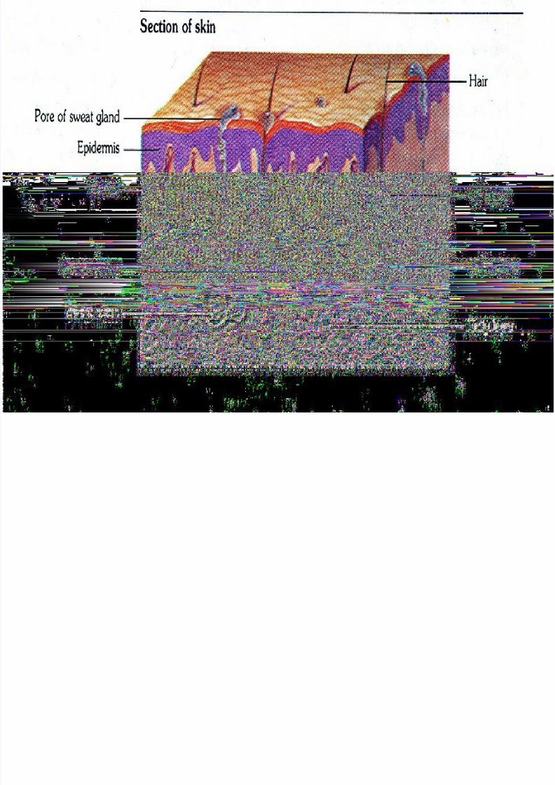
Integumentary System
• an anatomical barrier from pathogens anddamage between the internal and externalenvironment in bodily defense.
8/4/2019 3. Venipuncture
http://slidepdf.com/reader/full/3-venipuncture 4/42
Two Main Layers:
1. Epidermis – outer layer composed of
squamous cells.2. Dermis – inner, thicker layer consisting
of blood vessels, hair follicles, sweat
glands, small muscles, and nerves.
8/4/2019 3. Venipuncture
http://slidepdf.com/reader/full/3-venipuncture 5/42
8/4/2019 3. Venipuncture
http://slidepdf.com/reader/full/3-venipuncture 6/42
Sensory Receptors
–Mechanoreceptors – skin tactileperceptions
–Thermoreceptors – process cold,warmth, and pain
–Nociceptors – process pain

8/4/2019 3. Venipuncture
http://slidepdf.com/reader/full/3-venipuncture 7/42
The Vascular System

8/4/2019 3. Venipuncture
http://slidepdf.com/reader/full/3-venipuncture 8/42
Vascular System
•
is concerned with thetransport of blood andlymph through the body.
8/4/2019 3. Venipuncture
http://slidepdf.com/reader/full/3-venipuncture 9/42
Variations:
1. Arteries – carries blood from
the heart to the body.2. Veins – carries blood from
the capillaries towards the heart.3. Capillaries – resembles a hair
follicle.
8/4/2019 3. Venipuncture
http://slidepdf.com/reader/full/3-venipuncture 10/42
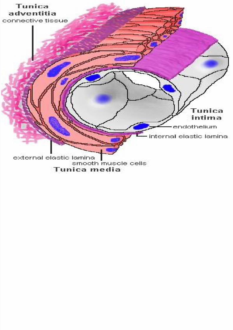
1. Tunica Adventitia – outermost layer;which consist mainly of connective tissue
fibers; blends with the connective tissue
surrounding the vessel; supports andsurrounds a vessel.
Layers of the Blood Vessel:
8/4/2019 3. Venipuncture
http://slidepdf.com/reader/full/3-venipuncture 11/42
2. Tunica Media – middle layer; is formed
by a layer of circumferential smoothmuscle and variable amounts of
connective tissue; collapses or distends as
pressure changes.
8/4/2019 3. Venipuncture
http://slidepdf.com/reader/full/3-venipuncture 12/42
3. Tunica Intima – innermost layer;
delimits the vessel wall towards thelumen of the vessel and comprises of
endothelial lining and connective tissue.
8/4/2019 3. Venipuncture
http://slidepdf.com/reader/full/3-venipuncture 13/42
8/4/2019 3. Venipuncture
http://slidepdf.com/reader/full/3-venipuncture 14/42
Peripheral Vascular
Vascular system that runs across the
periphery.
MAJOR TYPES OF VEINS (ARM)
1.) Digital Veins
2.) Metacarpal Veins – best choice
3.) Cephalic Veins
4.) Basilic Veins
8/4/2019 3. Venipuncture
http://slidepdf.com/reader/full/3-venipuncture 15/42
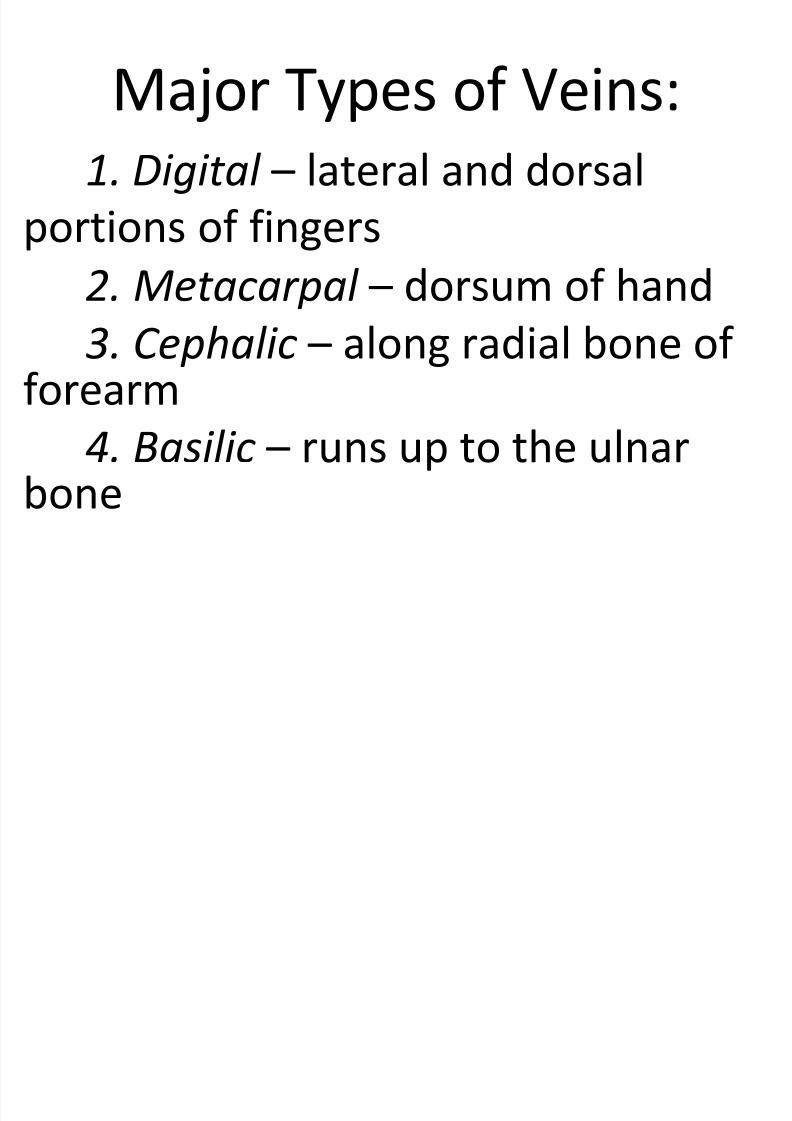
Major Types of Veins:
1. Digital – lateral and dorsal
portions of fingers
2. Metacarpal – dorsum of hand3. Cephalic – along radial bone of
forearm
4. Basilic – runs up to the ulnarbone

8/4/2019 3. Venipuncture
http://slidepdf.com/reader/full/3-venipuncture 16/42
8/4/2019 3. Venipuncture
http://slidepdf.com/reader/full/3-venipuncture 17/42
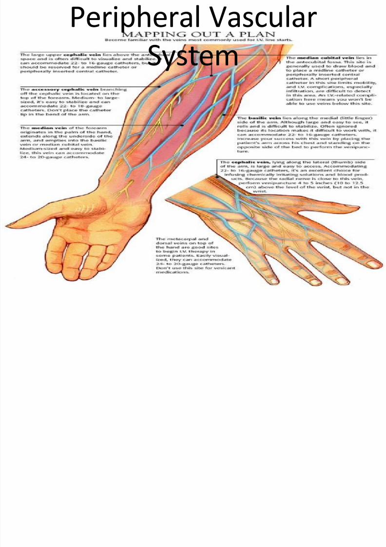
Peripheral Vascular
System
8/4/2019 3. Venipuncture
http://slidepdf.com/reader/full/3-venipuncture 18/42
Key Points Prior to IV Initiation
1. Physician’s order
2. Patient assessment3. IV set and equipment
preparation4. Medications
8/4/2019 3. Venipuncture
http://slidepdf.com/reader/full/3-venipuncture 19/42
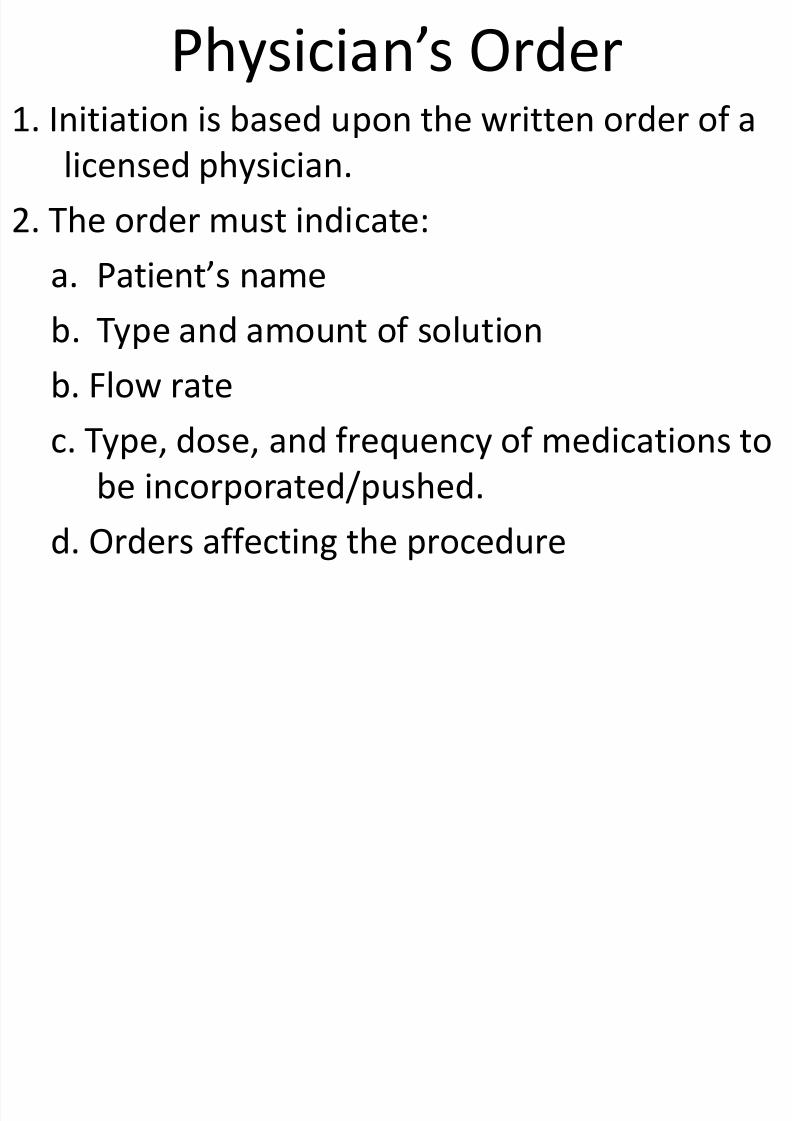
Physician’s Order 1. Initiation is based upon the written order of a
licensed physician.
2. The order must indicate:
a. Patient’s name b. Type and amount of solution
b. Flow rate
c. Type, dose, and frequency of medications to
be incorporated/pushed.
d. Orders affecting the procedure
8/4/2019 3. Venipuncture
http://slidepdf.com/reader/full/3-venipuncture 20/42
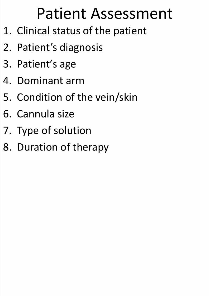
Patient Assessment1. Clinical status of the patient
2. Patient’s diagnosis
3. Patient’s age
4. Dominant arm
5. Condition of the vein/skin
6. Cannula size
7. Type of solution
8. Duration of therapy
8/4/2019 3. Venipuncture
http://slidepdf.com/reader/full/3-venipuncture 21/42
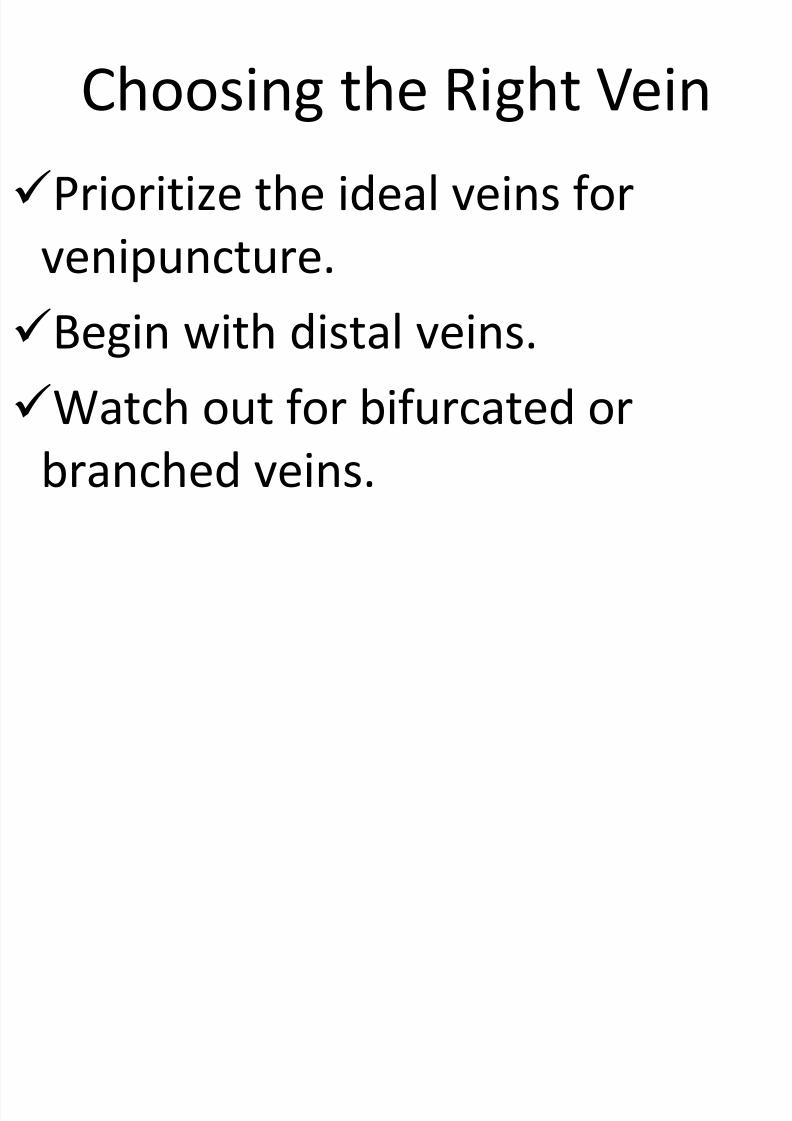
Choosing the Right Vein
Prioritize the ideal veins for
venipuncture.Begin with distal veins.
Watch out for bifurcated orbranched veins.
8/4/2019 3. Venipuncture
http://slidepdf.com/reader/full/3-venipuncture 22/42
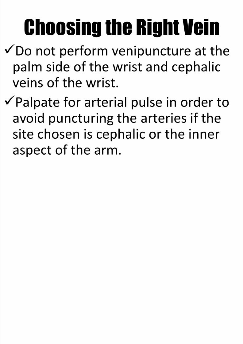
Choosing the Right VeinDo not perform venipuncture at the
palm side of the wrist and cephalic
veins of the wrist.Palpate for arterial pulse in order to
avoid puncturing the arteries if thesite chosen is cephalic or the inneraspect of the arm.
8/4/2019 3. Venipuncture
http://slidepdf.com/reader/full/3-venipuncture 23/42
Other sites to avoid include:
Veins below a previous IV infiltration.
Veins below a phlebitic area.Sclerosed or thrombosed veins.
Areas of skin inflammation, disease, bruising, or
breakdown.An arm affected by a radical mastectomy, edema,
blood clot, or infection.
An arm with an arteriovenous shunt or fistula.
Choosing the Right Vein
8/4/2019 3. Venipuncture
http://slidepdf.com/reader/full/3-venipuncture 24/42
IV Set and Equipment Preparation
1. Check for expiration date.2. Check for clarity.
3. Check label against physician’s writtenprescription.
4. Label any medications added.
5. Functionality of infusion pumps, PCA.

8/4/2019 3. Venipuncture
http://slidepdf.com/reader/full/3-venipuncture 25/42
Medications
1. Nurses should have a knowledge onall medications administered
including:a. Dosages
b. Drug interactionsc. Possible clinical effects
8/4/2019 3. Venipuncture
http://slidepdf.com/reader/full/3-venipuncture 26/42
Venipuncture Techniques (do
not include)1. Vein dilatation
2. Sitepreparation
3. Catheter
insertion4. Securing thecatheter
8/4/2019 3. Venipuncture
http://slidepdf.com/reader/full/3-venipuncture 27/42
Vein Dilatation
1. Tourniquet – place 6-8 inches abovethe venipuncture site.
2. Gravity – position the extremity below
the heart.3. Fist clenching – open and close his fist.
4. Warm compress – maximum of 10
minutes.5. Multiple tourniquet technique – use of 2-3 tourniquets.
8/4/2019 3. Venipuncture
http://slidepdf.com/reader/full/3-venipuncture 28/42
Site Preparation
1. Do not shave site. Remove hair with clippersonly.
2. Depilatories are not recommended.
3. Cleanse with one of the following solutions:a. 2% Chlorhexidine gluconate
b. Povidone-iodine
c. 70% Isoprophyl alcohol
4. Work from the center outward in a circular
motion.
8/4/2019 3. Venipuncture
http://slidepdf.com/reader/full/3-venipuncture 29/42
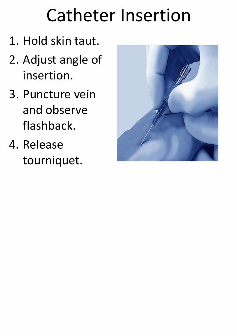
Catheter Insertion
1. Hold skin taut.2. Adjust angle of
insertion.
3. Puncture vein
and observe
flashback.4. Release
tourniquet.
8/4/2019 3. Venipuncture
http://slidepdf.com/reader/full/3-venipuncture 30/42
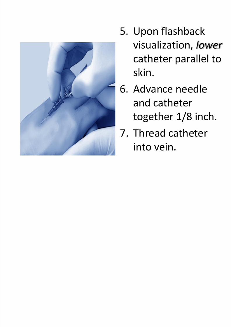
5. Upon flashback
visualization, lower
catheter parallel to
skin.
6. Advance needle
and catheter
together 1/8 inch.
7. Thread catheter
into vein.
8/4/2019 3. Venipuncture
http://slidepdf.com/reader/full/3-venipuncture 31/42
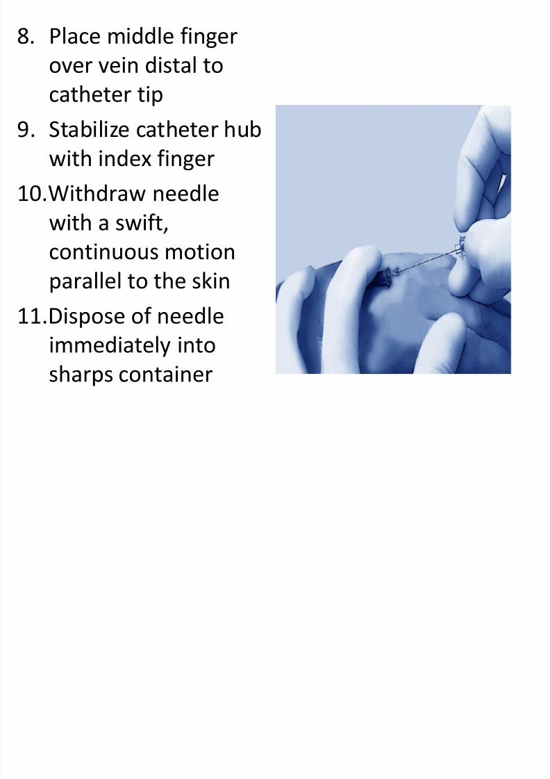
8. Place middle finger
over vein distal to
catheter tip9. Stabilize catheter hub
with index finger
10.Withdraw needlewith a swift,
continuous motion
parallel to the skin11.Dispose of needle
immediately into
sharps container
8/4/2019 3. Venipuncture
http://slidepdf.com/reader/full/3-venipuncture 32/42
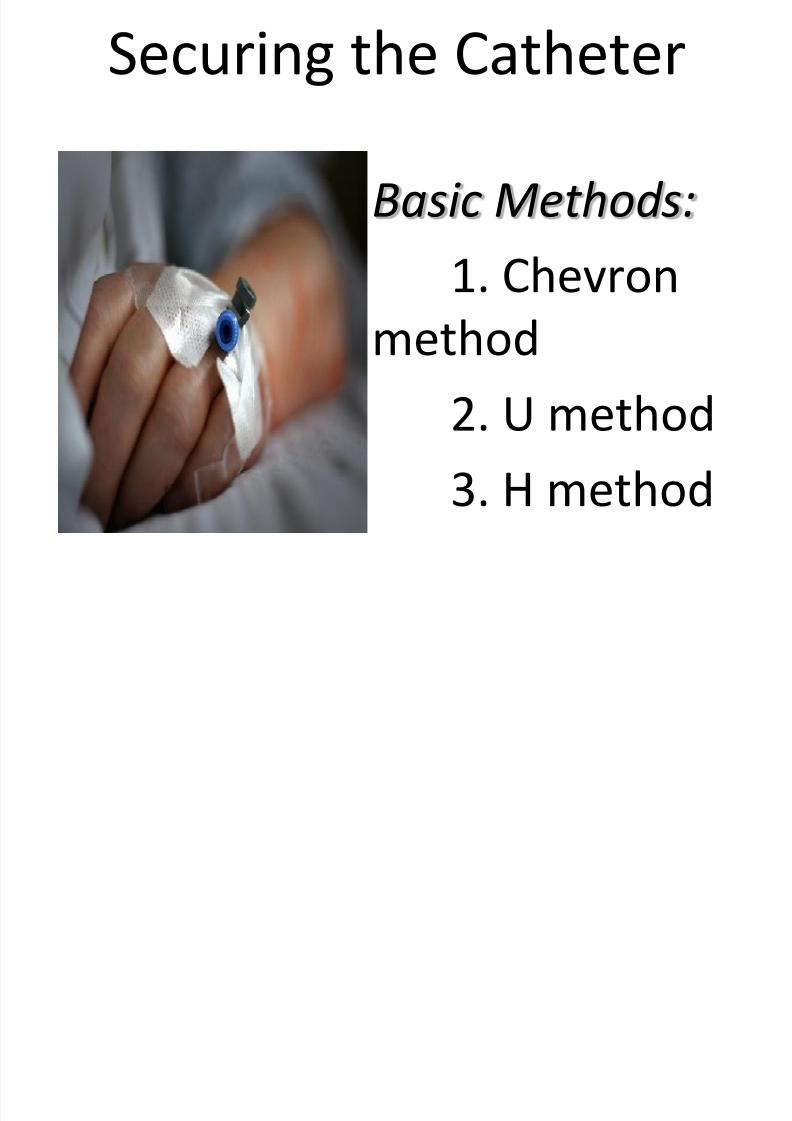
Securing the Catheter
Basic Methods:
1. Chevronmethod
2. U method3. H method
8/4/2019 3. Venipuncture
http://slidepdf.com/reader/full/3-venipuncture 33/42
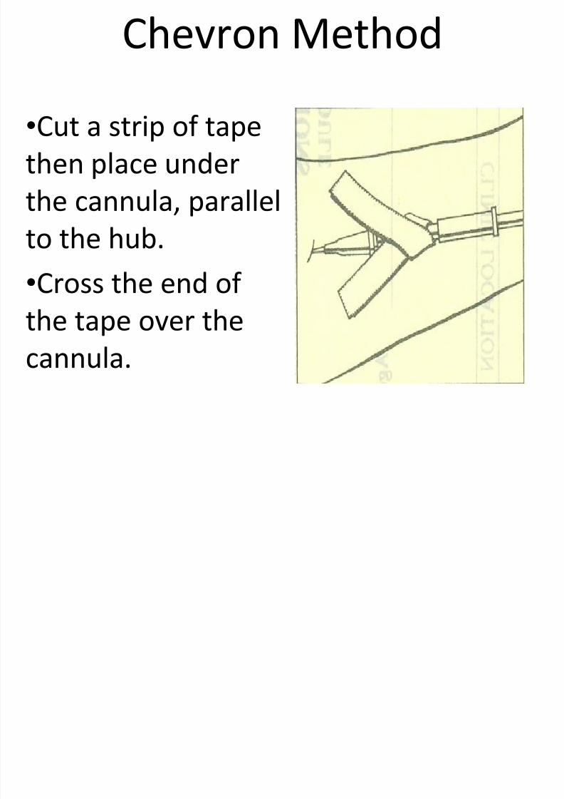
Chevron Method
•Cut a strip of tape
then place under
the cannula, parallelto the hub.
•Cross the end of
the tape over the
cannula.
U h d
8/4/2019 3. Venipuncture
http://slidepdf.com/reader/full/3-venipuncture 34/42
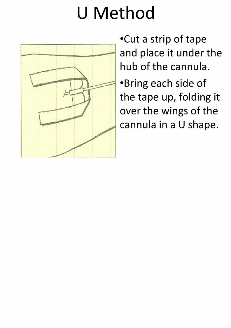
U Method
•
Cut a strip of tapeand place it under the
hub of the cannula.
•Bring each side of the tape up, folding it
over the wings of the
cannula in a U shape.
H M th d
8/4/2019 3. Venipuncture
http://slidepdf.com/reader/full/3-venipuncture 35/42
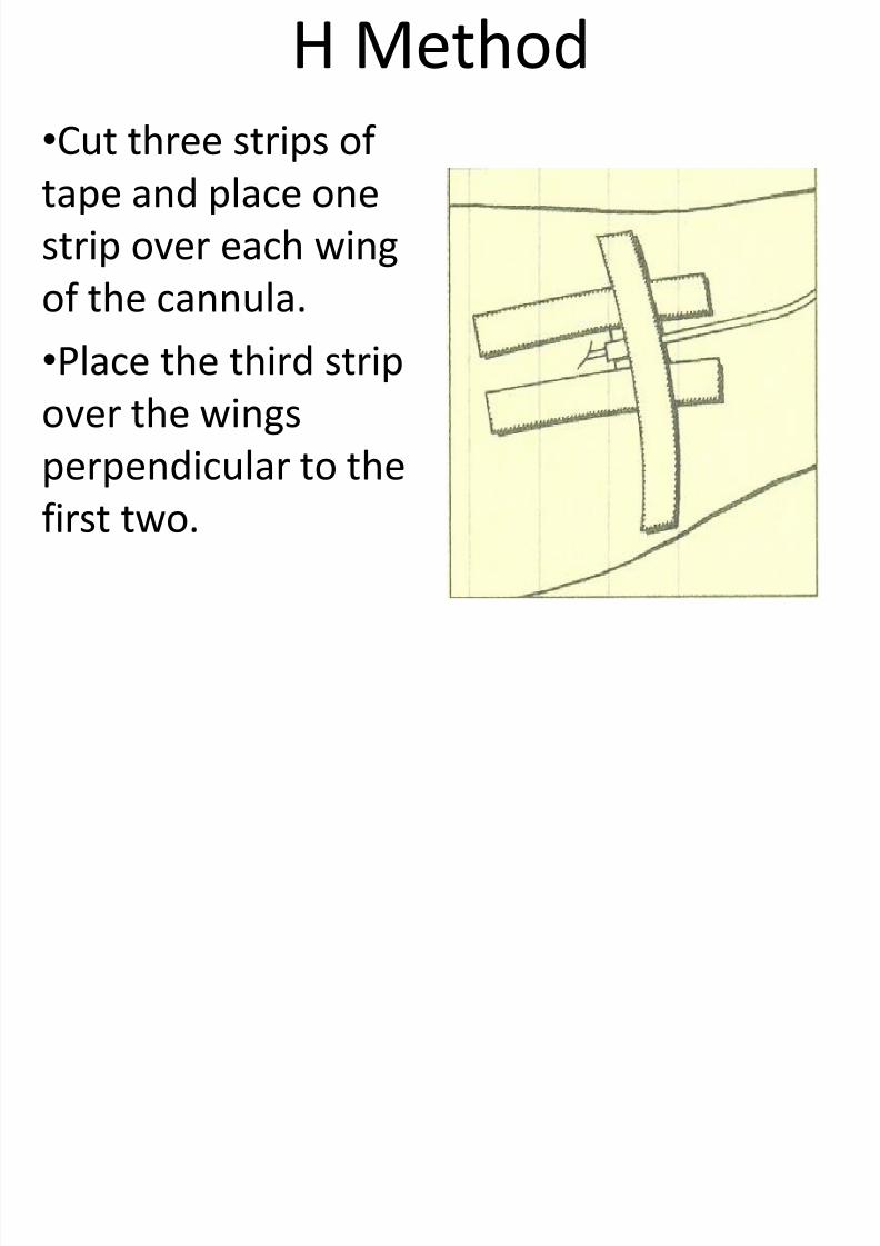
H Method
•
Cut three strips of tape and place one
strip over each wing
of the cannula.•Place the third strip
over the wings
perpendicular to thefirst two.
8/4/2019 3. Venipuncture
http://slidepdf.com/reader/full/3-venipuncture 36/42
•Always apply a label after securing thecatheter.
•On the label, write the following:
Date of insertion
Time of insertion
Type of catheter usedGauge used
Your initials
Reminder for all methods:
8/4/2019 3. Venipuncture
http://slidepdf.com/reader/full/3-venipuncture 37/42
Maintaining Peripheral IV
Therapy:
1. Changing the dressing
2. Changing the IV solution
3. Changing the administration set
4. Changing the IV site

8/4/2019 3. Venipuncture
http://slidepdf.com/reader/full/3-venipuncture 38/42
•The insertion site should be inspectedand palpated for tenderness daily,through intact dressing.
•Gauze dressing should be changed
routinely every 48 hours.
•A semipermeable dressing should bechanged whenever its integrity is
compromised.
Changing the Dressing:
8/4/2019 3. Venipuncture
http://slidepdf.com/reader/full/3-venipuncture 39/42
•Do not allow an IV container to hang formore than 24 hours.
•Before changing the IV container, check
the new one for cracks, leaks, and otherdamages.
•
Check the solution for discoloration,turbidity, and particulates.
•Note date and time the solution was mixed
and the expiration date.
Changing the IV solution:
8/4/2019 3. Venipuncture
http://slidepdf.com/reader/full/3-venipuncture 40/42
•Change the administration setevery 72 hours and whenever you
note or suspect contamination.•As much as possible, change the
administration set when you start anew venous access device duringroutine site rotation.
Changing the Administration Set:
8/4/2019 3. Venipuncture
http://slidepdf.com/reader/full/3-venipuncture 41/42
•
As a standard of care, rotate the siteevery 48-72 hours.
•If limited venous access will prevent
you from changing sites, notify thedoctor of the situation.
•
Be prepared to change the entiresystem when you detect signs ofthrombophlebitis, cellulitis, or IV
therapy related bacteremia.
Changing the IV Site:
8/4/2019 3. Venipuncture
http://slidepdf.com/reader/full/3-venipuncture 42/42
•
Be careful to avoid manipulating thedevice in the skin to prevent skinorganisms from entering the
bloodstream.
•Never use an alcohol pad to clean the
site when discontinuing an infusion.•If the patient feels lingering tendernessat the IV site, apply warm, moist packs.
Discontinuing Peripheral IV: