Embed Size (px)
DESCRIPTION
i don't know what it is!
Citation preview
Horizontal transfer of carbohydrate metabolism genes intoectomycorrhizal Amanita
Maryam Chaib De Mares1,2*, Jaqueline Hess2,3*, Dimitrios Floudas4, Anna Lipzen5, Cindy Choi5,
Megan Kennedy5, Igor V. Grigoriev5 and Anne Pringle6
1Department of Microbial Ecology, University of Groningen, 9747 AG Groningen, the Netherlands; 2Department of Organismic and Evolutionary Biology, Harvard University, Cambridge,
MA 02138, USA; 3Department of Biosciences, University of Oslo, 0371 Oslo, Norway; 4Department of Biology, Clark University, Worcester, MA 01610, USA; 5US Department of Energy
Joint Genome Institute, Walnut Creek, CA 94598, USA; 6Harvard Forest, 324 North Main Street, Petersham, MA 01366, USA
Author for correspondence:Maryam Chaib De Mares
Tel: +31 0 503632169Email: [email protected]
Received: 1 July 2014
Accepted: 24 September 2014
New Phytologist (2014)doi: 10.1111/nph.13140
Key words: Amanita, carbohydratemetabolism, comparative genomics,evolution of symbiosis, evolutionary novelty,horizontal gene transfer (HGT), saprotrophy.
Summary
� The genus Amanita encompasses both symbiotic, ectomycorrhizal fungi and asymbiotic
litter decomposers; all species are derived from asymbiotic ancestors. Symbiotic species are no
longer able to degrade plant cell walls. The carbohydrate esterases family 1 (CE1s) is a diverse
group of enzymes involved in carbon metabolism, including decomposition and carbon stor-
age. CE1 genes of the ectomycorrhizal A. muscaria appear diverged from all other fungal ho-
mologues, and more similar to CE1s of bacteria, suggesting a horizontal gene transfer (HGT)
event.� In order to test whether Amanita CE1s were acquired horizontally, we built a phylogeny of
CE1s collected from across the tree of life, and describe the evolution of CE1 genes among
Amanita and relevant lineages of bacteria.� CE1s of symbiotic Amanita were very different from CE1s of asymbiotic Amanita, and are
more similar to bacterial CE1s. The protein structure of one CE1 gene of A. muscaria matched
a depolymerase that degrades the carbon storage molecule poly((R)-3-hydroxybutyrate)
(PHB). Asymbiotic Amanita do not carry sequence or structural homologues of these genes.� The CE1s acquired through HGT may enable novel metabolisms, or play roles in signaling or
defense. This is the first evidence for the horizontal transfer of carbohydrate metabolism
genes into ectomycorrhizal fungi.
Introduction
Horizontal gene transfer (HGT) is the mobilization and stableintegration of genetic material between distinct, reproductivelyisolated genomes (Richards et al., 2011). HGT is ubiquitousamong bacteria (Brown & Doolittle, 1997; Lawrence & Och-man, 1997; Nelson et al., 1999; Koonin et al., 2001), and is alsoa major force in evolution among eukaryotes, enabling diversifi-cation and the adaptation of organisms to new environments.Horizontally transferred genes have facilitated changes in the hostranges of rumen and pathogenic fungi (Garcia-Vallv�e et al.,2000; Juhas et al., 2009; Mehrabi et al., 2011; Sun et al., 2013),the spread of antibiotic resistance (Weldhagen, 2004; Roberts,2005; Hanssen & Ericson-Sollid, 2006), and the evolution ofnovel metabolic capabilities (Lawrence & Ochman, 1998;Kanhere & Vingron, 2009; Marchetti et al., 2009; Ma et al.,2010; Christin et al., 2012).
Although HGT appears to be less frequent among eukaryotes,as compared with bacteria, there is ample evidence for the
movement of genes from bacteria to fungi, as well as among dif-ferent species of fungi and between plants and fungal pathogens(Richards et al., 2009, 2011; Fitzpatrick, 2011; Armijos-Jaramilloet al., 2013; Sun et al., 2013; Bruto et al., 2014). Ectomycorrhizal(EM) fungi form intimate associations with the roots of plants,but also extend into the surrounding soil, an environment teem-ing with bacteria (Gans et al., 2005). Nevertheless, to date thereis scant evidence for HGT into EM lineages. Research targettingthe EM fungus Amanita muscaria and transgenic poplar treesfound no evidence for HGT between fungus and plant in labora-tory settings (Zhang et al., 2005; Nehls et al., 2006). But agrobac-teria have been used to transform other EM fungi, includingHebeloma cylindrosporum (Combier et al., 2003), Tuber borchii(Grimaldi et al., 2005) and Laccaria bicolor (Kemppainen &Pardo, 2011); demonstrating that lateral acquisition of genesfrom bacteria is theoretically possible. Whether HGT enables themovement of genes into EM fungi in nature remains an openquestion.
The movement of carbohydrate metabolism genes from bacte-ria to fungi or between fungi may enable fungi to establish innovel habitats or niches. For example, the glycosyl hydrolases*These authors contributed equally to this work.
� 2014 The Authors
New Phytologist� 2014 New Phytologist Trust
New Phytologist (2014) 1www.newphytologist.com
Research
(GH) of rumen fungi are bacterial in origin, and allow the fungito degrade cellulose and hemicellulose in the rumens of herbivo-rous mammals (Garcia-Vallv�e et al., 2000). The transfer of a highaffinity nitrate assimilation gene cluster from a basidiomycete toan ancestor of the ascomycetous mold Trichoderma reesei mayhave facilitated a change in the mold’s nutritional mode, allowingit to become an efficient wood degrader (Slot & Hibbett, 2007).Moreover, an earlier HGT of nitrate assimilation genes into Dik-arya may have facilitated exploitation of nitrate in aerobic soils(Slot & Hibbett, 2007).
The genus Amanita encompasses a diversity of EM and closelyrelated saprotrophic (SAP) fungi. A recent phylogeny documentsa single origin of symbiosis within the Amanita; asymbioticAmanita form a strongly supported clade basal to a monophyleticclade of symbiotic species (Wolfe et al., 2012b). Comparativegenomics of EM and SAP Amanita reveal large-scale losses of car-bohydrate-active enzymes from symbiotic genomes (Nagendranet al., 2009; Wolfe et al., 2012b; Chaib De Mares et al., 2013;Hess & Pringle, 2014). The result appears to be a general one;plant cell wall degrading enzymes are frequently lost after fungibecome obligately dependent on plants for carbon (Martin et al.,2008, 2010).
The carbohydrate esterases family 1 (CE1s; Cantarel et al.,2009) are a diverse group of enzymes, encompassing at least sevenclasses within the CAZy database (http://www.cazy.org/). Theenzymes are heterogeneous with respect to both substrate speci-ficity and structure. Some CE1 enzymes target esters or amides,deacetylating the side group components of hemicellulose(Towler et al., 1988; Biely, 2012). These side groups covalentlylink and physically mask potentially fermentable substrates inplant cell walls, perhaps protecting them from degradation (Akin,2008). CE1s of this group are hemicellulose accessory enzymes(McDonald et al., 2009) and enable microorganisms to attackand partially degrade plant tissues, working with xylanases andpectinases to break apart plant cell walls (Kubicek et al., 2010).Other CE1 enzymes, structurally related to lipases and proteases,consist of depolymerases that degrade a bacterial polymer, poly((R)-3-hydroxybutyrate) (PHB; Dawes, 1988). PHB is built fromglucose, and is used by microorganisms to store energy; it ismetabolized when other carbon sources are unavailable.
Phylogenetic analyses by the Mycorrhizal Genomics Initiative(MGI, http://mycor.nancy.inra.fr/IMGC/MycoGenomes/) iden-tify three phylogenetically distinct clades of CE1 genes in fungalgenomes (genes annotated as CE1s identified by B. Henrissat,Cantarel et al., 2009). Two clades show sequence homology tohemicellulose accessory enzymes. But within the analyses, a thirdgroup of CE1 genes from the EM species A. muscaria appearsdiverged from other clades, and similar to CE1s of bacteria.Complex evolutionary relationships among CE1s of fungi, bacte-ria and plants are common (Udatha et al., 2011), and the patternssuggest a potential HGT event between A. muscaria and bacteria.
In order to explore and test for potential HGT events inA. muscaria and the genus Amanita, we identified four key ques-tions: are the CE1 genes described from A. muscaria structurallyand functionally integrated into the genome? What kind of CE1genes are found in other Amanita species, and are the CE1s of
ectomycorrhizal Amanita different from CE1s of saprotrophs?Do phylogenies built from a comprehensive dataset of CE1s sug-gest HGT? What do phylogenies tell us about the history ofCE1s within the lineage of ectomycorrhizal Amanita? We took avariety of genetic and bioinformatic approaches to answer thesequestions, and then more fully characterized the EM AmanitaCE1 genes, as well as the function of one predicted protein.
Materials and Methods
Identification of CE1s in Amanita genomes and a transcrip-tome
In order to investigate the origins of Amanita CE1 genes, we firstidentified the complete set of CE1 genes in A. muscaria, homo-logues present in available genomes of other Amanita species, andhomologues in the outgroup Volvariella volvacea. The genome ofA. muscaria var. guessowi (Koide BX008, Pennsylvania, USA;Hess & Pringle, 2014) has been sequenced twice; one genome isdeposited at Joint Genome Institute (JGI, genome.jgi.doe.gov/Amuscaria), and the other was sequenced by the Pringle labora-tory (Cambridge, MA, USA). The Pringle laboratory has alsosequenced genomes for the EM fungi A. brunnescens andA. polypyramis, and SAP fungi A. inopinata and V. volvacea (Hesset al., 2014). Genome sequences are available at NCBI under theaccession nos. PRJNA236753, PRJNA236755, PRJNA236758,PRJNA236757 and PRJNA236756. The genome of A. thiersii isalso available through JGI (genome.jgi.doe.gov/Athiersii, Wolfeet al., 2012a).
In addition to genomic data, a transcriptome of A. crenulatawas sequenced at JGI in the context of a different experiment andIllumina RNA-Seq data are available at NCBI SRA under theaccessions SRX141954 and SRX141955. Cultures were main-tained as in Wolfe et al. (2012b). Mycelia were collected andimmediately stored in RNAlater (Qiagen). RNA was isolatedwith the RNeasy Maxi Kit (Qiagen). Poly A RNA was isolatedfrom 10 ug total RNA using the Absolutely mRNA purificationkit (Stratagene, Santa Clara, CA, USA). This procedure wasrepeated twice, to ensure that the sample was free from rRNAcontamination. Detailed protocols for RNA isolation, sequencinglibraries preparation, sequencing and assembly are available inSupporting Information Methods S1.
In order to generate a catalogue of candidate CE1 loci, twopreviously annotated CE1 genes – one from A. muscaria (JGIprotein ID 166350) and a second from A. thiersii (JGI proteinID 1897), both annotated by B. Henrissat (pers. comm.) – wereused as probes to screen all available genomes and the transcrip-tome with TBLASTN (Altschul et al., 1990), using an E-valuecutoff of 10�5 (Table S1).
Naming conventions
We adopt the following naming conventions: CE1 genes inA. muscaria are labelled as CE1_AmX, where X is a number usedto distinguish among individual genes. The CE1 genes of otherspecies are named as CE1_Ab (A. brunnescens, where only one
New Phytologist (2014) � 2014 The Authors
New Phytologist� 2014 New Phytologist Trustwww.newphytologist.com
Research
NewPhytologist2
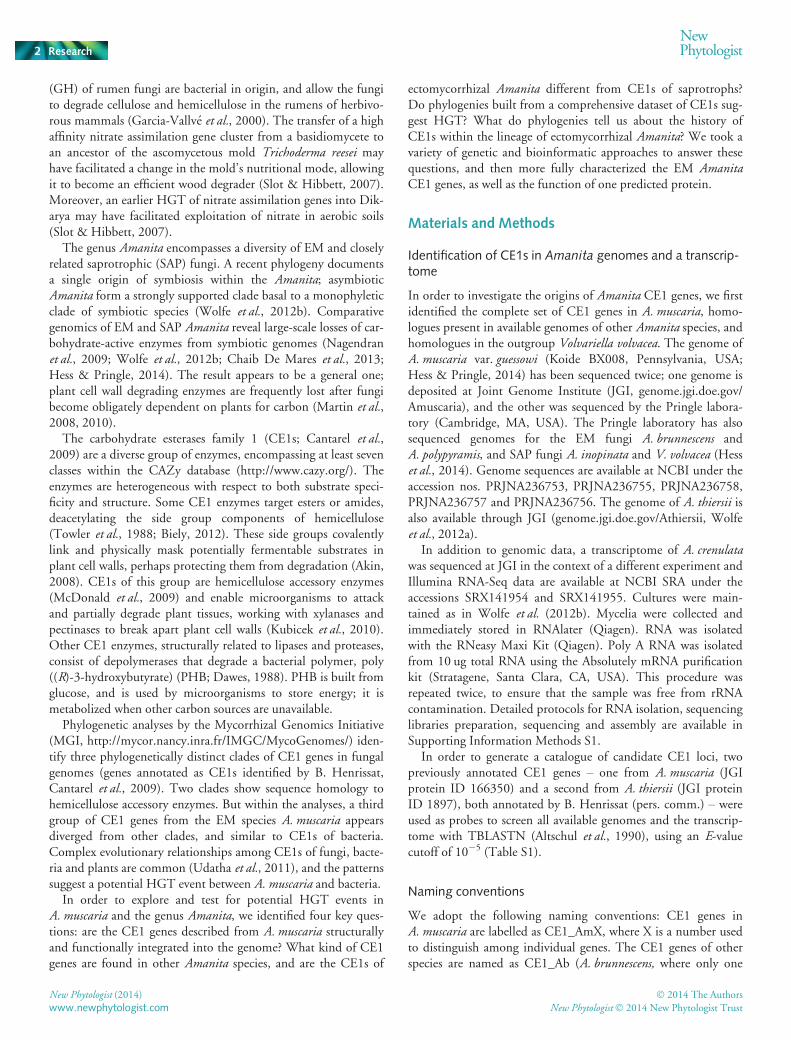
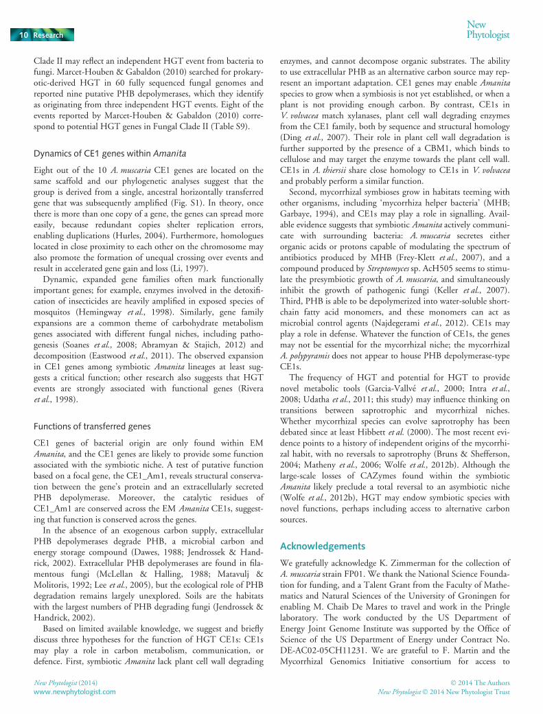
gene was identified), CE1_AcrX (A. crenulata) and CE1_AcoX(A. constricta). Carbohydrate-binding module 1 (CBM1) is anadditional domain found in CE1 genes of A. thiersii andV. volvacea, and so we named the CE1s of these saprotrophic spe-cies as CE1-CBM1_Ath (A. thiersii) and CE1-CBM1_VvX (X isa number). The fungal genes flanking CE1 genes of A. muscaria’sscaffold 57 (Fig. 1) are labelled as FX (X is a number).
Confirming physical integration of A. muscaria CE1 genes
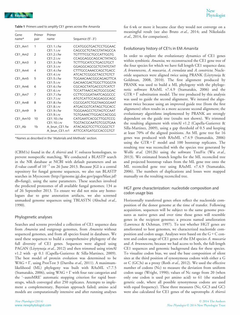
In order to confirm a subset of candidate A. muscaria genes asphysically linked and integrated within the genome, and not theresult of contaminant DNA, we used a single, long-range PCR toamplify a 24 435 bp region of A. muscaria JGI genome scaffold57, where eight CE1 and five fungal genes are found (Fig. 1). Weused primers spanning genes CE1_Am1 and CE1_Am3 (Table 1;see also the ‘Primer Design’ section below). To ensure high fidel-ity amplification, we used LongAmpTM Taq DNA polymerase(New England BioLabs Inc., Ipswich, MA, USA) and followedthe manufacturer’s protocol implementing 16-min extensioncycles. The resulting long-range construct was used as a templatefor subsequent PCR reactions, and was therefore diluted 1 : 1000.CE1 genes (CE1_Am1, CE1_Am5, CE1_Am9, CE1_Am10,CE1_Am2 and CE1_Am3; Table S2) and interspersed fungalgenes (Table S3) were successfully amplified from the long-rangeconstruct using specific primer pairs (Table 1), and confirmed byresequencing (Genewiz Inc., Cambridge, MA, USA).
Expression of A. muscaria CE1 genes
The expression of A. muscaria CE1 genes was confirmed usingRNAseq data available at JGI (http://genome.jgi-psf.org/cgi-bin/browserLoad/?db=Amamu1). Assembled transcripts were down-loaded and aligned to the genome using GMAP with default set-tings (Wu & Watanabe, 2005) and visualized using IGV(Thorvaldsd�ottir et al., 2013).
Identification of CE1s in Amanita without sequenced ge-nomes
We screened genomic DNA originally extracted by Wolfeet al. (2012b) from 11 species of Amanita subgenusAmanita (A. multisquamosa, A. subglobosa, A. parvipantherina,A. praecox, A. xylinivolva, A. altipes, A. crenulata, A. wellsii,A. murinoflammeum, A. umbrinella and A. constricta). We also
screened 13 species of Amanita subgenus Lepidella (A. flavoru-bens, A. novinupta, A. luteolovolvata, A. citrina, A. rubescensvar. congolensis, A. marmorata, A. virosa, A. smithiana, A. pelioma,A. clelandii, A. conicobulbosa, A. chlorinosoma and A. cokeri).
Genomic DNA was also newly isolated from the original,sequenced strain of A. muscaria var. guessowii (Koide BX008),two additional strains (FP01, collected in Cambridge, MA, USA,and PS #283, from the Penn State Spawn Collection, originallycollected in PA, USA), and A. brunnescens (BW HF10C), using amodified version of an extraction protocol developed forPhytophtora infestans (http://my.jgi.doe.gov/general/protocols/).We used a 1 : 1 phenol chloroform ratio in our extractions.
Primers (see the ‘Primer Design’ section below) were used totest for CE1 genes in each species and strain (Fig. 2). Amplifica-tion was performed using a touchdown PCR protocol, to increasesensitivity and specificity, as previously described (Don et al.,1991). The protocol resulted in unique, clear bands. Ampliconswere verified as CE1 genes using Sanger sequencing (GenewizInc.).
Primer design
CE1 genes found in sequenced genomes of A. muscaria andA. brunnescens, and the transcriptome of A. crenulata, are variableenough so that individual genes can be distinguished. To confirmthe presence of multiple individual CE1s in genomic DNA, wedesigned highly specific primers for each CE1 gene, based oneither A. muscaria or A. brunnescens sequences (Table 1). Toincrease the likelihood that each primer pair designed fromA. muscaria would amplify only one CE1 gene, primers weresimultaneously designed for all of the different CE1 loci identi-fied on scaffold 57 of the A. muscaria JGI assembly (Table S2)using Geneious v1.6 (http://www.geneious.com/, Biomatters,Newark, NJ, USA; Table 1). Our design strategy was successful,but because we designed these primers to target specific, variableregions, in some cases the primers do not amplify the full lengthof a gene. Moreover, successfully amplified fragments were moreeasily sequenced from species closely related to A. muscaria.
Identification of CE1s from across the tree of life
We next collected CE1 homologues from across the tree of life,using the predicted protein sequences of CE1s from A. muscaria,A. brunnescens, A. thiersii and V. volvacea as probes (Table S4).Before probing, we removed the carbohydrate binding modules
Fig. 1 Physical map of Amanita muscariascaffold 57. Carbohydrate esterase family 1(CE1) genes with close homology to bacterialCE1 genes shown in green. Blue, fungalgenes; brown, gene models; pink,transposable elements (TEs); grey, evidenceof gene expression from JGI EST data; black,a long-range PCR confirmed CE1 and fungalgenes are linked.
� 2014 The Authors
New Phytologist� 2014 New Phytologist TrustNew Phytologist (2014)
www.newphytologist.com
NewPhytologist Research 3
(CBM1s) found in the A. thiersii and V. volvacea homologues, toprevent nonspecific matching. We conducted a BLASTP searchin the NR database at NCBI with default parameters and anE-value cutoff of 10�5 on 26 June 2013. Because JGI is a centralrepository for fungal genome sequences, we also ran BLASTPsearches in Mycocosm (http://genome.jgi.doe.gov/pages/blast.jsf?db=fungi), using the same parameters. These searches involvedthe predicted proteomes of all available fungal genomes; 134 asof 26 September 2013. To ensure we did not miss any homo-logues due to gene annotation artifacts, we also screenedunmasked genome sequences using TBLASTN (Altschul et al.,1990).
Phylogenetic analyses
Searches and screens provided a collection of CE1 sequence datafrom Amanita and outgroup genomes, from Amanita withoutsequenced genomes, and from all species found in databases. Weused these sequences to build a comprehensive phylogeny of thefull diversity of CE1 genes. Sequences were aligned usingPAGAN (L€oytynoja et al., 2012) and then trimmed using trimAlv1.2 with –gt 0.1 (Capella-Gutierrez & Silla-Martinez, 2009).The best model of protein evolution was determined to beWAG + Γ, using ProtTest 3 (Darriba et al., 2011). A maximum-likelihood (ML) phylogeny was built with RAxML v7.7.5(Stamatakis, 2006), using WAG + Γ with four rate categories andthe ‘–autoMRE’ automatic stopping criterion for rapid boot-straps, which converged after 250 replicates. Attempts to imple-ment a complementary, Bayesian approach failed; amino acidmodels are computationally intensive and after running analyses
for 6 wk or more it became clear they would not converge on ameaningful result (see also Bruto et al., 2014; and Nikolaidiset al., 2014, for comparison).
Evolutionary history of CE1s in EM Amanita
In order to explore the evolutionary dynamics of CE1 geneswithin symbiotic Amanita, we reconstructed the CE1 gene tree ofthe four species for which we have full length CE1 sequence data;A. brunnescens, A. muscaria, A. crenulata and A. constricta. Nucle-otide sequences were aligned twice using PRANK (L€oytynoja &Goldman, 2008, 2010). The first alignment produced byPRANK was used to build a ML phylogeny with the phyloge-netic software RAxML v7.4.9 (Stamatakis, 2006) and theGTR + Γ substitution model. The tree produced by this analysiswas used to guide the second alignment. We iterated the align-ment twice because using an improved guide tree (from the firstalignment) often results in a more accurate second alignment; theevolutionary algorithms implemented by PRANK are stronglydependent on the guide tree (results not shown). We trimmedthe resulting alignment with trimAl v1.2 (Capella-Gutierrez &Silla-Martinez, 2009), using a gap threshold of 0.5 and keepingat least 70% of the aligned positions. An ML gene tree for 16genes was produced with RAxML v7.4.9 (Stamatakis, 2006)using the GTR + Γ model and 100 bootstrap replicates. Theresulting tree was reconciled with the species tree generated byWolfe et al. (2012b) using the software TreeFix (Wu et al.,2013). We estimated branch lengths for the ML reconciled treeand projected bootstrap values from the ML gene tree onto theML reconciled gene tree using RAxML v7.4.9 (Stamatakis,2006). The numbers of duplications and losses were mappedmanually on the resulting reconciled tree.
HGT gene characterization: nucleotide composition andcodon usage bias
Horizontally transferred genes often reflect the nucleotide com-position of the donor genome at the time of transfer. Followingacquisition, sequences will be subject to the same genome pres-sures as native genes and over time those genes will resemblegenes in the recipient genome; a process named amelioration(Lawrence & Ochman, 1997). To test whether HGT genes areameliorated to host genomes, we characterized nucleotide com-position and codon usage. Analyses were based on the G +C con-tent and codon usage of CE1 genes of the EM species A. muscariaand A. brunnescens, because we had access to both, the full-lengthCE1 sequences and genomic background data for these species.To visualize codon bias, we used the base composition of silentsites at the third position of synonymous codons with either a Gor C (GC3s) as a proxy (Roth et al., 2012). We used the effectivenumber of codons (Nc) to measure the deviation from uniformcodon usage (Wright, 1990); values of Nc range from 20 (whenonly one codon is used per amino acid) to 61 (the standardgenetic code, where all possible synonymous codons are usedwith equal frequency). These three measures (Nc, GC3 and GC)were also calculated for CE1 genes of the saprotrophs A. thiersii
Table 1 Primers used to amplify CE1 genes across the Amanita
Genename*
Primerpair
Primername Sequence (50–30)
CE1_Am1 1 CE1.1.i.fw CCATGGGTGACTCCTGGAACCE1.1.i.rv CAGCGCTGTACGTATAGCCA
CE1_Am2 2 CE1.2.i.fw TGTTTTCGCTGCCATTGGTGCE1.2.i.rv CCAGGAGGCAGCACTATACG
CE1_Am3 3 CE1.3.i.fw TCTTTGCATCCTGACGTGCTCE1.3.i.rv GGAGGCAGCGCTGTATGTAT
CE1_Am4 4 CE1.4.i.fw CTTTGCCAAACCAGCTGACCCE1.4.i.rv ATCACTCGGGCTACCTGTCT
CE1_Am5 5 CE1.5.i.fw TGGAACAACGGCAGAGTTCACE1.5.i.rv GACAACGACTGGCTTGGGTA
CE1_Am6 6 CE1.6.i.fw CGCAGCTATGACCGTCATCTCE1.6.i.rv TCCATTAACCAGTGGCGGAC
CE1_Am7 7 CE1.7.i.fw CCTTCCGGATAATCAGGCCCCE1.7.i.rv ATGTCATTCCAGGAGGCAGC
CE1_Am8 8 CE1.8.i.fw CGCGGATCTGGTAAGGGAATCE1.8.i.rv ATGACGGTCATAGCTGCACC
CE1_Am9 9 CE1.9.i.fw TGGGAAGCCTGTAGTCCAATCE1.9.i.rv TGTGAAACTTGGACCACGGG
CE1_Am10 10 CE1.10.i.fw CATGAATCACGCTTGGTCCGCE1.10.i.rv TGGTACGCAATGTCGGTGAA
CE1_Ab 1b A_brun_CE1.fw ATACAGGTCCTTCCGGCTCTA_brun_CE1.rv1 ATTCCATGATGCCACCGTGT
*Names as described in the ‘Materials and Methods’ section.
New Phytologist (2014) � 2014 The Authors
New Phytologist� 2014 New Phytologist Trustwww.newphytologist.com
Research
NewPhytologist4
and V. volvacea, the CE1 gene of the bacteria Ktedonobacterracemifer, the closest homologue to a potential donor lineage.Measures were calculated using CodonW (Peden, 2000; http://codonw.sourceforge.net/).
HGT gene characterization: evolution of gene structure
We reconstructed patterns of exon and intron structure for CE1gene sequences found in the EM A. muscaria, A. brunnescens, andthe saprotrophs A. thiersii and V. volvacea using WebScipio(Odronitz et al., 2013; Hatje et al., 2011; http://www.webscipio.org). These are the same species and sequences used in analyses ofnucleotide composition and codon usage bias. We then usedGenePainter (Hammesfahr et al., 2013) to align gene structures.
The analyses focused on comparing position and phase: positionis defined by where an intron is inserted, and phase is defined bywhat the intron interrupts. For example, phase 0 introns inter-rupt an ORF between two consecutive codons, phase 1 intronsare found between the first and second nucleotide of one codon,and phase 2 introns between second and third nucleotide of acodon.
HGT gene characterization: putative cellular location ofproteins
In order to explore whether the proteins coded for by CE1 genesin the fungi A. muscaria, A. brunnescens, A. thiersii and V. volvaceaare secreted, we searched for signal peptide cleavage sites in
(a)
(b)
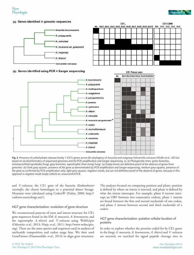
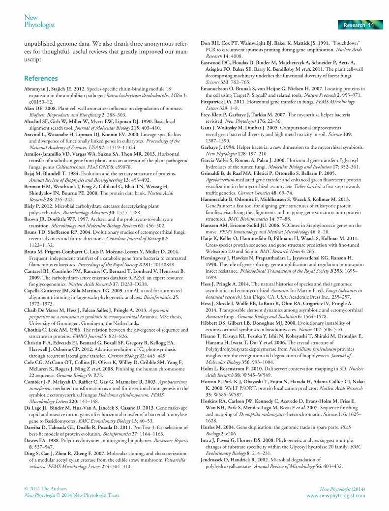
Fig. 2 Presence of carbohydrate esterase family 1 (CE1) genes across the phylogeny of Amanita and outgroup Volvariella volvacea (Wolfe et al., 2012a)based on (a) bioinformatics of sequenced genomes and (b) PCR amplification and Sanger sequencing. (a, b) Phylogenetic trees: green branches,ectomycorrhizal (symbiotic) fungi; grey branches, saprotrophic (free-living) fungi. (a) Empty boxes are definitive proof of the absence of genes fromgenomes. (b) Dark grey squares, presence of the gene as demonstrated by PCR amplification and Sanger sequencing; medium grey squares, presence ofthe gene as confirmed by PCR amplification only; light grey squares, negative results, but are not definitive proof of the absence of genes, because in thisapproach a negative result simply reflects an unsuccessful PCR.
� 2014 The Authors
New Phytologist� 2014 New Phytologist TrustNew Phytologist (2014)
www.newphytologist.com
NewPhytologist Research 5
amino acid sequences using Signal P v4.1 (Petersen et al., 2011;http://www.cbs.dtu.dk/services/SignalP/), and predicted theputative cellular location of the proteins using Target P v1.1(Emanuelsson et al., 2007) and WoLF PSORT (Horton et al.,2000; http://psort.hgc.jp/). We also scanned for transmembranea-helices using TMHMM v2.0 (Krogh et al., 2001; http://www.cbs.dtu.dk/services/TMHMM/). We labelled a protein as likelyto be secreted if it: possessed a signal peptide (Signal P); was pre-dicted to be extracellular (Target P or WoLF PSORT); and hadno transmembrane helices.
Structural analyses of a predicted protein
The horizontal acquisition of genes suggests functional relevancefor the recipient species. Function may be better predicted by ter-tiary structure, as opposed to primary sequence (Bajaj & Blun-dell, 1984; Chothia & Lesk, 1986), and so we reconstructed thetertiary structure of CE1_Am1 (see later Fig. 5) using Phyre2(Kelley & Sternberg, 2009; http://www.sbg.bio.ic.ac.uk/phyre2/html/page.cgi?id=index). We did not target the CE1_Am1 pro-tein for any particular reason, and chose it simply because wenamed this gene first. Next, we generated a list of proteins withstructural similarities to the CE1_Am1 protein model using theDali server (Holm & Rosenstrom, 2010; http://ekhidna.biocen-ter.helsinki.fi/dali_server/). A 3D model of the best match, pro-tein PDB 2D80 (Hisano et al., 2006), was retrieved from theProtein Data Bank (PDB; Berman et al., 2000; http://www.rcsb.org). Manipulations, structural alignments and comparisonsbetween the 3D models of CE1_Am1 and PDB 2D80 usedPyMOL (Schr€odinger, 2010; http://www.pymol.org).
Based on results found for CE1_Am1, and to explore whetherother CE1s of EM Amanita may be active esterases, we identifiedthe esterase domains of all EM Amanita CE1s through a searchof the Pfam database (http://pfam.sanger.ac.uk) (Punta et al.,2012). We then extracted the coordinates of the domain in eachof the genes, and aligned the domains to investigate the degree ofconservation of the active site and substrate interacting residues.
Results
Identification of CE1 genes in A. muscaria
We identified a total of 10 CE1 genes within the A. muscariagenome. Eight of the 10 genes are located on scaffold 57 of theJGI assembly, between positions 64 720 and 103 161 (Fig. 1,Table S2). One gene is found on scaffold 120, and the other onscaffold 547. Within the 35-kb region housing the majority ofCE1_Am genes, CE1 genes are interspersed with fungal genes(Fig. 1), and there are also five sequencing gaps (not shown). Iter-ated attempts to sequence these gaps failed, perhaps because theregions are rich in repeats (Hoskins et al., 2007; Cole et al., 2008).
CE1 genes are integrated into the A. muscaria genome
A long-range PCR approach confirmed the physical integrationof CE1 genes within the A. muscaria genome. We amplified a
section of scaffold 57 housing six CE1_Am, five fungal genes andall gaps (‘Long PCR’, Fig. 1). The PCR successfully generated afragment spanning from CE1_Am1 to the 30 end of CE1_Am 3;subsequent PCR and sequencing confirmed the presence of allother annotated CE1 and fungal genes expected from the frag-ment (Table 1; Fig. 1). BLAST searches based on fungal genesequences confirmed that in all cases the closest match in Gen-Bank is to another fungus (Table S3). The length of genesCE1_Am8, CE1_Am9 and CE1_Am10 was too short to allowconfirmation by sequencing (Table S2); however, the sizes ofPCR fragments were visualized by agarose gel electrophoresis,and sizes matched expectations (data not shown).
In addition to being structurally integrated into the genome ofA. muscaria, CE1 genes appear to be actively expressed. EST dataare evidence for transcription of these genes (Fig. 1). CE1_Am1,CE1_Am3, CE1_Am6 and CE1_Am4 are contained withinunambiguously mapped assembled transcripts. Transcriptsmapped across the region containing CE1_Am5, CE1_Am9,CE1_Am10 and CE1_Am2 often span multiple genes. Those arelikely to be mapping artifacts due to the presence of sequencinggaps in this region and high sequence similarity between genes.Further support for expression of CE1 genes in this region is evi-dent from aligned RNAseq data in the JGI genome browser(http://genome.jgi-psf.org/cgi-bin/browserLoad/?db=Amam-u1&position=scaffold_57:75706-85676).
CE1 genes are common among other Amanita species
In order to investigate the diversity of CE1 genes among other spe-cies in the genus, we sought to identify additional CE1 genes frommultiple, other Amanita species, and compare the CE1 repertoireof symbiotic and asymbiotic Amanita. Within species withsequenced genomes, one CE1 homologue was found in thegenome of A. brunnescens, and four CE1 gene copies were identi-fied in the transcriptome data of A. crenulata (Fig. 2a). By contrast,no homologues of CE1 genes were found in the genome of thesequenced EM fungus A. polypyramis. Homology BLAST searchesusing A. muscaria CE1 genes did not retrieve genes in the closelyrelated asymbiotic species A. thiersii or V. volvacea. Instead, highlydivergent homologues in these asymbiotic species were found fromgene annotations of sequenced genomes; these homologues includeCBM1-type carbohydrate binding modules (Fig. 2a). However,the divergence between CE1 genes of symbiotic and asymbioticAmanita species is apparent even when truncated sequences with-out CBM1 modules are used as the basis for comparisons.
We also verified the presence of CE1 genes in additionalAmanita lacking sequenced genomes, from newly extracted DNAof the sequenced A. muscaria, and from two additional strains ofA. muscaria var. guessowii collected from Massachusetts andPennsylvania, USA (Fig. 2). CE1 genes appear ubiquitous amongsymbiotic species of subgenus Amanita, although confirmationby Sanger sequencing was not possible for each successful PCRamplification (Fig. 2b). Because our aim was to use sequencing toconfirm at least one copy of a CE1 in every species where PCRwas successful, we did not attempt cloning and sequencing, orother approaches which would have enabled us to generate
New Phytologist (2014) � 2014 The Authors
New Phytologist� 2014 New Phytologist Trustwww.newphytologist.com
Research
NewPhytologist6
sequences for every PCR fragment. In fact, sequences may begreatly diverged in a subset of species. For this reason we do notinterpret unsuccessful amplifications as evidence for the absenceof genes. We used CE1_Ab primers to amplify DNA fragmentsfrom every species of Amanita subgenus Lepidella, the subgenushousing A. brunnescens. However, we were not able to confirmany of these products by sequencing and therefore did notinclude this subgenus or these species in Fig. 2; genes may behighly diverged CE1 homologues, or the result may reflect non-specific amplifications.
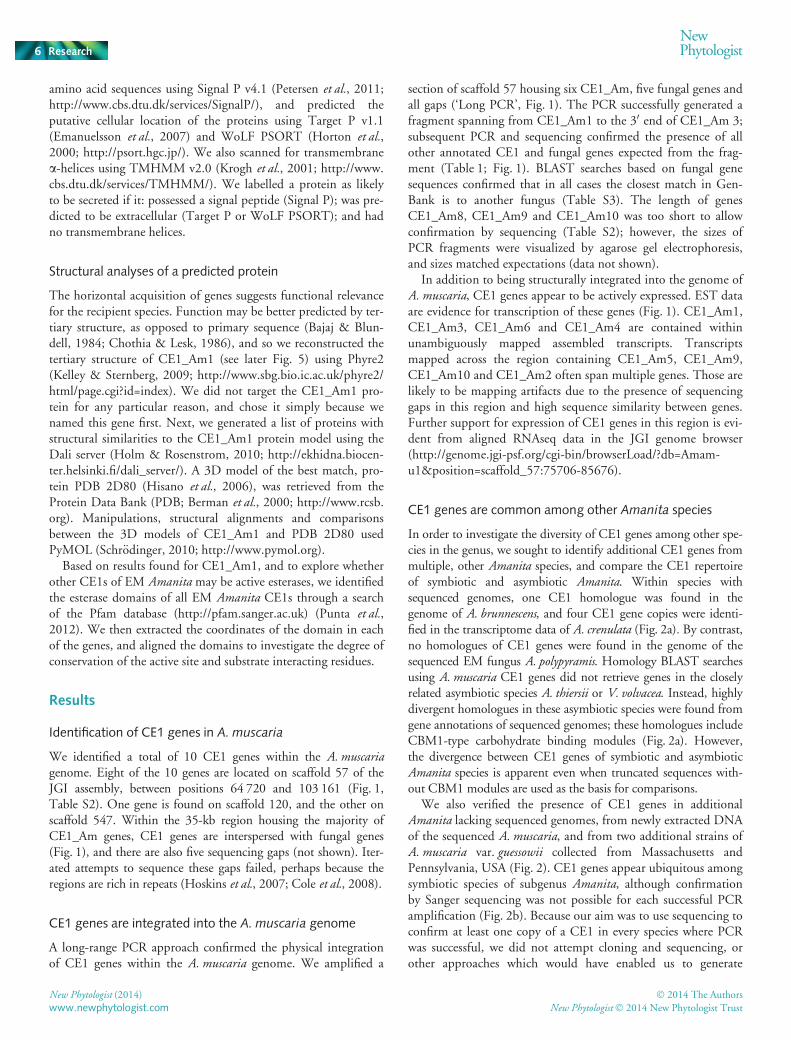
Phylogenetic analyses suggest CE1s of EM Amanita as HGT
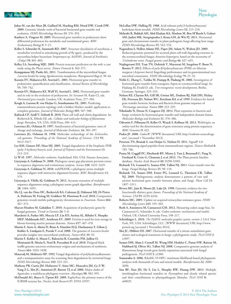
The CE1 sequences of EM Amanita are most similar to sequencesof soil bacteria of divergent phyla (Fig. 3, Table S4). The highestsequence identities are to PHB depolymerases in bothAlicyclobacillus pohliae (51%; gi|516850840 and gi|516850844,http://www.ncbi.nlm.nih.gov/protein/WP_018130341.1) andK. racemifer DSM 44963 (39–47%; gi|298250533 and gi|297548537) (J. A. Eisen, 2010, unpublished; http://genome.jgi.doe.gov/ktera/ktera.info.html). By contrast, the best-matchingCE1 sequences of EM Amanita are only a 32% match to sequencesin A. thiersii and 33% match to sequences in V. volvacea.
In order to test the hypothesis that EM CE1 genes were hori-zontally transferred from bacteria, we conducted a phylogeneticanalysis of over 1600 CE1 homologues identified throughBLAST searches in NCBI and JGI databases (Table S4). Weincluded CE1 gene products from bacteria, archaea, nonfungaleukaryotes, as well as basidiomycetes and ascomycetes in theanalysis. The fungal genes were distributed over four distinctclades (Fig. 3, Notes S1). The first we identify as ‘Fungal CladeI’, the largest fungal clade, which includes the CE1 genes ofA. thiersii and V. volvacea, and may represent a clade of genesunique to fungi. Three other clades are interspersed within bacte-rial lineages. One clade includes both ascomycetes and basidio-mycetes (‘Fungal Clade II’), the second groups a set of diverseascomycetes (‘Fungal Clade III’) and is near Fungal Clade I. Thethird is the symbiotic Amanita clade (‘EM Amanita’, Fig. 3) andit includes all of the CE1 genes identified from EM Amanita.They form a strongly supported monophyletic clade, embeddedwithin bacterial lineages, with 99% bootstrap support. The CE1genes present in asymbiotic Amanita may have been lost fromsymbiotic lineages of the genus. Copies of CE1 genes in symbi-otic and asymbiotic lineages are clearly highly divergent. A fewother CE1 genes from various other lineages (for example, othereukaryotes) are found scattered in apparently unusual places
Fig. 3 Phylogenetic evidence of horizontal gene transfer of carbohydrate esterase family 1 (CE1) genes. Lower left, complete CE1 phylogeny (available as alarger format in Supporting Information Notes S1). Inset, Clade containing CE1s in ectomycorrhizal (EM) Amanita. Numbers are bootstrap values above70; not all bootstrap values shown for larger phylogeny. Black branches, lineages leading to clades where species belong to different groups (Bacteria,Ascomycota, Basidiomycota and Other). Fungal Clade 1 houses saprotrophic (SAP) Amanita and Volvariella sequences, as well as sequences of otherfungi.
� 2014 The Authors
New Phytologist� 2014 New Phytologist TrustNew Phytologist (2014)
www.newphytologist.com
NewPhytologist Research 7
across the phylogeny (‘Others’, Fig. 3). These are described indetail in Table S5.
CE1 genes are dynamic elements of EM Amanita genomes
A maximum-likelihood reconciliation analysis of the Amanitaspecies tree with all CE1 genes of the EM species A. muscaria,A. crenulata and A. constricta (subgenus Amanita) andA. brunnescens (subgenus Lepidella) suggests a single HGT eventfollowed by a dynamic history of duplications and losses (Figs 2,3, S1). A conservative inference using only highly supportednodes reveals at least four duplication events, but up to 10 dupli-cations and six losses are possible (Fig. S1). The oldest duplica-tion occurred outside subgenus Amanita. Therefore, the HGTevent must have occurred before the split of subgenera Amanitaand Lepidella. The CE1 genes of A. muscaria are highly dynamic;four duplications have occurred within this genome alone.
Transferred CE1 genes have been ameliorated in their hostgenomes
Patterns of CE1_Am and CE1_Ab nucleotide composition andcodon usage are highly similar to patterns found in recipient ge-nomes (Table S6). For example, the average GC3 content of fun-gal genes of A. muscaria scaffold 57 is 0.45, similar to CE1 geneson scaffold 57 (0.46); by contrast, the average GC3 content ofCE1s of K. racemifer is high (0.65). Similar trends are observedfor codon usage (Table S6).
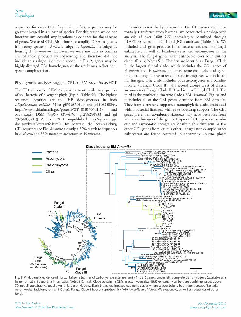
The numbers and placement of introns in A. muscaria andA. brunnescens are well conserved and distinct from introns of the
CE1-CBM1 genes of A. thiersii and V. volvacea (Fig. 4). Numbersof introns in A. muscaria and A. brunnescens CE1 genes rangefrom one to four. The density of introns per gene falls at thelower limit of values for basidiomycete genomes (3.8–5.7 intronsper gene; Da Lage et al., 2013), but corresponds well with themedian (3) and average (4.5) number of introns per gene inthe A. muscaria genome (http://genome.jgi.doe.gov/Amamu1/Amamu1.info.html).
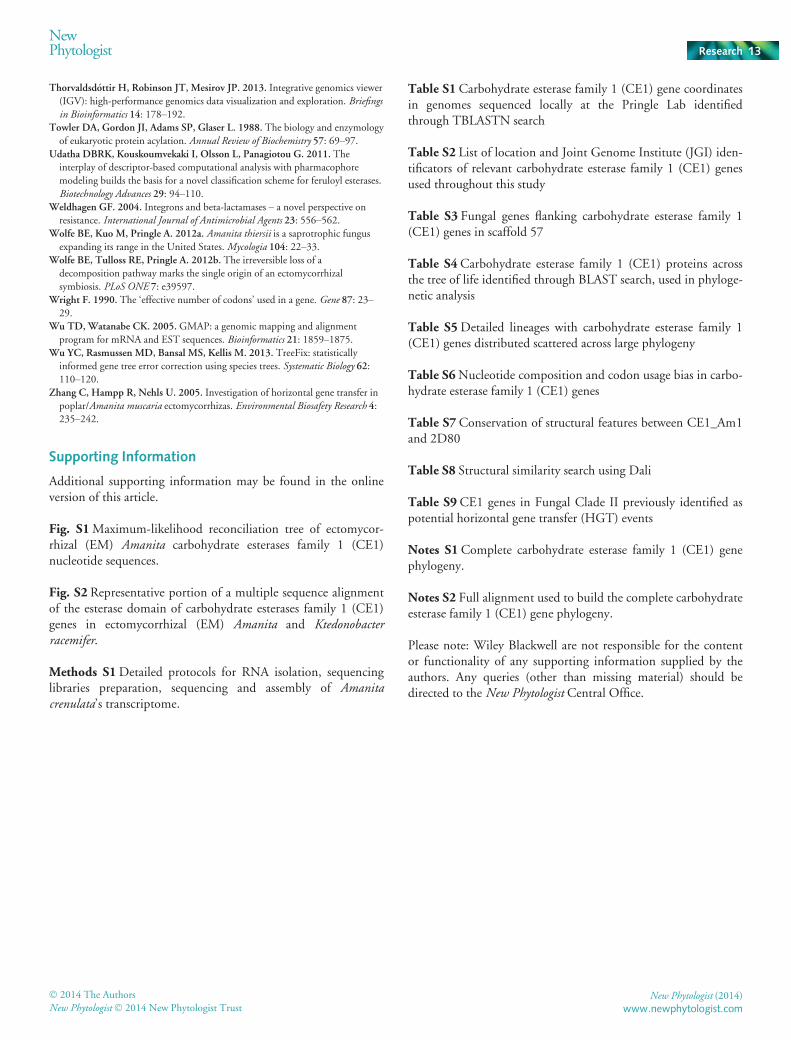
CE1_Am1 shows structural similarity to a PHBdepolymerase
In order to explore the potential function of the CE1 genes foundin EM Amanita, we predicted and analysed the structure of theinferred protein sequence of CE1_Am1. The predicted structureof CE1_Am1 shows close similarity to the crystal structure of aPHB depolymerase isolated from Penicillium funiculosum (fig. 6,PDB 2D80, Hisano et al., 2006). Despite the strong structuralsimilarities, the sequence of PDB 2D80 is sufficiently divergedfrom the sequence of CE1_Am1, and the other Amanita CE1s,that it was excluded by our BLAST cutoff in initial analyses. TheP. funiculosum protein is therefore not present in our phyloge-netic tree (Fig. 3).
The CE1_Am1 protein possesses the typical structural featuresof extracellular PHB depolymerases: catalytic (320–400 aa),linker (50–100 aa) and substrate-binding (40–60 aa) domains. Itwas also identified as a putatively secreted protein. The catalyticdomain houses a lipase-like catalytic triad (serine, aspartic acidand histidine residues; Fig. 5a). The substrate binding domain inthe PHB depolymerase of P. funiculosum possesses 14 binding
Fig. 4 Gene structure of horizontal genetransfer (HGT) carbohydrate esterase family1 (CE1) genes in ectomycorrhizal (EM)Amanita compared to CE1-CBM1 genesfrom A. thiersii and Volvariella volvacea.
Grey blocks, aligned exons; coloured bars,different introns. Introns sharing position andphase are shown in the same colour. Ab,A. brunnescens; Am, A. muscaria; Ath,A. thiersii; Vv, V. volvacea.
New Phytologist (2014) � 2014 The Authors
New Phytologist� 2014 New Phytologist Trustwww.newphytologist.com
Research
NewPhytologist8

residues and these combine to provide a hydrophobic environ-ment inside a pocket formed on the surface of the enzyme (Hisa-no et al., 2006); seven of these residues remain conserved inCE1_Am1, and five of the residues differ between the enzymesbut remain hydrophobic in CE1_Am1 (Table S7). The bindingresidues are essential for interaction with PHB chains and definethe substrate specificity of the enzyme (Fig. 5b; Hisano et al.,2006). This is likely to be conserved in CE1_Am1, because thesame catalytic residues are located within a pocket formed by thebinding domain (red spheres in Fig. 5b).
The Dali search also returned good matches to several carboxy-lesterases, lipases and peptidases (Table S8), reflecting the diver-sity of enzymatic functions within the CE1 class of carbohydrateesterases. However, PHB depolymerases are structurally very dif-ferent from carboxylesterases; for example, carboxylesterases lackregions of helices and coils (Fig. S2) typical in PHB depolymeras-es, and CE1_Am1 shows closer structural conservation to PHBdepolymerases (Fig. 5).
Catalytic residues of CE1_Am1 are conserved within the ester-ase domains of other CE1 genes found in other EM Amanita(Fig. S2). This suggests that all of the CE1s are potentially active,and have the same function: degrading PHB or PHB-like carbonstorage molecules.
Discussion
Carbohydrate metabolism genes of bacterial origin in EMAmanita
Phylogenetic analyses identify CE1 genes of bacterial originwithin symbiotic Amanita; the same genes are not found in asym-biotic species of the genus. Multiple, independent lines of evi-dence confirm the A. muscaria genes as integrated within the
genome, and expressed. CE1 homologues are found throughoutsubgenus Amanita, and are also found in A. brunnescens (subge-nus Lepidella). The distribution suggests an HGT event aroundthe time of the evolution of the EM niche in Amanita, and beforethe split of the subgenera. CE1 genes are ameliorated (Lawrence& Ochman, 1997) within host genomes, with nucleotide con-tents and exon/intron structures typical of basidiomycetes.
We considered and eliminated alternative explanations toHGT. For example, rapid evolution can blur homology relation-ships among orthologous genes. Although horizontally trans-ferred CE1 genes resemble basidiomycete genes, ameliorationdoes not always denote rapid evolution; amelioration may alsomark an ancient transfer event (Lawrence & Ochman, 1997).Moreover, sequence homology and gene structure analyses provethat CE1 genes in symbiotic Amanita are very different fromthose found in the asymbiotic species A. thiersii and V. volvacea(Notes S2). A scenario in which the CE1 copy in an ancient sym-biotic Amanita lineage diverged radically from its homologue inA. thiersii, and convergently evolved to be most similar to bacte-rial CE1s, is unlikely.
The selective loss of genes in specific lineages may also createincongruent gene and species trees (Aravind et al., 2000). How-ever, a gene loss hypothesis would require an ancient origin ofPHB depolymerase-type CE1 genes during the early diversifica-tion of eukaryotes, or fungi at least, followed by multiple lossesin every lineage except the lineage housing EM Amanita. The sce-nario is implausible, and not the most parsimonious explanationfor observed patterns. Moreover, phylogenies do not suggest avertical origin from within the fungi, and we are confident thatwe retrieved a comprehensive set of homologous CE1 genes fromavailable fungal genomes with our BLAST searches.
Although HGT is the most consistent and parsimonious expla-nation for observed patterns (Fig. 3), available data do not allowunequivocal determination of a donor lineage. The phylogeneticanalysis clusters symbiotic Amanita CE1 genes with CE1 genesfrom both spore-forming soil bacteria from the phylum Firmi-cutes (A. pohliae and Bacillus megaterium) and with a filamentoussoil bacterium from the phylum Chloroflexi (K. racemifer); boot-strap support is strong (99). However, these sequences share atmost 51% identity to CE1 genes in symbiotic Amanita, and indi-cate that either the true donor is absent from genome databases,that gene sequences have changed considerably over time, hinder-ing identification, or that perhaps the donor species is extinct. Toidentify the direction of HGT we rely on the widely acceptedassumption that the taxon of the broadest representation of thegene family is the most likely source (Koonin et al., 2002). In ourcase, CE1 genes in symbiotic Amanita are embedded within alarge bacterial clade, suggesting a bacteria to symbiotic AmanitaHGT event.
HGT events among bacteria, preceding the HGT to EMAmanita, may also have obscured the origin of symbioticAmanita CE1 genes; this idea is supported by the variety of dis-similar taxa found in phylogenetic proximity to the EM AmanitaCE1 clade, and also by previous research showing CE1 genes asprone to horizontal transfers (Marcet-Houben & Gabaldon,2010; Udatha et al., 2011). The pattern observed for Fungal
(a) (b)
Fig. 5 (a) Alignment between mature forms of poly((R)-3-hydroxybutyrate) (PHB) depolymerases of Penicillium funiculosum (PDB2D80) (black), and CE1_Am1 (colour). The match between the proteins isstrong, with a root-mean-square-deviation of 1.2�A and Z-score of 43.2.Colours in CE1_Am1 are as follows: cyan, a-helices; magenta, b-sheets;pink, coils. The catalytic triad residues Ser159, Asp235 and His309 ofCE1_Am1 are marked with red spheres and overlap with the dark redspheres of residues Ser39, Asp121 and His155 in PDB 2D80. Only namesof CE1_Am1 are shown. Unaligned sequence regions are shown in grey.(b) Representation of binding and catalytic sites in CE1_Am1 and PDB2D80. Protein structures of CE1_Am1 and 2D80 are represented as violetand grey mesh, respectively. Binding sites forming a pocket on the surfaceof the enzymes are represented: cyan spheres, CE1_Am1; blue spheres,2D80. Catalytic residues coloured as in (a).
� 2014 The Authors
New Phytologist� 2014 New Phytologist TrustNew Phytologist (2014)
www.newphytologist.com
NewPhytologist Research 9
Clade II may reflect an independent HGT event from bacteria tofungi. Marcet-Houben & Gabaldon (2010) searched for prokary-otic-derived HGT in 60 fully sequenced fungal genomes andreported nine putative PHB depolymerases, which they identifyas originating from three independent HGT events. Eight of theevents reported by Marcet-Houben & Gabaldon (2010) corre-spond to potential HGT genes in Fungal Clade II (Table S9).
Dynamics of CE1 genes within Amanita
Eight out of the 10 A. muscaria CE1 genes are located on thesame scaffold and our phylogenetic analyses suggest that thegroup is derived from a single, ancestral horizontally transferredgene that was subsequently amplified (Fig. S1). In theory, oncethere is more than one copy of a gene, the genes can spread moreeasily, because redundant copies shelter replication errors,enabling duplications (Hurles, 2004). Furthermore, homologueslocated in close proximity to each other on the chromosome mayalso promote the formation of unequal crossing over events andresult in accelerated gene gain and loss (Li, 1997).
Dynamic, expanded gene families often mark functionallyimportant genes; for example, enzymes involved in the detoxifi-cation of insecticides are heavily amplified in exposed species ofmosquitos (Hemingway et al., 1998). Similarly, gene familyexpansions are a common theme of carbohydrate metabolismgenes associated with different fungal niches, including patho-genesis (Soanes et al., 2008; Abramyan & Stajich, 2012) anddecomposition (Eastwood et al., 2011). The observed expansionin CE1 genes among symbiotic Amanita lineages at least sug-gests a critical function; other research also suggests that HGTevents are strongly associated with functional genes (Riveraet al., 1998).
Functions of transferred genes
CE1 genes of bacterial origin are only found within EMAmanita, and the CE1 genes are likely to provide some functionassociated with the symbiotic niche. A test of putative functionbased on a focal gene, the CE1_Am1, reveals structural conserva-tion between the gene’s protein and an extracellularly secretedPHB depolymerase. Moreover, the catalytic residues ofCE1_Am1 are conserved across the EM Amanita CE1s, suggest-ing that function is conserved across the genes.
In the absence of an exogenous carbon supply, extracellularPHB depolymerases degrade PHB, a microbial carbon andenergy storage compound (Dawes, 1988; Jendrossek & Hand-rick, 2002). Extracellular PHB depolymerases are found in fila-mentous fungi (McLellan & Halling, 1988; Matavulj &Molitoris, 1992; Lee et al., 2005), but the ecological role of PHBdegradation remains largely unexplored. Soils are the habitatswith the largest numbers of PHB degrading fungi (Jendrossek &Handrick, 2002).
Based on limited available knowledge, we suggest and brieflydiscuss three hypotheses for the function of HGT CE1s: CE1smay play a role in carbon metabolism, communication, ordefence. First, symbiotic Amanita lack plant cell wall degrading
enzymes, and cannot decompose organic substrates. The abilityto use extracellular PHB as an alternative carbon source may rep-resent an important adaptation. CE1 genes may enable Amanitaspecies to grow when a symbiosis is not yet established, or when aplant is not providing enough carbon. By contrast, CE1s inV. volvacea match xylanases, plant cell wall degrading enzymesfrom the CE1 family, both by sequence and structural homology(Ding et al., 2007). Their role in plant cell wall degradation isfurther supported by the presence of a CBM1, which binds tocellulose and may target the enzyme towards the plant cell wall.CE1s in A. thiersii share close homology to CE1s in V. volvaceaand probably perform a similar function.
Second, mycorrhizal symbioses grow in habitats teeming withother organisms, including ‘mycorrhiza helper bacteria’ (MHB;Garbaye, 1994), and CE1s may play a role in signalling. Avail-able evidence suggests that symbiotic Amanita actively communi-cate with surrounding bacteria: A. muscaria secretes eitherorganic acids or protons capable of modulating the spectrum ofantibiotics produced by MHB (Frey-Klett et al., 2007), and acompound produced by Streptomyces sp. AcH505 seems to stimu-late the presymbiotic growth of A. muscaria, and simultaneouslyinhibit the growth of pathogenic fungi (Keller et al., 2007).Third, PHB is able to be depolymerized into water-soluble short-chain fatty acid monomers, and these monomers can act asmicrobial control agents (Najdegerami et al., 2012). CE1s mayplay a role in defense. Whatever the function of CE1s, the genesmay not be essential for the mycorrhizal niche; the mycorrhizalA. polypyramis does not appear to house PHB depolymerase-typeCE1s.
The frequency of HGT and potential for HGT to providenovel metabolic tools (Garcia-Vallv�e et al., 2000; Intra et al.,2008; Udatha et al., 2011; this study) may influence thinking ontransitions between saprotrophic and mycorrhizal niches.Whether mycorrhizal species can evolve saprotrophy has beendebated since at least Hibbett et al. (2000). The most recent evi-dence points to a history of independent origins of the mycorrhi-zal habit, with no reversals to saprotrophy (Bruns & Shefferson,2004; Matheny et al., 2006; Wolfe et al., 2012b). Although thelarge-scale losses of CAZymes found within the symbioticAmanita likely preclude a total reversal to an asymbiotic niche(Wolfe et al., 2012b), HGT may endow symbiotic species withnovel functions, perhaps including access to alternative carbonsources.
Acknowledgements
We gratefully acknowledge K. Zimmerman for the collection ofA. muscaria strain FP01. We thank the National Science Founda-tion for funding, and a Talent Grant from the Faculty of Mathe-matics and Natural Sciences of the University of Groningen forenabling M. Chaib De Mares to travel and work in the Pringlelaboratory. The work conducted by the US Department ofEnergy Joint Genome Institute was supported by the Office ofScience of the US Department of Energy under Contract No.DE-AC02-05CH11231. We are grateful to F. Martin and theMycorrhizal Genomics Initiative consortium for access to
New Phytologist (2014) � 2014 The Authors
New Phytologist� 2014 New Phytologist Trustwww.newphytologist.com
Research
NewPhytologist10
unpublished genome data. We also thank three anonymous refer-ees for thoughtful, useful reviews that greatly improved our man-uscript.
References
Abramyan J, Stajich JE. 2012. Species-specific chitin-binding module 18
expansion in the amphibian pathogen Batrachochrytium dendrobatidis.MBio 3:e00150–12.
Akin DE. 2008. Plant cell wall aromatics: influence on degradation of biomass.
Biofuels, Bioproducts and Biorefining 2: 288–303.Altschul SF, Gish W, Miller W, Myers EW, Lipman DJ. 1990. Basic local
alignment search tool. Journal of Molecular Biology 215: 403–410.Aravind L, Watanabe H, Lipman DJ, Koonin EV. 2000. Lineage-specific loss
and divergence of functionally linked genes in eukaryotes. Proceedings of theNational Academy of Sciences, USA 97: 11319–11324.
Armijos-Jaramillo VD, Vargas WA, Sukno SA, Thon MR. 2013.Horizontal
transfer of a subtilisin gene from plants into an ancestor of the plant pathogenic
fungal genus Colletotrichum. PLoS ONE 8: e59078.
Bajaj M, Blundell T. 1984. Evolution and the tertiary structure of proteins.
Annual Review of Biophysics and Bioengineering 13: 453–492.Berman HM, Westbrook J, Feng Z, Gilliland G, Bhat TN, Weissig H,
Shindyalov IN, Bourne PE. 2000. The protein data bank. Nucleic AcidsResearch 28: 235–242.
Biely P. 2012.Microbial carbohydrate esterases deacetylating plant
polysaccharides. Biotechnology Advances 30: 1575–1588.Brown JR, Doolittle WF. 1997. Archaea and the prokaryote-to-eukaryote
transition.Microbiology and Molecular Biology Reviews 61: 456–502.Bruns TD, Shefferson RP. 2004. Evolutionary studies of ectomycorrhizal fungi:
recent advances and future directions. Canadian Journal of Botany 82:1122–1132.
Bruto M, Prigent-Combaret C, Luis P, Mo€enne-Loccoz Y, Muller D. 2014.
Frequent, independent transfers of a catabolic gene from bacteria to contrasted
filamentous eukaryotes. Proceedings of the Royal Society B 281: 20140848.
Cantarel BL, Coutinho PM, Rancurel C, Bernard T, Lombard V, Henrissat B.
2009. The carbohydrate-active enzymes database (CAZy): an expert resource
for glycogenomics. Nucleic Acids Research 37: D233–D238.
Capella-Gutierrez JM, Silla-Martinez TG. 2009. trimAl: a tool for automated
alignment trimming in large-scale phylogenetic analyses. Bioinformatics 25:1972–1973.
Chaib De Mares M, Hess J, Falcao Salles J, Pringle A. 2013. A genomicperspective on a transition to symbiosis in ectomycorrhizal Amanita. MSc thesis,
University of Groningen, Groningen, the Netherlands.
Chothia C, Lesk AM. 1986. The relation between the divergence of sequence and
structure in proteins. EMBO Journal 5: 823–826.Christin P-A, Edwards EJ, Besnard G, Boxall SF, Gregory R, Kellogg EA,
Hartwell J, Osborne CP. 2012. Adaptive evolution of C4 photosynthesis
through recurrent lateral gene transfer. Current Biology 22: 445–449.Cole CG, McCann OT, Collins JE, Oliver K, Willey D, Gribble SM, Yang F,
McLaren K, Rogers J, Ning Z et al. 2008. Finishing the human chromosome
22 sequence. Genome Biology 9: R78.Combier J-P, Melayah D, Raffier C, Gay G, Marmeisse R. 2003. Agrobacteriumtumefaciens-mediated transformation as a tool for insertional mutagenesis in the
symbiotic ectomycorrhizal fungus Hebeloma cylindrosporum. FEMSMicrobiology Letters 220: 141–148.
Da Lage JL, Binder M, Hua-Van A, Jane�cek S, Casane D. 2013. Gene make-up:
rapid and massive intron gains after horizontal transfer of a bacterial a-amylase
gene to Basidiomycetes. BMC Evolutionary Biology 13: 40–53.Darriba D, Taboada GL, Doallo R, Posada D. 2011. ProtTest 3: fast selection of
best-fit models of protein evolution. Bioinformatics 27: 1164–1165.Dawes EA. 1988. Polydroxybutyrate: an intriguing biopolymer. Bioscience Reports8: 537–547.
Ding S, Cao J, Zhou R, Zheng F. 2007.Molecular cloning, and characterization
of a modular acetyl xylan esterase from the edible straw mushroom Volvariellavolvacea. FEMS Microbiology Letters 274: 304–310.
Don RH, Cox PT, Wainwright BJ, Baker K, Mattick JS. 1991. “Touchdown”
PCR to circumvent spurious priming during gene amplification. Nucleic AcidsResearch 14: 4008.
Eastwood DC, Floudas D, Binder M, Majcherczyk A, Schneider P, Aerts A,
Asiegbu FO, Baker SE, Barry K, Bendiksby M et al. 2011. The plant cell-walldecomposing machinery underlies the functional diversity of forest fungi.
Science 333: 762–765.Emanuelsson O, Brunak S, von Heijne G, Nielsen H. 2007. Locating proteins in
the cell using TargetP, SignalP and related tools. Nature Protocols 2: 953–971.Fitzpatrick DA. 2011.Horizontal gene transfer in fungi. FEMS MicrobiologyLetters 329: 1–8.
Frey-Klett P, Garbaye J, Tarkka M. 2007. The mycorrhiza helper bacteria
revisited. New Phytologist 176: 22–36.Gans J, Wolinsky M, Dunbar J. 2005. Computational improvements
reveal great bacterial diversity and high metal toxicity in soil. Science 309:1387–1390.
Garbaye J. 1994.Helper bacteria: a new dimension to the mycorrhizal symbiosis.
New Phytologist 128: 197–210.Garcia-Vallv�e S, Romeu A, Palau J. 2000.Horizontal gene transfer of glycosyl
hydrolases of the rumen fungi.Molecular Biology and Evolution 17: 352–361.Grimaldi B, de Raaf MA, Filetici P, Ottonello S, Ballario P. 2005.
Agrobacterium-mediated gene transfer and enhanced green fluorescent protein
visualization in the mycorrhizal ascomycete Tuber borchii: a first step towards
truffle genetics. Current Genetics 48: 69–74.Hammesfahr B, Odronitz F, M€uhlhausen S, Waack S, Kollmar M. 2013.
GenePainter: a fast tool for aligning gene structures of eukaryotic protein
families, visualizing the alignments and mapping gene structures onto protein
structures. BMC Bioinformatics 14: 77–88.Hanssen AM, Ericson-Sollid JU. 2006. SCCmec in Staphylococci: genes on the
move. FEMS Immunology and Medical Microbiology 46: 8–20.Hatje K, Keller O, Hammesfahr B, Pillmann H, Waack S, Kollmar M. 2011.
Cross-species protein sequence and gene structure prediction with fine-tuned
Webscipio 2.0 and Scipio. BMC Research Notes 4: 265.Hemingway J, Hawkes N, Prapanthadara L, Jayawardenal KG, Ranson H.
1998. The role of gene splicing, gene amplification and regulation in mosquito
insect resistance. Philosophical Transactions of the Royal Society B 353: 1695–1699.
Hess J, Pringle A. 2014. The natural histories of species and their genomes:
asymbiotic and ectomycorrhizal Amanita. In: Martin F, ed. Fungi (advances inbotanical research). San Diego, CA, USA: Academic Press Inc., 235–257.
Hess J, Skrede I, Wolfe EB, LaButti K, Ohm RA, Grigoriev IV, Pringle A.
2014. Transposable element dynamics among asymbiotic and ectomycorrhizal
Amanita fungi. Genome Biology and Evolution 6: 1564–1578.Hibbett DS, Gilbert LB, Donoghue MJ. 2000. Evolutionary instability of
ectomycorrhizal symbioses in basidiomycetes. Nature 407: 506–510.Hisano T, Kasuya KI, Tezuka Y, Ishii N, Kobayashi T, Shiraki M, Oroudjev E,
Hansma H, Iwata T, Doi Y et al. 2006. The crystal structure ofPolyhydrohybutyrate depolymerase from Penicillium funiculosum provides
insights into the recognition and degradation of biopolyesters. Journal ofMolecular Biology 356: 993–1004.
Holm L, Rosenstrom P. 2010. Dali server: conservation mapping in 3D. NucleicAcids Research 38: W545–W549.
Horton P, Park K-J, Obayashi T, Fujita N, Harada H, Adams-Collier CJ, Nakai
K. 2000.WoLF PSORT: protein localization predictor. Nucleic Acids Research35: W585–W587.
Hoskins RA, Carlson JW, Kennedy C, Acevedo D, Evans-Holm M, Frise E,
Wan KH, Park S, Mendez-Lago M, Rossi F et al. 2007. Sequence finishingand mapping of Drosophila melanogaster heterochromatin. Science 316: 1625–1628.
Hurles M. 2004. Gene duplication: the genomic trade in spare parts. PLoSBiology 2: e206.
Intra J, Pavesi G, Horner DS. 2008. Phylogenetic analyses suggest multiple
changes of substrate specificity within the Glycosyl hydrolase 20 family. BMCEvolutionary Biology 8: 214–231.
Jendrossek D, Handrick R. 2002.Microbial degradation of
polyhydroxyalkanoates. Annual Review of Microbiology 56: 403–432.
� 2014 The Authors
New Phytologist� 2014 New Phytologist TrustNew Phytologist (2014)
www.newphytologist.com
NewPhytologist Research 11
Juhas M, van der Meer JR, Gaillard M, Harding RM, Hood DW, Crook DW.
2009. Genomic islands: tools of bacterial horizontal gene transfer and
evolution. FEMS Microbiology Reviews 33: 376–393.Kanhere A, Vingron M. 2009.Horizontal gene transfers in prokaryotes show
differential preferences for metabolic and translational genes. BMCEvolutionary Biology 9: 9–21.
Keller S, Schneider K, Sussmuth RD. 2007. Structure elucidation of auxofuran, a
metabolite involved in stimulating growth of fly agaric, produced by the
mycorrhiza helper bacterium Streptomyces sp. AcH505. Journal of Antibiotics(Tokyo) 59: 801–803.
Kelley LA, Sternberg MJE. 2009. Protein structure prediction on the web: a case
study using the Phyre server. Nature Protocols 4: 363–371.Kemppainen MJ, Pardo AG. 2011. Transformation of the mycorrhizal fungus
Laccaria bicolor by using Agrobacterium tumefaciens. Bioengineered Bugs 2: 38–44.Koonin EV, Makarova KS, Aravind L. 2001.Horizontal gene transfer in
prokaryotes: quantification and classification. Annual Review of Microbiology55: 709–742.
Koonin EV, Makarova KS, Wolf YI, Aravind L. 2002.Horizontal gene transfer
and its role in the evolution of prokaryotes. In: Syvanen M, Kado CI, eds.
Horizontal gene transfer. London, UK: Academic Press, 277–304.Krogh A, Larsson B, von Heijne G, Sonnhammer EL. 2001. Predicting
transmembrane protein topology with a hidden Markov model: application to
complete genomes. Journal of Molecular Biology 305: 567–580.Kubicek CP, Seidl V, Seiboth B. 2010. Plant cell wall and chitin degradation. In:
Borkovich K, Ebbole DJ, eds. Cellular and molecular biology of filamentousfungi. Herndon, VA, USA: ASM Press, 396–413.
Lawrence JG, Ochman H. 1997. Amelioration of bacterial genomes: rates of
change and exchange. Journal of Molecular Evolution. 44: 383–397.Lawrence JG, Ochman H. 1998. Molecular archaeology of the Escherichiacoli genome. Proceedings of the National Academy of Sciences, USA 95:
9413–9417.Lee KM, Gimore DF, Huss MJ. 2005. Fungal degradation of the bioplastic PHB
(poly-3-hydroxy-butyric acid. Journal of Polymers and the Environment 13:213–219.
Li W-H. 1997.Molecular evolution. Sunderland, MA, USA: Sinauer Associates.
L€oytynoja A, Goldman N. 2008. Phylogeny-aware gap placement prevents errors
in sequence alignment and evolutionary analysis. Science 320: 1632–1635.L€oytynoja A, Goldman N. 2010. webPRANK: a phylogeny-aware multiple
sequence aligner with interactive alignment browser. BMC Bioinformatics 11:579.
L€oytynoja A, Vilella AJ, Goldman N. 2012. Accurate extension of multiple
sequence alignments using a phylogeny-aware graph algorithm. Bioinformatics28: 1684–1691.
Ma LJ, van der Does HC, Borkovich KA, Coleman JJ, Daboussi MJ, Di Pietro
A, Dufresne M, Freitag M, Grabherr M, Henrissat B et al. 2010. Comparative
genomics reveals mobile pathogenicity chromosomes in Fusarium. Nature 464:367–373.
Marcet-Houben M, Gabaldon T. 2010. Acquisition of prokaryotic genes by
fungal genomes. Trends in Genetics 26: 5–8.Marchetti A, Parker MS, Moccia LP, Lin EO, Arrieta AL, Ribalet F, Murphy
MEP, Maldonado MT, Armbrust EV. 2009. Ferritin is used for iron storage in
bloom-forming marine pennate diatoms. Nature 457: 467–470.Martin F, Aerts A, Ahr�en D, Brun A, Danchin EGJ, Duchaussoy F, Gibon J,
Kohler A, Lindquist E, Pereda V et al. 2008. The genome of Laccaria bicolorprovides insights into mycorrhizal symbiosis. Nature 452: 88–92.
Martin F, Kohler A, Murat C, Balestrini R, Coutinho PM, Jaillon O,
Montanini B, Morin E, Noel B, Percudani R et al. 2010. P�erigord blacktruffle genome uncovers evolutionary origins and mechanisms of symbiosis.
Nature 464: 1033–1038.Matavulj M, Molitoris HP. 1992. Fungal degradation of polyhydroxyalkanoates
and a semiquantitative assay for screening their degradation by terrestrial fungi.
FEMS Microbiology Reviews 103: 323–331.Matheny PB, Curtis JM, Hofstetter V, Aime MC, Moncalvo JM, Ge Z-W,
Yang Z-L, Slot JC, Ammirati JF, Baroni TJ et al. 2006.Major clades of
Agaricales: a multilocus phylogeny overview.Mycologia 98: 982–995.McDonald AG, Boyce S, Tipton KF. 2009. ExplorEnz: the primary source of the
IUBMB enzyme list. Nucleic Acids Research 37: D593–D597.
McLellan DW, Halling PJ. 1988. Acid-tolerant poly(3-hydroxybutyrate)
hydrolases from moulds. FEMS Microbiology Letters 52: 215–218.Mehrabi R, Bahkali AH, Abd-Elsalam KA, Moslem M, Ben M’Barek S, Gohari
AM, Jashni MK, Stergiopoulos I, Kema GH, de Wit PJ. 2011.Horizontal
gene and chromosome transfer in plant pathogenic fungi affecting host range.
FEMS Microbiology Reviews 35: 542–554.Nagendran S, Hallen-Adams HE, Paper JM, Aslam N, Walton JD. 2009.
Reduced genomic potential for secreted plant cell-wall-degrading enzymes in
the ectomycorrhizal fungus Amanita bisporigera, based on the secretome of
Trichoderma reesei. Fungal genetics and Biology 46: 427–435.Najdegerami EH, Tran TN, Defoirdt T, Marzorati M, Sorgeloos P, Boon N,
Bossier P. 2012. Effects of poly-b-hydroxybutyrate (PHB) on Siberian
sturgeon (Acipenser baerii) fingerlings performance and its gastrointestinal tract
microbial community. FEMS Microbiology Ecology 79: 25–33.Nehls U, Zhang C, Tarkka M, Hampp R, Fladung M. 2006. Investigation of
horizontal gene transfer from transgenic Aspen to ectomycorrhizal fungi. In:
Fladung M, Ewald D, eds. Tree transgenesis: recent developments. Berlin,Germany: Springer, 323–333.
Nelson KE, Clayton RA, Gill SR, Gwinn ML, Dodson RJ, Haft DH, Hickey
EK, Peterson JD, Nelson WC, Ketchum KA et al. 1999. Evidence for lateralgene transfer between Archaea and Bacteria from genome sequence of
Thermotoga maritima. Nature 399: 323–329.Nikolaidis N, Doran N, Cosgrove DJ. 2014. Plant expansins in bacteria and
fungi: evolution by horizontal gene transfer and independent domain fusion.
Molecular Biology and Evolution 31: 376–386.Odronitz F, Pillmann H, Keller O, Waack S, Kollmar M. 2013.WebScipio: an
online tool for the determination of gene structures using protein sequences.
BMC Genomics 9: 422.Peden JF. 2000. CodonW. [WWW document] URL http://codonw.sourceforge.
net/. [accessed 1 November 2013].
Petersen TN, Brunak S, von Heijne G, Nielsen H. 2011. SignalP 4.0:
discriminating signal peptides from transmembrane regions. Nature Methods 8:785–786.
Punta M, Coggill PC, Eberhardt RY, Mistry J, Tate J, Boursnell C, Pang N,
Forslund K, Ceric G, Clements J et al. 2012. The Pfam protein families
database. Nucleic Acids Research 40: D290–D301.
Richards TA, Leonard G, Soanes DM, Talbot NJ. 2011. Gene transfer into the
fungi. Fungal Biology Reviews 25: 98–110.Richards TA, Soanes DM, Foster PG, Leonard G, Thornton CR, Talbot
NJ. 2009. Phylogenomic analysis demonstrates a pattern of rare and
ancient horizontal gene transfer between plants and fungi. Plant Cell 21:1897–1911.
Rivera MC, Jain R, Moore JE, Lake JA. 1998. Genomic evidence for two
functionally distinct gene classes. Proceedings of the National Academy ofSciences, USA 95: 6239–6244.
Roberts MC. 2005. Update on acquired tetracycline resistance genes. FEMSMicrobiology Letters 245: 195–203.
Roth A, Anisimova M, Cannarozzi GM. 2012.Measuring codon usage bias. In:
Cannarozzi G, Schneider A, eds. Codon evolution: mechanisms and models.Oxford, UK: Oxford University Press, 198–217.
Schr€odinger L. 2010. The PyMOL molecular graphics system, version 1.5.0.4. New
York, NY, USA: Schr€odinger, LLC. [WWW document] URL http://www.
pymol.org [accessed 1 November 2014].
Slot JC, Hibbett DS. 2007.Horizontal transfer of a nitrate assimilation gene
cluster and ecological transitions in fungi: a phylogenetic study. PLoS ONE 2:
e1097.
Soanes DM, Alam I, Cornell M, Wong HM, Hedeler C, Paton NW, Rattray M,
Hubbard SJ, Oliver SG, Talbot NJ. 2008. Comparative genome analysis of
filamentous fungi reveals gene family expansions associated with fungal
pathogenesis. PLoS ONE 3: e2300.
Stamatakis A. 2006. RAxML-VI-HPC: maximum likelihood-based phylogenetic
analyses with thousands of taxa and mixed models. Bioinformatics 22: 2688–2690.
Sun BF, Xiao JH, He S, Liu L, Murphy RW, Huang DW. 2013. Multiple
interkingdom horizontal transfers in Pyrenophora and closely related species
and their contributions to phytopathogenic lifestyles. PLoS ONE 8:
e60029.
New Phytologist (2014) � 2014 The Authors
New Phytologist� 2014 New Phytologist Trustwww.newphytologist.com
Research
NewPhytologist12
Thorvaldsd�ottir H, Robinson JT, Mesirov JP. 2013. Integrative genomics viewer
(IGV): high-performance genomics data visualization and exploration. Briefingsin Bioinformatics 14: 178–192.
Towler DA, Gordon JI, Adams SP, Glaser L. 1988. The biology and enzymology
of eukaryotic protein acylation. Annual Review of Biochemistry 57: 69–97.Udatha DBRK, Kouskoumvekaki I, Olsson L, Panagiotou G. 2011. The
interplay of descriptor-based computational analysis with pharmacophore
modeling builds the basis for a novel classification scheme for feruloyl esterases.
Biotechnology Advances 29: 94–110.Weldhagen GF. 2004. Integrons and beta-lactamases – a novel perspective onresistance. International Journal of Antimicrobial Agents 23: 556–562.
Wolfe BE, Kuo M, Pringle A. 2012a. Amanita thiersii is a saprotrophic fungusexpanding its range in the United States.Mycologia 104: 22–33.
Wolfe BE, Tulloss RE, Pringle A. 2012b. The irreversible loss of a
decomposition pathway marks the single origin of an ectomycorrhizal
symbiosis. PLoS ONE 7: e39597.
Wright F. 1990. The ‘effective number of codons’ used in a gene. Gene 87: 23–29.
Wu TD, Watanabe CK. 2005. GMAP: a genomic mapping and alignment
program for mRNA and EST sequences. Bioinformatics 21: 1859–1875.Wu YC, Rasmussen MD, Bansal MS, Kellis M. 2013. TreeFix: statistically
informed gene tree error correction using species trees. Systematic Biology 62:110–120.
Zhang C, Hampp R, Nehls U. 2005. Investigation of horizontal gene transfer in
poplar/Amanita muscaria ectomycorrhizas. Environmental Biosafety Research 4:235–242.
Supporting Information
Additional supporting information may be found in the onlineversion of this article.
Fig. S1Maximum-likelihood reconciliation tree of ectomycor-rhizal (EM) Amanita carbohydrate esterases family 1 (CE1)nucleotide sequences.
Fig. S2 Representative portion of a multiple sequence alignmentof the esterase domain of carbohydrate esterases family 1 (CE1)genes in ectomycorrhizal (EM) Amanita and Ktedonobacterracemifer.
Methods S1Detailed protocols for RNA isolation, sequencinglibraries preparation, sequencing and assembly of Amanitacrenulata’s transcriptome.
Table S1Carbohydrate esterase family 1 (CE1) gene coordinatesin genomes sequenced locally at the Pringle Lab identifiedthrough TBLASTN search
Table S2 List of location and Joint Genome Institute (JGI) iden-tificators of relevant carbohydrate esterase family 1 (CE1) genesused throughout this study
Table S3 Fungal genes flanking carbohydrate esterase family 1(CE1) genes in scaffold 57
Table S4Carbohydrate esterase family 1 (CE1) proteins acrossthe tree of life identified through BLAST search, used in phyloge-netic analysis
Table S5Detailed lineages with carbohydrate esterase family 1(CE1) genes distributed scattered across large phylogeny
Table S6Nucleotide composition and codon usage bias in carbo-hydrate esterase family 1 (CE1) genes
Table S7Conservation of structural features between CE1_Am1and 2D80
Table S8 Structural similarity search using Dali
Table S9CE1 genes in Fungal Clade II previously identified aspotential horizontal gene transfer (HGT) events
Notes S1Complete carbohydrate esterase family 1 (CE1) genephylogeny.
Notes S2 Full alignment used to build the complete carbohydrateesterase family 1 (CE1) gene phylogeny.
Please note: Wiley Blackwell are not responsible for the contentor functionality of any supporting information supplied by theauthors. Any queries (other than missing material) should bedirected to the New Phytologist Central Office.
� 2014 The Authors
New Phytologist� 2014 New Phytologist TrustNew Phytologist (2014)
www.newphytologist.com
NewPhytologist Research 13


