Embed Size (px)
Citation preview
9/11/2012
1
1
Chapter 36
Shock
2
Learning Objectives
• Define shock.
• Outline factors necessary to achieve adequate tissue oxygenation.
• Describe how the diameter of resistance vessels influences preload.
3
Copyright © 2013 by Jones & Bartlett Learning, LLC, an Ascend Learning Company
9/11/2012
2
Learning Objectives
• Calculate mean arterial pressure when given a blood pressure.
• Outline changes in the microcirculation during the progression of shock.
4
Learning Objectives
• List the causes of hypovolemic, cardiogenic, neurogenic, anaphylactic, and septic shock.
• Describe pathophysiology as a basis for signs and symptoms associated with the progression through the stages of shock.
5
Shock
• Defined by Gross in 1850– “Rude unhinging of the machinery of life”
• Robert M. Hardaway, professor of surgery at Texas Tech University School of Medicine in El Paso, Texas– I believe that the best definition of shock is inadequate capillary perfusion. As a corollary of this broad definition, almost anyone who dies, except one who is instantly destroyed, must go through a stage of shock—a momentary pause in the act of death
6
Copyright © 2013 by Jones & Bartlett Learning, LLC, an Ascend Learning Company
9/11/2012
3
Shock
• Shock is not single event
– Does not have one specific cause and treatment
• Complex group of physiological abnormalities
• Many complexities involved in shock, not adequately defined by pulse rate, blood pressure, cardiac function
7
• Causes– Healthy patient (adult)
• Coronary syndromes• Respiratory arrest• Anaphylaxis • Drowning• Traumatic hemorrhage• Spinal cord injury• Electrocution• Hypothermia• Toxic exposures• Pulmonary embolus
Shock
8
Shock
• Causes– Unhealthy patient (adult)
• Congestive heart failure
• Renal failure
• Uncontrolled hypertension
• Uncontrolled diabetes
• Obesity
• Electrolyte imbalance
• Drug toxicity
• Stroke
9
Copyright © 2013 by Jones & Bartlett Learning, LLC, an Ascend Learning Company
9/11/2012
4
Shock
• Causes
– Pediatric
• Trauma
• Chest wall injury
• Fluid loss
• Spinal cord injury
• Anaphylaxis
• Heart disease
10
Shock
• Cannot be reduced to loss of circulating blood or loss of pressure in vascular system
– May affect entire body
• May occur at tissue or cellular level, even with normal hemodynamics
– Understanding of cellular physiology is needed to recognize subtle aspects of shock
• Will aid in properly assessing severity of various stages of shock
11
Tissue Oxygenation
• Perfusion– Adequate oxygenation of tissue cells
– To achieve adequate oxygenation, three distinct components of cardiovascular system must work properly
• Heart
• Vasculature
• Lungs
– Hypoperfusion• Decrease in cellular oxygenation can occur
• Occurs when heart, vasculature, or lungs malfunction
12
Copyright © 2013 by Jones & Bartlett Learning, LLC, an Ascend Learning Company
9/11/2012
5
Heart
• Cardiac cycle– Pumping action produces pressure changes that circulate blood through body
• Cardiac output– Crucial determinant of organ perfusion
– Depends on• Strength of contraction
• Rate of contraction
• Amount of venous return available to ventricle (preload)
– Formula to determine cardiac output• Cardiac output (CO) = Heart rate (HR) × Stroke volume (SV)
13
Preload, Afterload, and MAP
• Preload
– Amount of venous return to ventricle
– Ventricular volume at end of diastole
– It is "load" that must be given to left ventricle prior to contraction
14
Preload, Afterload, and MAP
• Afterload
– Total resistance against which blood must be pumped
– It is "load" that must be given to heart to overcome resistance to ventricular ejection
• Total peripheral vascular resistance
– Determined by volume of blood in vascular system and by diameter of vessel walls
15
Copyright © 2013 by Jones & Bartlett Learning, LLC, an Ascend Learning Company
9/11/2012
6
Preload, Afterload, and MAP
• Mean arterial pressure (MAP)
– Function of total cardiac output and total peripheral resistance
– Represents average pressure in vascular system that perfuses tissues
– More time is spent in diastole than in systole
• Reflects relative time spent in each portion of cardiac cycle
• Can be calculated in several ways
16
Preload, Afterload, and MAP
• Common formula used in prehospital care uses diastolic pressure and pulse pressure (difference between systolic and diastolic pressure)– MAP = diastolic pressure + 1/3 pulse pressure
– Example: patient with blood pressure of 120/80 mm Hg
MAP= 80 + 120 ([120 – 80]/3)
= 80 + (40/3)
= 80 + 13.3
= 93.3, rounded down to 93
17
Vasculature
• Entire vascular system is lined with smooth, low‐friction endothelial cells– All vessels larger than capillaries have layers of tissue surrounding endothelium
• Layers known as tunicae
• Provide supporting connective tissue to counter pressure of blood contained in vascular system
• Have elastic properties to dampen pressure pulsations and minimize flow variations throughout cardiac cycle
• Have muscle fibers to control vessel diameter
– Vascular system maintains blood flow by changes in pressure and peripheral vascular resistance
18
Copyright © 2013 by Jones & Bartlett Learning, LLC, an Ascend Learning Company
9/11/2012
7
Peripheral Vascular Resistance
• Determined primarily by change in diameter of arterioles
– Arteriolar constriction raises mean arterial pressure by preventing free flow of blood into capillaries
– Dilation has opposite effect
– Reflex control of vasoconstriction and vasodilationis mediated by sympathetic nervous system
19
Peripheral Vascular Resistance
• Measure of friction between vessel walls and fluid, and between molecules of fluid themselves, both of which oppose flow
– When resistance to flow increases, blood pressure must increase for flow to remain constant
– Resistance to blood flow increases with increased fluid viscosity or vessel length and decreased vessel diameter
20
Peripheral Vascular Resistance
• Viscosity is physical property of liquid
– Characterized by degree of friction between its component molecules
• Example: between blood cells and between plasma proteins
– Normally plays minor role in blood flow regulation
• Remains fairly constant in healthy persons
– Vessel length in human body remains fairly constant
– Vessel diameter is main factor affecting resistance to blood flow
21
Copyright © 2013 by Jones & Bartlett Learning, LLC, an Ascend Learning Company
9/11/2012
8
How do firefighters use these principles of viscosity and vessel diameter when fighting a fire?
22
Peripheral Vascular Resistance
• Major arteries are large
– Offer little resistance to flow unless they have abnormal narrowing (stenosis)
• Arterioles have much smaller diameter than arteries
– Offer major resistance to blood flow
23
Peripheral Vascular Resistance
• Smooth muscle in arteriole walls can relax or contract, changing diameter of inside of arteriole as much as fivefold
– Vasoconstriction or vasodilation of these vessels primarily regulates arterial blood pressure
24
Copyright © 2013 by Jones & Bartlett Learning, LLC, an Ascend Learning Company
9/11/2012
9
Peripheral Vascular Resistance
• Fluid flows through tube in response to pressure gradients between two ends of tube
– Difference in pressure between ends determines flow, not absolute pressure in tube
– In many animals and human beings, ends are aorta and venae cavae
• Systemic pressure (left‐sided pressure) and pulmonic pressure (right‐sided pressure) are measurements of pressure in vascular system
25
Peripheral Vascular Resistance
• Systemic pressure
– Two phases: systolic and diastolic
• Difference between two pressures is pulse pressure
• Reflects tone of arterial system
• Pressure is greatest at its origin (heart), is least at its terminating point (venae cavae)
• Pulse pressure is more sensitive to changes in perfusion than systolic or diastolic pressures alone
26
Microcirculation
• Refers to circulation of blood from heart to arteries, capillaries, veins
• Divided into pulmonary microcirculation and peripheral microcirculation
– Separate pumps, right side and left side of heart, respectively, produce pressure in each of these divisions
27
Copyright © 2013 by Jones & Bartlett Learning, LLC, an Ascend Learning Company
9/11/2012
10
Microcirculation
• At any given moment, about 5 percent of total circulating blood is flowing through capillaries
– 5 percent is exchanging nutrients and picking up waste from cellular metabolism
28
Microcirculation
• Muscular arterioles
– Major resistance vessels
– Regulate regional blood flow to capillary beds
• Capacitance vessels
– Venules and veins serve as collecting channels and storage vessels
– Normally contain about 70 percent of blood volume
29
Microcirculation
• Mechanisms that control blood flow to tissues– Local control of blood flow by tissues
– Nervous control of blood flow
– Baroreceptor reflexes
– Chemoreceptor reflexes
– Central nervous system ischemia response
– Hormonal mechanisms
– Adrenal‐medullary mechanism
– Renin‐angiotensin‐aldosterone mechanism
– Vasopressin mechanism
– Reabsorption of tissue fluid
30
Copyright © 2013 by Jones & Bartlett Learning, LLC, an Ascend Learning Company
9/11/2012
11
Lungs
• Tissue cells require adequate O2 to function
– Adequate O2 must be available to red blood cells as they pass through capillary membranes in lungs
– Adequate oxygenation made possible by
• High partial pressure of O2 in inspired air
• Adequate depth and rate of ventilation
• Matching of pulmonary ventilation and perfusion
31
Can you think of what might impair each of these components of
adequate oxygenation?
32
• Healthy body can be viewed as smooth‐flowing fluid‐delivery system inside container– Container must be filled to achieve adequate preload and tissue oxygenation
– External size of container of any human body is relatively constant
• Volume of vascular component in container is related directly to diameter of resistance vessels
• Diameter can change rapidly• Any change in diameter of vessels changes volume of fluid that container holds
• Affects preload
Body as a Container
33
Copyright © 2013 by Jones & Bartlett Learning, LLC, an Ascend Learning Company
9/11/2012
12
Body as a Container
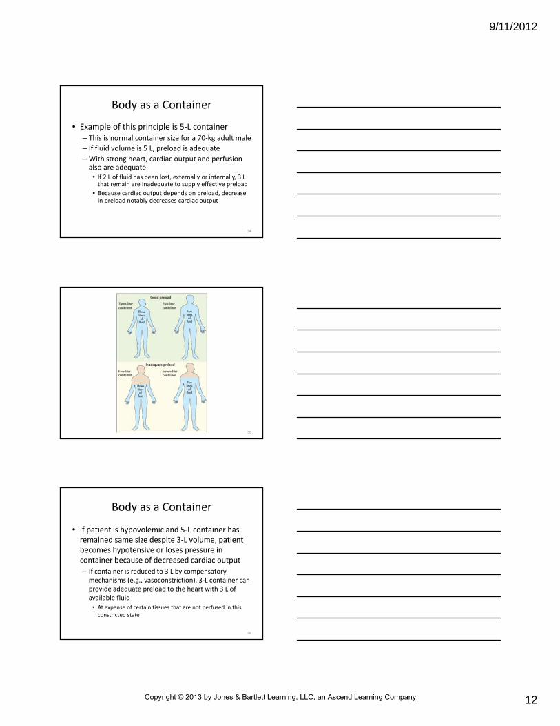
• Example of this principle is 5‐L container– This is normal container size for a 70‐kg adult male
– If fluid volume is 5 L, preload is adequate
– With strong heart, cardiac output and perfusion also are adequate
• If 2 L of fluid has been lost, externally or internally, 3 L that remain are inadequate to supply effective preload
• Because cardiac output depends on preload, decrease in preload notably decreases cardiac output
34
35
Body as a Container
• If patient is hypovolemic and 5‐L container has remained same size despite 3‐L volume, patient becomes hypotensive or loses pressure in container because of decreased cardiac output
– If container is reduced to 3 L by compensatory mechanisms (e.g., vasoconstriction), 3‐L container can provide adequate preload to the heart with 3 L of available fluid
• At expense of certain tissues that are not perfused in this constricted state
36
Copyright © 2013 by Jones & Bartlett Learning, LLC, an Ascend Learning Company
9/11/2012
13
Body as a Container
• If fluid is adequate for a 5‐L container but container size has been enlarged to 7 L by illness or injury
– Results in vasodilation: 5 L of fluid does not provide adequate preload for container (relative hypovolemia)
– Factors responsible for vasodilation
• Cardiac and BP medications
• Allergic reaction
• Heat‐ and cold‐related injuries
• Alcohol or other drug use
37
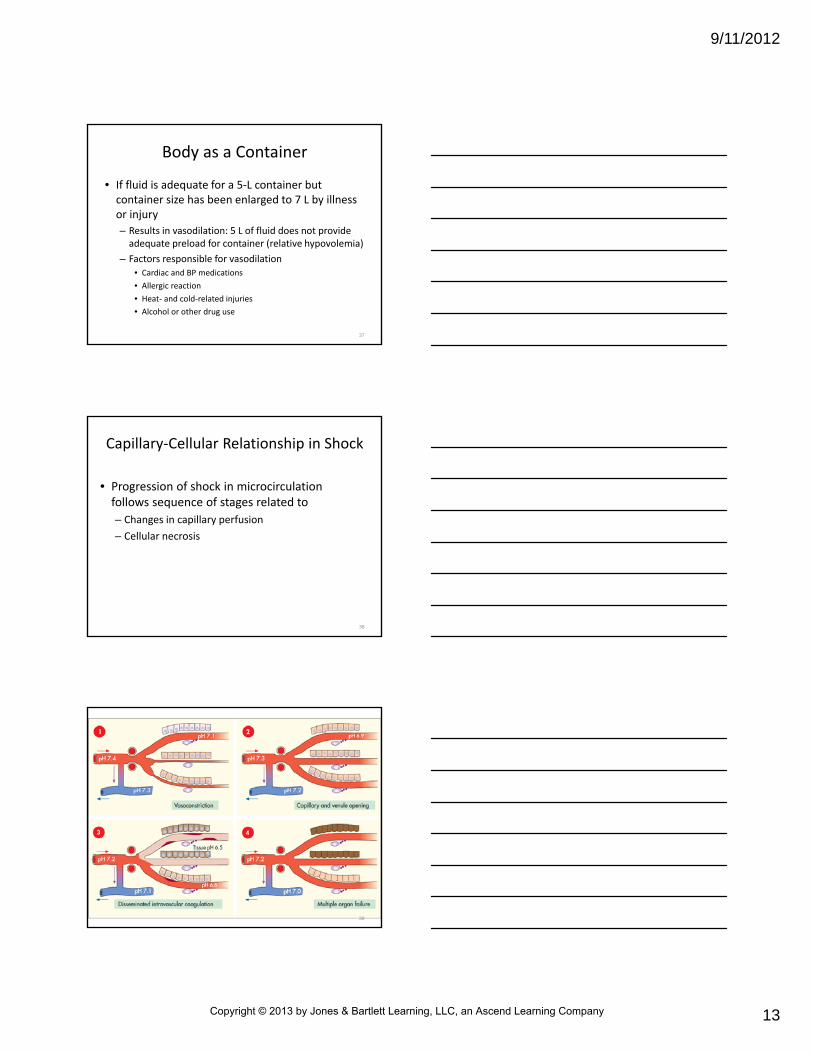
• Progression of shock in microcirculation follows sequence of stages related to
– Changes in capillary perfusion
– Cellular necrosis
Capillary‐Cellular Relationship in Shock
38
39
Copyright © 2013 by Jones & Bartlett Learning, LLC, an Ascend Learning Company
9/11/2012
14
Stage 1: Vasoconstriction
• In response to intravascular volume depletion (hypovolemia), precapillary arterioles and postcapillary venules constrict
– Constriction helps to maintain systemic BP
– Because of narrowing of entrance to microcirculation, velocity of blood passing through it increases
• Leads to increase in hydrostatic pressure in capillaries and allows fluid to be reabsorbed into circulation
• Fluid is reabsorbed as it shifts from extravascular space (transcapillary refill)
40
Stage 1: Vasoconstriction
• As shock progresses
– O2 and nutrient delivery to cells supplied by these capillaries decreases
– Anaerobic metabolism replaces aerobic metabolism
– Production of hydrogen ions and lactate increases
– Shortly thereafter, lining of capillaries can begin to lose ability to hold large molecules within capillary
– Capillary lining permits protein‐containing fluid to leak into interstitial spaces
• Known as leaky capillary syndrome
41
If this “leak” persists, what effect will it have on preload and cardiac
output?
42
Copyright © 2013 by Jones & Bartlett Learning, LLC, an Ascend Learning Company
9/11/2012
15
Stage 1: Vasoconstriction
• Arteriovenous (AV) shunts open, particularly in skin, kidneys, GI tract
– Shunts cause less flow to arterioles and thus less flow through capillaries
– Sympathetic stimulation produces
• Pale, sweaty skin
• Rapid, thready pulse (caused by hypovolemia and vasoconstriction)
• Elevation in blood glucose
43
Stage 1: Vasoconstriction
• Release of epinephrine dilates coronary, cerebral, and skeletal muscle arterioles and constricts other arterioles– As a result, blood is shunted to heart, brain, skeletal muscle
– Capillary flow to kidneys and abdominal organs decreases
– Vasoconstriction stage must be treated by prompt restoration of circulatory fluid volume
• Otherwise, shock progresses to next stage
44
Stage 2: Capillary and Venule Opening
• As shock progresses, precapillary sphincter relaxes
– Results in some expansion of vascular space
– Postcapillary sphincters resist relaxation effects
• Remain closed
• This causes blood to pool or stagnate in capillary system
• Capillaries become engorged with fluid
45
Copyright © 2013 by Jones & Bartlett Learning, LLC, an Ascend Learning Company
9/11/2012
16
• As shock progresses, precapillary sphincter relaxes
– Less blood flow caused by
• Arterial hypotension
• Secondary arteriolar vasoconstriction
• Opening of arteriovenous shunts
• Conditions also contribute to stagnation of blood flow in capillaries
Stage 2: Capillary and Venule Opening
46
• Vascular space expands greatly as increasing hypoxemia and acidosis lead to opening of more venules and capillaries– When occurs, even normal blood volume may be inadequate to fill container
– Capillary and venule capacity can increase to point that volume of available blood returning to great veins and venae cavae is reduced
• Results in decreased venous return and fall in cardiac output
Stage 2: Capillary and Venule Opening
47
• In addition, viscera (lungs, liver, kidneys, GI mucosa) can become congested with fluid
– Stagnant capillary flow caused by
• Low arterial BP
• Extremely constricted arterioles
• Presence of arteriovenous shunts
• Many open capillaries
Stage 2: Capillary and Venule Opening
48
Copyright © 2013 by Jones & Bartlett Learning, LLC, an Ascend Learning Company
9/11/2012
17
What happens to the function of the heart as acidosis increases?
49
• Sluggish blood flow and decrease in amount of O2 delivered to cell results in cell metabolism occurring without O2 (anaerobic metabolism)
• When tissue hypoxia is present
– Pyruvate oxidation decreases
– Lactate production increases
– ATP formation continues via glycolysis
• Results in metabolic acidosis
Stage 2: Capillary and Venule Opening
50
• Respiratory system attempts to compensate for acidosis by increasing ventilation to release CO2
– Produces partially compensated metabolic acidosis
– As acidosis increases and pH falls, red blood cells may cluster together
• Known as rouleaux formation
Stage 2: Capillary and Venule Opening
51
Copyright © 2013 by Jones & Bartlett Learning, LLC, an Ascend Learning Company
9/11/2012
18
• Rouleaux formation– Halts perfusion in vital organ capillaries
• Affects nutritional flow and prevents removal of waste products of metabolism
– Clotting mechanisms affected• Leads to hypercoagulability
• This stage of shock often advances to third stage if fluid resuscitation is inadequate or delayed– Also may progress if shock state is complicated by trauma or infection (sepsis)
Stage 2: Capillary and Venule Opening
52
Stage 3: Disseminated Intravascular Coagulation
• Stage 3 is resistant to treatment
– Refractory shock
– Still reversible early on with fluid replacement and support of vital functions
– Blood begins to coagulate in microcirculation, clogging capillaries
• Referred to as disseminated intravascular coagulation
– Lumps of red blood cells may occlude capillaries
53
• Occlusion
– Decreases capillary perfusion
– Prevents delivery of oxygenated substrates such as glucose
– Prevents removal of metabolites
• As a result, distal tissue cells switch to anaerobic metabolism, lactic acid production increases
Stage 3: Disseminated Intravascular Coagulation
54
Copyright © 2013 by Jones & Bartlett Learning, LLC, an Ascend Learning Company
9/11/2012
19
• As shock continues, lactic acid accumulates around cell
– Cell no longer has energy needed to maintain homeostasis, or balance to function normally
• Water and sodium leak into cell through cellular membrane
• Potassium leaks out
• Cells swell and die (known as washout phase)
Stage 3: Disseminated Intravascular Coagulation
55
• Microinfarcts (small areas of dead cells) develop in organs
– Microthrombi produce
• Capillary congestion
• Fluid leaks
• Rupture of cells
• Hemorrhage
Stage 3: Disseminated Intravascular Coagulation
56
• Pulmonary capillaries become permeable to fluid, which leads to pulmonary edema
– Edema decreases absorption of O2 and results in possible alterations in CO2 elimination
• Can lead to acute respiratory failure or adult respiratory distress syndrome
• If shock and disseminated intravascular coagulation continue, progresses to multiple organ failure
Stage 3: Disseminated Intravascular Coagulation
57
Copyright © 2013 by Jones & Bartlett Learning, LLC, an Ascend Learning Company
9/11/2012
20
Stage 4: Multiple Organ Failure
• Amount of cellular necrosis (death) required to produce organ failure varies with each organ– Depends on underlying condition of organ
• Usually hepatic failure occurs first
• Followed by renal failure and heart failure
– If any given area of capillary occlusion persists for more than 1 to 2 hours, cells nourished by that capillary undergo changes that rapidly become irreversible
58
Stage 4: Multiple Organ Failure
• In this stage, BP falls dramatically (to levels of 60 mm Hg or less)
– Even if BP is returned to normal after couple of hours, ability of cell to obtain energy from O2
through anaerobic metabolism fails
• Cell dies from inadequate capillary perfusion
• Inadequate tissue perfusion and cell death are results of irreversible shock
59
Stage 4: Multiple Organ Failure
• If cellular necrosis damages critical amount of a vital organ, organ soon fails
– Failure of liver and kidneys is common and often presents early in this stage
– Capillary blockage can cause heart failure
– GI bleeding and sepsis can result from GI mucosal necrosis
– Pancreatic necrosis can lead to further clotting disorders and severe pancreatitis
– Pulmonary thrombosis can produce hemorrhage and fluid loss into alveoli
• Can lead to death from respiratory failure
60
Copyright © 2013 by Jones & Bartlett Learning, LLC, an Ascend Learning Company
9/11/2012
21
Classifications of Shock
• Commonly classified based on initiating cause
– Two or more types often combined
– Underlying defect is inadequate tissue perfusion
61
Hypovolemic Shock
• In U.S., hypovolemic shock (shock that occurs from fluid loss) most often is caused by hemorrhage
– Can result from dehydration (commonly seen with severe diarrhea and vomiting)
– Loss of circulating volume occurs
62
Hypovolemic Shock
• Illnesses and injuries that can lead to hypovolemic shock
– Hemorrhage
– Burns
– Severe or prolonged diarrhea
– Vomiting
– Endocrine disorders
– Internal third space loss, as in peritonitis
63
Copyright © 2013 by Jones & Bartlett Learning, LLC, an Ascend Learning Company
9/11/2012
22
Hypovolemic Shock
• In addition to loss of circulating volume, tissue injury resulting from trauma can worsen shock
– Tissue injury causes microemboli and further activates inflammatory and coagulation systems
64
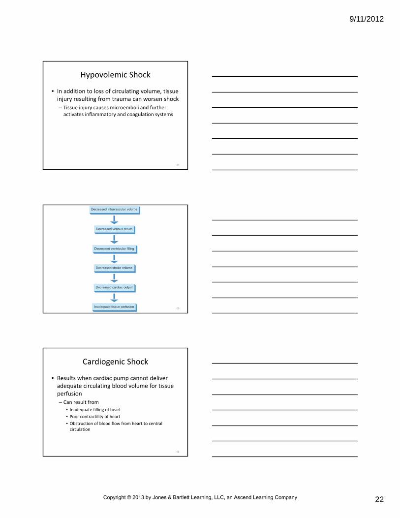
65
Cardiogenic Shock
• Results when cardiac pump cannot deliver adequate circulating blood volume for tissue perfusion
– Can result from
• Inadequate filling of heart
• Poor contractility of heart
• Obstruction of blood flow from heart to central circulation
66
Copyright © 2013 by Jones & Bartlett Learning, LLC, an Ascend Learning Company
9/11/2012
23
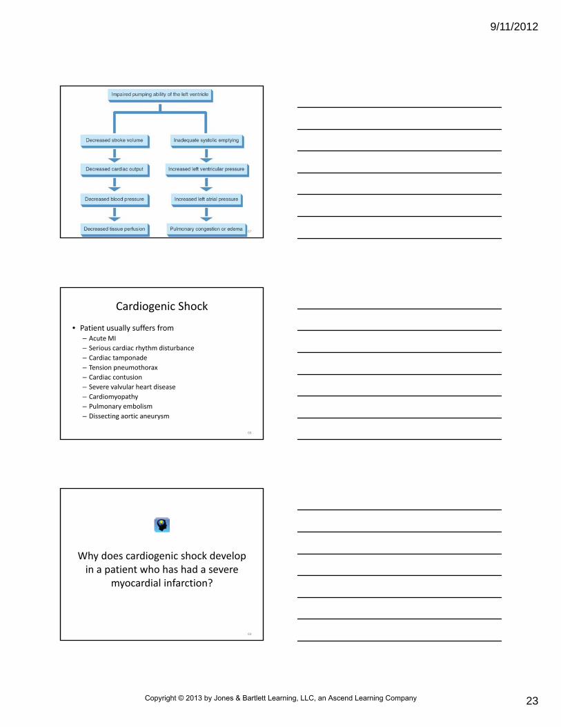
67
Cardiogenic Shock
• Patient usually suffers from– Acute MI
– Serious cardiac rhythm disturbance
– Cardiac tamponade
– Tension pneumothorax
– Cardiac contusion
– Severe valvular heart disease
– Cardiomyopathy
– Pulmonary embolism
– Dissecting aortic aneurysm
68
Why does cardiogenic shock develop in a patient who has had a severe
myocardial infarction?
69
Copyright © 2013 by Jones & Bartlett Learning, LLC, an Ascend Learning Company
9/11/2012
24
Neurogenic Shock
• Also known as spinal cord, distributive, or vasogenic shock
– Results from vasomotor paralysis below level of injury
– Normal vasomotor tone through sympathetic nervous system control is lost
• Results in decrease in peripheral vascular resistance
70
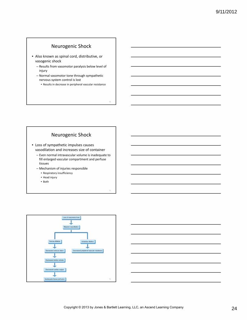
Neurogenic Shock
• Loss of sympathetic impulses causes vasodilation and increases size of container
– Even normal intravascular volume is inadequate to fill enlarged vascular compartment and perfuse tissues
– Mechanism of injuries responsible
• Respiratory insufficiency
• Head injury
• Both
71
72
Copyright © 2013 by Jones & Bartlett Learning, LLC, an Ascend Learning Company
9/11/2012
25
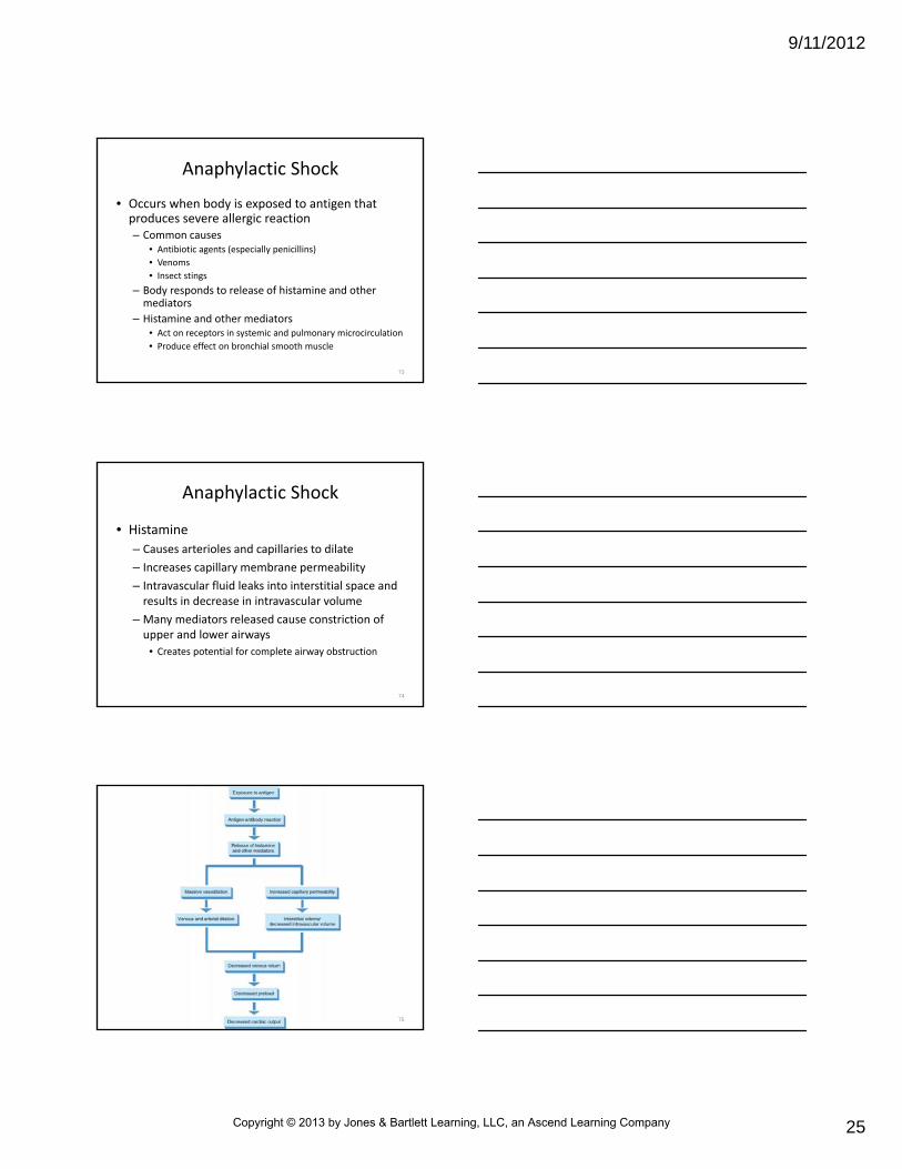
Anaphylactic Shock
• Occurs when body is exposed to antigen that produces severe allergic reaction– Common causes
• Antibiotic agents (especially penicillins)
• Venoms
• Insect stings
– Body responds to release of histamine and other mediators
– Histamine and other mediators• Act on receptors in systemic and pulmonary microcirculation
• Produce effect on bronchial smooth muscle
73
• Histamine
– Causes arterioles and capillaries to dilate
– Increases capillary membrane permeability
– Intravascular fluid leaks into interstitial space and results in decrease in intravascular volume
– Many mediators released cause constriction of upper and lower airways
• Creates potential for complete airway obstruction
Anaphylactic Shock
74
75
Copyright © 2013 by Jones & Bartlett Learning, LLC, an Ascend Learning Company
9/11/2012
26
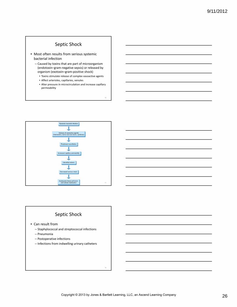
Septic Shock
• Most often results from serious systemic bacterial infection
– Caused by toxins that are part of microorganism (endotoxin–gram‐negative sepsis) or released by organism (exotoxin–gram‐positive shock)
• Toxins stimulate release of complex vasoactive agents
• Affect arterioles, capillaries, venules
• Alter pressure in microcirculation and increase capillary permeability
76
77
Septic Shock
• Can result from
– Staphylococcal and streptococcal infections
– Pneumonia
– Postoperative infections
– Infections from indwelling urinary catheters
78
Copyright © 2013 by Jones & Bartlett Learning, LLC, an Ascend Learning Company
9/11/2012
27
Septic Shock
• Between 40,000 and 100,000 persons develop septic shock each year
– Leading cause of death in intensive care units
– Most often occurs in
• Older adults (particularly nursing home residents)
• Alcoholics
• Neonates
• Patients who are immunosuppressed
79
Stage of Shock
• Degree of hypoperfusion and anaerobic metabolism can be categorized by stages in the response of body to shock syndrome
• Stages
– Compensated shock
– Uncompensated (or decompensated) shock
– Irreversible shock
80
Compensated Shock
• Associated with some decreased blood flow and perfusion to tissues
– Compensatory responses of body can overcome decrease in available fluid
– Increase in catecholamine production maintains cardiac output and normal systolic BP
81
Copyright © 2013 by Jones & Bartlett Learning, LLC, an Ascend Learning Company
9/11/2012
28
82
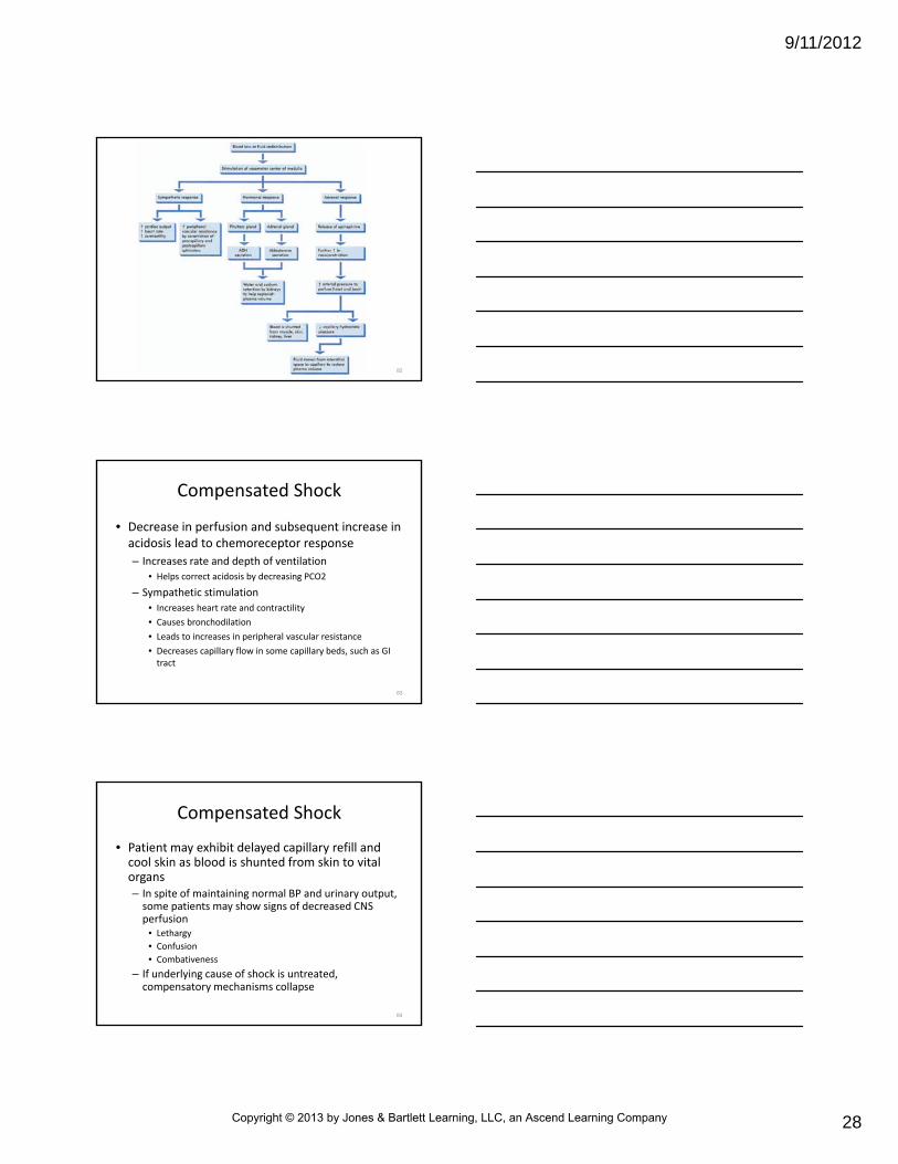
Compensated Shock
• Decrease in perfusion and subsequent increase in acidosis lead to chemoreceptor response
– Increases rate and depth of ventilation
• Helps correct acidosis by decreasing PCO2
– Sympathetic stimulation
• Increases heart rate and contractility
• Causes bronchodilation
• Leads to increases in peripheral vascular resistance
• Decreases capillary flow in some capillary beds, such as GI tract
83
Compensated Shock
• Patient may exhibit delayed capillary refill and cool skin as blood is shunted from skin to vital organs– In spite of maintaining normal BP and urinary output, some patients may show signs of decreased CNS perfusion
• Lethargy
• Confusion
• Combativeness
– If underlying cause of shock is untreated, compensatory mechanisms collapse
84
Copyright © 2013 by Jones & Bartlett Learning, LLC, an Ascend Learning Company
9/11/2012
29
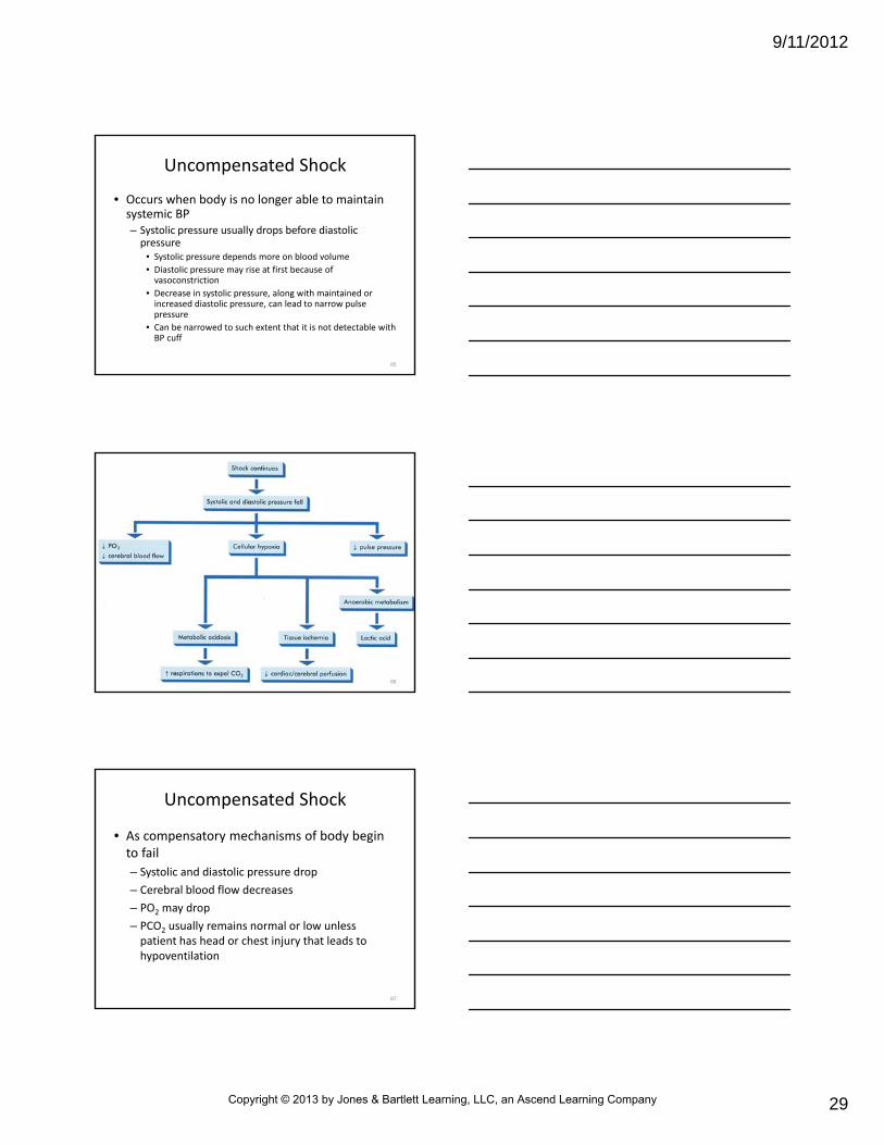
Uncompensated Shock
• Occurs when body is no longer able to maintain systemic BP– Systolic pressure usually drops before diastolic pressure
• Systolic pressure depends more on blood volume
• Diastolic pressure may rise at first because of vasoconstriction
• Decrease in systolic pressure, along with maintained or increased diastolic pressure, can lead to narrow pulse pressure
• Can be narrowed to such extent that it is not detectable with BP cuff
85
86
Uncompensated Shock
• As compensatory mechanisms of body begin to fail
– Systolic and diastolic pressure drop
– Cerebral blood flow decreases
– PO2 may drop
– PCO2 usually remains normal or low unless patient has head or chest injury that leads to hypoventilation
87
Copyright © 2013 by Jones & Bartlett Learning, LLC, an Ascend Learning Company
9/11/2012
30
Uncompensated Shock
• Clinical signs
– Hypotension
– Tachycardia
– Tachypnea
– Delayed capillary refill
– Decreased urinary output
88
Uncompensated Shock
• Shunting of blood and tissue hypoxia may cause patient to have cold extremities and cyanosis
– Effects on cardiovascular system
• Decreased preload
• Increased rate of contraction caused by catecholamine stimulation
• Myocardial contractions initially can be stronger as result of catecholamine release
89
Uncompensated Shock
• In latter phases, myocardial strength may decrease as result of following factors
– Ischemia can result from
• Reduction of circulating red blood cells
• Lower PO2
• Decreased coronary perfusion because of hypotension (especially diastolic hypotension)
– Cardiodepressant substances can depress heart function in late shock
90
Copyright © 2013 by Jones & Bartlett Learning, LLC, an Ascend Learning Company
9/11/2012
31
Uncompensated Shock
• In latter phases, myocardial strength may decrease as result of following factors– Necrosis of myocardium (essentially simulating myocardial infarction) can result from associated ischemia
– Decreased preload can lead to decreased contractility
– Acidosis can lead to decreased contractility
– Cardiac rhythm disturbances can result from hypoxia
91
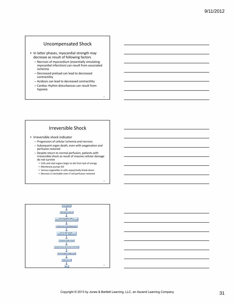
Irreversible Shock
• Irreversible shock indicator– Progression of cellular ischemia and necrosis
– Subsequent organ death, even with oxygenation and perfusion restored
– Despite return to normal perfusion, patients with irreversible shock as result of massive cellular damage do not survive
• Cells and vital organs begin to die from lack of energy
• Membrane pumps fail
• Various organelles in cells sequentially break down
• Necrosis is inevitable even if cell perfusion restored
92
93
Copyright © 2013 by Jones & Bartlett Learning, LLC, an Ascend Learning Company
9/11/2012
32
Irreversible Shock
• Decompensation may occur suddenly or may be delayed from 1 day to 3 weeks after onset of shock– Clinical signs of irreversible shock
• Bradycardia
• Serious dysrhythmias
• Frank hypotension
• Evidence of multiple organ failure
• Pale, cold, clammy skin
– Cardiopulmonary collapse usually is imminent
94
Variations in Physiological Response to Shock
• Many variations in physiological response occur among patients who are in shock
• Determining factors
– Age
• Older adults are less able to compensate
• Children compensate longer but deteriorate faster
95
Variations in Physiological Response to Shock
• Determining factors
– General health
• Preexisting disease
• Other injuries
– Ability to activate compensatory mechanisms
– Medications, some of which can interfere with compensatory mechanisms
– Specific organ system(s) affected
96
Copyright © 2013 by Jones & Bartlett Learning, LLC, an Ascend Learning Company
9/11/2012
33
What diseases can influence a patient’s response to shock?
97
Management and Treatment Plan
• Focuses on assessment of oxygenation and perfusion of body organs
• Goals of treatment
– Ensure patent airway
– Provide adequate oxygenation and ventilation
– Restore perfusion
98
Primary Survey
• Can help to identify whether cell perfusion is adequate– Five‐step description of primary survey focuses on evaluating shock victim
• Be aware of common objectives in evaluating any patient with other types of serious illness or injury
• Shock evaluation– Airway
• Must be opened
• Patency must be maintained
99
Copyright © 2013 by Jones & Bartlett Learning, LLC, an Ascend Learning Company
9/11/2012
34
Primary Survey
• Shock evaluation
– Breathing
• Respiratory pattern often reflects adequacy of ventilation
• Can offer clues to presence of shock
• Closely monitor pulse oximetry
100
Primary Survey
• Shock evaluation– Circulation
• Assess patient’s circulatory status
• Check for any uncontrolled arterial bleeding
• In cases of external hemorrhage, apply direct pressure
• If direct pressure does not immediately control vigorous bleeding in extremity, tourniquet should be applied
• Pressure dressings can be applied to maintain control of hemorrhage
• If suspected internal bleeding, after securing airway and ensuring adequate ventilation, rapid transport is highest priority
101
Primary Survey
• Internal bleeding should be suspected in any trauma patient with signs of shock
– Especially trauma patients without evidence of external blood loss
– Treatment must be directed at definitive care to stop bleeding
• Rapid transport to proper facility is critical
• IV fluid therapy, if initiated in field, should be performed en route to avoid delay of definitive care
102
Copyright © 2013 by Jones & Bartlett Learning, LLC, an Ascend Learning Company
9/11/2012
35
Consider a patient with early signs and symptoms of shock. However, the patient’s SaO2 reading is normal.
Should you administer oxygen?
103
Primary Survey
• Evaluate rate, character, location of pulse as part of circulatory assessment
– Pulse rates increase fairly early in shock
• This helps maintain adequate cardiac output
– Strength of contraction also may increase
– These attempts to maintain cardiac output may be negated by decrease in preload
104
Primary Survey
• Tachycardia usually will not occur until patient has suffered 10 to 15 percent volume depletion (relative to container size) as result of blood loss or increase in container size
– Character of pulse can be strong or weak
– Strength of pulse provides an estimate of filling volume of artery being palpated and indirect measurement of systolic pressure
105
Copyright © 2013 by Jones & Bartlett Learning, LLC, an Ascend Learning Company
9/11/2012
36
Primary Survey
• Tissue perfusion sometimes can be estimated by evaluating color, moisture, temperature of skin
– Can be unreliable in
• Older patients
• Those who have been exposed to extremes of temperature
• Those suffering from septicemia and shock caused by neurological injury
106
Primary Survey
• Evaluation of fingers and toes (most distal points of circulation) is crucial
– Can be the first to show signs of inadequate tissue perfusion (cyanosis, cool skin)
– If ambient temperatures are moderate and tissue perfusion is adequate, will be pink, warm, dry
107
Primary Survey
• Capillary refill test can offer useful details on pediatric patient’s tissue perfusion
– Use only as guide
– Accuracy can be affected by environment and patient’s general health, age, gender
108
Copyright © 2013 by Jones & Bartlett Learning, LLC, an Ascend Learning Company
9/11/2012
37
Primary Survey
• Disability
– Evaluation of patient’s level of consciousness is crucial in assessing cerebral oxygenation
– Can become restless, agitated, confused as cerebral ischemia develops
– In addition to shock, cerebral edema and intracranial hemorrhage from head injury can compromise cerebral perfusion
109
Primary Survey
• Any significant change in patient’s sensoriumshould be considered indicator of critical perfusion deficit
– True whether decrease in cerebral circulation is from shock or from increase in intracranial pressure
– Measure patient’s level of consciousness with AVPU scale or other evaluation methods
110
Primary Survey
• Exposure of body surfaces
– Only as indicated by scenario or mechanism of injury
– Visual inspection can reveal conditions that may be life threatening
• Can be hidden by clothing
111
Copyright © 2013 by Jones & Bartlett Learning, LLC, an Ascend Learning Company
9/11/2012
38
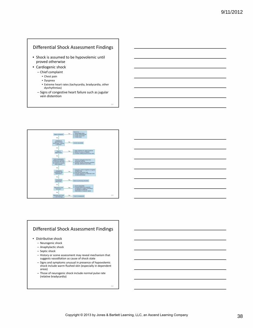
Differential Shock Assessment Findings
• Shock is assumed to be hypovolemic until proved otherwise
• Cardiogenic shock– Chief complaint
• Chest pain
• Dyspnea
• Extreme heart rates (tachycardia, bradycardia, other dysrhythmias)
– Signs of congestive heart failure such as jugular vein distention
112
113
Differential Shock Assessment Findings
• Distributive shock– Neurogenic shock
– Anaphylactic shock
– Septic shock
– History or scene assessment may reveal mechanism that suggests vasodilation as cause of shock state
– Signs and symptoms unusual in presence of hypovolemicshock include warm flushed skin (especially in dependent areas)
– Those of neurogenic shock include normal pulse rate (relative bradycardia)
114
Copyright © 2013 by Jones & Bartlett Learning, LLC, an Ascend Learning Company
9/11/2012
39
Differential Shock Assessment Findings
• Obstructive shock (caused by obstruction to blood flow)
– Often are victims of major chest injury (usually penetrating type of injury)
– History consistent with pulmonary embolism
– Patients with cardiac tamponade or tension pneumothoraxoften have jugular vein distention
– Patients with tension pneumothorax almost always have decreased breath sounds on affected side
115
Detailed Physical Examination
• Systematic approach offers way to evaluate potentially life‐threatening conditions and allows paramedic to further assess patient’s perfusion status
– Begin with baseline measurements of vital signs
– Evaluate patient’s ECG
116
Can blood donation cause a fluid deficit large enough to cause shock?
If so, how is that fluid deficit managed?
117
Copyright © 2013 by Jones & Bartlett Learning, LLC, an Ascend Learning Company
9/11/2012
40
Detailed Physical Examination
• Expect pulse rate to increase above normal limits after fluid volume drops 10 to 15 percent
– Some continue to have normal pulse rates even though volume deficit of this extent exists
– Patient’s pulse rate should be only one factor in evaluating patient’s level of perfusion
118
Detailed Physical Examination
• Bradycardia can result from
– Hypoxemia
– Existing neurological injury
– Increased vagal tone
– Preexisting illness
– Prior medication use
119
Detailed Physical Examination
• Can indicate severe myocardial ischemia (primary cause of cardiogenic shock)
• Bradycardic rhythms often occur just before cardiac arrest
– If rhythm is bradycardic, optimize oxygenation by increasing fraction of inspired oxygen (FiO2) and by assisting ventilations if needed
120
Copyright © 2013 by Jones & Bartlett Learning, LLC, an Ascend Learning Company
9/11/2012
41
Detailed Physical Examination
• Diastolic pressure– At first rises as peripheral vascular resistance increases with increased vascular tone
– Changes decrease container size
– Blood is shunted away selectively from certain portions of body
– When heart can no longer pump blood to keep container full on arterial side, diastolic pressure begins to drop
• Expect this when blood loss is greater than 20 to 25 percent of normal circulating blood volume
121
Detailed Physical Examination
• Systolic pressure
– Falls when heart can no longer pump enough blood to fill container at end of cardiac contraction
– Usually is more sensitive to volume depletion than is diastolic pressure
• Systolic pressure drops first
• As fluid deficit approaches 25 percent, systolic and diastolic pressures both begin to drop
122
Detailed Physical Examination
• Consider evaluation of orthostatic vital signs in conscious patients suspected of having lost circulating blood volume– Should only be performed in absence of suspected spinal injury or another condition that precludes this assessment
– Indicators of significant (at least 10 percent) volume depletion (postural hypotension) and decrease in perfusion status
• Rise from recumbent position to a sitting or standing position associated with fall in systolic pressure (after 1 minute) of 10 to 15 mm Hg
• Concurrent rise in pulse rate (after 1 minute) of 10 to 15 bpm
123
Copyright © 2013 by Jones & Bartlett Learning, LLC, an Ascend Learning Company
9/11/2012
42
Detailed Physical Examination
• Fluid deficit still can exist even after systolic pressure returns to normal following fluid replacement
– Continuing IV fluids after indicators of adequate tissue perfusion are present is controversial
• Improved skin color
• Capillary refill of less than 2 seconds in pediatric patients
• Normal pulse oximetry readings
– Aggressive fluid resuscitation can result in hemodilution(diluting blood of elements), disruption of clots, and renewed hemorrhage
124
Detailed Physical Examination
• With suspected internal hemorrhage in their chest, abdomen, or pelvis should have fluids titrated to maintain systolic BP of 90 mm Hg (MAP of 60 to 65 mm Hg)
• Permissive hypotension can be protective and may prevent further blood loss
• Follow local protocol established by medical direction
125
Resuscitation
• Resuscitation of shock victim is aimed at restoring adequate peripheral tissue oxygenation as quickly as possible
– Ensure adequate oxygenation
– Maintain effective ratio of volume to container size
– Rapidly transport victim to appropriate medical facility
126
Copyright © 2013 by Jones & Bartlett Learning, LLC, an Ascend Learning Company
9/11/2012
43
Red Blood Cell Oxygenation
• Adequate oxygenation of red blood cells is required for adequate tissue oxygenation
– For RBC oxygenation to be adequate, must have patent airway
– Ventilation must be supported with high fraction of inspired O2
– If needed, assist ventilation with positive pressure
127
Red Blood Cell Oxygenation
• Adequate oxygenation of red blood cells is required for adequate tissue oxygenation
– Any abnormality that interferes with adequate ventilation should be corrected if possible
• Obstructed airway
• Pneumothorax
• Hemothorax
• Open chest wound
• Unstable chest wall
128
• Second component necessary to maintain adequate oxygen‐carrying capacity requires container be full of fluid
– Achieve this by decreasing size of container
– Especially the case in shock states not associated with hemorrhage
– In some cases of distributive shock, vasoconstricting drugs can be used to manage when reduction of container size is main concern
Ratio of Volume to Container Size
129
Copyright © 2013 by Jones & Bartlett Learning, LLC, an Ascend Learning Company
9/11/2012
44
• Volume replacement also may be necessary in these patients
• Vasoconstricting drugs are not recommended to treat patients in hypovolemic shock until fluid volume replacement is complete
• Complete volume replacement rarely occurs in prehospital setting
Ratio of Volume to Container Size
130
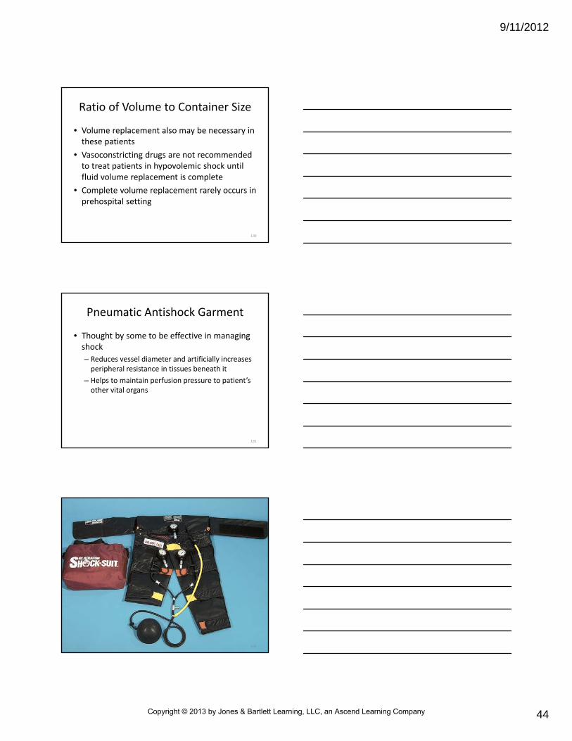
Pneumatic Antishock Garment
• Thought by some to be effective in managing shock
– Reduces vessel diameter and artificially increases peripheral resistance in tissues beneath it
– Helps to maintain perfusion pressure to patient’s other vital organs
131
132
Copyright © 2013 by Jones & Bartlett Learning, LLC, an Ascend Learning Company
9/11/2012
45
Pneumatic Antishock Garment
• Thought by some to be effective in managing shock
– Can arrest hemorrhage by tamponading any bleeding vessels in abdomen, pelvis, or lower extremities
– Can help to stabilize pelvic and lower‐extremity fractures when it is inflated
– Decreases movement and subsequent blood loss
133
Pneumatic Antishock Garment
• Decisions to use are left to local protocol and medical direction
– Contraindications
• Pulmonary edema
• Cardiogenic shock
• Ruptured diaphragm
• Hemorrhage within chest cavity
134
Pneumatic Antishock Garment
• Some medical direction authorities feel it is contraindicated in following situations
– Impaled objects in the abdomen
• Precluding inflation of abdominal section of garment
– Advanced pregnancy
• Third trimester pregnancy precludes inflation of abdominal compartment
– Evisceration
• No inflation of abdominal compartment
135
Copyright © 2013 by Jones & Bartlett Learning, LLC, an Ascend Learning Company
9/11/2012
46
General Pneumatic AntishockGarment Guidelines
• Apply when indicated after lower extremities and abdomen have been inspected for major wounds
– Must always position below level of patient’s lowest rib
– Patient’s BP and lung sounds should be monitored before, during, and after inflation
136
General Pneumatic AntishockGarment Guidelines
• Inflation is stopped when adequate BP is obtained
– Compartments are inflated before or with abdominal compartment
– Never inflate abdominal section before inflating leg sections
• Can cause abdominal compartment to act as constrictive band that reduces venous return from legs
137
Consider that the abdominal compartment of the pneumatic
antishock garment is inflated mistakenly before the leg segments. What effect can this have on cardiac output?
138
Copyright © 2013 by Jones & Bartlett Learning, LLC, an Ascend Learning Company
9/11/2012
47
General Pneumatic AntishockGarment Guidelines
• After inflation, garment should seldom if ever be deflated in prehospital setting
– Only with physician’s direction
– Monitor patient closely during deflation process
– Removal of garment before fluid replacement commonly results in rapid fall of BP and cardiac output, which can lead to cardiac arrest
139
General Pneumatic AntishockGarment Guidelines
• Changes in temperature and atmospheric pressure can cause notable change in pressure within PASG
– Monitor patient constantly when patient is moved from cold environment to warm one or when transported by air
140
General Pneumatic AntishockGarment Guidelines
• Relationship among temperature, atmospheric pressure, pressure within PASG is as follows
– Rise in temperature raises pressure within PASG
• Fall in temperature decreases pressure
– Fall in atmospheric pressure causes increase in PASG pressure
• Rise in atmospheric pressure produces decrease in garment pressure
141
Copyright © 2013 by Jones & Bartlett Learning, LLC, an Ascend Learning Company
9/11/2012
48
General Pneumatic AntishockGarment Guidelines
• PASG is not without complications even with appropriate use
• Sustained inflation of garment for more than 1 to 2 hours can lead to
– Decreased tissue perfusion
– Ischemia of underlying tissues
– Loss of limb, even without underlying fracture
142
Fluid Resuscitation in Shock
• Almost every shock victim requires volume expanders as part of resuscitation
– Except for patients in cardiogenic shock
• Selection of intravenous (IV) fluids for initial volume replacement varies according to medical direction
– Most common emergency requiring fluid replacement is loss of volume caused by hemorrhage or dehydration
143
Fluid Resuscitation in Shock
• Type of needed fluid replacement depends on nature and extent of volume loss
– Two main categories of fluids used in resuscitation
• Crystalloids
• Colloids
– Follow recommendations for fluid resuscitation provided by medical direction
144
Copyright © 2013 by Jones & Bartlett Learning, LLC, an Ascend Learning Company
9/11/2012
49
Crystalloids
• Created by dissolving crystals such as salts and sugars in water
– Do not have as much osmotic pressure as colloid solutions
– Expected to equilibrate more quickly between vascular and extravascular spaces
145
Crystalloids
• Created by dissolving crystals such as salts and sugars in water
– 2/3 of infused crystalloid fluid leaves vascular space within 1 hour
• 3 mL is needed to replace 1 mL of blood
– Examples
• Lactated Ringer’s solution
• Normal saline
• Glucose solutions in water
146
Crystalloids
• Hypertonic solutions
– Have higher osmotic pressure than that of body cells
– Include 5% dextrose in 0.9% sodium chloride, 7.5% saline, and 5% dextrose in 0.45% sodium chloride
– Hypotonic solutions have lower osmotic pressure than that of body cells
• Distilled water and 0.45% sodium chloride
147
Copyright © 2013 by Jones & Bartlett Learning, LLC, an Ascend Learning Company
9/11/2012
50
Crystalloids
• Lactated Ringer’s solution– Fluid of choice for resuscitating patients
– Solution is well balanced and contains many chemicals found in human blood
– Lactated Ringer’s solution contains
• Sodium chloride
• Small amounts of potassium and calcium
• 28 mEq of lactate, which can act as buffer to neutralize acidity when metabolized by liver
• 1/3 of infused solution remains in vascular space after 1 hour
148
Crystalloids
• Normal saline
– Contains 154 mEq/L of sodium
– Has no buffering capabilities
– Higher chloride content of normal saline is less desirable than more balanced lactated Ringer’s solution
– 1/3 of infused normal saline remains in vascular space after 1 hour
• Makes equally effective volume expander
– Follow local protocol when choosing IV fluids
149
Crystalloids
• Glucose‐containing solutions (e.g., 5% dextrose in water)
– Have immediate volume expansion effects
– Glucose leaves intravascular compartment rapidly with resultant free water increase
– Volume‐replacement benefits only last 5 to 10 minutes while glucose is metabolized
• Use of 5% dextrose in water should not be used to replace volume deficit
• Most often used to maintain vascular access for administration of IV medications
150
Copyright © 2013 by Jones & Bartlett Learning, LLC, an Ascend Learning Company
9/11/2012
51
Colloids
• Contain molecules (usually protein) too large to pass through capillary membrane– Exhibit osmotic pressure
– Remain within vascular compartment for considerable time
– Examples• Whole blood
• Packed red blood cells
• Blood plasma
• Plasma substitutes
151
Colloids
• Whole blood replacement is rarely given in U.S. for management of shock and is usually unavailable in emergency department
– Packed red cells are transfused, other blood components are transfused as necessary
• Packed RBC have volume of hemoglobin per unit that is almost twice that of whole blood
• Because there is no plasma in packed red cells, circulatory overload is less likely and transfusion reactions are less frequent
152
Colloids
• Type and crossmatch should be obtained when possible before patient is given blood products to determine patient’s ABO group and Rh type
– Will determine whether other antibodies are present that may cause transfusion reaction
– Type‐specific blood should be used for resuscitation when patient’s condition and time permits
153
Copyright © 2013 by Jones & Bartlett Learning, LLC, an Ascend Learning Company
9/11/2012
52
Colloids
• Uncrossmatched blood is usually given immediately for patients with hypotension and uncontrolled hemorrhage– Group O universal donor blood does not have A or B antigens on their surface
• Not agglutinated by anti‐A or anti‐B antibodies
– O‐negative blood is used for women of childbearing age who are at risk for Rhcomplications with future pregnancies
– O‐positive blood is used in all other patients
154
Colloids
• Blood plasma may be given without concern for ABO compatibility– Blood plasma contains
• Fibrinogen• Albumin• Gamma globulins• Hemagglutinins (agglutinin that clumps red blood corpuscles)
• Prothrombin (chemical that is part of clotting cascade)• Other clotting factors• Sugar• Salts
155
Colloids
• Blood plasma
– Sometimes used to restore effective blood volume in circulatory failure associated with
• Burns
• Traumatic shock
• Hemorrhage
– Blood plasma more commonly used to correct clotting deficiencies
• Often supplied as fresh frozen plasma
156
Copyright © 2013 by Jones & Bartlett Learning, LLC, an Ascend Learning Company
9/11/2012
53
Colloids
• Plasma substitutes– Do not increase oxygen‐carrying capacity by replacing RBCs
– Do not improve clotting by addition of plasma protein
– Used to restore circulating blood volume as emergency treatment for hypovolemia caused by blood loss
– Plasma substitutes such as dextran and hetastarchhave osmotic properties similar to those of plasma
– Stay in intravascular space longer than crystalloid solution
157
Colloids
• Plasma substitutes– Do not carry HIV or hepatitis viruses
– Do not require type and crossmatching before administration
– Readily available
– Have some adverse effects, including increased bleeding tendencies and immune suppression
– Emergency vehicles can carry plasma substitutes• Expense and storage issues make them impractical for general use in prehospital setting
158
Theory of Fluid Flow
• Flow of fluid through catheter is related directly to its diameter (to 4th power) and inversely related to its length
– Catheter with large diameter has a much greater flow than catheter with small diameter
– Short catheters provide faster flow rates than longer catheters of equal diameter
159
Copyright © 2013 by Jones & Bartlett Learning, LLC, an Ascend Learning Company
9/11/2012
54
Theory of Fluid Flow
• Other factors that affect the flow of fluid– Diameter and length of tubing
– Size of vein
– Height of fluid bag
– Viscosity and temperature of IV fluid• Temperature affects viscosity
• Warm fluids generally flow better than cold ones
• Pressure bags available that pressurize IV system to 300 mm Hg to maximize rate of fluid administration
160
Theory of Fluid Flow
• When aggressive fluid resuscitation is indicated
– Use short, large‐diameter catheters
– Use warm fluids of low viscosity (if possible)
– Keep tubing short, and pressurize IV system
161
Aside from flow, what other advantages do warmed fluids offer
for the patient in shock who requires a large‐volume fluid bolus?
162
Copyright © 2013 by Jones & Bartlett Learning, LLC, an Ascend Learning Company
9/11/2012
55
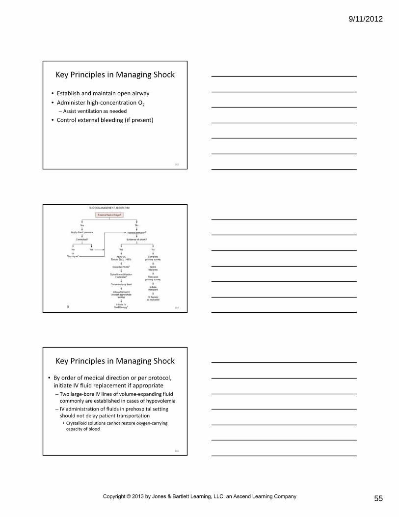
• Establish and maintain open airway
• Administer high‐concentration O2
– Assist ventilation as needed
• Control external bleeding (if present)
Key Principles in Managing Shock
163
164
• By order of medical direction or per protocol, initiate IV fluid replacement if appropriate
– Two large‐bore IV lines of volume‐expanding fluid commonly are established in cases of hypovolemia
– IV administration of fluids in prehospital setting should not delay patient transportation
• Crystalloid solutions cannot restore oxygen‐carrying capacity of blood
Key Principles in Managing Shock
165
Copyright © 2013 by Jones & Bartlett Learning, LLC, an Ascend Learning Company
9/11/2012
56
• By order of medical direction or per protocol, initiate IV fluid replacement if appropriate
– Best served by
• Rapid assessment
• Airway stabilization
• Immobilization
• Rapid transportation to appropriate medical facility
• Many EMS authorities recommend that IV therapy for shock resuscitation be initiated en route to hospital
Key Principles in Managing Shock
166
• Consider use of PASG (per protocol)
– Indications
• Transportation time is long
• Pelvic fractures are suspected
• Patient is deteriorating despite IV therapy
• Maintain patient’s normal body temperature
– Patients in shock often are unable to conserve body heat
– Can become hypothermic easily
Key Principles in Managing Shock
167
• In absence of spine or head injury and if hypovolemia is suspected and ventilation is adequate– Consider positioning patient in modified Trendelenburg position
• Legs elevated 15 to 18 inches
• Monitor cardiac rhythm and O2 saturation
• Frequently reassess vital signs en route to emergency department
Key Principles in Managing Shock
168
Copyright © 2013 by Jones & Bartlett Learning, LLC, an Ascend Learning Company
9/11/2012
57
Hypovolemic Shock
• Management
– Not considered complete until volume is replaced and cause of shock are corrected
– Crystalloid fluid replacement in cases of simple dehydration
– Volume replacement because of
• Hemorrhage
• Definitive surgery
• Critical care support
• Postoperative rehabilitation
– Fluid amount replaced in trauma is controversial, should be guided by medical direction
169
Hypovolemic Shock
• Stable trauma patients should not receive aggressive fluid resuscitation
– Volume given to trauma patients depends on type of trauma and patient’s condition
• Large volumes of fluid to maintain a systolic BP ≥ 90 mm Hg (MAP 60 to 65 mm Hg) should only be given to patients with isolated head or extremity injuries
• Aggressive fluid resuscitation may increase blood loss and can delay arrival to surgical care at trauma center
170
Cardiogenic Shock
• Management focuses on improving pumping action of heart and on managing cardiac rhythm irregularities
– Initiate fluid resuscitation in adult with 100 to 200 mL of volume‐expanding fluid
– Fluid resuscitation should be initiated as long as patient has no crackles in lung fields that would indicate pulmonary edema
171
Copyright © 2013 by Jones & Bartlett Learning, LLC, an Ascend Learning Company
9/11/2012
58
Cardiogenic Shock
• If patient improves, fluid therapy should be continued cautiously
– Continue until BP stabilizes and pulse rate decreases
– Assess lung sounds often
– If patient shows signs of increased lung congestion, adjust rate of infusion to keep vein open
172
Cardiogenic Shock
• Drug therapy varies according to cause– Can include
• Vasopressors
• Vasodilators
• Inotropic drugs
• Antidysrhythmics (usually after fluid infusion)
– Patients with cardiogenic shock caused by MI or infarction require reperfusion strategies (clot busting drugs or surgery) and possible circulatory support
– Paramedic must manage obstructive causes of cardiogenicshock immediately, including tension pneumothorax and cardiac tamponade
173
Neurogenic Shock
• Management is similar to management for hypovolemia
– Take care during fluid therapy to avoid circulatory overload
– Throughout resuscitation phase, monitor patient’s lung sounds closely for signs of pulmonary congestion
– Patients may respond to administration of vasopressors
174
Copyright © 2013 by Jones & Bartlett Learning, LLC, an Ascend Learning Company
9/11/2012
59
Anaphylactic Shock
• Treatment
– Intramuscular administration of epinephrine
– IV or intramuscular administration of antihistamines such as diphenhydramine
– Bronchodilators to treat bronchospasm that persist after administration of epinephrine
– Steroids used to reduce inflammatory response
175
Anaphylactic Shock
• Crystalloid volume replacement also indicated
– May compensate for increased container size caused by vasodilation resulting from histamine release during reaction
– Administration of 1 to 3 L of normal saline may be indicated in patients who have signs of shock after administration of epinephrine
– Anticipate need for aggressive airway management in any allergic reaction
176
Septic Shock
• Prehospital management
– Management of hypovolemia (if present)
– Correction of metabolic acid‐base imbalance
– Fluid resuscitation
– Respiratory support
– Vasopressors to improve cardiac output
177
Copyright © 2013 by Jones & Bartlett Learning, LLC, an Ascend Learning Company
9/11/2012
60
Septic Shock
• Obtain thorough patient history to help identify cause of sepsis
– Any immunocompromised group of patients has an increased risk of septic shock
– Examples
• HIV infection
• Some cancer patients receiving chemotherapy
• Patients with indwelling urinary or vascular catheters
178
Integration of Patient Assessment and Treatment Plan
• Prehospital care goals
– Rapid recognition of event
– Initiation of treatment
– Prevention of additional injury
– Rapid transport to medical facility by ground or air ambulance
– Advanced notification of receiving facility
179
Integration of Patient Assessment and Treatment Plan
• Follow guidelines established by local protocol and medical direction
– Determine appropriate prehospital level of care
– Identifying appropriate medical facility for patient transport
180
Copyright © 2013 by Jones & Bartlett Learning, LLC, an Ascend Learning Company
9/11/2012
61
Summary
• Shock is inadequate tissue perfusion
– Is not a single event but rather the culmination of a complex group of physiological abnormalities
• Perfusion is adequate oxygenation of tissue cells
– Heart, lungs, and blood vessels (and blood volume) must all be working effectively to achieve normal perfusion
181
Summary
• Blood vessels form body’s container
– Container must be able to shrink and grow and must be filled with an adequate volume to achieve normal tissue perfusion
• Uncorrected shock progresses through series of stages
– Vasoconstriction, capillary and venous opening, disseminated intravascular coagulation, and multiple organ failure
182
Summary
• Shock can be categorized using many methods
– Hypovolemic shock occurs when excess blood or body fluid is lost
• Cardiogenic shock results from pump failure related to heart muscle, valve, or rhythm problem
• Neurogenic shock occurs when there is vasomotor paralysis high on spinal cord
183
Copyright © 2013 by Jones & Bartlett Learning, LLC, an Ascend Learning Company
9/11/2012
62
Summary
• Anaphylactic shock is type of severe allergic reaction that causes impaired vasomotor tone, fluid volume loss, airway obstruction, and bronchospasm
• Septic shock occurs as a result of a systemic infection
– Chemical toxins released from infectious agent cause cascade of events that impair cardiac output
• Three stages of shock are compensated, uncompensated, and irreversible shock
184
Summary
• Treatment of shock aims to ensure a patent airway, provide adequate oxygenation, and restore perfusion– Means to achieve each of those objectives varies according to type of shock and condition of patient
• Fluid resuscitation in shock varies according to cause– If patient has uncorrected internal hemorrhage, isotonic crystalloid solution should be infused to maintain systolic blood pressure of 90 mm Hg
185
Summary
• Treatment of cardiogenic shock is aimed at normalizing heart rate and improving pumping action of the heart
• During neurogenic shock, fluids should be administered cautiously with frequent monitoring of lung sounds
186
Copyright © 2013 by Jones & Bartlett Learning, LLC, an Ascend Learning Company
9/11/2012
63
Summary
• Anaphylactic shock is treated with epinephrine, diphenhydramine, and fluid bolus
• Treatment for patients with septic shock includes fluid resuscitation and possibly administration of vasopressors
187
Questions?
188
Copyright © 2013 by Jones & Bartlett Learning, LLC, an Ascend Learning Company


![Chapter 20ems.jbpub.com/aehlert/paramedic/docs/PPT_Lectures/Chapter_020.pdfEpistaxis Nose foreign ... Microsoft PowerPoint - Chapter_020 [Compatibility Mode] Author: Jennifer.Meltz](https://img.pdfslide.net/doc/110x75/5ae60d537f8b9a9e5d8d33f1/chapter-20emsjbpubcomaehlertparamedicdocspptlectureschapter020pdfepistaxis.jpg)