Embed Size (px)
Citation preview

9/11/2012
1
1
Chapter 42
Chest Trauma
2
Learning Objectives
• Discuss mechanism of injury associated with chest trauma.
• Describe the mechanism of injury, signs and symptoms, and management of skeletal injuries to the chest.
• Describe the mechanism of injury, signs and symptoms, and prehospital management of pulmonary trauma.
3
Copyright © 2013 by Jones & Bartlett Learning, LLC, an Ascend Learning Company

9/11/2012
2
Learning Objectives
• Describe the mechanism of injury, signs and symptoms, and prehospital management of injuries to the heart and great vessels.
• Outline the mechanism of injury, signs and symptoms, and prehospital care of the patient with esophageal and tracheobronchial injury and diaphragmatic rupture.
4
Skeletal Injury
• May be caused by blunt and/or penetrating trauma
• Thoracic cage protects vital organs within chest
– Prevents collapse of thorax during respiration
5
Skeletal Injury
• Skeletal components of the thoracic cage– 12 thoracic vertebrae
– 12 ribs (with their associated costal cartilages)
– Sternum• Superior 7 ribs (true ribs) are attached by cartilage to sternum
• Inferior 5 ribs (false ribs) articulate with vertebrae, but do not attach directly to sternum
• Ribs 8, 9, 10 are joined to common cartilage, which is attached to sternum
• Ribs 11 and 12 are “floating ribs,” no attachment to sternum
6
Copyright © 2013 by Jones & Bartlett Learning, LLC, an Ascend Learning Company

9/11/2012
3
Skeletal Injury
• Sternum has three parts– Manubrium
• Jugular notch is located at superior end
• Joins body of sternum at sternal angle (angle of Louis)
– Body
– Xiphoid process
• Clavicles are part of appendicular skeleton– Attach upper limbs to the axial skeleton
– Made at sternoclavicular joint between clavicles and sternum
7
8
Clavicular Fractures
• Clavicle accounts for 5 percent of all fractures and is most frequently fractured bone in children– Isolated clavicular fracture is seldom significant injury
– Common in children who fall on their shoulders or outstretched arms
– Common in athletes involved in contact sports
– Treatment usually involves applying clavicle strap or sling and swathe that immobilizes affected shoulder and arm
– Usually heal well within 4 to 6 weeks
9
Copyright © 2013 by Jones & Bartlett Learning, LLC, an Ascend Learning Company

9/11/2012
4
10
Clavicular Fractures
• Signs and symptoms– Pain
– Point tenderness
– Evident deformity
• Rare complication is injury to subclavian vein or artery– Vascular injury can occur when bony fragments from fracture puncture vessel
• Results in hematoma or venous thrombosis
11
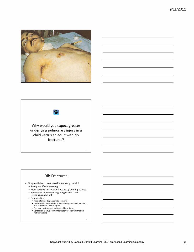
Rib Fractures
• Most often occur on lateral aspect of 3rd to 8th ribs, where ribs are least protected by musculature– More likely to occur in adults than in children
• Younger patients have more resilient cartilage that is not fully calcified
• When blunt forces are applied to ribs of children, energy is transmitted to lung, where pulmonary contusion is more frequent injury than rib fracture
– Morbidity or mortality from rib fractures depends on patient’s age and number and location of fractures
12
Copyright © 2013 by Jones & Bartlett Learning, LLC, an Ascend Learning Company
9/11/2012
5
13
Why would you expect greater underlying pulmonary injury in a child versus an adult with rib
fractures?
14
Rib Fractures
• Simple rib fractures usually are very painful– Rarely are life‐threatening– Most patients can localize fracture by pointing to area– Sometimes movement or grating of bone ends (crepitus) can be felt
– Complications• Respiratory or diaphragmatic splinting• Occurs when patient uses breath holding or minimizes chest wall movement to lessen pain
• Can lead to atelectasis (collapse of lung tissue)• Ventilation–perfusion mismatch (perfused alveoli that are not ventilated)
15
Copyright © 2013 by Jones & Bartlett Learning, LLC, an Ascend Learning Company

9/11/2012
6
Rib Fractures
• Goals of treatment
– Relieve pain
• May be relieved by splinting arm against chest wall with sling and swathe
• Circumferential splinting should not be used as it may not allow complete expansion of chest wall during respiration
• Administration of analgesics per protocol
16
Rib Fractures
• Goals of treatment– Maintain pulmonary function to prevent atelectasis
• Encourage patient to cough and to breathe deeply
– Based on mechanism of injury, consider possibility of more serious trauma
• Closed pneumothorax
• Internal bleeding
– Fractures to lower ribs (8‐2) may be associated with injuries to spleen, kidneys, or liver
17
Rib Fractures
• Great force is required to fracture 1st and 2nd ribs
– Because of their shape and protection provided by scapulae, clavicles, and upper chest musculature
– May be associated with
• Myocardial contusion
• Bronchial tears
• Vascular injury
18
Copyright © 2013 by Jones & Bartlett Learning, LLC, an Ascend Learning Company

9/11/2012
7
Flail Chest
• May occur when three or more adjacent ribs are fractured in two or more places– May be difficult to detect in prehospital setting because of muscle spasm that often accompanies injury
– Within 2 hours after injury, muscle spasm subsides
– At that point, injured segment of chest wall may begin to move in contrary fashion (paradoxical motion) with inspiration and expiration
– Interrupts normal mechanics of breathing and decreases effective ventilation
19
20
Ribfracture
21
Copyright © 2013 by Jones & Bartlett Learning, LLC, an Ascend Learning Company

9/11/2012
8
22
Flail Chest
• Causes
– Vehicle crashes
– Falls
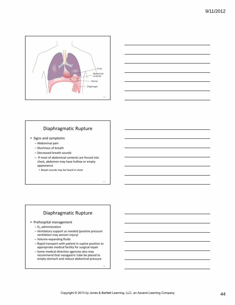
– Industrial accidents
– Assault
– Birth trauma
23
Flail Chest
• Mortality rate is 8 to 35 percent because of underlying, associated injuries
– Increases with
• Advanced age
• Seven or more rib fractures
• Three or more associated injuries
• Shock
• Head injury
24
Copyright © 2013 by Jones & Bartlett Learning, LLC, an Ascend Learning Company

9/11/2012
9
Flail Chest
• Diaphragm descends during inspiration
• Lowers intrapleural pressure
– Unstable chest wall is pushed (“sucked”) inward by negative intrathoracic pressure as rest of chest wall expands
– During expiration, diaphragm rises, and intrapleural pressure exceeds atmospheric pressure
• Causes unstable chest wall to move outward
25
Flail Chest
• Often develop hypoxia
– Because of lung contusions usually related to this injury
– Bleeding from alveoli and lung tissue causes contusion
– Associated with decreased vital capacity and vascular shunting of deoxygenated blood
26
Flail Chest
• Signs and symptoms
– Bruising
– Tenderness
– Bony crepitus on palpation
– Paradoxical motion (late sign)
27
Copyright © 2013 by Jones & Bartlett Learning, LLC, an Ascend Learning Company

9/11/2012
10
Flail Chest
• Prehospital management– Assisting ventilation with high‐concentration supplemental O2
– Fluid replacement as needed
– Field stabilization of flail segment is not recommended
– Many authorities recommend intubation and positive‐pressure ventilation (internal splinting) in patients with severe respiratory distress and flail chest
28
Flail Chest
• Prehospital management
– Intubation may be indicated if chest injury is associated with
• Shock
• Other severe injuries
• Head injury
• Pulmonary disease
• Patient over 65 years of age
29
Flail Chest
• Most conservative methods for obtaining adequate oxygenation and ventilation should be used to manage patients with flail chest
– Large percentage of patients with significant chest injury will progress to respiratory failure
• Requires long‐term ventilatory support and hospitalization
30
Copyright © 2013 by Jones & Bartlett Learning, LLC, an Ascend Learning Company
9/11/2012
11
Why is positive‐pressure ventilation the management of choice for this
injury?
31
Sternal Fractures
• Uncommon but serious injury
– Usually result from direct blow to chest
– Usually very painful
– May be associated with
• Unstable chest wall
• Myocardial injury
• Cardiac tamponade
32
Sternal Fractures
• Occur in only 5 to 8 percent of patients with blunt chest trauma
– Mortality rate is 25 to 45 percent
– Signs and symptoms
• History of significant anterior chest trauma
• Tenderness
• Abnormal motion or crepitation over sternum
33
Copyright © 2013 by Jones & Bartlett Learning, LLC, an Ascend Learning Company

9/11/2012
12
Sternal Fractures
• Prehospital management
– Maintaining high degree of suspicion for associated injuries
– Airway maintenance
– Ventilatory support
– Pulse oximetry
– ECG monitoring
– Rapid transport
34
Sternal Fractures
• Associated injuries that often contribute to serious disability or death
– Pulmonary and myocardial contusion
– Flail chest
– Vascular disruption of thoracic vessels (rare)
– Intra‐abdominal injuries
– Head injury
35
36
Copyright © 2013 by Jones & Bartlett Learning, LLC, an Ascend Learning Company

9/11/2012
13
Pulmonary Injury
• Classified as
– Closed pneumothorax
– Tension pneumothorax
– Open pneumothorax
– Hemothorax
– Pulmonary contusion
– Traumatic asphyxia
37
Pulmonary Injury
• Any of these injuries can result in difficulty in breathing and respiratory insufficiency
• Prehospital treatment
– Ensure open airway
– Ventilatory support
– Correct immediately life‐threatening ventilatoryproblems (e.g., tension pneumothorax)
– Rapid transport for definitive care
38
Closed Pneumothorax
• Simple pneumothorax caused by presence of air in pleural space– Causes lung to partially or totally collapse– Common causes
• Fractured rib that penetrates underlying lung• May occur without rib fractures• Excessive pressure on chest wall against closed glottis (paper bag effect)
• Rupture or tearing of lung tissue and visceral pleura from no apparent cause (e.g., spontaneous pneumothorax)
– Occurs in 15 to 50 percent of patients with severe blunt chest trauma
– 100 percent of patients with penetrating chest trauma
39
Copyright © 2013 by Jones & Bartlett Learning, LLC, an Ascend Learning Company

9/11/2012
14
How do you think that high‐flow oxygen promotes faster resolution
of a closed pneumothorax?
40
41
Closed Pneumothorax
• Signs and symptoms
– Dependent on severity of hypoxia, ventilation impairment, percentage of lung that has collapsed
– Chest pain
– Dyspnea
– Tachypnea
– Diminished/absent breath sounds on affected side
42
Copyright © 2013 by Jones & Bartlett Learning, LLC, an Ascend Learning Company
9/11/2012
15
Closed Pneumothorax
• Treatment
– Ventilatory support with high‐concentration O2
– Carefully monitor for signs of tension pneumothorax
– Transport in semisitting position of comfort unless contraindicated by mechanism of injury
– If patient’s respiratory rate is less than 12 or greater than 28 beats/minute, ventilatoryassistance with a bag‐valve‐mask may be indicated
43
Closed Pneumothorax
• Most healthy patients have large circulatory and ventilatory reserve capacities
– Closed pneumothoraces usually do not pose threat to life
– Life‐threatening consequences may develop if
• Pneumothorax is tension pneumothorax
• It occupies more than 40 percent of hemithorax
• Occurs in patient with shock or preexisting pulmonary or cardiovascular
44
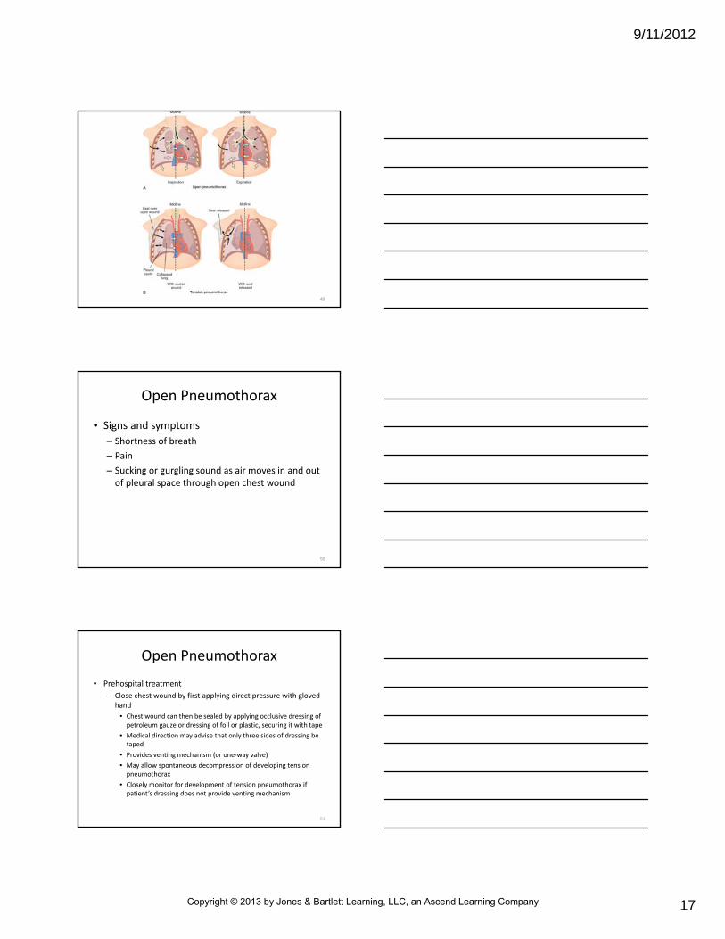
Open Pneumothorax
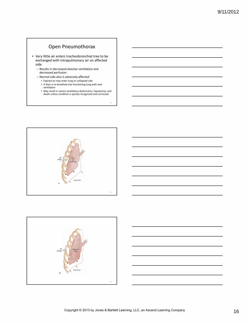
• Communicating pneumothorax develops when chest injury exposes pleural space to atmospheric pressure– Severity of injury is directly proportional to size of wound
– When chest wound is larger than normal pathway for air through nose and mouth, atmospheric pressure forces air through open wound and into thoracic cavity during inspiration
• As air accumulates in pleural space, lung on injured side collapses
• Lung begins to shift toward uninjured side
45
Copyright © 2013 by Jones & Bartlett Learning, LLC, an Ascend Learning Company
9/11/2012
16
Open Pneumothorax
• Very little air enters tracheobronchial tree to be exchanged with intrapulmonary air on affected side– Results in decreased alveolar ventilation and decreased perfusion
– Normal side also is adversely affected• Expired air may enter lung on collapsed side
• It then is re‐breathed into functioning lung with next ventilation
• May result in severe ventilatory dysfunction, hypoxemia, and death unless condition is quickly recognized and corrected
46
47
48
Copyright © 2013 by Jones & Bartlett Learning, LLC, an Ascend Learning Company
9/11/2012
17
49
Open Pneumothorax
• Signs and symptoms
– Shortness of breath
– Pain
– Sucking or gurgling sound as air moves in and out of pleural space through open chest wound
50
Open Pneumothorax
• Prehospital treatment
– Close chest wound by first applying direct pressure with gloved hand
• Chest wound can then be sealed by applying occlusive dressing of petroleum gauze or dressing of foil or plastic, securing it with tape
• Medical direction may advise that only three sides of dressing be taped
• Provides venting mechanism (or one‐way valve)
• May allow spontaneous decompression of developing tension pneumothorax
• Closely monitor for development of tension pneumothorax if patient’s dressing does not provide venting mechanism
51
Copyright © 2013 by Jones & Bartlett Learning, LLC, an Ascend Learning Company

9/11/2012
18
Open Pneumothorax
• Prehospital treatment
– Provide ventilatory support with high‐concentration O2 and monitor O2 saturation
• Airway management includes assisting ventilations with bag‐mask device and intubation
– Treat patient for shock by administering crystalloid per protocol
– Rapidly transport
52
53
Tension Pneumothorax
• When air in thoracic cavity cannot exit pleural space, a tension pneumothorax may develop
– True emergency
– Results in profound hypoventilation and impaired perfusion
– May result in death if not immediately recognized and managed
54
Copyright © 2013 by Jones & Bartlett Learning, LLC, an Ascend Learning Company

9/11/2012
19
55
56
Tension Pneumothorax
• When air is allowed to leak into pleural space during inspiration and becomes trapped during expiration, pleural pressure increases
– Produces shift in mediastinum
– Further compresses lung on uninjured side
– Compression of vena cava reduces venous return to heart
• Results in decrease in cardiac output
57
Copyright © 2013 by Jones & Bartlett Learning, LLC, an Ascend Learning Company

9/11/2012
20
• Signs and symptoms– Anxiety
– Cyanosis
– Increasing dyspnea
– Tracheal deviation (late sign)
– Tachycardia
– Hypotension or unexplained signs of shock
– Diminished or absent breath sounds on injured side
– Distended neck veins (unless patient is hypovolemic)
– Unequal expansion of chest (tension does not fall with respiration)
– Subcutaneous emphysema
Tension Pneumothorax
58
Why may the neck veins be distended in a patient with tension
pneumothorax?
59
• Should be managed aggressively– Evidenced by
• Increasing dyspnea• Compromised ventilation• Tachycardia• Tachypnea• Unilateral decreased or absent breath sounds• Hyper‐resonance on percussion
– Emergency care• Directed at reducing pressure in pleural space• Returning intrapleural pressure to atmospheric or subatmospheric levels
Tension Pneumothorax
60
Copyright © 2013 by Jones & Bartlett Learning, LLC, an Ascend Learning Company

9/11/2012
21
• Sealing open pneumothorax with occlusive dressing may produce tension pneumothorax– In such cases, increased pleural pressure can be relieved by momentarily removing dressing
– When dressing is lifted from wound, audible release of air from thoracic cavity should be noted
– If this does not occur and patient’s condition remains unchanged, wound should be gently spread open with gloved fingers
• May allow trapped air to escape
Tension Pneumothorax Associated with Penetrating Trauma
61
• Sealing open pneumothorax with occlusive dressing may produce tension pneumothorax
– After pressure has been released, wound should again be sealed
• Dressing may need to be removed more than once to relieve pleural pressure during transport
• If tension is not relieved with this procedure, needle decompression of thorax (needle thoracentesis; needle thoracostomy) should be performed
Tension Pneumothorax Associated with Penetrating Trauma
62
• Needle decompression should be performed when three findings are present
– Worsening respiratory distress or increasing difficulty ventilating with BVM device
– Unilateral decreased or absent breath sounds
– Decompensated shock (systolic BP less than 90 mm Hg)
Tension Pneumothorax Associated with Penetrating Trauma
63
Copyright © 2013 by Jones & Bartlett Learning, LLC, an Ascend Learning Company

9/11/2012
22
• Tension pneumothorax that develops in patient with closed chest trauma
– Must be relieved through thoracic decompression
– Can be done with large‐bore needle or commercially available thoracic decompression kit
Tension Pneumothorax Associated with Closed Trauma
64
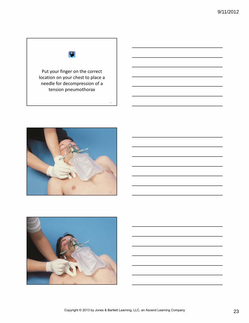
• For needle decompression, large‐bore, 10‐ or 14‐gauge hollow catheter‐over‐needle is inserted into affected pleural space– Needle can be inserted anteriorly in 2nd intercostalspace in midclavicular line
– May be placed in 4th or 5th intercostal space laterally on involved side
– Needle should be inserted just above rib
– After insertion of needle, audible rush of air should be noted
Tension Pneumothorax Associated with Closed Trauma
65
• Audible rush of air– Pressure escaping from pleural space (confirming tension pneumothorax)
– At this point, patient should show signs of improvement
• Patient will be easier to ventilate• Person’s breathing will be less labored
– Needle should be withdrawn and catheter secured in place with tape
– Needle decompression may need to be repeated if catheter becomes occluded blood clot and tension pneumothorax reoccurs
Tension Pneumothorax Associated with Closed Trauma
66
Copyright © 2013 by Jones & Bartlett Learning, LLC, an Ascend Learning Company
9/11/2012
23
Put your finger on the correct location on your chest to place a needle for decompression of a
tension pneumothorax
67
68
69
Copyright © 2013 by Jones & Bartlett Learning, LLC, an Ascend Learning Company
9/11/2012
24
70
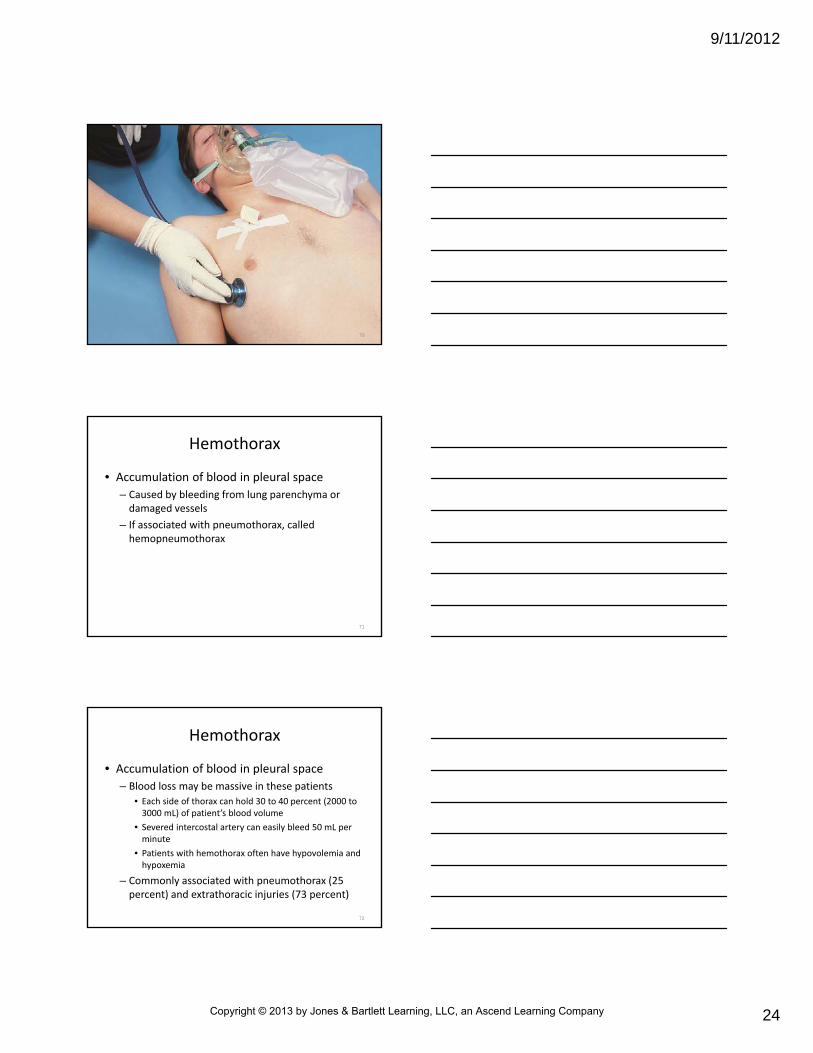
Hemothorax
• Accumulation of blood in pleural space
– Caused by bleeding from lung parenchyma or damaged vessels
– If associated with pneumothorax, called hemopneumothorax
71
Hemothorax
• Accumulation of blood in pleural space
– Blood loss may be massive in these patients
• Each side of thorax can hold 30 to 40 percent (2000 to 3000 mL) of patient’s blood volume
• Severed intercostal artery can easily bleed 50 mL per minute
• Patients with hemothorax often have hypovolemia and hypoxemia
– Commonly associated with pneumothorax (25 percent) and extrathoracic injuries (73 percent)
72
Copyright © 2013 by Jones & Bartlett Learning, LLC, an Ascend Learning Company

9/11/2012
25
73
74
75
Copyright © 2013 by Jones & Bartlett Learning, LLC, an Ascend Learning Company

9/11/2012
26
Hemothorax
• As blood continues to fill pleural space, lung on affected side may collapse
– In rare cases, mediastinum may even shift away from hemothorax
• Would compress unaffected lung
76
Hemothorax
• As blood continues to fill pleural space, lung on affected side may collapse
– Resultant effects of respiratory and circulatory compromise are responsible for the following signs and symptoms
• Tachypnea
• Dyspnea
• Cyanosis (often not evident in hemorrhagic shock)
• Diminished or decreased breath sounds (dullness on percussion)
• Hypovolemic shock
• Narrow pulse pressure
• Tracheal deviation to the unaffected side (rare)
77
Hemothorax
• Prehospital care– Directed at correcting ventilatory and circulatory problems
• High‐concentration O2
• Ventilatory support with bag‐mask device, intubation, or both
• Administration of volume‐expanding fluids to correct hypovolemia
• Rapid transport
• Hemothorax associated with great vessel or cardiac injury has a high mortality rate– 50 percent die within 1 hour of injury
78
Copyright © 2013 by Jones & Bartlett Learning, LLC, an Ascend Learning Company
9/11/2012
27
Why is hemothorax associated with a higher mortality rate than simple,
closed pneumothorax?
79
Pulmonary Contusion
• Most often caused by rapid deceleration forces
– Push lung against chest wall
• Results in rupture of alveoli, with hemorrhage and swelling of lung tissue
– More than 50 percent of patients with blunt chest trauma have pulmonary contusion
80
Pulmonary Contusion
• During sudden inertial deceleration and direct impact, fixed and mobile parts of lung move at varying speeds– Result is stretching and shearing of alveoli and intravascular structures (inertial effect)
• This kinetic wave of energy is partly reflected at alveolar membrane surface
• Remainder causes localized release of energy (spaldingeffect)
• Overexpansion of air in lungs occurs after primary energy wave has passed (implosion effect)
81
Copyright © 2013 by Jones & Bartlett Learning, LLC, an Ascend Learning Company
9/11/2012
28
Pulmonary Contusion
• Low‐pressure rebound shock waves cause overstretching and damage to lung tissue– Combination of these events results in alveolar and capillary damage with bleeding into lung tissue and alveoli
– Contused area of lung is unable to function properly after injury
• Profound hypoxemia may develop
• Degree of respiratory complication is directly related to size of contused area
82
Pulmonary Contusion
• Signs and symptoms are subtle at first
– Should be suspected based on kinematics of event and presence of associated injuries
– Tachypnea
– Tachycardia
– Cough
– Hemoptysis
– Apprehension
– Respiratory distress
– Dyspnea
– Evidence of blunt chest trauma
– Cyanosis
83
Will you always be able to distinguish between simple pneumothorax and
pulmonary contusion in the prehospitalsetting? Why?
84
Copyright © 2013 by Jones & Bartlett Learning, LLC, an Ascend Learning Company
9/11/2012
29
Pulmonary Contusion
• Emergency care– Ventilatory support and administration of high‐concentration O2
– Patients with associated injuries or preexisting pulmonary or cardiovascular disease
• Should be closely monitored in case ventilations need to be assisted with a bag‐valve device, intubation, or both
– May be associated with major chest injury
– Generally heal spontaneously over several weeks
85
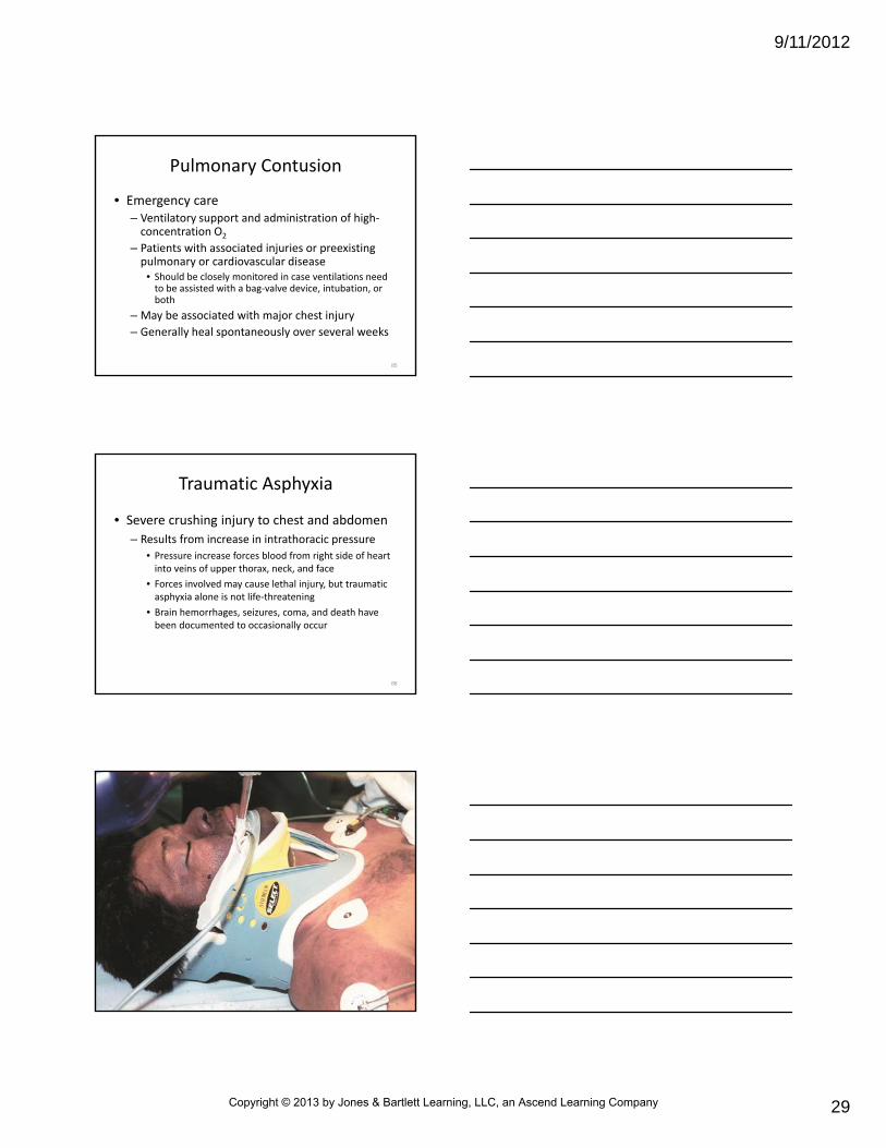
Traumatic Asphyxia
• Severe crushing injury to chest and abdomen
– Results from increase in intrathoracic pressure
• Pressure increase forces blood from right side of heart into veins of upper thorax, neck, and face
• Forces involved may cause lethal injury, but traumatic asphyxia alone is not life‐threatening
• Brain hemorrhages, seizures, coma, and death have been documented to occasionally occur
86
87
Copyright © 2013 by Jones & Bartlett Learning, LLC, an Ascend Learning Company
9/11/2012
30
Traumatic Asphyxia
• Signs and symptoms
– Reddish purple discoloration of face and neck
• Skin below area remains pink
– Jugular vein distention
– Swelling or hemorrhage of conjunctiva (subconjunctival petechiae may appear)
88
Traumatic Asphyxia
• Emergency care
– Ensure open airway
– Provide adequate ventilation
– Care for associated injuries
– Be ready to manage hypovolemia and shock when compressive force is released
89
Heart and Great Vessel Injury
• Trauma to heart and great vessels may result from blunt or penetrating trauma and associated
• Potentially fatal complications of these injuries– Life‐threatening dysrhythmias
– Conduction abnormalities
– Congestive heart failure
– Cardiogenic shock
– Cardiac tamponade
– Cardiac rupture
– Coronary artery occlusion
90
Copyright © 2013 by Jones & Bartlett Learning, LLC, an Ascend Learning Company
9/11/2012
31
Myocardial Contusion
• Usually caused by vehicle collision
– Chest wall strikes dashboard or steering column
– Deformed dashboard or steering column should alert paramedic to possibility of cardiac injury
– Blunt myocardial injury occurs in as many as 55 percent of patients who suffer blunt trauma to chest
91
How would you manage a cardiac rhythm disturbance resulting from a
myocardial contusion?
92
Myocardial Contusion
• Extent of injury may vary– May be only localized bruise
– May be full‐thickness injury to wall of heart with hemorrhage and edema
– Blood may accumulate in pericardium (hemopericardium) as result of tear in epicardiumor endocardium
• May result in cardiac rupture or traumatic MI
• Fibrinous reaction at contusion site may lead to delayed rupture or ventricular aneurysm
93
Copyright © 2013 by Jones & Bartlett Learning, LLC, an Ascend Learning Company

9/11/2012
32
Myocardial Contusion
• Patients may have no symptoms, or they may complain of chest pain similar to that seen with MI– Other signs and symptoms
• ECG abnormalities
• New cardiac murmur
• Pericardial friction rub (late)
• Persistent tachycardia (sinus tachycardia occurs in 70 percent of patients)
• Palpitations
94
Myocardial Contusion
• Emergency care
– Similar to that for MI
– O2 administration
– ECG monitoring
– Pharmacological therapy for dysrhythmias and hypotension
– Any intervention that increases myocardial O2
demand should be avoided
95
Pericardial Tamponade
• Penetrating trauma may cause tears in heart chamber walls– Allows blood to leak from heart
– If pericardium has been torn sufficiently, blood can leak into thoracic cavity and patient rapidly dies from hemorrhage
– Often, pericardium remains intact• In such cases, blood enters pericardial space
• Causes increase in pericardial pressure and volume (pericardial tamponade)
96
Copyright © 2013 by Jones & Bartlett Learning, LLC, an Ascend Learning Company

9/11/2012
33
Pericardial Tamponade
• Increased pressure prevents heart from expanding and refilling with blood
– 60 to 100 mL of blood and clots in pericardial sac can cause tamponade
– Results in decrease in stroke volume and cardiac output
– Myocardial perfusion decreases because of pressure effects on walls of heart and decreased diastolic pressures
97
Pericardial Tamponade
• Increased pressure prevents heart from expanding and refilling with blood
– Associated ischemic dysfunction may result in MI
– Pericardial tamponade occurs in fewer than 2 percent of patients who suffer blunt chest trauma
– 60 to 80 percent of patients with stab wounds involving heart develop tamponade
98
Pericardial Tamponade
• At first, most patients with pericardial tamponade have peripheral vasoconstriction
– Diastolic BP rises > systolic BP
– Causes decrease in pulse pressure
– These patients are also tachycardic
– Increase in heart rate compensates for decrease in cardiac output
99
Copyright © 2013 by Jones & Bartlett Learning, LLC, an Ascend Learning Company
9/11/2012
34
Pericardial Tamponade
• At first, most patients with pericardial tamponade have peripheral vasoconstriction
– Up to this point, pericardial tamponade and hemorrhagic shock have similar signs
• Key clinical finding often allows differentiation of two forms of shock
• First described by Beck in 1935
• It and two other clinical clues make up Beck triad
100
Pericardial Tamponade
• Beck triad
– Consists of
• Elevated central venous pressure (evidenced by jugular vein distention)
• Muffled heart sounds
• Hypotension
– 1st element: elevated central venous pressure, is single best way to distinguish pericardial tamponade from hemorrhagic shock
101
Pericardial Tamponade
• Beck triad
– Other signs and symptoms
• Tachycardia
• Respiratory distress
• Narrow pulse pressure
• Cyanosis of head, neck, upper extremities
102
Copyright © 2013 by Jones & Bartlett Learning, LLC, an Ascend Learning Company

9/11/2012
35
Pericardial Tamponade
• Two other findings in pericardial tamponade
– Pulsus paradoxus
• Systolic BP that drops more than 10‐15 mm Hg during inspiration compared with expiration
• Excessive decline in systolic pressure occurs in cardiac tamponade when pleural pressure is reduced during inspiration
• Reduction of pleural pressure provides some relief from tamponade and causes inspiratory fall in arterial flow and systolic pressure
103
Pericardial Tamponade
• Two other findings in pericardial tamponade
– Electrical alternans
• Refers to change in amplitude of patient’s ECG waveforms that decrease with every other cardiac cycle
• Rare finding in cardiac tamponade
104
105
Copyright © 2013 by Jones & Bartlett Learning, LLC, an Ascend Learning Company

9/11/2012
36
• True emergency
– Pericardial blood must be removed
– Bleeding must be stopped if patient is to survive injury
• Prehospital management
– Careful monitoring
– O2 administration
– Aggressive fluid replacement to maintain adequate preload (if transport time is short)
– Rapid transport
Pericardial Tamponade
106
• Definitive care
– Needle pericardiocentesis to remove blood from pericardial sac
– Removal of as little as 20 mL may drastically improve cardiac output
Pericardial Tamponade
107
Myocardial Rupture
• Occurs when blood‐filled chambers of ventricles are compressed with enough force to rupture chamber wall, septum, or valve– Nearly always immediately fatal
– About 20 percent of patients will survive 30 minutes or longer, allowing time for surgical repair
• May allow time for rapid transport and surgical repair
– Motor vehicle crashes are responsible for most cases of myocardial rupture, accounting for 15 percent of fatal thoracic injuries
108
Copyright © 2013 by Jones & Bartlett Learning, LLC, an Ascend Learning Company

9/11/2012
37
Myocardial Rupture
• Other proposed mechanisms
– Deceleration or shearing forces that disrupt the inferior and superior venae cavae
– Upward displacement of blood (causing increase in intracardiac pressure) after abdominal trauma
– Direct compression of heart between sternum and vertebrae
– Laceration from rib or sternal fracture
– Complications of myocardial contusion
109
Myocardial Rupture
• Patients often present with significant mechanism of injury
– Signs and symptoms of congestive heart failure and cardiac tamponade may be present
• Monitor closely for signs of pericardial tamponade
110
Myocardial Rupture
• Prehospital care
– Mainly supportive
– Airway and ventilatory support
– Rapid transport
– Consider possibility of tension pneumothorax
• Mimic those of myocardial rupture with tamponade
111
Copyright © 2013 by Jones & Bartlett Learning, LLC, an Ascend Learning Company

9/11/2012
38
• Thought to be result of shearing forces– Develop between tissues that decelerate at different rates
– Common mechanisms of injury• Rapid deceleration in high‐speed motor vehicle crashes
• Falls from great heights
• Crushing injuries
– Estimated that 1 in 6 people who die in motor vehicle crashes has rupture of aorta
• Of these patients, 80 to 90 percent die at scene as result of massive hemorrhage
Traumatic Aortic Rupture
112
Traumatic Aortic Rupture
• About 10 to 20 percent survive first hour
– Bleeding is tamponaded by surrounding adventitia of aorta and intact visceral pleura
– Of these individuals, 30 percent have ruptures within 6 hours
• For these reasons, rapid and pertinent evaluation and transport to appropriate medical facility are critical
– Aortic rupture is responsible for 15 percent of all deaths from blunt trauma
113
Traumatic Aortic Rupture
• Usual site of damage to aorta is in distal arch– Just beyond takeoff of left subclavian artery and proximal to ligamentum arteriosum
– Ligamentum arteriosum and descending thoracic arch are somewhat fixed
• Transverse portion of arch is somewhat mobile
• If shearing forces exceed tensile strength of arch, junction of mobile and fixed points of attachment may be partly torn
• If outer layer of tissue around aorta remains intact, patient may survive long enough for surgical repair
114
Copyright © 2013 by Jones & Bartlett Learning, LLC, an Ascend Learning Company

9/11/2012
39
115
Traumatic Aortic Rupture
• Aortic rupture is severe injury– About 85 percent of patients die before reaching hospital
– Any trauma patient who has unexplained shock and appropriate mechanism of injury (rapid deceleration) should be suspected of having ruptured aorta
– BP may be normal or elevated, with significant difference between two arms
– Upper extremity hypertension with absent or weak femoral pulses can occur in these patients
• Thought to result from compression of aorta by expanding hematoma
116
Traumatic Aortic Rupture
• Other patients have hypertension because of increased activity of sympathetic nervous system– About 25 percent have harsh systolic murmur that can be heard over pericardium or between scapulae
– In rare cases, may have paraplegia without cervical or thoracic spine injury
• Occurs as consequence of decreased blood flow through anterior spinal artery
– Anterior spinal artery is in thoracic region• Composed of branches from posterior intercostal arteries
• Are branches of thoracic aorta
117
Copyright © 2013 by Jones & Bartlett Learning, LLC, an Ascend Learning Company

9/11/2012
40
Traumatic Aortic Rupture
• Prehospital management
– Advising medical direction of suspected rupture
– Administration of high‐concentration O2
– Ventilatory support with spinal precautions
– Judicious fluid replacement (avoiding overhydration)
– Rapid transport for surgical repair
118
• Usually involve injury to chest, abdomen, or neck
– Often accompanied by
• Massive hemothorax
• Hypovolemic shock
• Cardiac tamponade
• Enlarging hematomas that may cause compression of vena cava, trachea, esophagus, great vessels, and heart
Penetrating Wounds of the Great Vessels
119
• Prehospital care
– Provide airway and ventilatory support
– Managing hypovolemia with judicious fluid therapy (guided by medical direction)
– Rapid transport for definitive care
Penetrating Wounds of the Great Vessels
120
Copyright © 2013 by Jones & Bartlett Learning, LLC, an Ascend Learning Company
9/11/2012
41
Other Thoracic Injuries
• Other injuries that may be associated with blunt or penetrating trauma to thorax
– Esophageal and tracheobronchial injuries
– Diaphragmatic rupture
121
• Esophageal injuries most often are caused by penetrating trauma
– Can result from
• Spontaneous perforation caused by cancer
• Anatomic distortions caused by diverticula or gastric reflux, both of which can lead to violent vomiting
Esophageal and TracheobronchialInjuries
122
• Assessment findings
– Pain
– Fever
– Hoarseness
– Dysphagia
– Respiratory distress
– Shock
Esophageal and TracheobronchialInjuries
123
Copyright © 2013 by Jones & Bartlett Learning, LLC, an Ascend Learning Company
9/11/2012
42
• If esophageal perforation occurs in cervical region– Local tenderness
– Subcutaneous emphysema
– Resistance to neck movement
• Esophageal perforation that occurs lower in thoracic region may result in– Mediastinal and subcutaneous emphysema
– Inflammation of mediastinum
– Splinting of chest wall
Esophageal and TracheobronchialInjuries
124
• Tracheobronchial injuries (tracheobronchialdisruptions) are rare– Occur in fewer than 3 percent of victims of blunt or penetrating chest trauma
– Mortality rate for these injuries is about 10 percent, depending on associated injuries, early diagnosis, and surgical repair
– Most injuries occur within 3 cm (about 1½ inches) of carina
• Can occur anywhere along tracheobronchial tree
Esophageal and TracheobronchialInjuries
125
• Tracheobronchial injuries (tracheobronchialdisruptions) are rare– Signs and symptoms
• Severe hypoxia
• Tachypnea
• Tachycardia
• Massive subcutaneous emphysema
• Dyspnea
• Respiratory distress
• Hemoptysis
Esophageal and TracheobronchialInjuries
126
Copyright © 2013 by Jones & Bartlett Learning, LLC, an Ascend Learning Company
9/11/2012
43
• Emergency care
– Provide airway, ventilatory, and circulatory support
– Rapid transport for definitive care
Esophageal and TracheobronchialInjuries
127
Diaphragmatic Rupture
• Diaphragm is sheet of dome‐shaped muscle
– Separates abdominal cavity from thoracic cavity
– Sudden compression of abdomen (such as with blunt trauma to trunk) results in sharp increase in intra‐abdominal pressure
– When this occurs, pressure differences may cause abdominal contents to rupture through thin diaphragmatic wall and enter chest cavity
128
Diaphragmatic Rupture
• Detected more often on left side than on right side– Rupture on either side may allow intra‐abdominal organs to enter thoracic cavity
– There they may compress lung, resulting in• Reduced ventilation
• Decreased venous return
• Decreased cardiac output
• Shock
– Because of mechanical forces involved, patients with diaphragmatic rupture often have multiple injuries
129
Copyright © 2013 by Jones & Bartlett Learning, LLC, an Ascend Learning Company
9/11/2012
44
130
Diaphragmatic Rupture
• Signs and symptoms
– Abdominal pain
– Shortness of breath
– Decreased breath sounds
– If most of abdominal contents are forced into chest, abdomen may have hollow or empty appearance
• Bowel sounds may be heard in chest
131
Diaphragmatic Rupture
• Prehospital management– O2 administration
– Ventilatory support as needed (positive pressure ventilation may worsen injury)
– Volume‐expanding fluids
– Rapid transport with patient in supine position to appropriate medical facility for surgical repair
– Some medical direction agencies also may recommend that nasogastric tube be placed to empty stomach and reduce abdominal pressure
132
Copyright © 2013 by Jones & Bartlett Learning, LLC, an Ascend Learning Company

9/11/2012
45
Summary
• Chest injuries are caused by blunt or penetrating trauma– Often results from motor vehicle crashes, falls from heights, blast injuries, blows to the chest, chest compression, gunshot wounds, and stab wounds
• Fractures of clavicle, ribs, or sternum, as well as flail chest, may be caused by blunt or penetrating trauma– Complications of skeletal trauma of chest may include cardiac, vascular, or pulmonary injuries
133
Summary
• Closed pneumothorax may be life‐threatening if (1) it is a tension pneumothorax, (2) it occupies more than 40 percent of the hemithorax, or (3) it occurs in a patient in shock or with a preexisting pulmonary or cardiovascular disease
• Open pneumothorax may result in severe ventilatory dysfunction, hypoxemia, and death unless it is quickly recognized and corrected
134
Summary
• Tension pneumothorax is a true emergency
– Results in profound hypoventilation
– May result in death if it is not quickly recognized and managed
• Hemothorax may result in massive blood loss
– These patients often have hypovolemia and hypoxemia
135
Copyright © 2013 by Jones & Bartlett Learning, LLC, an Ascend Learning Company
9/11/2012
46
Summary
• Pulmonary contusion results when trauma to lung causes alveolar and capillary damage
– Severe hypoxemia may develop
• Degree is directly related to size of contused area
• Traumatic asphyxia results from forces that cause increase in intrathoracic pressure
– When it occurs alone, it is often not lethal
• Brain hemorrhages, seizures, coma, and death have been reported after these injuries
136
Summary
• Extent of injury from myocardial contusion may vary
– Injury may be only localized bruise
• Also may be full‐thickness injury to wall of heart
– Full‐thickness injury may result in cardiac rupture, ventricular aneurysm, or traumatic myocardial infarction
137
Summary
• Pericardial tamponade occurs if 150 to 200 mLof blood enters the pericardial space suddenly
– Results in decrease in stroke volume and cardiac output
– Myocardial rupture refers to acute traumatic perforation of ventricles or atria
– Nearly always immediately fatal
• Death may be delayed for several weeks after blunt trauma
138
Copyright © 2013 by Jones & Bartlett Learning, LLC, an Ascend Learning Company

9/11/2012
47
Summary
• Aortic rupture is severe injury
– There is an 80 to 90 percent mortality rate in first hour
– Paramedic should consider possibility of aortic rupture in any trauma patient who has unexplained shock after rapid deceleration injury
139
Summary
• Esophageal injuries most frequently are caused by penetrating trauma (e.g., missile projectile and knife wounds)– Tracheobronchial injuries are rare
• Occur in fewer than 3 percent of victims of blunt or penetrating chest trauma, but mortality rate is over 30 percent
– Tension pneumothorax that does not improve following needle decompression or absence of continuous flow of air from needle following decompression should alert paramedic to possibility of tracheobronchial injury
140
Summary
• Diaphragmatic ruptures may allow abdominal organs to enter thoracic cavity
– There they may cause compression of the lung, resulting in reduction in ventilation, decrease in venous return, decrease in cardiac output, and shock
141
Copyright © 2013 by Jones & Bartlett Learning, LLC, an Ascend Learning Company

9/11/2012
48
Questions?
142
Copyright © 2013 by Jones & Bartlett Learning, LLC, an Ascend Learning Company