Embed Size (px)
Citation preview

CardiacConsult
3D TEE of the Tricuspid Valve – p. 3
WRAP-IT Yields Insights into CIED Infections – p. 8
Barlow’s Disease: Treat the Chords, Not the Leaflets – p. 10
INSIDE THIS ISSUE
Heart and Vascular News from Cleveland Clinic | 2019 | Issue 2
How Data Science Is Shaping Cardiothoracic Surgery– p. 4

Page 2 | Cardiac Consult | 2019 | Issue 2 |
Dear Colleagues,The clinicians who began building Cleveland Clinic’s Cardiovascu-
lar Information Registry back in 1971 were farsighted individuals,
but there was no way they could have imagined their efforts would
one day serve as the underpinning of advanced data techniques
like machine learning. Yet that’s precisely what happened, as
outlined in the cover story of this issue of Cardiac Consult.
Beyond growing into one of the world’s largest and longest-running
cardiovascular databases, the registry has provided invaluable
longitudinal data to fuel sophisticated algorithms offering insights
on everything from how to intervene in ischemic cardiomyopathy
to advanced analysis of valve disease.
An essential player in these innovative uses of our registry has
been our longtime Head of Clinical Investigations, Eugene Black-
stone, MD. The cover story outlines how Gene’s passion for new
fields of statistical endeavor has led him to assemble a team of
data experts who collaborate with our cardiothoracic surgeons to
generate fresh practice insights from novel analyses of reams of
clinical data. The beneficiaries have been patients here at Cleve-
land Clinic and around the world.
This issue also shares other developments from across our Heart
& Vascular Institute, from the results of the Cleveland Clinic-led
global WRAP-IT trial of an antibiotic-eluting envelope for cardiac
implantable electronic devices to the management of a fascinating
case of thoracic dystrophy. As always, we welcome your feedback
and inquiries on collaboration.
Respectfully,
Lars G. Svensson, MD, PhD
CHAIRMAN | Sydell and Arnold Miller Family Heart & Vascular Institute
Stay Connectedconsultqd.clevelandclinic.org/cardiovascular
clevelandclinic.org/cardiacconsult
@CleClinicMD
clevelandclinic.org/heartlinkedin
clevelandclinic.org/cardiacconsultpodcast
24/7 Referrals855.REFER.123 | clevelandclinic.org/heartreferrals
Outcomes Onlineclevelandclinic.org/hvioutcomes
Clinical Trialsclevelandclinic.org/clinicaltrials
Cardiac Consult is produced by Cleveland Clinic’s Sydell and Arnold Miller Family Heart & Vascular Institute.
Medical Editor Lars G. Svensson, MD, PhD Institute Chair [email protected]
Managing Editor Glenn R. Campbell
Art Director Michael Viars
Marketing Jackie Riggle | Colleen Burke
Photography & Illustrations Cleveland Clinic Center for Medical Art & Photography Russell Lee Photography
In 2018, Cleveland Clinic was ranked a top U.S. hospital in U.S. News & World Report’s “Best Hospitals” survey. The survey ranks Cleveland Clinic among the nation’s top 5 hospitals in 12 specialty areas, and the top hospital in heart care (for the 24th consecutive year) and urologic care.
© 2019 The Cleveland Clinic Foundation

| Cardiac Consult | 2019 | Issue 2 | Page 3Visit clevelandclinic.org /heart
Image of the Issue
The tricuspid valve is a complex, highly variable struc-
ture that has been historically challenging to image with
transesophageal echocardiography (TEE). As percutaneous
options for tricuspid valve interventions have increased, so
has the need for high-quality tricuspid imaging for use in
preprocedural planning and intraprocedural guidance.
Cleveland Clinic is using state-of-the-art three-dimensional
(3D) TEE to obtain high-resolution images to detail tricuspid
anatomy. The number of leaflets, orientation and subvalvular
apparatus can be identified clearly, as demonstrated in the
representative images above.
Furthermore, the use of live 3D multiplanar reconstruction
has allowed us to provide highly detailed and accurate si-
multaneous 2D and 3D imaging for intraprocedural guidance
of transcatheter tricuspid valve interventions, as reflected
in the sample images to the right.
For patients, 3D TEE technology translates into provision of
the highest-quality imaging available for both the diagnosis
and treatment of complex valvular heart disease. ■
Images and text supplied by Rhonda Miyasaka, MD, a staff physician in Cleveland Clinic’s Section of Cardiovascular Imaging. Contact her at 216.442.2304.
3D TEE OF THE TRICUSPID VALVE: A NEW WINDOW INTO COMPLEX, HIGHLY VARIABLE ANATOMY
Anterior
Anterior chords
Septal
Posterior Posterior chords
Septal chords
3D transesophageal echo images showing atrial (left) and ventricular (right) views of the tricuspid valve in systole.
Live 3D multiplanar reconstruction allows for accurate, real-time 3D image guidance.

Page 4 | Cardiac Consult | 2019 | Issue 2 |
› CARDIAC CONSULT FEATURE
How Data Science Is Shaping Cardiothoracic SurgeryITS ROOTS RUN DEEP, AND ITS REACH KEEPS GROWING.
The uses of data science in cardiothoracic
surgery seem to know no bounds:
• Creation of comparative prediction models of
survival with various surgical interventions for
ischemic cardiomyopathy
• Continuously updated estimates of mortality risk
for patients on the heart transplant waitlist
• Selection of variables for esophageal cancer staging
These wide-ranging examples are just from
Cleveland Clinic alone, and they’re only a sampling
of the applications of data science achieved by the
multidisciplinary clinical research team directed by
cardiac surgeon Eugene Blackstone, MD, Head of
Clinical Investigations in Cleveland Clinic’s Miller
Family Heart & Vascular Institute.

| Cardiac Consult | 2019 | Issue 2 | Page 5Visit clevelandclinic.org /heart
CARDIAC CONSULT FEATURE ‹
Seeds planted decades ago
Yet they all can be traced back to a handful of
consequential developments several decades ago.
First was the launch of Cleveland Clinic’s Cardiovascu-
lar Information Registry, established in 1971 to collect
data on every cardiovascular surgery patient at the
institution. The result is the oldest and one of the larg-
est computerized databanks of cardiovascular informa-
tion, which has served ever since as the foundation of
Cleveland Clinic’s pioneering cardiovascular outcomes
research. For instance, the registry was instrumental
in confirming the internal thoracic artery as the con-
duit of choice for coronary bypass grafting, as reported
by Loop and colleagues in a landmark 1986 New
England Journal of Medicine publication.
Next was Dr. Blackstone’s recruitment to Cleveland
Clinic in 1997 as Head of Clinical Research for the
Department of Thoracic and Cardiovascular Surgery.
He arrived after many years at the University of
Alabama at Birmingham, where he collaborated with
John Kirklin, MD, a pioneer of data-driven cardiotho-
racic surgery research and practice who helped put
the subspecialty in the vanguard of clinical outcomes
analysis. Dr. Blackstone brought extensive statistical
expertise and a passion for novel mathematical mod-
els and algorithmic approaches, which he was eager
to apply to Cleveland Clinic’s treasure trove of data.
Another key development was the start of a long-
standing collaboration between Cleveland Clinic and
IBM to construct an optimal medical record based
on ontology, or the metaphysical concept of grouping
things according to similarities and differences. “The
aim was to capture information on patients in terms
of values or variables, rather than as long stories, to
promote interoperability and use anywhere in the world,
regardless of language,” explains Dr. Blackstone. IBM’s
efforts in this realm began at the University of Alabama
in 1993 when Dr. Blackstone was still working there,
but they continued at an accelerated pace starting in
1997 when Dr. Blackstone began to help pair IBM’s
data capabilities with Cleveland Clinic’s Cardiovascular
Information Registry.
“Cleveland Clinic was one of only perhaps five medical
centers in the world with a serious interest in data sci-
ence dating back to the early 1970s,” he says. “This in-
stitution was ahead of the curve in carving out a role like
mine and consistently supporting it for so many years.”
The wide reach of data science in CT surgery
That support has allowed Dr. Blackstone, who is also
staff in Cleveland Clinic’s Department of Quantitative
Health Sciences, to assemble a team of statisticians,
computer scientists, mathematicians and other data
experts to support his cardiothoracic surgery colleagues
in a wealth of research and outcomes endeavors. They
draw on methodologies ranging from machine learning
to data management to artificial intelligence to help
surgeons improve clinical decision-making and assess
appropriateness of care through long-term follow-up.
The following paragraphs present a sampling of a few
projects the group has undertaken in recent years to
shape practice.Continued next page ›

Page 6 | Cardiac Consult | 2019 | Issue 2 |
› CARDIAC CONSULT FEATURE› CARDIAC CONSULT FEATURE
› Prediction models for decision support in
surgical management of ischemic cardiomyopathy.
About a decade ago, Dr. Blackstone’s team worked
with clinical colleagues to develop and validate
comparative prediction models of survival follow-
ing four different surgical intervention strategies for
ischemic cardiomyopathy (J Thorac Cardiovasc Surg.
2010;139:283-293). Using a robust, nonparametric
algorithmic method called Random Survival Forests,
they transformed the models into a computer-based
strategic decision aid to facilitate personalized
decision-making. “The idea was to quantitatively
make decisions with a model that could go down
many different paths rather than the norm of going
down each path individually,” Dr. Blackstone explains.
“These methods have since morphed into a formal set
of machine learning tools that allow clinicians to test
alternative treatment scenarios to recommend the one
that maximizes an outcome, such as length of life.”
› Classification of bicuspid aortopathy. In work
published just last year (J Thorac Cardiovasc Surg.
2018;155:461-469), Dr. Blackstone and colleagues
applied unsupervised clustering algorithms to a data
set of 656 patients with bicuspid aortic valves who
underwent ascending aorta surgery at Cleveland Clinic
over a 12-year period. This method finds similarities in
uncategorized data and then groups similar data points,
allowing processing of a large number of variables to
uncover underlying patterns. The result was a new clas-
sification of bicuspid aortopathy that for the first time
established a statistical relationship between the shapes
of bicuspid valves and patterns of aortic aneurysms.
› Selection of variables for esophageal cancer staging
and precision cancer care. Cleveland Clinic thoracic
surgeons and data scientists gathered data on patients
with esophageal cancer from all six inhabited conti-
nents to develop a data-driven approach to staging for
the seventh and eighth editions of the cancer staging
manual of the American Joint Committee on Cancer.
“Survival of individual patients with esophageal cancer
is all over the map, which means ‘average survival time’
is not very meaningful,” Dr. Blackstone says. “We used
machine learning algorithms — specifically, Random
Survival Forests — to enable much more precise predic-
tion of which treatments will promote longer survival at
the individual patient level. This approach is generally
applicable in many other conditions as well.”
› Longitudinal assessment of valve function. In the
past, emphasis has been on clinical events, like death
and stroke, that occur during follow-up after heart
surgery. Today, however, fully half of the analyses done
by Dr. Blackstone’s group focus on longitudinal data.
Examples include series of follow-up echocardiographic
assessments of pressure gradient developing across a
repaired heart valve or an artificial heart valve, or serial
assessments of the degree of leakiness of such valves.
This information can inform clinicians about the durabil-
ity of the valve repair or replacement device, but it
also serves to identify factors that increase or decrease
the speed of developing gradients or leakage, thereby
informing clinicians of who is at risk and who needs
reintervention. These analytic methods, developed at
Cleveland Clinic with National Institutes of Health (NIH)
funding, harken back to Dr. Blackstone’s early career
at the University of Chicago in the late 1960s, where
he helped develop the field of digital signal processing,
whereby complex longitudinal data can be broken down
into simple components, much like white light is broken
into a rainbow of colors by a prism.
› Continuously updated heart transplant waitlist
mortality estimation. In a recent publication (J Am
Coll Cardiol. 2018;72:650-659), Cleveland Clinic
clinicians and statisticians shared initial findings from
a dynamic model that continuously updates predicted
mortality based on laboratory values and other clini-
cal measures as the conditions of patients on the
heart transplant waitlist change over time. Using a
newly developed method for analyzing time-related
mortality, the model recomputes patients’ mortality
risk with each new clinical event or change in key
lab values or organ function. “A model of this type
that uses time-varying mortality risk estimation could
reduce mortality on the waitlist and promote better
utilization of the limited supply of donor hearts,” Dr.
Blackstone explains. “It represents a combination of
traditional statistics with machine learning.”
› Identification of variables most predictive of death
on the heart transplant waitlist. In related work,

| Cardiac Consult | 2019 | Issue 2 | Page 7Visit clevelandclinic.org /heart
CARDIAC CONSULT FEATURE ‹ CARDIAC CONSULT FEATURE ‹
In the words of a longtime colleague…“Gene Blackstone has made a huge contribution to our
understanding of the management of cardiothoracic sur-
gery patients,” says surgeon Lars Svensson, MD, PhD,
Chair of Cleveland Clinic’s Heart & Vascular Institute.
“His work has resulted in considerably better care of
patients, not only here but around the world. As we look
at new fields of statistical endeavor — such as machine
learning, voice recognition and interpolation for the EMR,
and ultimately automated artificial intelligence — we
know that Gene and his team will be at the forefront of
implementing these new methods to better understand
what makes for world-class cardiovascular care.”
Dr. Blackstone and Cleveland Clinic heart transplant
researchers have joined with colleagues from several
other institutions on a project funded by a $2.8 million
NIH grant to develop new machine learning methods
to examine and reduce disparities in survival among
heart failure patients before and after transplant. The re-
searchers are applying machine learning to the national
Scientific Registry of Transplant Recipients database to
identify major risk factors for death on the waitlist and
how these factors interact. The group’s first publication
(Am J Transplant. 2019 Jan 19 [Epub ahead of print])
identified the most important variables in predicting
mortality on the waitlist, including a couple that are not
in the current allocation system for donor hearts. “This
work involves some new mathematics well suited to
handling complex interactions among variable and large
amounts of missing data,” Dr. Blackstone observes.
What’s ahead
Dr. Blackstone sees emerging heart valve therapies
as one of the most active frontiers for future data sci-
ence applications in cardiothoracic surgery. “I expect
that we’ll be doing a lot of work to support decisions
around when to do valve procedures surgically versus
percutaneously, such as which approach and which
valve type are best for a given patient,” he says.
He notes that Cleveland Clinic can draw on the troves
of data it collected as one of the data analysis centers
for the PARTNER trial program for transcatheter
aortic valve replacement. “We also benefit from the
considerable follow-up we have done with so many
cardiovascular patients at Cleveland Clinic dating
back to the early 1970s,” he adds. “Good data sci-
ence requires the coupling of short-term data with
long-term outcomes.”
Challenges and opportunities
Yet he sees follow-up as the biggest challenge now
facing data science. “Patient follow-up today is much
more difficult than it was 20-some years ago, in part
because of patient privacy laws,” he says. The result is
inferior follow-up data, which he notes is exacerbated
by a continuing lack of interconnectivity of health data
information systems. “Our methods have ended up
being better than our data.”
Another challenge for a leader in his role is keeping
top data science talent around for the next project.
“The people who are good at this work come from a
diversity of backgrounds — from traditional statistics
to computer science to mathematics to computational
fluid dynamics,” he says. “Whoever they are, it can
be a challenge to keep good data scientists around,
as industry is always looking to recruit them.”
Cleveland Clinic hopes its recent establishment of a
cross-disciplinary Center for Clinical Artificial Intelligence
will assist in attracting and keeping top talent, and Dr.
Blackstone sees the center as a promising new partner.
Meanwhile, he spends time mentoring other clinician-
researchers who have a shared interest in champion-
ing novel data methods to help shape cardiothoracic
surgery. One current protégé is new Cleveland Clinic
congenital heart surgeon Tara Karamlou, MD, who
trained with him in the past.
“One way Cleveland Clinic tries to share the wealth in
terms of data science expertise is through cardiothoracic
surgery fellows and residents who train here,” Dr. Black-
stone says. “They can go back to their home institution
with knowledge of how to apply new tools or methodolo-
gies. But the most fundamental step any institution can
take toward advanced data techniques is to first get a
handle on their data. Start by understanding your data
and focusing on the quality of your outcomes. That’s
where Cleveland Clinic started many decades ago.
Success and sophistication follow from that.” ■
Contact Dr. Blackstone at 216.444.6712.

Page 8 | Cardiac Consult | 2019 | Issue 2 |
WRAP-IT Reveals 40% Reduction in CIED Infections with Antibiotic EnvelopeHuge postmarketing study also shows no increase in complication rate.
Placement of cardiac implantable electronic devices (CIEDs) inside an absorbable, antibiotic-eluting
envelope reduced the incidence of major infections by 40% relative to standard-of-care CIED infection
prevention strategies alone, according to results of the international WRAP-IT randomized controlled trial.
“Rates of CIED infection are generally low, but when these
infections occur, they can be devastating for patients, result-
ing in prolonged hospitalization, removal of the device system,
possible vascular injuries and sometimes fatality,” says Cleve-
land Clinic electrophysiologist Khaldoun Tarakji, MD, MPH,
global principal investigator of WRAP-IT, who reported the
results in a late-breaking trials presentation at the American
College of Cardiology (ACC) scientific session in March. The
study was published simultaneously in the New England
Journal of Medicine. “These findings provide strong evidence
for use of the envelope to reduce CIED infection risk.”
First randomized controlled trial of the envelope
This evidence may expand adoption of the envelope (TYRX™
Absorbable Antibacterial Envelope, Medtronic), which was
approved by the FDA in 2013. The product was designed
to surround a CIED implanted in the chest. It consists of a
knitted mesh body coated with an absorbable polymer mixed
with the antibiotics minocycline and rifampin, which are
eluted into local tissue for at least seven days. The envelope
is absorbed by the body within about nine weeks.
“Evidence on prophylactic strategies to reduce CIED infection be-
yond the use of preoperative antibiotics has been limited,” notes
Cleveland Clinic electrophysiologist Bruce Wilkoff, MD, who
served as the WRAP-IT trial’s steering committee chair. “Several
studies have yielded supportive evidence for local antibiotic
delivery, but a large randomized trial has been lacking.”
Nearly 7,000 patients at increased infection risk
WRAP-IT, which was supported by Medtronic, was undertak-
en to help address that deficiency through its size and design.
The study randomly assigned 6,983 patients from 25 nations
to receive the envelope or not receive the envelope in addition
to standard-of-care infection prevention (pre-procedure intra-
venous antibiotics plus sterile technique) during their CIED
procedure.
Randomization was done on a 1:1 basis with patients stratified
by device type — either a pacemaker/cardiac resynchronization
therapy (CRT) pacemaker or an implantable cardioverter defib-
rillator/CRT defibrillator. Patients (mean age, 70.1 years) were
blinded to their treatment group assignment, and the groups
were well matched for baseline and procedural characteristics.
“Our enrollment strategy was to include individuals at in-
creased risk for CIED infection based on literature evidence,”
notes Dr. Tarakji. This included those undergoing CIED
generator replacement or system upgrade with or without
new leads, as well as de novo CRT defibrillator implantation
or pocket or lead revisions.
“Management of patients with a CIED involves more than
just the implantation — there are follow-up procedures for
upgrades, battery or lead changes, and the like,” Dr. Wilkoff
adds. “Every time the CIED pocket is revisited, there is a
risk of infection.”
“ This study, which is the largest global cardiac device trial
ever completed, provides a realistic look at the number of
device infections associated with CIEDs.”— Bruce Wilkoff, MD

| Cardiac Consult | 2019 | Issue 2 | Page 9Visit clevelandclinic.org /heart
The primary end point was the incidence of major CIED infec-
tion within 12 months of the procedure, with major infections
defined as those requiring system extraction or revision, requir-
ing long-term antibiotic therapy with infection recurrence, or
resulting in death. A secondary safety outcome was procedure-
or system-related complications within 12 months.
Results: Fewer infections, comparable safety
Major CIED infections occurred in 25 patients in the envelope
group compared with 42 patients in the control group, for
12-month Kaplan-Meier event rate estimates of 0.7% and
1.2%, respectively (hazard ratio [HR] = 0.60; 95% CI, 0.36-
0.98; P = 0.041), representing a 40% relative reduction in
the risk of developing a major CIED infection.
On the safety front, procedure- or system-related complica-
tions occurred in 201 patients in the envelope group and 236
patients in the control group, for 12-month Kaplan-Meier
event rate estimates of 6.0% and 6.9%, respectively (HR =
0.87; 95% CI, 0.72-1.06; P < 0.001 for noninferiority).
Mean follow-up was 20.7 months, with 89.4% of enrollees
completing at least 12 months of follow-up. Across the entire
follow-up period, rates of major CIED infections were again sig-
nificantly lower in the envelope group than in the control group,
with 36-month Kaplan-Meier event rate estimates of 0.9% and
1.6%, respectively (HR = 0.63; 95% CI, 0.40-0.98).
Efficacy despite a low overall infection rate
“In a population at increased risk of CIED pocket infection,
the antibiotic envelope was significantly more effective at
preventing infection than standard infection control strategies
by themselves,” observes Dr. Tarakji. “The envelope was safely
implanted in 99.7% of procedure attempts, and it did not
increase complications despite the fact that use of the envelope
may require dissection of a slightly larger CIED pocket.”
The researchers called attention to two additional findings:
• The envelope’s benefits were especially pronounced
in patients with high-power devices relative to patients
with low-power devices or those receiving de novo
CRT defibrillators.
• The envelope demonstrated its treatment benefit despite a
low overall infection rate across the entire study.
“This study, which is the largest global cardiac device trial ever
completed, provides a realistic look at the number of device in-
fections associated with CIEDs,” says Dr. Wilkoff. “While overall
infection rates were low, the envelope still provided significant
benefit to patients. That’s important in view of the fact that well
over 1 million patients receive CIEDs worldwide every year.” ■
Contact Dr. Tarakji at 216.445.9225 and Dr. Wilkoff at 216.444.4975.
Photo of TYRX envelope above is courtesy of Medtronic.
WRAP-IT: Largest global cardiac device trial to date
6,983 patients | 181 centers | 25 countries
Patients undergoing cardiac implantable electronic device (CIED) procedures randomized 1:1 to:
Standard-of-care infection prevention
Standard of care + TYRX™ antibiotic-eluting
envelope to surround CIED
After 12 months…• 40% relative reduction in major CIED infections
in envelope group
• Comparable rates of complications in envelope and no-envelope groups
With >1 million people worldwide receiving CIEDs annually, the antibiotic envelope is an effective strategy for reducing risk of devastating CIED infections.
BOTTOMLINE
Tarakji KG, et al. N Engl J Med. 2019 Mar 17 [Epub ahead of print].SOURCE
Page 10 | Cardiac Consult | 2019 | Issue 2 |
Barlow’s Disease: Treating the Chords, Not the Leaflets, Is KeyThe case for a minimally invasive robotic approach to this form of mitral valve disease
Minimally invasive robotic, nonresectional mitral valve repair using neochords in Barlow’s disease is safe,
effective and durable. That’s the consensus among Cleveland Clinic cardiothoracic surgeons, who are now
using the technique on a regular basis.
“The technique avoids the fibrosis and scarring caused by
extensive cutting and suturing,” says one of those surgeons,
Per Wierup, MD, PhD. “It results in a larger opening with
a lower gradient.”
The procedure can be completed more quickly with a
robotic approach than a conventional repair performed with a
sternotomy. “Patients tolerate it very well,” Dr. Wierup notes.
“They require much less inotropic support and routinely only
stay overnight in the ICU. Typically they can be discharged
from the hospital after a few days.”
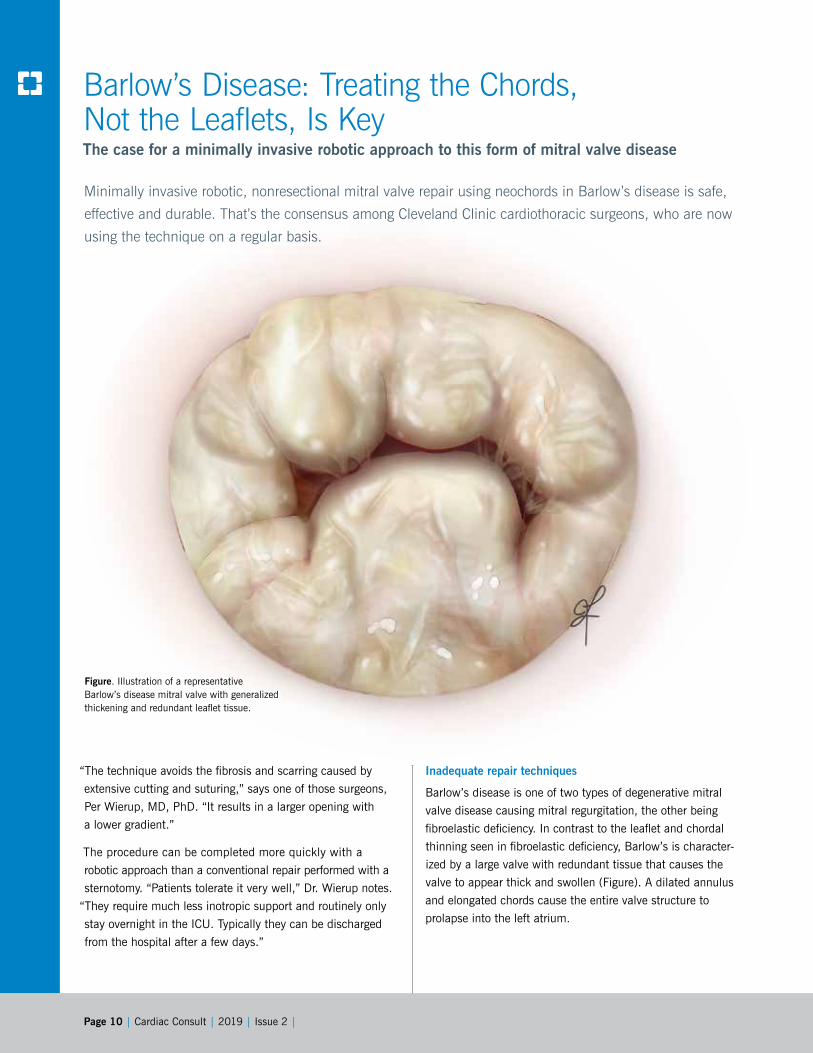
Inadequate repair techniques
Barlow’s disease is one of two types of degenerative mitral
valve disease causing mitral regurgitation, the other being
fibroelastic deficiency. In contrast to the leaflet and chordal
thinning seen in fibroelastic deficiency, Barlow’s is character-
ized by a large valve with redundant tissue that causes the
valve to appear thick and swollen (Figure). A dilated annulus
and elongated chords cause the entire valve structure to
prolapse into the left atrium.
Figure. Illustration of a representative Barlow’s disease mitral valve with generalized thickening and redundant leaflet tissue.

| Cardiac Consult | 2019 | Issue 2 | Page 11Visit clevelandclinic.org /heart
Surgical correction of the regurgitation in Barlow’s disease
offers a survival benefit when compared with optimal
medical therapy or valve replacement. Conventional repair
techniques used for fibroelastic deficiency have been
problematic, however.
“In a conventional repair, the prolapsed sections of the valve
are resected,” Dr. Wierup says. “A chordal transfer may be
performed, and the remaining valve is reattached.”
A better approach
More than a decade ago, Dr. Wierup began exploring how
to improve on nonresectional techniques for Barlow’s
disease. The approach treats the chords while leaving the
leaflets intact.
“Leakage is caused by valve prolapse due to elongated chords,
so we replace these with Gore-Tex® neochords,” he explains.
“Making the chords on the anterior and posterior leaflets
different lengths allows the zone of coaptation to be adjusted
for perfect results.”
Supportive data from a recent review
Performing the procedure using a minimally invasive, video-
assisted robotic technique offers multiple advantages over
conventional sternotomy, as demonstrated by outcomes from
a recent retrospective review of 102 patients who underwent
repair of Barlow’s disease. The results, which Dr. Wierup
presented at the 2018 American Heart Association Scientific
Sessions last November, included the following:
• No patient in either the sternotomy group (n = 38) or the
minimally invasive robotic repair group (n = 64) required
valve replacement, and no patient who underwent the
minimally invasive robotic procedure required intraoperative
conversion to sternotomy.
• There were no perioperative deaths or cases of >1+ mitral
regurgitation at discharge in either group.
• There were no significant differences in cardiopulmonary
bypass times between the groups.
• More than six hours of mechanical ventilation was required
by 21% of patients in the sternotomy group versus no
patients in the minimally invasive robotic repair group.
• Whereas 21% of patients in the sternotomy group remained
in the ICU more than 24 hours, no patients in the minimally
invasive robotic repair group did.
• Average hospital stay was 4 days following minimally
invasive robotic repair versus 9 days following sternotomy.
• Postoperative atrial fibrillation occurred in 16% of the
minimally invasive robotic repair group versus 42% of the
sternotomy group.
• Freedom from the composite end point of death,
reoperation or >1+ mitral regurgitation did not differ
between the two cohorts.
New standard of care for most patients
“Since he joined the Cleveland Clinic staff in 2017, Dr. Wierup
has shared his techniques with other members of our mitral
valve surgery team, increasing the options we make available
to treat patients with complex mitral valve disease,” says
A. Marc Gillinov, MD, Chair of Thoracic and Cardiovascular
Surgery.
“Minimally invasive repair using robotic assistance is now
standard treatment for Barlow’s disease at our institution,” Dr.
Wierup notes. “Patients must have a healthy aortic valve and
groin blood vessels that are large enough to accommodate the
cannulas required for robotic surgery. If their aortic valve is
leaking or their groin vessels are too small, it is safer to perform
the repair using a minimally invasive alternative approach.
However, this is necessary in only a minority of patients.” ■
Contact Dr. Wierup at 216.445.1652.
“ The technique avoids the fibrosis and scarring caused
by extensive cutting and suturing. It results in a larger
opening with a lower gradient.” — Per Wierup, MD, PhD

Page 12 | Cardiac Consult | 2019 | Issue 2 |
› CASE STUDY IN COLLABORATION
Launching an LAAO Program: How Cleveland Clinic Assisted an Affiliated InstitutionA Missouri center turns lessons from a site visit into program success.
Not long after Saint Francis Medical Center entered into a cardiology and cardiovascular surgery affiliation
with Cleveland Clinic in early 2014, the Southeast Missouri facility turned to Cleveland Clinic to help it
create a transcatheter aortic valve replacement (TAVR) program.
That experience (profiled at consultqd.clevelandclinic.org/
tavrtogether) was a success, prompting the 306-bed non-
profit tertiary care institution to turn to Cleveland Clinic for
guidance in launching another offering in its structural heart
disease program — this time for left atrial appendage
occlusion (LAAO) with the Watchman™ device.
Site visit for firsthand observation
The effort started with a February 2018 site visit to Cleveland
Clinic’s main campus by several key players in the planned
Saint Francis LAAO program: three physicians (an intervention-
al cardiologist, an imaging cardiologist and an electrophysiolo-
gist) plus an advanced practice nurse serving as coordinator
of the institution’s structural heart program.
During the visit, the Saint Francis staff observed the Cleveland
Clinic LAAO team perform a couple of Watchman proce-
dures in the electrophysiology lab as well as manage several
LAAO patients at different stages in the course of care, from
workup through post-procedure discharge. The structural
heart program coordinator consulted with nurse practitioners
before and after LAAO procedures and visited extensively with
Cleveland Clinic Heart & Vascular Institute registry, coding
and billing specialists to learn the finer points of data abstrac-
tion relating to LAAO procedures for purposes of compliance,
coding, billing, quality reporting and patient follow-up.
“We came prepared with questions to allow us to implement
processes from Cleveland Clinic’s program in a smaller organi-
zation like ours,” explains Jennifer Cotner, MSN, APRN, ACNP,
Structural Heart Program Coordinator at Saint Francis.
Implementation and outcomes
The Saint Francis team applied their learnings in short order,
performing their first LAAO procedure within a month after
the site visit. A full-fledged program was soon up and running,
with 42 patients evaluated for LAAO within the first year and
25 LAAO procedures performed. Saint Francis now performs
LAAO procedures every other Tuesday. Outcomes to date have
been excellent, as detailed in the table below.
Initial LAAO Outcomes at Saint Francis Medical CenterInitial Episode of Care
(n = 15)45-Day Follow-Up
(n = 13)6-Month Follow-Up
(n = 4)
Mortality 0% 0% 0%
Stroke (ischemic or hemorrhagic) 0% 0% 0%
Systemic embolism 0% 0% 0%
Evaluated for stroke using CHA2DS2-VASc score
100% 100% 100%
Evaluated for bleed using HAS-BLED score
100% 100% 100%
LAAO procedures that met FDA indications 100% 100% 100%
Source: American College of Cardiology LAAO Registry™: Standard Report ending 2018 Q3; 45-Day Follow-Up Report ending 2018 Q3; 6-Month Follow-Up Report ending 2018 Q1.
| Cardiac Consult | 2019 | Issue 2 | Page 13Visit clevelandclinic.org /heart
CASE STUDY IN COLLABORATION ‹
Ongoing support
Key Cleveland Clinic team members provide ongoing support
to their Saint Francis counterparts as needed. For instance,
electrophysiologist Mohamed Kanj, MD, who met with Saint
Francis staff during their site visit, provided at least six second
opinions on Saint Francis patients who were considering an
LAAO procedure during the first year of the Saint Francis
program’s operation. Additionally, Cleveland Clinic’s LAAO
registry team continues to advise and support their Saint
Francis colleagues on questions of coding and reporting.
Indeed, a key aspect of the training provided to Saint Fran-
cis by Cleveland Clinic involved education relating to the
American College of Cardiology’s LAAO Registry™. Cleveland
Clinic data specialists provided templates to assist with proper
abstracting, documentation and reporting for the registry.
Additionally, as a Cleveland Clinic Heart & Vascular Institute
affiliate organization, Saint Francis is entitled to complimen-
tary attendance at Cleveland Clinic’s annual Cardiac Registry
and Analytics Boot Camp, a three-day event designed to share
best practices in maintaining cardiac registries, data valida-
tion and accuracy, analytic outcomes reporting and physician
engagement efforts. This year’s boot camp is scheduled for
Nov. 13-15 on Cleveland Clinic’s main campus.
Reflections on the partnership
For its part, Saint Francis is delighted with the progress it
has made in just one year of offering LAAO services. “The
support we have received from Cleveland Clinic has been in-
valuable to the development and expansion of our structural
heart program,” says Saint Francis interventional cardiolo-
gist Steven Joggerst, MD, who leads the LAAO program. “Dr.
Kanj and his team readily shared their expertise, experience
and ideas, allowing us to make a smooth transition to this
technology. They have always provided timely responses to
our clinical questions and concerns. It is a privilege to be
affiliated with Cleveland Clinic.”
“It has been gratifying to see the Saint Francis team flourish
in their use of LAAO technology,” observes Oussama Wazni,
MD, Cleveland Clinic’s Section Head of Cardiac Electrophysi-
ology and Pacing, who met with the team during their site
visit. “Through Cleveland Clinic’s affiliate program, we’ve
gained considerable experience in helping smaller cen-
ters adapt new technologies to the size and scope of their
programs. This helps the affiliate centers distinguish their
programs in their local markets, and it also sharpens our use
of the technology by guiding another center in its use. The
collaboration is inevitably a win-win.” ■
For information on affiliation and alliance opportunities with Cleveland Clinic’s Heart & Vascular Institute, visit clevelandclinic.org/heartaffiliates.

Page 14 | Cardiac Consult | 2019 | Issue 2 |
Making Space to Breathe: A Case of Successful Surgery for Acquired Thoracic DystrophyA recent case from Cleveland Clinic’s Section of Thoracic Surgery underscores the need for vigilance
for acquired thoracic dystrophy when an adult with childhood pectus excavatum repair presents with
exercise intolerance. This article recaps the case and presents a Q&A with the managing thoracic
surgeon, Daniel Raymond, MD, on resulting insights about this rare condition.
Case vignette
A 43-year-old man presented to Cleveland Clinic’s Center
for Chest Wall Disease with significant exercise intolerance.
He was very short of breath and limited in his daily activi-
ties. Attempts to exercise led to palpitations and profuse
sweating. On cardiopulmonary testing, his maximal oxygen
uptake (VO2max) was 42% of predicted.
At age 7 years, he had undergone a modified Ravitch procedure
to correct a pectus excavatum defect, which entailed removal
of the cartilage between the sternum and ribs. This procedure
resulted in destruction of the growth plates so that his ribs
stopped growing, which essentially left him with adult-sized
organs squeezed within the rib cage of a 7-year-old.
Clinicians he had seen previously believed he had a recur-
rence of the pectus excavatum, as his sternum looked low on
imaging, as if the defect had not been corrected entirely. How-
ever, CT scans at Cleveland Clinic revealed the telltale sign of
acquired thoracic dystrophy: a bell-shaped chest (Figure 1)
associated with a restrictive pattern on his lung function stud-
ies. Moreover, axial images showed his organs to be visibly
crowded in the chest (Figure 2), restricting space for his lung
tissue. In addition, the normal cartilage between his ribs had
been replaced by bone, reducing his chest wall flexibility.
The patient was operated on at Cleveland Clinic in November
2017. The rib portions that had calcified were resected, with
care taken to preserve the soft tissue underneath. His sternum
was cut and bent forward since there was still a residual com-
ponent of his pectus excavatum. A titanium plate was inserted
to hold the sternum in its new, normal position.
Because his organs were so restricted, a bar was also used to
cross his chest and push the sternum forward to create even
more space in the chest (Figure 3). While bars are generally
to be avoided in adults when correcting previously untreated
pectus excavatum, in this case the need for additional chest
wall expansion was clear. Removal of the bar is planned after
two years. The plate will remain permanently.
Figure 1. Preoperative image showing the classic bell-shaped chest of acquired thoracic dystrophy.
Figure 2. Preoperative axial image showing calcification of the medial ribs with some residual pectus deformity.

| Cardiac Consult | 2019 | Issue 2 | Page 15Visit clevelandclinic.org /heart
Q: How has the patient fared since the operation?
A: He has worked hard at exercising and has improved
dramatically. On repeat cardiopulmonary testing, his VO2max
is now up to 78% of predicted. This is due not simply to the
surgery but also to his vigilance: He walks every day, goes
to the gym and does all the exercise we prescribed. Since he
has been able to exercise, he also has lost about 30 pounds.
While we made his chest wall better, he had to be the one
to improve his cardiopulmonary fitness. Unfortunately, many
patients don’t do that. This dedication was likely central to
our patient’s positive result.
Interestingly, despite the improved VO2max, his forced vital
capacity and forced expiratory volume at 1 minute have not
changed dramatically, remaining around 50%. We do not
currently have effective technology to surgically increase the
thoracic cage volume to a significant degree, and this offers
an opportunity for research.
Q: Exactly how can a modified Ravitch procedure for
pectus excavatum in childhood contribute to development
of acquired thoracic dystrophy?
A: Pectus excavatum is a congenital chest wall deformity in
which several ribs and the sternum grow abnormally, produc-
ing a concave appearance in the anterior chest wall. It occurs
in about 1 in 300 births, with a male-to-female predomi-
nance of about 3:1.
In most cases, the defect is mild and doesn’t require correc-
tion, but sometimes cosmetic treatment is justified in children
for psychological reasons. In adults, we do not correct pectus
excavatum unless there are problems with exercise intoler-
ance, which can be overlooked. Patients are often told that
pectus excavatum is only a cosmetic problem when in fact
there can be real physiologic manifestations.
In the mid-1980s, pediatric surgeons recognized that pectus
excavatum could be corrected without damage to the growth
plates if dissection of the interface between cartilage and
bone were done less aggressively. The modified Ravitch is still
used, but a newer, less invasive approach called the Nuss
procedure has largely replaced it.
With the Nuss, the cartilage stays in place while a bar is
pushed under the sternum to force it forward. The Nuss works
well in children and teens, but it tends to be more painful
than the Ravitch. My practice is to not perform the Nuss in
adults out of concern that rigidity of the adult skeletal system
leads to rib fractures without effective correction of the defect.
However, other surgeons will perform the Nuss in adults.
Q: Does this mean acquired thoracic dystrophy will
disappear as a clinical entity?
A: Current awareness of the need for greater care in cartilage
dissection with the modified Ravitch, along with elimination
of dissection altogether with greater use of the Nuss, means
we should see fewer and fewer cases of acquired thoracic
dystrophy as time goes by. But we can’t predict when we’ll
stop seeing them, since we don’t know how many cases are
out there.
Q: What’s needed to manage restrictive chest wall
diseases like this?
A: At Cleveland Clinic’s Center for Chest Wall Disease,
we are fortunate to be able to pool the expertise of thoracic
surgeons along with clinicians in orthopaedics, radiation
oncology, musculoskeletal radiology, pulmonary physiology
and pain management to help these patients. We are now
working on developing new technologies to more effectively
expand the chest volume to treat acquired thoracic dystro-
phy and other restrictive diseases. ■
Contact Dr. Raymond at 216.636.1623.
Figure 3. Postoperative image showing the plate and bar placed to hold the sternum in its normal position and further increase chest volume.
WITH Daniel Raymond, MD
Page 16 | Cardiac Consult | 2019 | Issue 2 |
Stress Test Performance Beats Chronological Age in Predicting LongevityThe cliché that you’re only as old as you feel just got an evidentiary boost from findings of the largest
cohort of exercise stress test participants reported to date. The cohort study, published by Cleveland
Clinic researchers in the European Journal of Preventive Cardiology in February, shows that physiological
age, based on exercise stress test performance, is a better predictor of survival than chronological age.
The study evaluates a formula the researchers developed to
calculate exercise performance, which they dub A-BEST (Age
Based on Exercise Stress Testing), or “physiological age.” It
is sex-specific and consists of three components, taking into
account the use of negative chronotropic medications:
• Exercise capacity (number of peak estimated metabolic
equivalents of task [METs])
• Chronotropic reserve index (heart rate response to exercise)
• Heart rate recovery
“These three exercise parameters are readily available in stress
test reports,” explains lead author and Cleveland Clinic cardi-
ologist Serge Harb, MD. “Our goal was to develop a mortality
risk estimate that’s more practical and easy to understand for
patients and clinicians. The A-BEST measure can be used as
a surrogate for physiological age, as it incorporates factors as-
sociated with diminishing treadmill exercise performance.”
Study essentials
The researchers applied the A-BEST formula to 126,356 con-
secutive patients who underwent exercise stress testing (ECG,
echocardiography or myocardial perfusion imaging) at Cleve-
land Clinic from 1991 to 2015. Univariable and multivariable
regression analyses were used to determine the association
of A-BEST with all-cause mortality throughout the duration of
available follow-up.
Mean patient age was 53.5 years. During mean follow-up
of 8.7 years across the cohort, a total of 9,929 patients
(7.9%) died.
Following adjustment for clinical comorbidities, each of
the three A-BEST components was significantly associated
with mortality, as follows:
• Greater exercise capacity (higher METs) was associated
with lower mortality (P < 0.001).
• Higher chronotropic reserve index was associated
with lower mortality (P = 0.0135).
• Abnormal heart rate recovery was associated with
higher mortality (P < 0.001).
Consistent with the above results, the A-BEST measure itself
was significantly associated with mortality, with a higher
A-BEST correlating with higher mortality (P < 0.001).
Moreover, A-BEST was significantly more predictive of death
than was a patient’s chronological age (P < 0.001).
The above findings applied to both the overall cohort and
to men and women when analyzed separately.
Helping patients better understand risk
“These findings show that estimated physiological age,
based on readily available exercise stress testing parameters,
performs better than chronological age in predicting all-cause
mortality,” Dr. Harb observes.
“The primary advantage of A-BEST is that it reliably and con-
veniently translates exercise variables to a risk estimate that’s
easily appreciated by both patients and their providers,” adds
senior author Wael Jaber, MD. “The hope is that this can help
motivate patients to exercise more.”
Simple formulas for calculating A-BEST for both men and
women are presented in the European Journal of Preventive
Cardiology study report (2019 Feb 13 [Epub ahead of print]). ■
Contact Dr. Harb at 216.444.3316 and Dr. Jaber at 216.444.8305.

| Cardiac Consult | 2019 | Issue 2 | Page 17Visit clevelandclinic.org /heart
Research RoundupQuick Takes on Recent Cardiovascular Studies of Note
Increasing Inflammation Correlates with Residual Risk After ACSSerial measurements of high-sensitivity C-reactive protein
(hsCRP) following acute coronary syndrome (ACS) may help
identify patients at higher risk for morbidity and mortality. So
concludes a secondary analysis of the multicenter VISTA-16 trial
published by a Cleveland Clinic-led group in JAMA Cardiology.
VISTA-16 randomized 5,145 patients to 16 weeks of the experi-
mental anti-inflammatory drug varespladib or placebo within 96
hours of ACS. The trial was stopped early due to futility, but it
lasted long enough for hsCRP levels to be obtained at baseline
and weeks 1, 2, 4, 8 and 16 in over 4,200 patients. The new
analysis showed that each standard-deviation increment in
longitudinal hsCRP concentration was associated with a 15%
increased risk of a major adverse cardiovascular event, a 25%
increased risk of all-cause death and a 26% increased risk of
cardiovascular death. “Monitoring hsCRP in addition to lipids
after ACS may help us better identify patients at increased risk
for recurrent events or death, which may prompt more intensive
treatment,” says senior author Rishi Puri, MD, PhD.
More at consultqd.clevelandclinic.org/vista16.
For Ideal STEMI Care, Look Beyond Door-to-Balloon TimeA comprehensive systems-based strategy for acute ST-segment
elevation myocardial infarction (STEMI) care improves outcomes
compared with a singular focus on door-to-balloon time, accord-
ing to research from Cleveland Clinic investigators in Circulation:
Cardiovascular Interventions. The study reviewed the care of
1,272 consecutive patients with STEMI treated with percutane-
ous coronary intervention (PCI) at Cleveland Clinic from 2011
through 2016 to assess the respective contributions of three
care components: use of guideline-directed medical therapy, use
of transradial primary PCI and prompt door-to-balloon time.
Each component was found to yield significant incremental out-
come improvements, showing a graded association with reduced
ejection fraction and reduced rates of bleeding, cardiogenic
shock, and cardiovascular and all-cause mortality. “This study
indicates that it’s time to move beyond focusing solely on door-
to-balloon time for providing quality acute STEMI care,” says
corresponding author Umesh Khot, MD. “We found that the best
approach is to consistently achieve a range of STEMI care best
practices.” More at consultqd.clevelandclinic.org/stemi.
Surgery for Marfan Syndrome: Support for Earlier Intervention Guidelines recommend prophylactic aortic root replacement
when the aortic diameter reaches 5 cm or larger (or ≥ 4 to 4.5
cm with certain risk factors), but a new observational study
suggests that size threshold needs to be revisited. The study
reviewed 491 adults with suspected Marfan syndrome who
presented for aortic surgery at Cleveland Clinic from 1990 to
2016 who met the revised Ghent criteria and hadn’t had prior
aortic surgery. It found that the majority of patients who un-
derwent emergency aortic root replacement had an aortic root
diameter smaller than guideline cutoffs for prophylactic surgery.
It also found that prophylactic root replacement was associated
with significantly improved survival and freedom from aortic
dissection compared with emergency surgery among the cohort.
“These findings suggest that Marfan syndrome patients with
ascending aortopathy may benefit from prophylactic aortic
root replacement surgery earlier than is indicated by current
guidelines based on aortic root dilation,” says senior author
and cardiac surgeon Lars Svensson, MD, PhD. The study was
published in the Journal of the American College of Cardiology.
More at consultqd.clevelandclinic.org/marfan.
LAA Closure Device Safe and Effective Despite Prior ICHPlacement of the Watchman left atrial appendage (LAA)
closure device followed by short-term anticoagulation is effec-
tive and well tolerated in patients with atrial fibrillation and a
history of intracranial hemorrhage (ICH), a Cleveland Clinic
feasibility study shows. The study of 38 consecutive patients,
published in Heart Rhythm, found that no strokes, intracranial
bleeds, major cardiovascular events or deaths occurred over
mean follow-up of more than a year after implantation.
“Patients with atrial fibrillation who’ve had a previous ICH
are challenging to treat because they’re at elevated risk for
both stroke and recurrent bleeding,” says co-author Ayman
Hussein, MD, a Cleveland Clinic electrophysiologist. Yet ICH
was an exclusion criterion in pivotal trials of the Watchman
device. “Our study, which we believe is the largest to date in
patients with ICH, suggests that short-term anticoagulation
therapy, which is required perioperatively for the LAA closure
device, can safely be provided to these patients.” More at
consultqd.clevelandclinic.org/ichlaa.

Page 18 | Cardiac Consult | 2019 | Issue 2 |
› CME PREVIEW
We’re Bringing a Valve Disease Update to Boston
Contemporary Management of Valvular Disease: Diagnosis, Imaging and InterventionFri.-Sat., Sept. 6-7, 2019 | InterContinental Boston
› ccfcme.org/bostonvalve2019
For a comprehensive update on the latest in heart valve
disease, there’s no better place to be than Boston in
early September for this 1.5-day CME-certified course
sponsored by Cleveland Clinic. Fifteen expert faculty
from Cleveland Clinic, Boston Children’s Hospital and
other leading U.S. centers will share insights and prac-
tice tips across the spectrum of valve care.
“Management of heart valve disease is constantly evolv-
ing,” says course director Lars Svensson, MD, PhD,
Chair of Cleveland Clinic’s Miller Family Heart & Vas-
cular Institute. “We see this in the steady emergence of
new techniques, devices and imaging approaches and
in the rapid expansion of the evidence base on patient
assessment and treatment outcomes from valve inter-
ventions. This program is designed to bring together
cardiologists, interventionalists, surgeons and other
providers to review the latest research and technical
advances and assess their likely impact on practice.”
A deep dive on the mitral valve
The full-day Friday program is devoted to the mitral
valve, beginning with discussions of various imag-
ing modalities in evaluating and quantifying mitral
regurgitation and an exploration of the “valve center
of excellence” model.
Next comes a detailed survey of degenerative mitral re-
gurgitation, from when and how to intervene to consid-
eration of various special situations, such as Barlow’s
valve, managing concomitant tricuspid regurgitation
and issues around concurrent atrial fibrillation ablation.
All of Friday afternoon is dedicated to functional mitral
regurgitation, with focused explorations of a range of
topics, from the role of atrial dilatation to integrating
other therapies with the MitraClip® device for func-
tional mitral regurgitation.
Aortic and tricuspid valves on Saturday
The aortic valve takes center stage for much of Saturday
morning, which is packed with 15- or 20-minute up-
dates on a range of timely subtopics. A few examples:
• Trends in transcatheter (TAVR) versus surgical
aortic valve replacement, including the impact
of the new PARTNER 3 trial
• Valve-in-valve TAVR
• Management of aortic regurgitation
• Treatment for the bicuspid aortic valve: Surgery
or TAVR, and do we need a randomized trial?
The final few segments before 12:30 adjournment
on Saturday will explore the latest in management of
tricuspid regurgitation, including an update on trans-
catheter therapies.
“Our overarching goal will be to clarify optimal manage-
ment strategies across the spectrum of valvular disease,
whether it’s choosing between mitral valve repair or
replacement or how best to quantify various types of
valve disease,” says Dr. Svensson. ■
Visit ccfcme.org/bostonvalve2019 for registration and details. Early-bird registration ends July 7.
This activity has been approved for AMA PRA Category 1 Credit™.

| Cardiac Consult | 2019 | Issue 2 | Page 19Visit clevelandclinic.org /heart
› CME PREVIEW
We’re Teaming with Boston Children’s in NYC to Keep You Current on CHD
Innovations in the Management of Congenital Heart Disease: Old Problems, New ApproachesFri.-Sat., Oct. 4-5, 2019 | JW Marriott Essex House,
New York City
› ccfcme.org/congenitalheart19
Given that congenital heart disease is one of the most
complex realms of cardiovascular practice, why not
come at it with the strengths of two powerhouse heart
programs instead of just one? That’s the thinking
behind this CME course jointly offered by Cleveland
Clinic and Boston Children’s Hospital.
The 1.5-day program aims to extend the knowledge
and experience of 25 expert clinician faculty from
Cleveland Clinic and Boston Children’s to adult and
pediatric providers in cardiology, cardiac surgery,
pediatrics, internal medicine and beyond.
“Outcomes of both pediatric and adult patients with
congenital heart disorders have improved substantially,
thanks to innovations in imaging technology, research
studies and surgical techniques,” says course co-
director Lars Svensson, MD, PhD, Chair of Cleveland
Clinic’s Miller Family Heart & Vascular Institute. “Yet
these advances have been so rapid that it can be chal-
lenging to keep up, particularly in view of the patho-
physiologic complexity of congenital heart disease.”
“These challenges have informed the design of this
course, which aims to improve attendees’ competence
and skills in diagnosing congenital heart disorders and
choosing appropriate treatment strategies,” adds course
co-director Pedro del Nido, MD, Chair of the Department
of Cardiovascular Surgery at Boston Children’s. One of
his Boston Children’s cardiovascular surgery colleagues,
John Mayer, Jr., MD, is a third co-director of the course.
The course relies on a mix of formats — short lectures,
case studies, moderated discussions and more — to
address its six sessions’ broad topical range in a highly
tailored way:
• Hypoplastic left heart syndrome, which is explored
through cases focused on imaging and periopera-
tive care as well as reviews of surgical and medical
management, outcomes and issues that matter
most to patients
• Innovative approaches to complex problems, includ-
ing the biventricular pathway, complex heterotaxy,
double-switch and cone procedures, and more
• Outcomes in congenital heart surgery, including
discussions of multicenter database analyses, risk
scores, quality ratings and big data
• More innovative approaches to complex problems,
including 3D printing applications in interventional
and surgical management, blood conservation,
preoperative rhythm assessment and more
• A range of issues in preserving and restoring
valvular function in congenital heart disease
• Discussions of three high-interest topics in the
care of adult congenital heart disease, each with
perspectives from a medical and a surgical expert
“The program was conceived to be highly multidisci-
plinary,” says Dr. del Nido. “Most topics are addressed
from a multiplicity of perspectives, including surgi-
cal, medical, imaging, interventional, perioperative,
outcomes and more.”
“Planning this course has been a delight,” adds Dr.
Svensson, “as it allows us to combine the expertise of
Cleveland Clinic and Boston Children’s in highly comple-
mentary ways to address congenital heart disease in
both adult and pediatric populations. Attendees will
benefit from the synergy that results.” ■
Visit ccfcme.org/congenitalheart19 for registration and details. Early-bird registration ends Aug. 4.
This activity has been approved for AMA PRA Category 1 Credit™.
19-HRT-138
The Cleveland Clinic Foundation9500 Euclid Ave./AC311Cleveland, OH 44195
CardiacConsult
Live CME Events from Cleveland Clinic
20th Annual Intensive Review of CardiologySat.-Wed., Aug. 17-21, 2019 InterContinental Hotel & Conference Center | Cleveland
Info/registration: ccfcme.org/cardioreview19
Contemporary Management of Valvular Disease: Diagnosis, Imaging and InterventionFri.-Sat., Sept. 6-7, 2019 InterContinental Boston | Boston
Info/registration: ccfcme.org/bostonvalve2019 (see page 18 for more detail)
CLE: Comprehensive Lifelong Expeditious Care of Aortic DissectionThu.-Fri., Sept. 19-20, 2019
InterContinental Hotel & Conference Center | Cleveland
Info/registration: ccfcme.org/aorticdissection19
Is Now a Podcast Too
Listen at clevelandclinic.org/
cardiacconsultpodcast or
subscribe from your favorite
podcast source.
Global EP Summit 2019Fri.-Sat., Sept. 27-28, 2019 Hilton Cleveland Downtown | Cleveland
Info/registration: ccfcme.org/globalep19
Innovations in the Management of Congenital Heart Disease: Old Problems, New ApproachesFri.-Sat., Oct. 4-5, 2019 JW Marriott Essex House | New York City
Info/registration: ccfcme.org/congenitalheart19 (see page 19 for more detail)
Congenitally Corrected Transposition of the Great Arteries: Management and Outcomes from Infancy to AdulthoodThu.-Sat., Oct. 17-19, 2019 InterContinental Hotel & Conference Center | Cleveland
Info/registration: ccfcme.org/pediatricheart19
These activities have been approved for AMA PRA Category 1 Credit™.