Embed Size (px)
Citation preview
Anthozoa
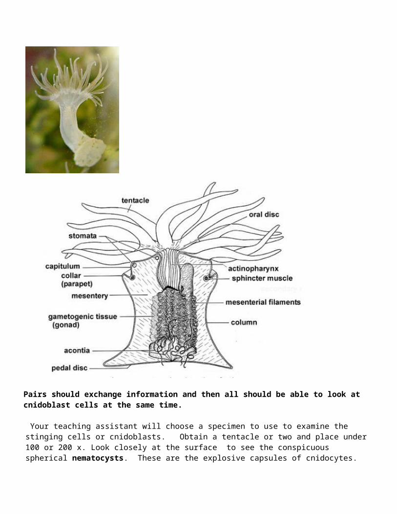
1. Sea Anemones: The external morphology of anemones is limited to a column; an oral disk, in the center of which the mouth is located, and on which the tentacles are located; and either a pedal disk, that affixes the anemone to the substrate, or a bulb-like physa, used by burrowing anemones to anchor in soft substrate.
.
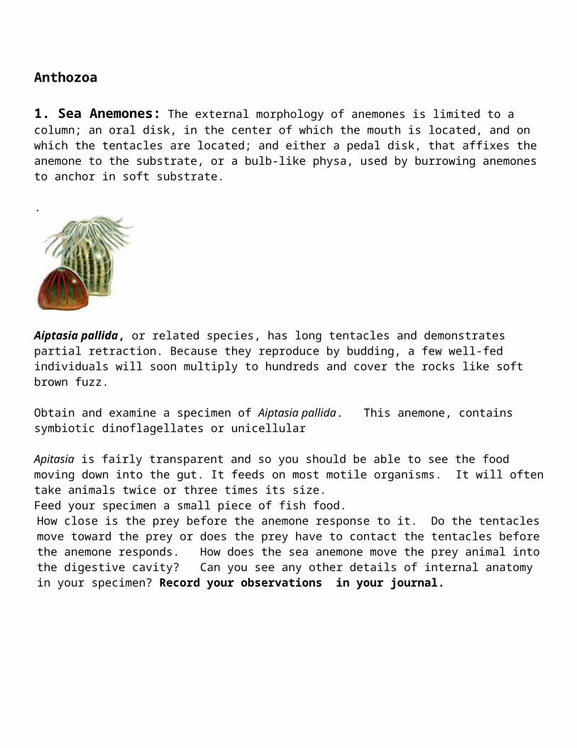
Aiptasia pallida, or related species, has long tentacles and demonstrates partial retraction. Because they reproduce by budding, a few well-fed individuals will soon multiply to hundreds and cover the rocks like soft brown fuzz.
Obtain and examine a specimen of Aiptasia pallida. This anemone, contains symbiotic dinoflagellates or unicellular Apitasia is fairly transparent and so you should be able to see the food moving down into the gut. It feeds on most motile organisms. It will often take animals twice or three times its size.Feed your specimen a small piece of fish food.How close is the prey before the anemone response to it. Do the tentacles move toward the prey or does the prey have to contact the tentacles before the anemone responds. How does the sea anemone move the prey animal into the digestive cavity? Can you see any other details of internal anatomy in your specimen? Record your observations in your journal.
Pairs should exchange information and then all should be able to look at cnidoblast cells at the same time.
Your teaching assistant will choose a specimen to use to examine the stinging cells or cnidoblasts. Obtain a tentacle or two and place under 100 or 200 x. Look closely at the surface to see the conspicuous spherical nematocysts. These are the explosive capsules of cnidocytes. The cnidocytes themselves will probably not be discernable but their capsules are abundant and
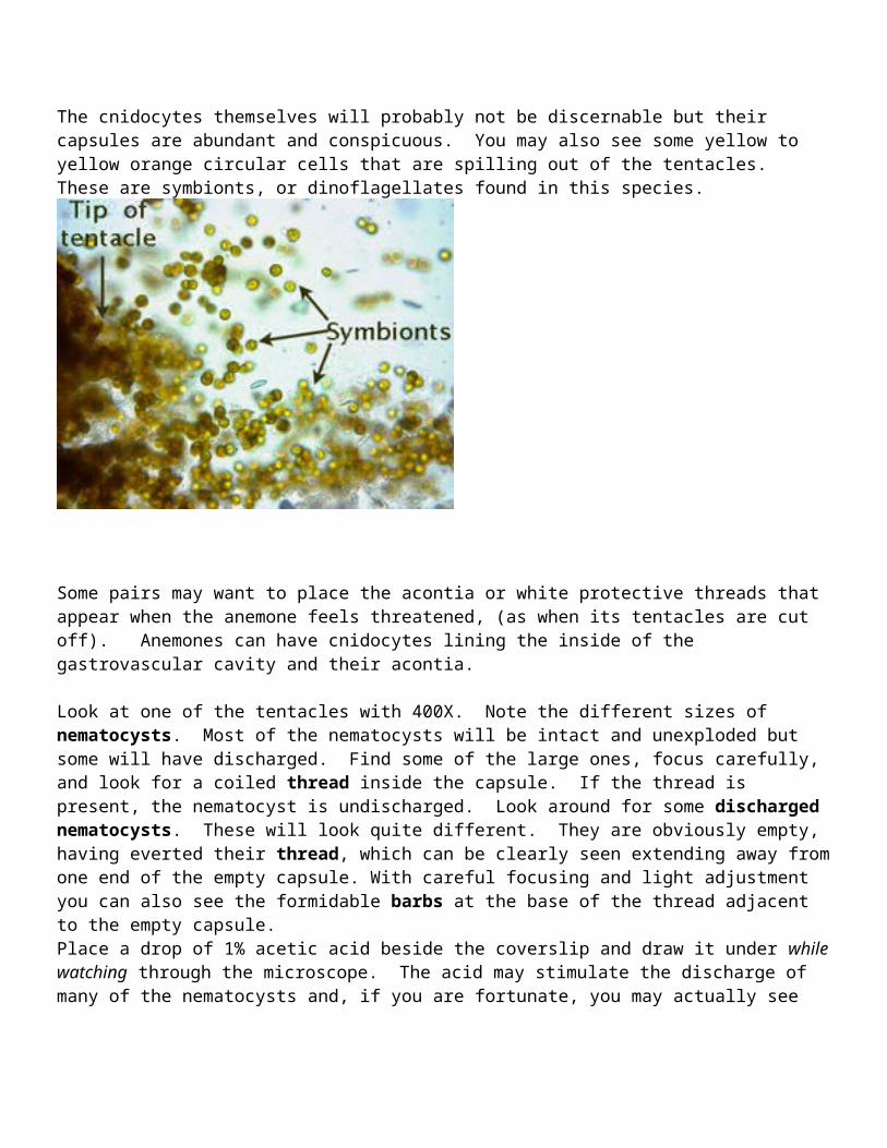
conspicuous. You may also see some yellow to yellow orange circular cells that are spilling out of the tentacles. These are symbionts, or dinoflagellates found in this species.
Some pairs may want to place the acontia or white protective threads that appear when the anemone feels threatened, (as when its tentacles are cut off). Anemones can have cnidocytes lining the inside of the gastrovascular cavity and their acontia.
Look at one of the tentacles with 400X. Note the different sizes of nematocysts. Most of the nematocysts will be intact and unexploded but some will have discharged. Find some of the large ones, focus carefully, and look for a coiled thread inside the capsule. If the thread is present, the nematocyst is undischarged. Look around for some discharged nematocysts. These will look quite different. They are obviously empty, having everted their thread, which can be clearly seen extending away from one end of the empty capsule. With careful focusing and light adjustment you can also see the formidable barbs at the base of the thread adjacent to the empty capsule. Place a drop of 1% acetic acid beside the coverslip and draw it under while watching through the microscope. The acid may stimulate the discharge of many of the nematocysts and, if you are fortunate, you may actually see one of them discharge as you watch. A drop of toluene blue applied the same way will stain the nematocysts, making them easier to see.
Obtain (a.) a photograph of a tentacle, discharged nematocysts and symbionts. Identify symbiotic algae and nematocysts on the photograph.
2. Gorgonians,Gorgonians are also known as octocorals, so named because the individual polyps have eight tentacles that they use to feed. If you look closely at the branches of a soft coral (gorgonians are probably the most obvious), you may see the tiny tentacles of the individual polyps. Each of these tentacles may looks like it is feathered
because it can bears numerous outfoldings. The main purpose of this exercise is for you to observe these animals and be able to compare them to the hydroid colonies you observed previously (Hydractinia and any hydroids available today), Many gorgonians, sea pansies or pens look superficially like hydroid colonies, but on close examination, you can see their more “sea anemone” or anthozoan morphology. One of the lab questions on the next exam will focus on distinguishing between the two.
------------------------------------------------------------------------------------------Available Octocorals Gorgonia sp. red or yellow
Look at a portion of the available Gorgonian colony under the dissecting scope. Be patient as it may take a while for the individuals to relax and expand. Focus on the tentacles and see if you can observe their “pinnate” nature. Count number of tentacle, etc. a. Obtain a photograph and indicate “octocoral” features, b. Compare the structure of these colonies to that of an hydrozoan colony and Aiptasia in your journal.