Embed Size (px)
Citation preview
101
4.1. PRELUDE
An antibiotic is a substance produced by various species of
microorganisms (bacteria, fungi, actinomycetes), that suppresses the growth
of other microorganisms and may eventually destroy them. However,
common usage often extends the term antibiotic to include synthetic
antibacterial agents, such as sulfonamides and quinolones, which are not
products of microbes. Therapeutic science has come a long way from the
insight and experiments of Ehrlich and Flemming. In the last two decades
the list of antimicrobials has expanded exponentially, with the introduction
of many new groups like quinolones, polypeptides etc. or improved
derivatives of the major groups.
The introduction of antibiotics in 1940’s revolutionized the practice of
medicine. The discovery, development and clinical exploitation of antibiotics
can arguably be counted as one of the most significant medical advances of
the twentieth century. Their potential for misuse was also recognized soon.
It is therefore sobering that at the beginning of the twenty first century,
articles abound concerning resistance. These super bugs with no new drugs
to treat them, question the prospects of a post antibiotic era! Thereafter this
has steadily increased with each passing decade. Widespread emergence of
resistant organisms is the biggest fallout of irrational antibiotic use.
Penicillin resistant pneumococcal infections, methicillin resistant
Staphylococcus aureus, multi-drug resistant Klebsiella pneumoniae, vancomycin
102
resistant Enterococci, extended spectrum beta-lactamase producing
Enterobacteriaceae complicate therapy immensely in hospitalized patients
(New, 1992). Antibiotic resistance is believed to have evolved millions of
years ago in soil bacteria as a means of protecting themselves against other,
antibiotic-producing bacteria or their own antimicrobial products (Davis et
al., 1971).
At present, medical practice at social and private level cannot be
understood without taking into account the quality control and cost-benefit
of medical activities. These parameters are essential for program assessment,
implementation of therapies and for individual or collective decisions.
Antibiotic resistance is believed to have evolved millions of years ago in soil
bacteria as a means of protecting themselves against other, antibiotic-
producing bacteria or their own antimicrobial products (Davies et al., 1971).
However, most researchers believe that the current crisis of antibiotic
resistance is bred from the intense selection pressure posed by wide spread
use and misuse of antibiotics; indeed, organism archived prior to the current
age of antibiotic excess do not display nearly the number of diversity of
hydrolyzing enzymes present in recent clinical isolates (Hughes et al., 1983).
Among the many factors that have contributed to the emergence and
spread of multiple-resistant organisms, three are of key importance:
mutations in common resistance genes that have extended their spectrum of
resistance; exchange of genetic information among microorganisms,
103
transferring well-known genes into new hosts and the increase in selective
pressure in hospitals, other institutional settings and communities that allow
resistant organisms to proliferate. The first two reflect the ability of bacteria
to adapt to changing environments, while the last emphasizes that
environmental conditions often enhance the emergence of novel phenotypes.
All three are clearly interrelated (New, 1992; Murray, 1991).
Mutations in common genes:
One example of the role of mutations in emerging resistance concerns
changes in -lactamases that extend their spectrum of activity. -Lactamases
are enzymes that inactivate, -lactam drugs, such as penicillin, ampicillin,
and cephalothin (Bush et al., 1995). Until recently, these enzymes were not
able to hydrolyze newer extended-spectrum cephalosporins such as
cefotaxime and ceftazidime. However, in 1982, mutant forms of the
-lactamases were reported that were capable of inactivating the extended-
spectrum cephalosporins and monobactams, such as aztreonam (Knothe et
al., 1983). The enzymes called extended-spectrum -lactamases (ESBLs), have
now been isolated from organisms throughout Europe, Japan and the United
States. At least 30 such ESBLs have now been reported (Phillipon et al., 1989,
Jacoby et al., 1997). When the amino acid sequences of the first ESBLs were
examined, only three differences were found between the ESBL and the
wild-type -lactamase that is found in 70% of ampicillin-resistant, but
extended-spectrum cephalosporin susceptible, E. coli (Agarwal et al., 1997).
104
These amino acid changes were the reflection of point mutations in the
coding sequence of the -lactamase gene. Recent experience suggests that
under the selective pressure of high cephalosporin usage in a hospital ward,
organisms with mutations that extend the spectrum of activity
of -lactamases can emerge and disseminate (Meyer et al., 1993).
The development and spread of ceftazidime-resistant Klebsiella pnuemoniae
is perhaps the best example of this phenomenon (Rice et al., 1990).
These organisms, which contain variations of common blasTEM or blaSHV
-lactamase genes (bla for beta-lactamase), have caused a number of
outbreaks in the United States (Burwen et al., 1994). Mutations in -lactamase
genes, however, are but one-way mutations can lead to resistance.
Reduced membrane permeability limiting a drug’s access into the cell and
changes in a drug’s target site can also be a result of mutation.
Genetic exchange:
Bacteria can exchange genetic information by transformation (the
uptake of naked DNA), transduction (transfer of DNA by bacteriophage),
and conjugation (cell-to-cell contact) (Johnson et al., 1998). Although
conjugation was previously though to be limited to Gram-negative bacilli,
extensive data now confirm the existence of a similar transfer process among
Gram-positive organisms whereby plasmids or independent transposable
elements, often carrying multiple-resistance genes, move from one organism
to another. The transfer process extends even to highly unrelated groups of
105
organisms, such as Campylobacter coli and Enterococci (Trieu et al., 1985),
which have been shown to exchange aminoglycoside resistance genes.
Enterococci demonstrate how organisms can gradually accumulate
resistance genes by genetic exchange and develop into multi-resistant
nosocomial pathogens causing untreatable infections. Enterococci accounted
for 16571 nosocomial infections in hospitals reporting to the national
nosocomial infection surveillance system during the period from January 1,
1989, to March 31, 1993 (CDC, 1993). Over the last decade, Enterococci have
become increasingly resistant to multiple antimicrobial agents. For example,
the genes encoding resistance to gentamicin and the production of
-lactamase were acquired from staphylococci and when coupled with
resistance to streptomycin and vancomycin, resulted in organisms that were
very difficult, if not impossible (Handwerger et al., 1993, Montecalvo et al.,
1994; Vashishtha, 2000), to treat. Streptomycin resistance is mediated
primarily by an aminoglycoside-modifying enzyme ANT, although
resistance due to ribosomal mutation has also been noted. Vancomycin
resistance can be mediated by several genes, including van A, van B, van C1,
or van C2 (Handwerger et al., 1993, Montecalvo et al., 1994), that function in
concert with other genetic loci frequently located on the same transposable
element. The alarming proportion of nosocomial enterococcal isolates
resistant to vancomycin, particularly from patients intensive care units,
106
which rose from 0.4% to 13.6% in the 4-year period from 1989 to 1993,
continued to rise in 1994 and 1995.
Although resistance genes from gram-positive organisms may be
transferred into Gram-negative organisms or vice versa, differences in the
genetic control mechanisms present in various species may limit the ability
of the new resistance genes to be expressed and to mediate (Davies et al.,
1971) resistance. Thus, there appear to be some limits to the constellation of
resistance genes that can be assembled and expressed in bacteria.
Nonetheless, the ability of bacteria to acquire resistance genes from other
members of the normal bacterial flora under the selective pressure of
antimicrobial use should not be underestimated. It has been shown, for
example, that the van A vancomycin resistance gene cluster in Enterococcus
faecalis can transfer to aureus via conjugation and can express high-level
resistance (Noble et al., 1992). Although an S. aureus isolate with high-level
vancomycin resistance has yet to be encountered in nature, the possibility
clearly exists that the genetic unit that includes van A, or perhaps one of the
other vancomycin resistance genes, will be transferred under natural
conditions to S. aureus. Such an occurrence would have serious public health
implications, since nosocomial strains of S. aureus, which tend to be resistant
to multiple drugs, could acquire the vancomycin resistance gene, rendering
current treatment ineffective.
107
Selective pressures in institutional and community settings
The concept of selective pressure refers to the environmental
conditions that encourages or enhance the proliferation of strains of bacteria
that develop resistance to antimicrobial agents through spontaneous
mutation or by acquisition of new DNA. It is hypothesized that organisms
with new mutations or genes would likely not survive were it not for the
selective pressures that encourage their emergence. In addition to
communications or hospital settings expanded use of antimicrobial agents in
sites out side the hospital, including nursing homes, day care centers and
animal feedlots, increases the selective pressure for resistant organisms to
emerge (American Society for Microbiology, 1995). The use of broad-
spectrum antimicrobial agents is increasing in outpatient settings.
Antimicrobial drugs are often used for common conditions for which their
effectiveness is unclear (Knothe et al., 1983; Gonzales et al., 1995).
Streptococcus pneumoniae serves as an example of a community-
acquired organism that has become increasingly resistant to a wide
variety of antimicrobial agents (Knothe et al., 1983; Reinert et al., 1995).
The metropolitan Atlanta, Ga, area noted that 25% of invasive pneumococcal
isolates from that region were no longer susceptible to penicillin and
9% were no longer susceptible to extended spectrum cephalosporins
(Hofmann et al., 1995) and pneumococcal isolates in Franklin Country, Ohio,
were penicillin-resistant. The development and spread of multiply-resistant
108
Pneumococci can be a major problem among children in day care centers
when antimicrobial agent use is often high (Knothe et al., 1983), in part
because children have clinically confirmed, or suspected, otitis media
receiving antimicrobial agents the likelihood increases that multi-resistant
organisms will be found in their throats that may disseminate to other
children. Among three children with pneumococcal meningitis caused by
strains resistant to cefotaxime and ceftriaxone, all had received prior
cephalosporin therapy for otitis media (Sloas et al., 1992). Thus, control of
antimicrobials, particularly those used prophylactically in children attending
day care centers, is in issue that must be reassessed.
While resistance is increasing in many pathogens, the number of new
antimicrobial agents approved for use in the United States has slowed.
In 1993 only a single antibacterial agent was approved for use in the United
States by the Food and Drug Administration and in 1994, no new
antibacterial agents were approved (National Committee for Clinical
Laboratory Standards, 1993). Thus, our ability to control outbreaks of
infectious diseases through antimicrobial use alone is diminishing. A recent
report from the Hospital Infection Control Practices Advisory Committee on
preventing the spread of vancomycin resistance (FDA, 1994) stresses the
need for professional and public educational programs, enhanced
microbiological surveillance, enhanced surveillance among patients, effective
implementation of infection control procedures and perhaps most important,
109
prudent use of antimicrobial agents for treatment and prophylaxis.
Whether these recommendations can be generalized to aid in the control of
other types of resistant organisms found in the hospital environment
remains to be seen. Studies to validate the effectiveness of the guidelines in
controlling the spread of vancomycin-resistant Enterococci are in progress.
The report also notes that our major surveillance system in the United States
for the detection of novel resistant organisms is the network of hospital
microbiology laboratories, since it is these data that feed into the National
Nosocomial Infections Surveillance System and other such programs.
However, with the exception of M. tuberculosis and N. gonorrhoeae, there are
no national surveillance systems that systematically monitor resistance in
community-acquired infections. While some states are initiating surveillance
of penicillin resistance in Pneumococci, H. influenzae, it is up to infectious
disease specialists, clinical microbiologists, pharmacists and public health
personnel to be vigilant for organisms with novel resistance patterns so that
control measures can be effectively implemented. Emergence of drug-
resistant organisms also has clear implications for vaccine and antimicrobial
drug development priorities.
Certain non-fastidious, Gram-negative bacilli possess the ability to
rapidly develop resistance to many of the newer “enzyme stable” -lactum
antibiotics. This finding poses many clinical problems including emergence
of resistance during therapy with the drugs. Therapeutic alternatives for
110
patients are severely limited which this problem occurs because multiple
drug resistance may arise simultaneously. To date, two mechanisms have
been found to be responsible for this resistance. The first, which produces
multiple -lactum resistance, is the induction of chromosomal -lactamases
that mediate resistance to non-substrate drugs by the creation of a non-
hydrolytic barrier that blocks access to target patients within the cell.
The second mechanism, which produces -lactam/aminoglycoside
resistance, involves a change in outer membrane permeability.
Many problems have been encountered with the newer generation of
-lactam antibiotics. Most of these problems have involved the ability of
certain non-fastidious, Gram-negative bacilli to rapidly develop resistance to
the drugs. Such resistance was unanticipated when these antibiotics were
developed since they had been designed to solve the problems posed by the
-lactamase that had limited efficacy of older penicillin’s and cephalosporins.
However, stability of -lactamases does not guarantee producing bacteria
rather; this characteristic appears to be responsible for many of the problems
that have occurred with these new drugs.
Many major improvements in antimicrobial agents have been based
upon knowledge of the mechanisms responsible for microbial drug
resistances, microbial resistance due to production of drug inactivating
enzymes has been circumvented by the design of new enzyme stable
compounds. Such “antibiotic engineering” led to the development of
111
penicillinase resistant penicillin and several new aminoglycosides that are
resistant to the diver’s aminoglycosides inactivating enzymes of bacteria.
These drugs represented major improvements in our antimicrobial
armamentarium. When the new -lactamase-stable cephalosporins were
developed, it was only logical to expect a similar improvement with these
agents. However, is much respect they have not lived up to expectations?
Extended Spectrum Beta Lactamase Mediated Resistance
ESL was first detected in Europe in 1978, in USA 1981 and in
Germany during 1983 (Medeiros, 1997). These resistant strains are
established in many hospitals, producing epidemic diseases. These ESL
producing strains have become more and more important because of their
ability to adopt in the hospital environment and cause more outbreaks in
hospitals (Guillume et al., 1993, Chaibi et al., 1999).
Gram-negative bacteria are an important hospital acquired pathogen
that causes severe morbidity and mortality in neonatal and pediatric
patients. Several out breaks of infection caused by K. pneumoniae isolates that
are simultaneously resistant to broad-spectrum cephalosporins and
aminoglycosides have been reported (Arlet et al., 1990; Bush et al., 1995,
2001). Over the last 20 years, many new -lactam antibiotics have been
developed that were specifically designed to be resistant to the hydrolytic
action of -lactamases. However, with each new class that has been used to
treat patients, new -lactamases emerged that caused resistance to this class
112
of drug. Not surprisingly, resistance to these expanded spectrums -lactam
antibiotics due to -lactamases emerged quickly. The first of these enzymes
capable of hydrolyzing the newer -lactam, SHV-2, was found in a single
strain of Klebsiella ozaenae isolated in Germany (Kim et al., 2000). Because of
their increased spectrum of activity, especially against the oxyimino-
cephalosporins these enzymes were called extended spectrum -lactamases
(ESLs). The organisms that are resistant to third generation cephalosporins
also shows high degree of resistance to other class of antibiotic such as
cepholothin, piperacillin and ampicillin etc, therefore they were named as
extended broad-spectrum -lactamases. Although the original definition of
what constituted an ESL was primarily based on the substrates hydrolyzed
by the enzymes, more recently the term ESL has been limited to those
-lactamases that are inhibited by clavulanic acid in addition to showing the
enhanced spectrum of activity. During early 1980, strains of Klebsiella
with reduced susceptibility to third generation cephalosporins were
noted in Europe. Today over 150 different ESLs have been described.
These -lactamases have been found worldwide in many different genera of
Enterobacteriaceae and Pseudomonas aeruginosa. Resistance was mediated by
new -lactamases present on plasmids. These resistances were soon passed
on to E. coli, which later showed resistance to the same antibiotic for which
Klebsiella was resistant. These enzymes were derived from plasmid mediated
113
class A, B lactamase in Gram-negative bacilli such as TEM-1 and SHV-1
(Chaibi et al., 1999; Heritage et al., 1999).
In the absence of specific surveillance ESL producing strains in our
country are probably more prevalent than currently recognized, because
they are often undetected by routine susceptibility testing methods (Agarwal
et al., 1997). Recent reports have highlighted the emergency of ESL
producing strains endowed with an extremely wide spectrum of antibiotic
resistance including resistance to trimethoprim, amikacin, gentamicin and
streptomycin. The increased prevalence of Enterobacteriaceae producing ESL
creates a great need for laboratory testing methods that will accurately
identify the presence of these enzymes in clinical isolates.
Functional group 1 -lactamases are described as cephalosporinases
which are not inhibited by clavulanate (Bush et al., 1995). Originally, these
enzymes were naturally occurring, chromosomally encoded AmpC
-lactamases found in genera such as Enterobacter, Citrobacter, Serratia and
Pseudomonas. The genes encoding these enzymes were found on the
chromosome and are inducible by certain beta lactam antibiotics (Sanders et
al., 1988). However, in recent years some of these genes have found their way
onto plasmids and are being expressed constitutively at high levels in
Klebsiella pneumoniae and Escherichia coli. Whereas the extended-spectrum
β-lactamases in these organisms confer resistance to the expanded-spectrum
β-lactam antibiotics such as ceftazidime, cefotaxime and aztreonam, the
114
plasmid-mediated AmpC-type enzymes also confer resistance to the
cephamycins. The first of these to be described was the MIR-1 enzyme
which, on the basis of a partial DNA sequence, appears to have originated
from the AmpC β-lactamase of Enterobacter cloacae (Agarwal et al., 1997).
Since that time a number of other enzymes such as BIL-1, CMY-1, CMY-2,
LAT-1, LAT-2 (Gazouli et al., 1997), FOX-1, and MOX-1 (Horii et al., 1992)
have been described. These enzymes are now being found on plasmids with
increasing frequency.
Metallo beta lactamases
The increase in antibiotic resistance among Gram-negative bacteria is
a notable example of how bacteria can procure, maintain and express new
genetic information that can confer resistance to one or several antibiotics.
This genetic plasticity can occur both inter- and intragenerically.
Gram-negative bacterial resistance possibly now equals or usurps that of
gram positive bacterial resistance and has prompted calls for similar
infection control measures to curb their dissemination (Poole, 2003).
Reports of resistance vary, but a general consensus appears to prevail that
quinolone and broad-spectrum β-lactam resistance increasing in members of
the family Enterobacteriaceae and Acinetobacter spp. and that treatment
regimes for the eradication of Pseudomonas aeruginosa infections are
becoming increasingly limited (Maniatis et al., 2003, Neuhauser et al., 2003).
For example, a 5-year longitudinal study involving many centres from Latin
115
America indicated that year after year, P. aeruginosa resistance has
continually risen to the point where approximately 40% are resistant to
“antipseudomonal” drugs, including carbapenems (Andrade et al., 2003).
While the advent of carbapenems in the 1980s heralded a new treatment
option for serious bacterial infections, carbapenem resistance can now be
observed in Enterobacteriaceae and Acinetobacter spp. and is becoming
commonplace in P. aeruginosa. Gram-negative bacteria have at their disposal
a plethora of resistance mechanisms that they can sequester and/or evince,
eluding the actions of carbapenems and other β-lactams. The common form
of resistance is either through lack of drug penetration (i.e., outer membrane
protein [OMP] mutations and efflux pumps), hyper-production of an AmpC-
type-lactamase and/or carbapenem-hydrolyzing-lactamases. Based on
molecular studies, two types of carbapenem-hydrolyzing enzymes have been
described: serine enzymes possessing a serine moiety at the active site, and
metallo-β-lactamases (MBLs), requiring divalent cations, usually zinc, as
metal cofactors for enzyme activity (Bush et al., 1995; Bush, 2001, Buynak et
al., 2004).
This is the first extensive study to prospectively look for ESL, AmpC
and metallo beta lactamase producing bacteria in pyogenic meningitis cases.
116
Detection of bacterial drug resistance in the clinical laboratory:
Several of the new resistance mechanisms recognized in Gram-
positive and Gram-negative organisms are difficult to detect with current
laboratory methods. For example, vancomycin resistance in Enterococci can
be difficult to detect by automated methods, such as Vitek (BioMerieux USA,
Hazelwood, Mo) and Microscan walk-away (Dade International, West
Sacramento, Calif), but is readily detected by most other broth and agar-
based methods (Tenover 1991). A vancomycin resistance agar screen test has
been developed as a backup to traditional susceptibility testing methods for
the detection of vancomycin-resistant Enterococci (Knothe et al., 1983).
With regard to resistance in Gram-negative bacilli, a major problem
noted recently is the detection of cefotaxime ceftriaxone and ceftazidime
resistance in K. pneumoniae and E. coli, particularly when resistance is
mediated by ESBLs. In part, the inability of the laboratory to recognize
resistant strains is due to the minimal inhibitory concentration (MIC)
breakpoints chosen by the National committee for Clinical Laboratory
standards to define “resistance” (1993). Then breakpoints, which for
extended-spectrum cephalosporins are usually 32 or 64 g/mL, were chosen
based on population distributions of bacteria before ESBLs became widely
disseminated. Depending on the antimicrobial agent tested in the laboratory,
MICs for ESBL-containing strains may vary anywhere from 4 g/mL
(susceptible) to 256 g/mL (highly resistant). Variation depends on the type
117
of ESBL (of which there are over 30), the antimicrobial agent tested, and the
method of testing. Given that the usual MIC of K. pneumoniae to ceftazidime
is 0.06 g/mL, it is possible to achieve a 50-fold increase in the MIC and still
be in the “susceptible” MIC range. However, if cefotaxime were tested in lieu
of ceftazidime, resistance may well be detected. New laboratory tests to
confirm the presence of ESBLs in K. pneumoniae and E. coli, such as disk
diffusion assays performed in parallel with and without the addition of the
enzyme inhibitor clavulanic acid may aid in the detection of such organisms,
other commercially prepared tests are under evaluation. Studies sponsored
by the National Committee for Clinical Laboratory Standards are currently
under way to develop alternate testing procedures to detect ESBL-mediated
resistance. Because laboratories need to screen a variety of bacterial
pathogens that were once considered uniformly susceptible, a guide to
appropriate susceptibility testing methods is presented in the Table.
While new testing methods may aid in identifying organisms with novel
resistance genes, growing restraints on the personnel and supply budgets in
hospital based microbiology laboratories may hinder the widespread
implementation of the tests. In fact, new laboratory information systems
often mask important quantitative information, such as the actual MICs of
antibiotics, in lieu of reporting only the interpretive categories of susceptible,
intermediate and resistant. Thus, physicians who suspect the presence of
new resistant organisms in their hospitals should consult with the hospital’s
118
microbiologists regarding the optimal approach to identifying and testing of
such organisms.
Objectives
1. To evaluate the susceptibility pattern of the organisms isolated from
pyogenic meningitis.
2. To evaluate extended spectrum of beta lactamase production.
3. To evaluate AmpC production in gram negative bacilli.
4. To evaluate metal beta lactamase production.
5. To evaluate methicillin resistant among Staphylococcus aureus.
4.2. MATERIALS AND METHODS
The primary purpose of antimicrobial susceptibility testing is to
provide clinicians with information that will assist in the choice of an
appropriate antibiotic for the patients. Laboratory report indicating that an
organism is sensitive to an antibiotic implies that the organism is relevant to
the patient’s clinical condition and that the infection is treatable under a
given set of pharmacological conditions. Reporting of resistance implies that
infection is not treatable with the antibiotic.
A total of 61 consecutive clinical isolates of which Streptococci
pneumoniae (89), H. influenzae (n=51), Group B Streptococci (19), N. meningitides
(13), Staphylococcus aureus (n=11), Klebsiella pneumoniae (n=08), E. coli (n=05)
and Pseudomonas aeruginosa (n=03), obtained from pyogenic meningitis cases
119
were identified by standard methods were included in this study.
Susceptibility to antibiotics was tested by disk diffusion method (Kirby
Bauer’s disc diffusion method). Extended spectrum beta lactamases was
detected by double disk diffusion synergy test (Bauer et al., 1966, NCCLS,
1993, 1997, 1998, 2000, 2004).
4.2.1. Disk diffusion by Kirby Bauer’s disc diffusion method:
The basic principle of disc diffusion method is the antibiotic-
impregnated disc absorbs moisture from the agar and antibiotic diffuses into
the agar medium. The rate of extraction of antibiotic from the disc is greater
than the rate of diffusion. As the distance from the disc increases, there is a
logarithmic reduction in the antibiotic concentration. The depth of the agar
affects the extent of antimicrobial diffusion. Visible growth of bacteria occurs
on the surface of the agar where the concentration of antibiotic has fallen
below its inhibitory level of the test strain. The results were interpreted as
per Kirby-Bauer’s chart provided by the procured company (Hi-media Ltd.
Mumbai).
All the organisms isolated from pyogenic meningitis were subjected
for antimicrobial susceptibility testing. The medium (Muller-Hinton) was
prepared and sterilized and was dispensed into flat-bottomed 9 cm glass
petri dishes. The pH of the medium was checked when medium was
prepared. Inoculum was prepared by picking 3–4 morphologically similar
120
colonies from the culture plates and inoculating into a test tube containing
peptone water. The tubes were incubated for 2 hrs at 370C to produce a
bacterial suspension of moderate turbidity. The inoculum size was adjusted
by comparison with a barium sulfate standard, 0.5 Mac Farlands standard
units (Bauer et al., 1966).
A sterile swab was dipped into the inoculum and excess of inoculum
was removed by pressing and rotating the swab firmly against the side of the
tube, above the level of the liquid. Swab was streaked all over the surface of
the medium three times, rotating the plate through an angle of 600 after each
application. The inoculum was left to dry for few minutes at room
temperature with the lid closed. The antibiotics used were penicillin,
ampicillin, erythromycin, gentamycin, amikacin, ciprofloxacin, cephalexin,
cefotaxime, ceftazidime, cefotaxime, ceftriaxone, cefuroxime and ofloxacin
(Hi-media laboratories, Mumbai). Discs were placed on the inoculated
plates using a pair of sterile forceps. The plates were incubated at 350C for
18-24 hrs. After overnight incubation, the diameter of each zone (including
the diameter of the disc) was measured and recorded in mm. The size of the
zone of inhibition is an indication of the susceptibility of the pathogen.
The zone sizes were compared with the standard chart and reported
accordingly.
121
4.2.2. Antimicrobial susceptibility testing of H. influenzae by the disk
diffusion method
Prepare the inoculum for seeding the antimicrobial susceptibility
media with H. influenzae from fresh, pure cultures of H. influenzae (i.e., from
isolates grown overnight on supplemented chocolate agar). Prepare cell
suspensions of the bacteria to be tested in broth or sterile physiological
saline; use a suspension equal to a density of a 0.5 McFarland turbidity
standard for the inoculum.
a) Suspend viable colonies from an overnight chocolate agar plate in a
tube of broth to achieve a bacterial suspension equivalent to a
0.5 McFarland turbidity standard; be careful not to form froth or
bubbles in the suspension when mixing the cells with the broth.
This suspension should be used within 15 minutes.
b) Compare the suspension to the 0.5 McFarland turbidity standard by
holding the suspension and the McFarland turbidity standard in front
of a light against a white background with contrasting black lines and
compare the density. If the density of the suspension is too heavy, the
suspension should be diluted with additional broth. If the density of
the suspension is too light, additional bacteria should be added to the
suspension.
122
c) When the proper density is achieved, dip a cotton swab into the
bacterial suspension. Press the swab on the side of the tube to drain
excess fluid.
d) Use the swab to inoculate the entire surface of the HTM plate three
times, rotating the plate 60 degrees between each inoculation. Use the
same swab with each rotated streak, but do not re-dip the swab in the
inoculum (i.e., the bacterial cell suspension).
e) Allow the inoculum to dry before the disks are placed on the HTM
plates. Drying usually takes only a few minutes and should take no
longer than 15 minutes. (If drying takes longer than 15 minutes, use a
smaller volume of inoculum in the future).
f) After the plate is dry, antimicrobial disks should be placed on the
HTM plate. The disks should be placed on the agar with sterile
forceps and tapped gently to insure adherence to the agar.
Diffusion of the drug in the disk begins immediately; therefore, once a
disk contacts the agar surface, the disk should not be moved.
g) Invert the plate and incubate it in a CO2-enriched atmosphere
(5% CO2- incubator or candle-extinction jar) for 16–18 hours at 350C.
h) After overnight incubation, measure the diameter of each zone of
inhibition. The zones of inhibition on the media containing blood are
measured from the top surface of the plate with the top removed.
123
Use either calipers or a ruler with a handle attached for these
measurements, holding the ruler over the center of the surface of the
disk when measuring the inhibition zone. Care should be taken not to
touch the disk or surface of the agar. Sterilize the ruler occasionally to
prevent transmission of the bacteria. In all measurements, the zones of
inhibition are measured as the diameter from the edges of the last
visible colony. Record the results in millimeters (mm).
i) Interpretation of the antimicrobial susceptibility is obtained by
comparing the results obtained and recorded (in the manner
described in this protocol) to the NCCLS standard inhibition zone
diameter sizes.
4.2.3. Antimicrobial susceptibility testing of N. meningitidis by E test
All the isolates were subjected to antibiotic susceptibility testing based
on the almost 100% susceptibility of N. meningitidis to commonly used
antibiotics. The antibiotic susceptibility patterns of 100 Neisseria meningitidis
strains to cotromixazole, chloramphenicol, cefotaxime and penicillin were
determined by a disc diffusion method using Mueller Hinton II agar with 5%
sheep blood. The table used for interpretation of disc diffusion results was
that recommended by the Clinical and Laboratory Standards Institute (CLSI)
2005.
124
Either 150-mm or 100-mm plates can be used for the E-test®,
depending on the number of antimicrobial agents to be tested per isolate.
Two different E-test® antimicrobial strips can be placed in opposite gradient
directions on a 100-mm plate and although the manufacturer states that up
to six E-test® strips can be used on a 150-mm plate, this laboratory manual
suggests that in order to avoid overlapping zones of inhibition of growth, not
more than five E-test® strips be used on a 150-mm plate. Mueller Hinton
chocolate agar was used.
4.2.4. Tests for ESBL production
Double disk approximation test for screening:
The test organisms were applied on to a Mueller Hinton agar plate by
adjusting turbidity to McFarland no 0.5 tube. Antibiotic discs of amoxicillin/
clavulanic acid (20/10 µg) and cefotaxime (30 µg) were placed at a distance
of 15 mm apart and incubated. Organisms that showed a clear extension of
cefotaxime inhibition zone towards the disc containing Clavulanate were
considered as ESBL producer. The organisms which were screened and
found positive for ESBL production were subjected to confirmatory test.
NCCLS phenotypic confirmatory test:
Ceftazidime (30 µg) and ceftazidime plus Clavulanic acid (30/10 µg)
were placed on Mueller Hinton agar and incubated. Organism was
considered as ESBL producer if there was a ≥ 5mm increase in diameter of
ceftazidime plus clavulanic disc and that of ceftazidime disc alone.
125
4.2.5. Amp C Disk Test:
Isolates were tested for AmpC activity by a three-dimensional extract
method, which was an adaptation of procedures described previously for the
detection of extended-spectrum beta-lactamases (ESBLs) (Agarwal, 1997).
Briefly, 50 µl of a 0.5 McFarland bacterial suspension prepared from an
overnight blood agar plate was inoculated into 12 ml of tryptic soy broth and
the culture was grown for 4 h at 35°C. The cells were concentrated by
centrifugation and freezing-thawing the cell pellets five times made crude
enzyme preparations. The surface of a Mueller-Hinton agar plate was
inoculated with one each of two E. coli strains (ATCC 25922 and ATCC
11775) as described for the standard disk diffusion method (NCCLS, 1999);
a 30-µg-cefoxitin disk was placed on the inoculated agar. With a sterile
scalpel blade, a slit beginning 5 mm from the edge of the disk was cut in the
agar in an outward radial direction. By using a pipette, 25 to 30 µl of enzyme
preparation was dispensed into the slit, beginning near the disk and moving
outward. Slit overfill was avoided. The inoculated media were incubated
overnight at 35°C. Enhanced growth of the surface organism at the point
where the slit intersected the zone of inhibition was considered as positive
for three-dimensional test result and was interpreted as evidence for the
presence of AmpC beta-lactamase.
126
4.2.6. Metallo β-lactamase (MBL) production
Gram negative organisms that showed resistance to Imipenem were
selected for MBL production.
Imipenem-EDTA combined disc test
This test was performed according to Yong et al. test organisms were
inoculated onto Mueller Hinton agar plates as per the CLSI
recommendations. Two 10µg imipenem disks were placed on the plate and
10 µl of sterile 0.5 M EDTA solution was added to one of the imipenem disk.
The inhibition zones of the imipenem and imipenem plus EDTA disks were
compared after inoculation. If the increase in inhibition zone with the
imipenem plus EDTA disc was ≥ 7mm than the imipenem disc alone, it was
considered as MBL positive.
4.2.7. Detection of methicillin resistant Staphylococcus aureus
Methicillin resistance was detected by disc diffusion technique using
30µg cefoxitin disc (Hi-Media, Mumbai). The suspension for inoculation was
prepared from the colonies from an overnight growth on nutrient agar plate.
The growth was suspended in 0.5 ml of sterile saline and the turbidity was
adjusted to 0.5 Mcfarland’s units. A sterile swab dipped into this suspension
and the excess of inoculum was removed by pressing it against the sides of
the tube. This swab was used to inoculate on Mueller-Hinton agar plate.
Cefoxitin disc was applied within 15 min after the inoculation. The plates
were incubated for 24 hrs at 370C. The diameter of the zone around the disc
127
was measured and the results were interpreted. Staphylococcus aureus NCTC
12493 was used as the control strain.
4.3. RESULT
Of 236 samples cultured, 199-yielded growth of microorganisms.
Of these, 68 were Gram-negative bacilli and 119 were Gram-positive cocci and
12 were Gram-negative cocci. Gram-negative bacteria isolated were H. influenzae
(n=51), Klebsiella sps (n=08), E. coli (n=06), Pseudomonas aeruginosa (n=03).
Gram negative cocci isolated is N. meningitides (02). Among Gram-positive
bacteria Streptococci pneumoniae (89), Gp B Streptococci sps (19) were the
predominant organism followed by Staphylococcus aureus (11).
6.3.1. Disc diffusion method (Kirby Bauer’s disc diffusion method)
Antibiotic resistant pattern of Gram-negative organisms isolated from
pyogenic meningitis cases (Table 4.1) (Plate 4.1, 4.2 and 4.3).
Out of 51 H. influenzae, 66.7% were resistance to more then
three groups of antibiotics. Among cephalosporins, all the isolates of
H. influenzae were sensitive to cefotaxime, ceftriaxone and ceftazidime.
Among aminoglycosides, maximum sensitivity was observed to amikacin
(84.6 %). 34 (66.7%) of H. influenza was resistant to gentamicin, 34 (66.7%) to
tetracycline, 42 (82.3%) to ampicillin and 43 (84.6%) to amoxicillin.
Out of 08 isolates of Klebsiella sps, 07 (87.5%) to tetracycline, 04 (50.0%)
to amikacin, 05 (62.5%) to chloramphenicol, 07 (87.51%) to ceftazidime.
Maximum sensitivity was seen to cefotaxime 06 (75.0%).
128
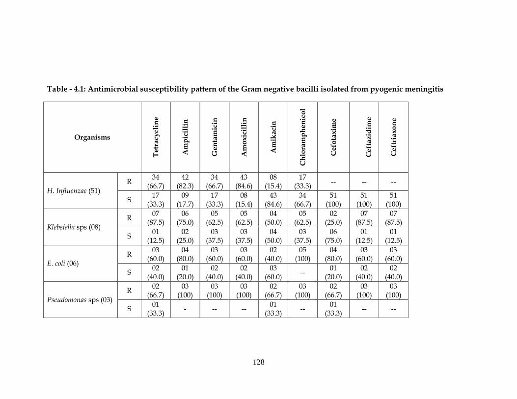
Table - 4.1: Antimicrobial susceptibility pattern of the Gram negative bacilli isolated from pyogenic meningitis
Organisms
Tetr
acy
clin
e
Am
pic
illi
n
Gen
tam
icin
Am
oxi
cill
in
Am
ikaci
n
Ch
lora
mp
hen
ico
l
Cefo
taxi
me
C
eft
azi
dim
e
Ceft
riaxo
ne
H. Influenzae (51) R
34 (66.7)
42 (82.3)
34 (66.7)
43 (84.6)
08 (15.4)
17 (33.3)
-- -- --
S 17
(33.3) 09
(17.7) 17
(33.3) 08
(15.4) 43
(84.6) 34
(66.7) 51
(100) 51
(100) 51
(100)
Klebsiella sps (08) R
07 (87.5)
06 (75.0)
05 (62.5)
05 (62.5)
04 (50.0)
05 (62.5)
02 (25.0)
07 (87.5)
07 (87.5)
S 01
(12.5) 02
(25.0) 03
(37.5) 03
(37.5) 04
(50.0) 03
(37.5) 06
(75.0) 01
(12.5) 01
(12.5)
E. coli (06) R
03 (60.0)
04 (80.0)
03 (60.0)
03 (60.0)
02 (40.0)
05 (100)
04 (80.0)
03 (60.0)
03 (60.0)
S 02
(40.0) 01
(20.0) 02
(40.0) 02
(40.0) 03
(60.0) --
01 (20.0)
02 (40.0)
02 (40.0)
Pseudomonas sps (03) R
02 (66.7)
03 (100)
03 (100)
03 (100)
02 (66.7)
03 (100)
02 (66.7)
03 (100)
03 (100)
S 01
(33.3) - -- --
01 (33.3)
-- 01
(33.3) -- --
129
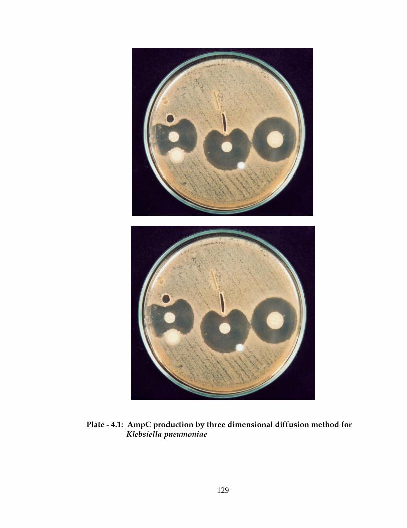
Plate - 4.1: AmpC production by three dimensional diffusion method for Klebsiella pneumoniae
130
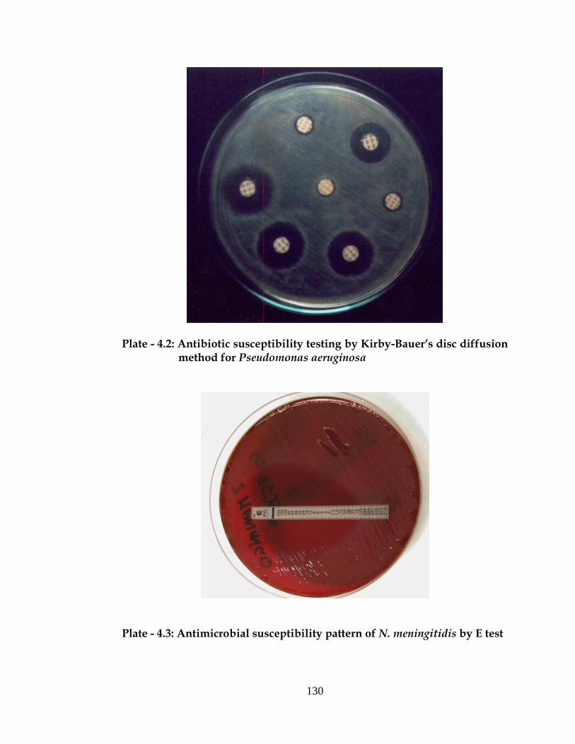
Plate - 4.2: Antibiotic susceptibility testing by Kirby-Bauer’s disc diffusion method for Pseudomonas aeruginosa
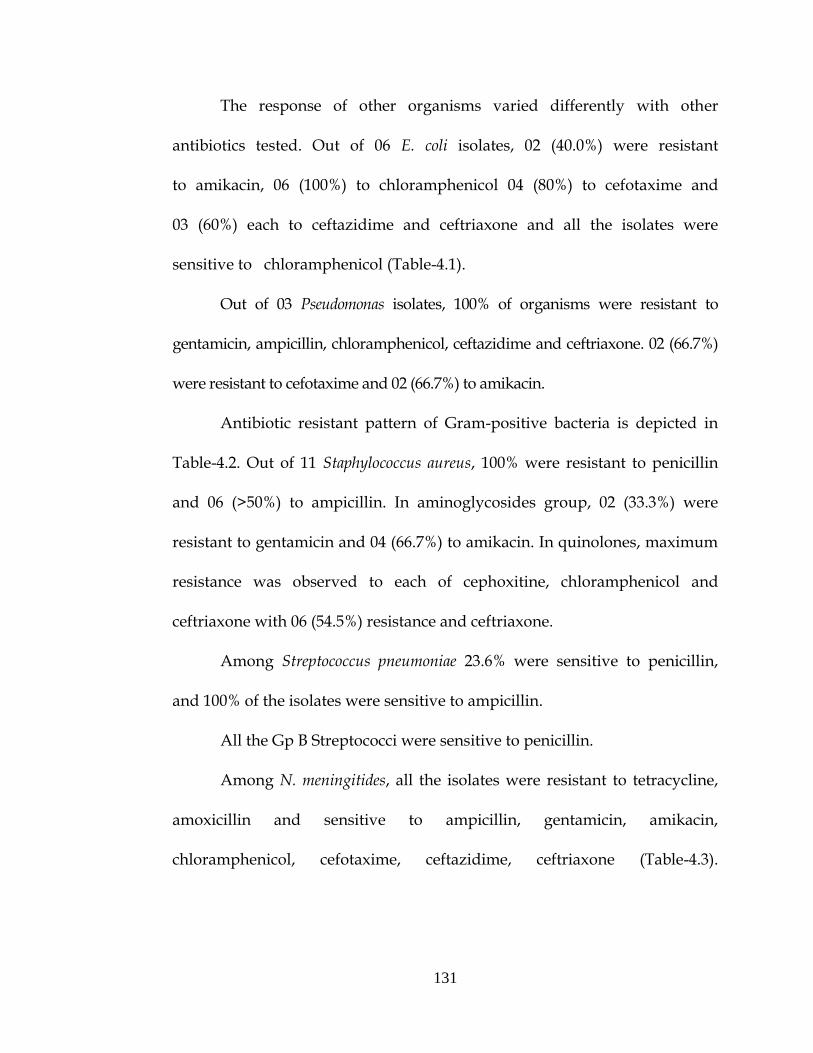
Plate - 4.3: Antimicrobial susceptibility pattern of N. meningitidis by E test
131
The response of other organisms varied differently with other
antibiotics tested. Out of 06 E. coli isolates, 02 (40.0%) were resistant
to amikacin, 06 (100%) to chloramphenicol 04 (80%) to cefotaxime and
03 (60%) each to ceftazidime and ceftriaxone and all the isolates were
sensitive to chloramphenicol (Table-4.1).
Out of 03 Pseudomonas isolates, 100% of organisms were resistant to
gentamicin, ampicillin, chloramphenicol, ceftazidime and ceftriaxone. 02 (66.7%)
were resistant to cefotaxime and 02 (66.7%) to amikacin.
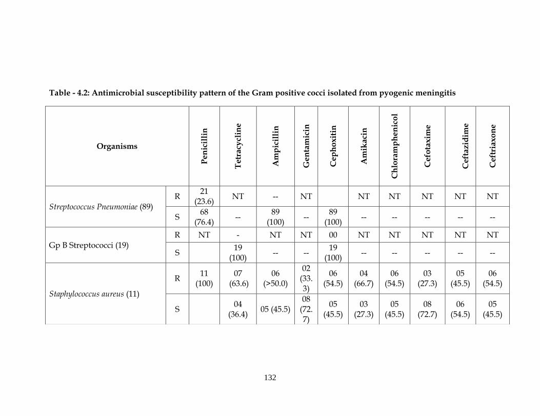
Antibiotic resistant pattern of Gram-positive bacteria is depicted in
Table-4.2. Out of 11 Staphylococcus aureus, 100% were resistant to penicillin
and 06 (>50%) to ampicillin. In aminoglycosides group, 02 (33.3%) were
resistant to gentamicin and 04 (66.7%) to amikacin. In quinolones, maximum
resistance was observed to each of cephoxitine, chloramphenicol and
ceftriaxone with 06 (54.5%) resistance and ceftriaxone.
Among Streptococcus pneumoniae 23.6% were sensitive to penicillin,
and 100% of the isolates were sensitive to ampicillin.
All the Gp B Streptococci were sensitive to penicillin.
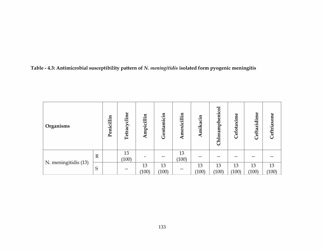
Among N. meningitides, all the isolates were resistant to tetracycline,
amoxicillin and sensitive to ampicillin, gentamicin, amikacin,
chloramphenicol, cefotaxime, ceftazidime, ceftriaxone (Table-4.3).
132
Table - 4.2: Antimicrobial susceptibility pattern of the Gram positive cocci isolated from pyogenic meningitis
Organisms
Pen
icil
lin
Tetr
acy
clin
e
Am
pic
illi
n
Gen
tam
icin
Cep
ho
xit
in
Am
ikaci
n
Ch
lora
mp
hen
ico
l
Cefo
taxim
e
Ceft
azid
ime
Ceft
riaxo
ne
Streptococcus Pneumoniae (89)
R 21
(23.6) NT -- NT NT NT NT NT NT
S 68
(76.4) --
89 (100)
-- 89
(100) -- -- -- -- --
Gp B Streptococci (19) R NT - NT NT 00 NT NT NT NT NT
S 19
(100) -- --
19 (100)
-- -- -- -- --
Staphylococcus aureus (11)
R 11
(100) 07
(63.6) 06
(>50.0)
02 (33.3)
06 (54.5)
04 (66.7)
06 (54.5)
03 (27.3)
05 (45.5)
06 (54.5)
S 04
(36.4) 05 (45.5)
08 (72.7)
05 (45.5)
03 (27.3)
05 (45.5)
08 (72.7)
06 (54.5)
05 (45.5)
133
Table - 4.3: Antimicrobial susceptibility pattern of N. meningitidis isolated form pyogenic meningitis
Organisms
Pen
icil
lin
Tetr
acy
clin
e
Am
pic
illi
n
Gen
tam
icin
Am
oxic
illi
n
Am
ikaci
n
Ch
lora
mp
hen
ico
l
Cefo
taxim
e
Ceft
azid
ime
Ceft
riaxo
ne
N. meningitidis (13)
R 13
(100) - --
13 (100)
-- -- -- -- --
S -- 13
(100) 13
(100) --
13 (100)
13 (100)
13 (100)
13 (100)
13 (100)
134
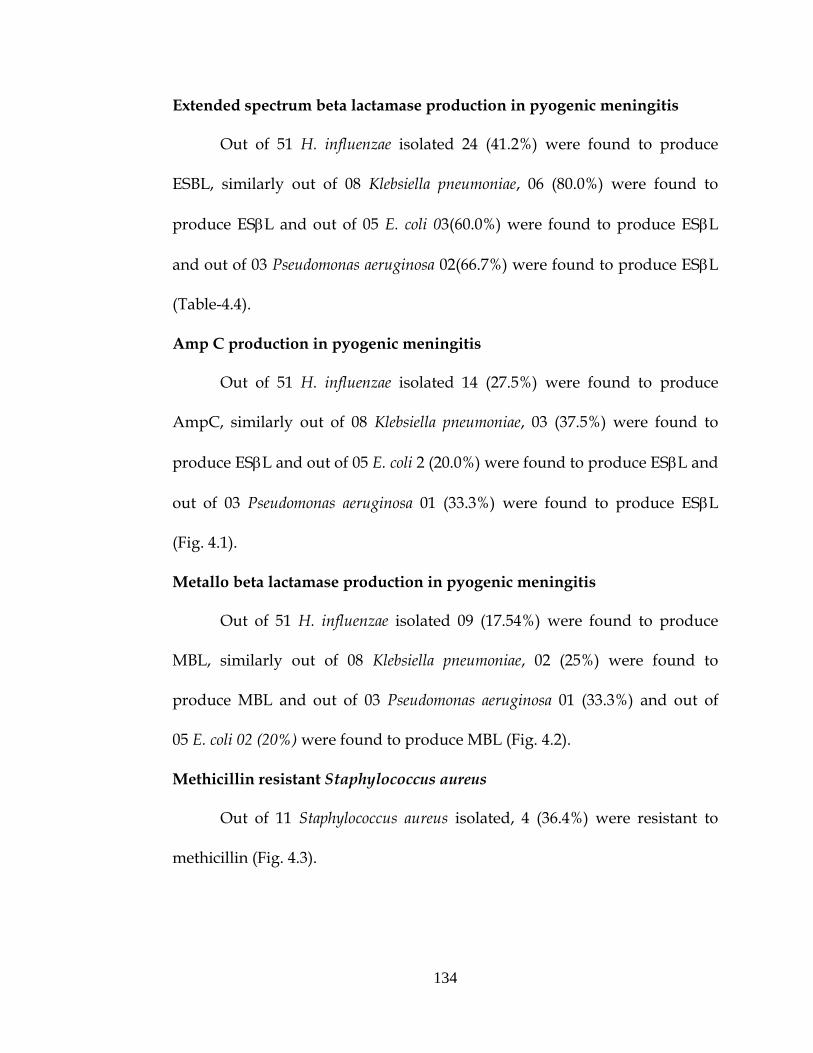
Extended spectrum beta lactamase production in pyogenic meningitis
Out of 51 H. influenzae isolated 24 (41.2%) were found to produce
ESBL, similarly out of 08 Klebsiella pneumoniae, 06 (80.0%) were found to
produce ESL and out of 05 E. coli 03(60.0%) were found to produce ESL
and out of 03 Pseudomonas aeruginosa 02(66.7%) were found to produce ESL
(Table-4.4).
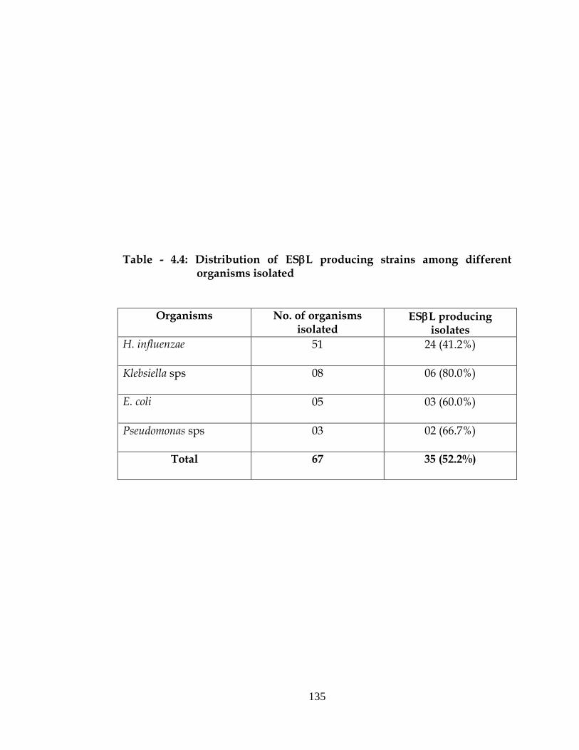
Amp C production in pyogenic meningitis
Out of 51 H. influenzae isolated 14 (27.5%) were found to produce
AmpC, similarly out of 08 Klebsiella pneumoniae, 03 (37.5%) were found to
produce ESL and out of 05 E. coli 2 (20.0%) were found to produce ESL and
out of 03 Pseudomonas aeruginosa 01 (33.3%) were found to produce ESL
(Fig. 4.1).
Metallo beta lactamase production in pyogenic meningitis
Out of 51 H. influenzae isolated 09 (17.54%) were found to produce
MBL, similarly out of 08 Klebsiella pneumoniae, 02 (25%) were found to
produce MBL and out of 03 Pseudomonas aeruginosa 01 (33.3%) and out of
05 E. coli 02 (20%) were found to produce MBL (Fig. 4.2).
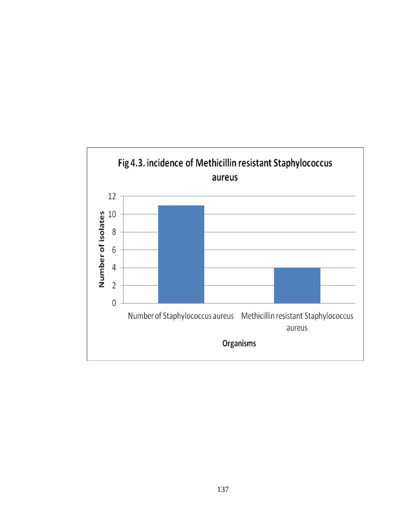
Methicillin resistant Staphylococcus aureus
Out of 11 Staphylococcus aureus isolated, 4 (36.4%) were resistant to
methicillin (Fig. 4.3).
135
Table - 4.4: Distribution of ESL producing strains among different organisms isolated
Organisms
No. of organisms isolated
ESL producing isolates
H. influenzae 51 24 (41.2%)
Klebsiella sps 08 06 (80.0%)
E. coli 05 03 (60.0%)
Pseudomonas sps 03 02 (66.7%)
Total 67 35 (52.2%)
136
0
5
10
15
20
25
30
35
40
Percen
tag
e
H. influenzae Klebsiella
pneumoniae
Pseudomonas
aeruginosa
E. coli
Organisms
Fig. 4.1: Incidence of Amp C production among Gram negative bacilli
0
5
10
15
20
25
30
35
Percen
tag
e
H. influenzae Klebsiella
pneumoniae
Pseudomonas
aeruginosa
E. coli
Organisms
Fig. 4.2: Percentage of metallo beta lactamase among Gram negative bacilli
137
138
4.4. DISCUSSION
Bacterial meningitis is fatal infectious disease in majority of the cases,
caused a variety of microorganisms. Even among survivors, morbidity rates
in terms of neurological sequel are fairly high. Chemotherapy for bacterial
meningitis has steadily progressed through successive eras beginning with
sulphonamide followed by penicillin’s, Beta-lactams, amino glycosides and
quinolones. The new potent agents offered a wide choice for therapy with
their broad spectrum and effectiveness against multi-resistant organisms.
However, clinicians by and large, would agree that despite the developments
in the management, the improvement in the outcome from bacterial
meningitis is marginally significant. Some of the attributable reasons could
be approaches in diagnosis, delay in the management, newer emergence of
resistant bacterial strains, choice of empirical antimicrobial regimens and
path physiological events that occur due to host inflammatory reactions.
These results signify the varying levels of drug resistance amongst the
gram positive and the gram negative microbes and the need to control the
spread of these resistant strains before they reach the alarming levels in this
region. It is particularly useful for the clinicians to possess the susceptibility
data on Gram positive and Gram negative bacteria rather than for particular
organisms only.
This problem of antibiotic misuse may lend credence to the increasing
resistance of the etiology to the common anti-meningitic drugs i.e.,
139
ampicillin, penicillin and chloramphenicol, in Nigeria (Akpede et al., 1999).
The high resistance of the pathogens to penicillin and ampicillin and the
sensitivity to Chloramphenicol was not strange in Nigeria (Akpede et al.,
1999; Green et al., 1993). Chloramphenicol appears to be the only one of the
three traditional drugs which can safely be used for empirical treatment of
meningitis without significant risk of treatment failure. In our experience,
there had been no major untoward effect of chloramphenicol use in children.
Ciprofloxacin achieves good CSF penetration in meningitis (Green et al.,
1993) and this had been shown to translate to less mortality among
meningitic children (Kiwanuka et al., 2001). On the other hand, the major
problem with ceftriaxone and ciprofloxacin is the high cost which technically
reduces their usefulness in the developing world where most patients are
poor.
Ampicillin resistance was found in 82.3% of H. influenzae isolates.
Resistance to chloramphenicol was noted in 33.3% of isolates. Resistance to
chloramphenicol was associated with resistance to ampicillin in 14.3% of
strains. These findings are similar to prevalences among isolates from
different types of specimen in nine European countries (1988-89) and South
Africa (1991-92) (Hussey et al., 1994). In the USA ampicillin resistance in
H. influenzae occurred twice as often, yet chloramphenicol resistance was rare
(Kayser et al., 1990). A significantly higher prevalence of ampicillin resistance
was found among our type b as compared with non-type b strains. This was
140
also seen in the USA, but not in previous studies from Europe and South
Africa (Kayser et al., 1990; Hussey et al., 1994). One study mentioned highest
rates of ampicillin resistance among children aged less than 4 years,
comparable to our findings. As the outcome of patients with H. influenzae
meningitis caused by strains which were resistant to initial therapy
consisting of ampicillin and chloramphenicol appeared to be worse in one
study, this regimen cannot be recommended now for H. influenzae meningitis
(Green et al., 1993).
Analysis of our N. meningitidis isolates revealed an overall prevalence
of intermediate penicillin resistance of 30.7%, comparable to prevalences
found in the UK (1980s), USA (1991) and Canada (1991-1992), but far lower
than the 20-40% prevalence in Spain (1989-1990) (Jones and Sutcliffe, 1990;
Saez-Nieto et al., 1992; Ringuette et al., 1995). Among our intermediately
resistant strains, serogroup B:4:P1.15 was relatively often present, as found in
Spain (Saez-Nieto et al., 1992). Penicillin is still regarded as first choice for the
treatment of meningococcal meningitis with intermediate resistant strains
(Jones and Sutcliffe, 1990). N. meningitidis was sensitive to chloramphenicol,
amikacin, but strains resistant to tetracycline and amoxicillin have been
reported.
Resistance to penicillin or chloramphenicol in S. pneumoniae CSF
isolates was very rare. With the recently suggested change in susceptibility
categories for pneumococci, 6 (6.7%) strains would be classified as
141
intermediately resistant and 21 (23.6%) as resistant (Scheel et al., 1995).
The rare occurrence of resistance cannot be explained by a different sero-
distribution compared with other regions, as the four serogroups known for
high rates of resistance (6, 14, 19, 23) represent 40% of CSF isolates. A more
likely explanation is that in India antibiotics are available without
prescription, and guidelines recommending restricted use of antibiotics in
various bacterial infections are not widely used.
In fact, the prevalence of resistance found in our pneumococcal CSF
isolates is among the lowest in the world. In other countries, the prevalence
of penicillin resistance in CSF and blood isolates were 4.3% (Belgium, 1986-
1993) and 1.8% (Germany, 1992-94) as compared with 0.5% in our isolates.
Chloramphenicol resistance was found in 2.7% and 1.9% of isolates from
Belgium and Germany, respectively, as compared with 0.3% of isolates in
this study (Verhaegen et al., 1995). The prevalence of penicillin resistance in
CSF isolates was 40% and 25% in Spain (1979-89) and France (1993),
respectively; 25% of isolates were chloramphenicol resistant (Linares et al.,
1992; Olivier et al., 1994). In a recent population-based surveillance of
invasive pneumococcal infections in the USA 25% of isolates were resistant
to penicillin and 3% to chloramphenicol (Hofmann et al., 1995). The findings
in the present study do not justify a change in initial therapy of presumptive
pneumococcal meningitis.
142
Present work showed relatively high incidence of MRSA infection in
meningitis. The prevalence rate of MRSA was found to be 36.4%, which was
slightly higher, compared to other reports where it ranged between 20 to
32.8% (Udaya et al., 1997). Methicillin resistance in Staphylococci is due to the
production of an additional non-native penicillin-binding protein PBP2a).
PBP2a is coded by the MecA gene and has low affinity for beta-lactam
antibiotics. The epidemiology of MRSA is gradually changing since its
emergence. Initially, there were occasional reports, but now it has become
one of the established pathogens. Moreover, association of multidrug
resistance with MRSA has added to the prevailing problem. -lactam
antibiotics like penicillin and cephalexin were not found to be effective
against MRSA. Penicillin was 100% resistance. Over 72.7% of MRSA strains
were resistant to amikacin however, 72.7% of the strains were susceptible to
cefotaxime. To date, there have been no isolates of MRSA with documented
in-vitro resistance to Vancomycin in India. Even in our study, none of the
MRSA isolates was resistant to vancomycin. Hence, it may be used as the
drug of choice for treating multidrug resistant MRSA strains isolated from
pyogenic meningitis.
The high rate of ESBLs among hospitalized patients is a global
problem. It is generally thought that patients infected by an ESBL-producing
organism are at an increased risk of treatment failure with an expanded-
spectrum beta-lactam. ESL producing K. pneumoniae have emerged as one of
143
the major multidrug resistant organisms. ESL mediated resistance to 3GC
was found in 52.2% in the pyogenic meningitis. This prevalence rate is higher
than other reports from India and abroad. Since the isolates were obtained
from infection in different age group they might be wide disparity in the
prevalence rate of ESL producing Gram-negative bacteria when compared
to other reports. During past decade, ESL producing K. pneumoniae have
emerged as one of the major multi drug organism (Agarwal et al., 1997;
Jerestin et al., 1997). The incidence of ESL producing Klebsiella isolated in the
United States has been reported to be 5% in France and England 14-16%
ESL producers among clinical, Klebsiella isolates has been reported. In a
previous study in central India, 76.5% of Klebsiella isolates resistant to 36oC
antibiotics was found to produce ESL by DDST. In our study, 80.0% of
Klebsiella pneumoniae were ESBL producers.
The incidence of ESL producing strains among clinical Klebsiella
isolates has been steadily increasing over the past years and accounts for
6-17% of all nosocomial urinary tract infection. In the present study Klebsiella
accounted for 80.0% ESL producer. According to Vinodkumar et al., 2004
the total 96 strains isolated from blood specimen were found to be resistant
to a minimum of 3 antibiotics, hence these were considered multidrug
resistant. 93.8% of isolates showed resistance or decreased susceptibility to at
least one of the 3GC and 68.8% to all the 360C.
144
We found that the ESL producing isolates were conferred with
resistance or decreased susceptibility to various third generation
cephalosporins. The DDST detected ESL in 52.2% of the isolates.
The specificity of DDST is well documented. In view of its simplicity, it may
be undertaken in a routine diagnostic laboratory for detecting ESL
producing strains with due consideration to factors like precise placement of
the discs, correct storage of the clavulanate containing discs and performance
of appropriate control tests, which are critical to the sensitivity of the DDST.
The other Gram negative bacteria producing ESBL are E. coli, Pseudomonas
aeruginosa and H. influenzae. Study conducted by Enting at Netherlands,
showed 30% of H. influenzae produced beta lactamases, but in our study the
incidence of beta lactamases was 41.2%.
Study carried out by Vishwanath et al., the influenza isolates were
sensitive to ciprofloxacin, 66% of the isolates were sensitive to ampicillin and
cefotaxime and 50% of the isolates were sensitive to cephalexin, gentamicin
and co-trimoxazole. 83% of Streptococci pneumoniae were resistant to
gentamicin and 67% to ciprofloxacin. All the isolates were resistance to
cephalexin, ampicillin and co-trimoxazole and all the isolates were sensitive
to amikacin and vancomycin.
AmpC β-lactamases are clinically important cephalosporinases
encoded on chromosomes of many of the Enterobacteriaceae and a few other
organisms, where they mediate resistance to cephalothin, cefazolin, cefoxitin,
145
most of the penicillins and β-lactamase inhibitor. In many bacteria Amp C
enzymes are inducible and can be expressed at high levels by mutation.
Over expression confers resistance to broad spectrum cephalosporins. In the
present study 17.9% were AmpC producers and H. influenzae was the
predominant Amp C producer followed by Klebsiella pneumonia. There are no
reports to compare the incidence of Amp C mediated resistance among
pyogenic meningitis infection in India and aboard.
Information on the prevalence of Amp C -lactamase producing
strains in India is very limited, and no data’s are available on the prevalence
of Amp C production in pediatric group. In a recent study showed that
20.7% of the clinical isolates were found to harbor Amp C enzymes in
contrast to reported values of 1.2 and 4.2% (Philippon et al., 1989) by other
workers in different clinical conditions. In the present study 12 (17.9%)
isolates were resistant to cefoxitin were positive by 3–dimensional test,
negative for inducible -lactamases by disc diffusion test and sensitive to
imipenem. Due to above phenotypic characters these isolates were
considered to be harboring plasmid encoded Amp C -lactamases. It has
been stated that detection of AmpC enzymes is quiet challenging since;
hyper-production of chromosomal Amp C with OMPF porin loss in E. coli or
porin deficiency in K. pneumoniae can produce similar resistant phenotype.
Further not all strains with AmpC enzymes meet NCCLS criteria for
resistance to cephamycins and oxyimino-cephalosporins (Philippon et al.,
146
1989). Hence a reference laboratory for -lactamase iso-electric focusing or
gene localization is needed. This will help us to know the actual prevalence
of these enzymes and characterize them.
Metallo β-lactamase (MBL) is a group of carbapenem hydrolysing
β-lactamase. They have been reported from many countries, as well as from
different parts of Indian subcontinent, particularly in multidrug resistance
pathogens like Pseudomonas aeruginosa and Acinetobacter species. The MBLs
are inhibited in-vitro by CuCl3, FeCl3, EDTA and thiol compounds like
2 mercaptopropionic acid, sodium mercaptoacetoic acid and
2 mercaptoethanal, but not by β-lactamase inhibitors like clavulanic acid,
sulbactum or tazobactam. Detection of MBL production in MDR organisms
from burn infection has tremendous therapeutic consequences, as the
treatment option for such isolates are aztreonam or potentially toxic
polymyxin B and colistin. In the present study not all gram negative bacteria
were tested for MBL production. Only those gram negative bacilli resistant to
imipenem were screened for MBL production and 29.5% of them were
metallo β-lactamase producers. Pseudomonas aeruginosa and Klebsiella
pneumoniae, were the predominant MBL producers.
Thus, the study clearly highlights the rising level of drug resistance
amongst the meningitis pathogens and hence the need to update and
formulate newer drug policies. Good infection control practices, rational
antibiotic policies, judicious use of interventions and implementation of
147
standard of isolation precautions are of vital importance today. Unless there
are strategies to optimize effective use of antibiotics, very few options will be
left in future in the antibiotic armamentarium and it might herald an era of
medical disaster with strains virtually untreatable with current spectrum of
antimicrobials.
Stress should be given on the restrained and rationale use of
antimicrobials both in and outside the hospital. This study also indicates the
urgent need for more of such studies in the patients of meningitis vis-a-vis
aetiology and drug resistance along with the need for the in-house review of
drug policy within hospitals at least once in every five years. There is also an
urgent need to develop institutional programs to enhance antimicrobial
stewardship thus minimizing the emergence and spread of antimicrobial
resistance.