Embed Size (px)
Citation preview
7/30/2019 4653.full
http://slidepdf.com/reader/full/4653full 1/10
4653
Introduction
MicroRNAs (miRNAs) are about 22-nucleotide, short, non-coding RNAs that are thought to regulate gene expressionthrough sequence-specific base pairing with target mRNAs.Hundreds of microRNAs have been identified in worms, flies,fish, frogs, mammals and flowering plants using molecularcloning and bioinformatics prediction strategies (Lagos-Quintana et al., 2001; Lau et al., 2001; Lee and Ambros, 2001;Lim et al., 2003a; Llave et al., 2002; Reinhart et al., 2002;Watanabe et al., 2005). MicroRNAs are transcribed as longRNA precursors (pri-miRNAs) that contain a stem-loopstructure of about 80 bases. Pri-miRNAs are processed in the
nucleus by the RNase III enzyme Drosha and DGCR8/Pasha,which excises the stem-loop to form the pre-miRNA (Denli etal., 2004; Gregory et al., 2004; Han et al., 2004; Landthaler etal., 2004; Lee et al., 2003). Pre-miRNAs are exported from thenucleus by Exportin-5 (Bohnsack et al., 2004; Lund et al.,2003; Yi et al., 2003). In the cytoplasm, another RNase IIIenzyme, Dicer, cuts the pre-miRNA to generate the maturemicroRNA as part of a short RNA duplex. The RNA issubsequently unwound by a helicase activity and incorporatedinto a RNA-induced silencing complex (RISC). For moreinformation on microRNA biogenesis and maturation, pleasesee the accompanying article by Du and Zamore (Du andZamore, 2005).
Most microRNAs in animals are thought to function through
the inhibition of effective mRNA translation of target genesthrough imperfect base pairing with the 3Ј-untranslated region(3ЈUTR) of target mRNAs (Bartel, 2004). The underlyingmechanism is still poorly understood, but it appears to involvethe inhibition of translational initiation (Pillai et al., 2005).MicroRNA targets are largely unknown, but estimates rangefrom one to hundreds of target genes for a given microRNA,based on target predictions using a variety of bioinformaticsapproaches (Brennecke et al., 2005; John et al., 2004;Kiriakidou et al., 2004; Lewis et al., 2005; Lewis et al., 2003;Rajewsky and Socci, 2004; Stark et al., 2003; Xie et al., 2005).In addition, at least one microRNA, miR-196, can cleave atarget mRNA, HOXB8, in the same manner as a short
interfering RNA (siRNA) does during the process of RNAinterference (RNAi) (Mansfield et al., 2004; Yekta et al., 2004).This mechanism is the preferred one for plant microRNAs(Meins et al., 2005). MicroRNAs may also play a role in AU-rich element-mediated mRNA degradation (Jing et al., 2005).See Du and Zamore for a further discussion of this (Du andZamore, 2005). Finally, the involvement of microRNAs intranscriptional gene silencing (TGS), which has been observedin plants, remains an intriguing possibility (Baulcombe, 2004).
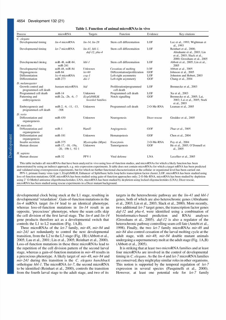
This review focuses on the function of animal microRNAsonly. For a recent review of our current understanding of theroles of microRNAs in plants, please see (Kidner andMartienssen, 2005), and for recent accounts of the history of this field, please see recent articles by the pioneers of the fieldthemselves (Lee et al., 2004a; Ruvkun et al., 2004). Here, wedescribe how microRNAs contribute to different aspects of animal development and what we know of their involvementin human disease (see Table 1).
Developmental timing
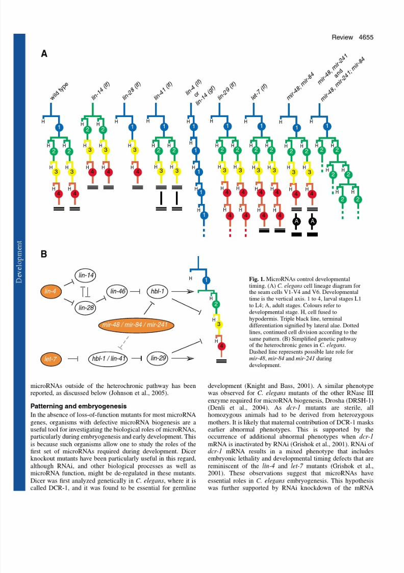
Interest in the genes controlling developmental timing in C.elegans (Ambros and Horvitz, 1984; Chalfie et al., 1981;Horvitz and Sulston, 1980) led to the cloning of the firstmicroRNA, lin-4 miRNA (Lee et al., 1993), and theidentification of the first microRNA target, lin-14 mRNA(Wightman et al., 1993). The developmental-timing, or
heterochronic, pathway regulates stage-specific processesduring C. elegans larval development. For a recent, detailedreview of this pathway, please see Rougvie (Rougvie, 2005).One focus of the study of the heterochronic pathway in C.elegans has been the developmental fate of several stem cellsin the lateral hypodermis, collectively known as the seam cells.The seam cells undergo a cell division pattern that issynchronised with the four larval molts of the animal (Fig. 1A).Only at the adult stage will the seam cells exit mitosis andterminally differentiate. In lin-4 mutant animals, the seam cellsrepeat the cell division pattern that characterises the first larvalstage (L1) and fail to differentiate. This mutant phenotype hasbeen interpreted as a heterochronic change with the
Five years into the ‘small RNA revolution’ it is hard not toshare in the excitement about the rapidly unravellingbiology of microRNAs. Since the discovery of the firstmicroRNA gene, lin-4, in the nematode Caenorhabditiselegans, many more of these short regulatory RNA geneshave been identified in flowering plants, worms, flies, fish,
frogs and mammals. Currently, about 2% of the knownhuman genes encode microRNAs. MicroRNAs are essentialfor development and this review will summarise ourcurrent knowledge of animal microRNA function. We willalso discuss the emerging links of microRNA biology tostem cell research and human disease, in particular cancer.
Summary
MicroRNA functions in animal development and human diseaseInes Alvarez-Garcia and Eric A. Miska*
The Wellcome Trust/Cancer Research UK Gurdon Institute and Department of Biochemistry, The Henry Wellcome Building ofCancer and Developmental Biology, University of Cambridge, Tennis Court Road, Cambridge CB2 1QN, UK*Author for correspondence (e-mail: [email protected])
Development 132, 4653-4662
Published by The Company of Biologists 2005
doi:10.1242/dev.02073
Review
7/30/2019 4653.full
http://slidepdf.com/reader/full/4653full 2/10
7/30/2019 4653.full
http://slidepdf.com/reader/full/4653full 3/10
7/30/2019 4653.full
http://slidepdf.com/reader/full/4653full 4/10
4656
transcripts for the two C. elegans argonaute proteins requiredfor miRNA biogenesis, ALG-1 and ALG-2 (Grishok et al.,2001). RNAi-treated worms showed a mixed phenotype thatincluded embryonic and larval lethality and heterochronicdefects.
The fruitfly Drosophila melanogaster has two Dicer genes, Dicer-1 and Dicer-2, and genetic analysis suggests that Dicer-1 is the major Dicer gene required for microRNA biogenesis(Lee et al., 2004b). Although the phenotype of Dicer-1 mutant
D. melanogaster has not been fully reported, it appears thatDicer-1 is required for wild-type development of both somatictissues and the germline (Hatfield et al., 2005; Lee et al.,2004b) (for details see below).
In the zebrafish Danio rerio, a likely null allele of Dicerleads to a developmental arrest at 7 to 10 days post-fertilization(Wienholds et al., 2003). This late terminal phenotype is againlikely to be due to maternal provision of Dicer and/or of microRNAs. Indeed, removal of the maternal Dicercontribution through the generation of germline clones leads toa more severe defect (Giraldez et al., 2005). In maternal-zygotic Dicer mutants, axis formation and early differentiationare normal, but many embryos have morphogenesis defectsaffecting gastrulation, brain formation, somitogenesis andheart development.
In the mouse, Mus musculus, Dicer1 mutants die around 7.5days of gestation (Bernstein et al., 2003). A maternalcontribution of Dicer is likely to have a much smaller effect in
M. musculus due to the much smaller size of the egg.Homozygous Dicer1 null mutants from heterozygous mothersdie around 7.5 days of gestation (Bernstein et al., 2003).Mutant embryos have defects in axis formation andgastrulation, and are depleted of Oct4-positive stem cells(Bernstein et al., 2003). In all cases where only Dicer1 mutantshave been analysed, one cannot easily distinguish betweendefects that are due to a loss of microRNA processing andthose that are due to a loss of endogenous RNAi or otherpathways regulated by Dicer. For example, Dicer appears tohave important roles in heterochromatin formation andchromosome segregation in the fission yeastSchizosaccharomyces pombe, in the ciliated protozoan
Tetrahymena and in vertebrate cells (Fukagawa et al., 2004;Mochizuki and Gorovsky, 2005; Provost et al., 2002). As S.
pombe does not encode any known microRNAs, these defectsare unlikely to be caused by their loss.
A more direct approach to investigating the role of microRNAs during embryogenesis has been taken in D.melanogaster , where 2Ј O-Methyl antisense
oligoribonucleotides were used in microRNA depletion studies(see Box 1) (Leaman et al., 2005).
A single injection of 2Ј O-Methyl antisenseoligoribonucleotides complementary to the 46 microRNAsknown to be expressed in the D. melanogaster embryo resultedin a total of twenty-five different, abnormal phenotypes. Thesephenotypes included defects in blastoderm cellularization andpatterning, morphogenesis and cell survival. Increasedprogrammed cell death was observed in embryos injected with2Ј O-Methyl antisense oligoribonucleotides that targeted the D.melanogaster miR-2 family, and miR-6, miR-11 and miR-308.
Clearly, our current understanding of microRNA functionduring embryogenesis is only rudimentary. However, it isnoteworthy that the only evidence for a role of miRNAs intissue patterning during embryogenesis to date comes fromdepletion studies of miR-31 in D. melanogaster (Leaman et al.,2005). Could pattern formation be largely independent of regulation by microRNAs? With this question in mind, it willbe exciting to see functional studies of mammalian miR-196,a microRNA that is located in a HOX cluster and can cleaveHOXB8 mRNA (Mansfield et al., 2004; Yekta et al., 2004).
Differentiation and organogenesis
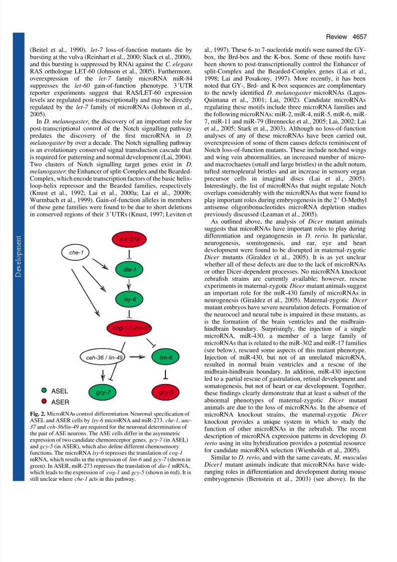
The heterochronic phenotypes of the lin-4 microRNA and thelet-7 family of microRNAs in C. elegans are clear examples of cell differentiation defects. However, an example of amicroRNA regulating differentiation that is uncoupled from
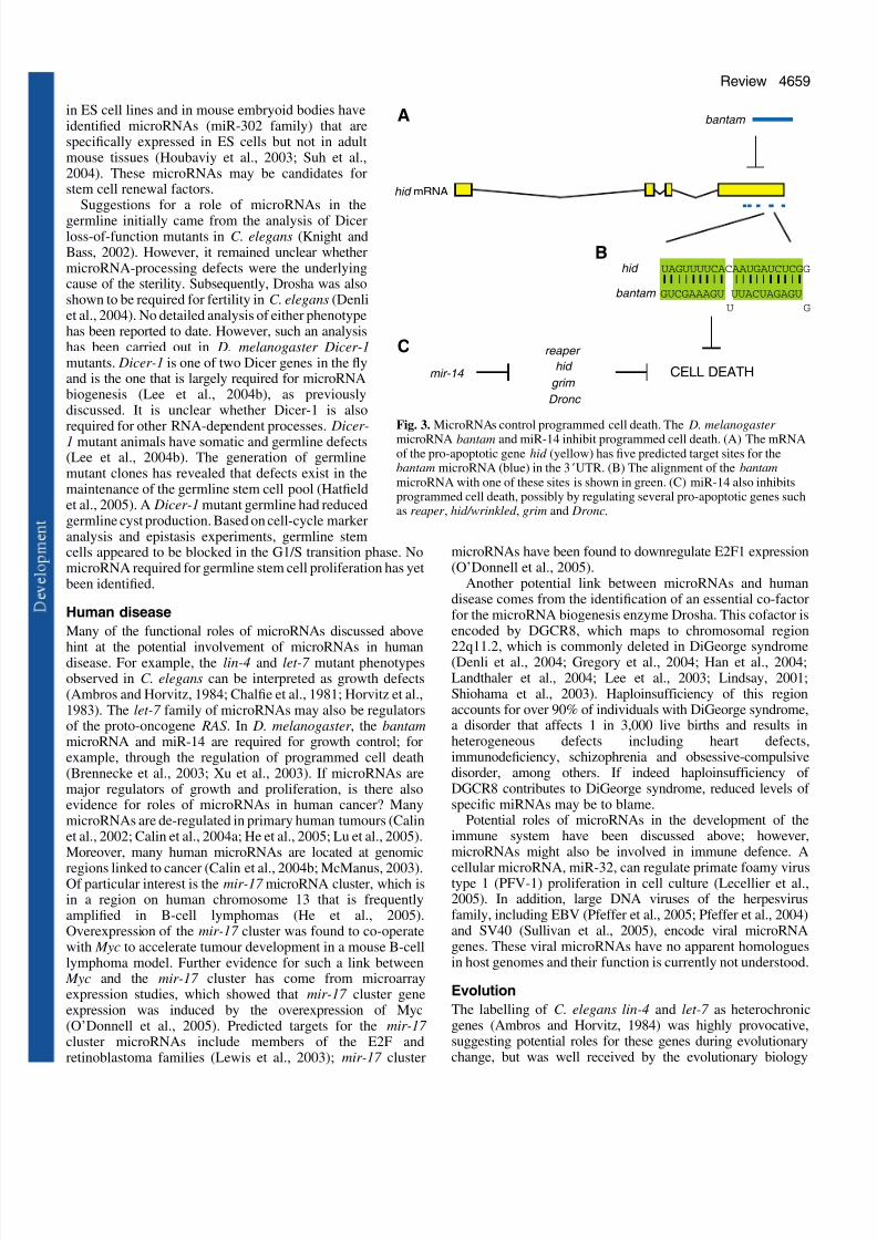
cell division was first uncovered through the study of left-rightasymmetry in C. elegans (Johnston and Hobert, 2003). In theworm, two bilateral taste receptor neurons, ASE left (ASEL)and ASE right (ASER), display a left/right asymmetricalexpression pattern of gcy-5, gcy-6 and gcy-7 , three putativechemoreceptor genes (Chang et al., 2003; Hobert et al., 2002)(Fig. 2). In a genetic screen for mutants in which the normallyASEL-specific expression of gcy-7 is disrupted, the microRNAgene lsy-6 was isolated (Chang et al., 2003). In lsy-6 mutants,ASEL neurons do not express gcy-7 , but instead express theASER-specific gcy-5 gene. Genetic interaction and GFPreporter studies showed that lsy-6 is a negative regulator of theNKX-type homeobox gene cog-1, which was identified in thesame genetic screen. Interestingly, a second microRNA, miR-
273, might act upstream in the same pathway as a regulator of die-1, which encodes a C2H2 zinc finger transcription factor(Chang et al., 2004). The transcription factor die-1 showsASEL-specific expression and acts upstream of lsy-6 .
Evidence for a role of microRNAs in organogenesis hascome from studies of vulval development in C. elegans. TheC. elegans vulva is a ring-like structure that forms theconnection between the hermaphrodite gonadal arms and theexterior, and is essential for egg-laying and sperm entry. Itderives from a group of cells in the ventral hypodermis that areinduced to undergo a series of cell divisions and differentiationby a signal from the gonadal anchor cell (Sulston and Horvitz,1977). Vulval induction requires RAS/LET-60 signalling
Development 132 (21)
Box 1. miRNA knockdown
To study loss-of-function phenotypes of miRNAs in the absenceof knockout strains, miRNA knockdown approaches are beingdeveloped. Two strategies are based on artificialoligonucleotides, 2Ј O-Methyl RNA and locked nucleotide RNA(LNA). The principle for both reagents is the same: an excess of
modified RNA complimentary to a miRNA is injected ortransfected so that it can compete for the miRNA interaction withits target mRNAs. 2Ј O-Methyl RNA offers an increased in vivohalf-life over RNA and was initially used in human cells and inC. elegans (Hutvagner et al., 2004; Meister et al., 2004; Poy etal., 2004) for the sequence-specific inhibition of small RNAs fora limited time-span, but it suffers from some pleiotropic effects(Leaman et al., 2005). LNA is a modified ribonucleic acid inwhich the ribose ring is constrained by a methylene bridgebetween the 2Ј O and the 4Ј C atoms. This modification leads toa higher thermal stability and discriminative power due to agreater difference in the melting temperature of a Watson-Crick base pair versus a mismatch (Petersen and Wengel, 2003).
7/30/2019 4653.full
http://slidepdf.com/reader/full/4653full 5/10
4657Review
(Beitel et al., 1990). let-7 loss-of-function mutants die bybursting at the vulva (Reinhart et al., 2000; Slack et al., 2000),and this bursting is suppressed by RNAi against the C. elegansRAS orthologue LET-60 (Johnson et al., 2005). Furthermore,overexpression of the let-7 family microRNA miR-84suppresses the let-60 gain-of-function phenotype. 3ЈUTRreporter experiments suggest that RAS/LET-60 expression
levels are regulated post-transcriptionally and may be directlyregulated by the let-7 family of microRNAs (Johnson et al.,2005).
In D. melanogaster , the discovery of an important role forpost-transcriptional control of the Notch signalling pathwaypredates the discovery of the first microRNA in D.melanogaster by over a decade. The Notch signalling pathwayis an evolutionary conserved signal transduction cascade thatis required for patterning and normal development (Lai, 2004).Two clusters of Notch signalling target genes exist in D.melanogaster : the Enhancer of split-Complex and the Bearded-Complex, which encode transcription factors of the basic helix-loop-helix repressor and the Bearded families, respectively(Knust et al., 1992; Lai et al., 2000a; Lai et al., 2000b;Wurmbach et al., 1999). Gain-of-function alleles in membersof these gene families were found to be due to short deletionsin conserved regions of their 3ЈUTRs (Knust, 1997; Leviten et
al., 1997). These 6- to 7-nucleotide motifs were named the GY-box, the Brd-box and the K-box. Some of these motifs havebeen shown to post-transcriptionally control the Enhancer of split-Complex and the Bearded-Complex genes (Lai et al.,1998; Lai and Posakony, 1997). More recently, it has beennoted that GY-, Brd- and K-box sequences are complimentaryto the newly identified D. melanogaster microRNAs (Lagos-
Quintana et al., 2001; Lai, 2002). Candidate microRNAsregulating these motifs include three microRNA families andthe following microRNAs: miR-2, miR-4, miR-5, miR-6, miR-7, miR-11 and miR-79 (Brennecke et al., 2005; Lai, 2002; Laiet al., 2005; Stark et al., 2003). Although no loss-of-functionanalyses of any of these microRNAs have been carried out,overexpression of some of them causes defects reminiscent of Notch loss-of-function mutants. These include notched wingsand wing vein abnormalities, an increased number of micro-and macrochaetes (small and large bristles) in the adult notum,tufted sternopleural bristles and an increase in sensory organprecursor cells in imaginal discs (Lai et al., 2005).Interestingly, the list of microRNAs that might regulate Notchoverlaps considerably with the microRNAs that were found toplay important roles during embryogenesis in the 2Ј O-Methylantisense oligoribonucleotides microRNA depletion studiespreviously discussed (Leaman et al., 2005).
As outlined above, the analysis of Dicer mutant animalssuggests that microRNAs have important roles to play duringdifferentiation and organogenesis in D. rerio. In particular,neurogenesis, somitogenesis, and ear, eye and heartdevelopment were found to be disrupted in maternal-zygotic
Dicer mutants (Giraldez et al., 2005). It is as yet unclearwhether all of these defects are due to the lack of microRNAsor other Dicer-dependent processes. No microRNA knockoutzebrafish strains are currently available; however, rescueexperiments in maternal-zygotic Dicer mutant animals suggest
an important role for the miR-430 family of microRNAs inneurogenesis (Giraldez et al., 2005). Maternal-zygotic Dicer mutant embryos have severe neurulation defects. Formation of the neurocoel and neural tube is impaired in these mutants, asis the formation of the brain ventricles and the midbrain-hindbrain boundary. Surprisingly, the injection of a singlemicroRNA, miR-430, a member of a large family of microRNAs that is related to the miR-302 and miR-17 families(see below), rescued some aspects of this mutant phenotype.Injection of miR-430, but not of an unrelated microRNA,resulted in normal brain ventricles and a rescue of themidbrain-hindbrain boundary. In addition, miR-430 injectionled to a partial rescue of gastrulation, retinal development andsomatogenesis, but not of heart or ear development. Together,
these findings clearly demonstrate that at least a subset of theabnormal phenotypes of maternal-zygotic Dicer mutantanimals are due to the loss of microRNAs. In the absence of microRNA knockout strains, the maternal-zygotic Dicer knockout provides a unique system in which to study thefunction of other microRNAs in the zebrafish. The recentdescription of microRNA expression patterns in developing D.rerio using in situ hybridization provides a potential resourcefor candidate microRNA selection (Wienholds et al., 2005).
Similar to D. rerio, and with the same caveats, M. musculus Dicer1 mutant animals indicate that microRNAs have wide-ranging roles in differentiation and development during mouseembryogenesis (Bernstein et al., 2003) (see above). In the
Fig. 2. MicroRNAs control differentiation. Neuronal specification of ASEL and ASER cells by lsy-6 microRNA and miR-273. che-1, unc-37 and ceh-36/lin-49 are required for the neuronal determination of the pair of ASE neurons. The ASE cells differ in the asymmetricexpression of two candidate chemoreceptor genes, gcy-7 (in ASEL)and gcy-5 (in ASER), which also define different chemosensoryfunctions. The microRNA lsy-6 represses the translation of cog-1
mRNA, which results in the expression of lim-6 and gcy-7 (shown ingreen). In ASER, miR-273 represses the translation of die-1 mRNA,which leads to the expression of cog-1 and gcy-5 (shown in red). It isstill unclear where che-1 acts in this pathway.
mir-273
cog-1 / unc-37
gcy-5 gcy-7
lim-6
lsy-6
die-1
ceh-36 / lin-49
ASELASER
che-1
7/30/2019 4653.full
http://slidepdf.com/reader/full/4653full 6/10
4658
mouse, additional insights can be gained from studyingconditional-knockout strains, which allow one to the study therequirement for Dicer and microRNAs in different tissues atdifferent developmental time points. In vitro, Dicer mutantembryonic stem (ES) cells, derived from conditional genetargeting, have severe differentiation defects (Kanellopoulou etal., 2005). One study that analyzed the effects of genetically
inactivating Dicer1 early during T-cell development foundevidence for the functioning of microRNAs in ␣ cell, but notin CD4/CD8, lineage commitment (Cobb et al., 2005). Anotherstudy found that knocking out Dicer1 during T-celldevelopment blocked peripheral CD8+ T-cell development,whereas CD4+ T cells, although reduced in numbers, wereviable; however, upon stimulation, these CD4+ T cellsproliferated poorly and underwent increased programmed celldeath (Muljo et al., 2005).
The particular caveat with these conditional-knockoutstudies in mice is that it is often unclear how efficiently Dicerand any existing microRNA pools are depleted upon thesomatic deletion of Dicer1. Indeed, microRNAs seem to persistfor some time (Cobb et al., 2005). One specific microRNA thathas been directly implicated in B-cell development is miR-181(Chen et al., 2004). This microRNA is highly expressed in B-lymphoid cells of mouse bone marrow. When overexpressed inhematopoietic progenitor cells, it leads to an increase in thefraction of B-lineage cells in in vitro differentiationexperiments and in vivo in adult mice. Conditionallyinactivating Dicer1 in discrete areas of the limb mesoderm inmice led to severe growth defects in the limbs of mutantembryos, but no defect in basic limb patterning or in tissue-specific differentiation was observed (Harfe et al., 2005). Thisis a striking finding that is somewhat reminiscent of the Dicer1knockout in D. rerio. However, it remains unknown whetherresidual Dicer activity or microRNA pools could have
disguised earlier roles of microRNAs in limb development.MicroRNA expression analysis has led to the discovery of a potential role for the microRNA miR-1 in mammalian heartdevelopment. The microRNA miR-1, which is the product of two genes, mir-1-1 and mir-1-2, is highly expressed in mouseheart and muscle (Lagos-Quintana et al., 2001; Lee andAmbros, 2001). An analysis of the upstream-regulatorysequence of these two genes has led to the identification of serum response factor (Srf), myocardin, Mef2 and Myod astranscriptional regulators of miR-1 expression in vitro (Zhaoet al., 2005). Of these, Srf was found to be required for miR-1 expression in the developing mouse heart, using aconditional Srf-knockout strain (Zhao et al., 2005).Overexpression of miR-1 under the -myosin heavy chain
promoter resulted in developmental arrest at embryonic day13.5, after heart failure. Transgenic embryos developed thinventricle walls and ventricular cardiomyocyte proliferationdefects. One candidate target for miR-1 in myocardialdevelopment is the transcription factor Hand2, which wasfound to be reduced in transgenic mice overexpressing miR-1 without an apparent change in Hand2 mRNA levels (Zhaoet al., 2005).
Growth control and programmed cell death
The D. melanogaster bantam gene was identified in a gain-of-function screen for regulators of cell growth (Hipfner et al.,2002). Overexpression of bantam causes the overgrowth of
wing and eye tissue, whereas bantam loss-of-function mutantanimals are smaller than wild-type animals and have reducedcell numbers. bantam was found to interact with the growthregulatory gene expanded , but was epistatic to the CycD/Cdk4pathway. Subsequent cloning of bantam identified it as amicroRNA-encoding gene (Brennecke et al., 2003). Thebantam microRNA regulates tissue growth cell autonomously.
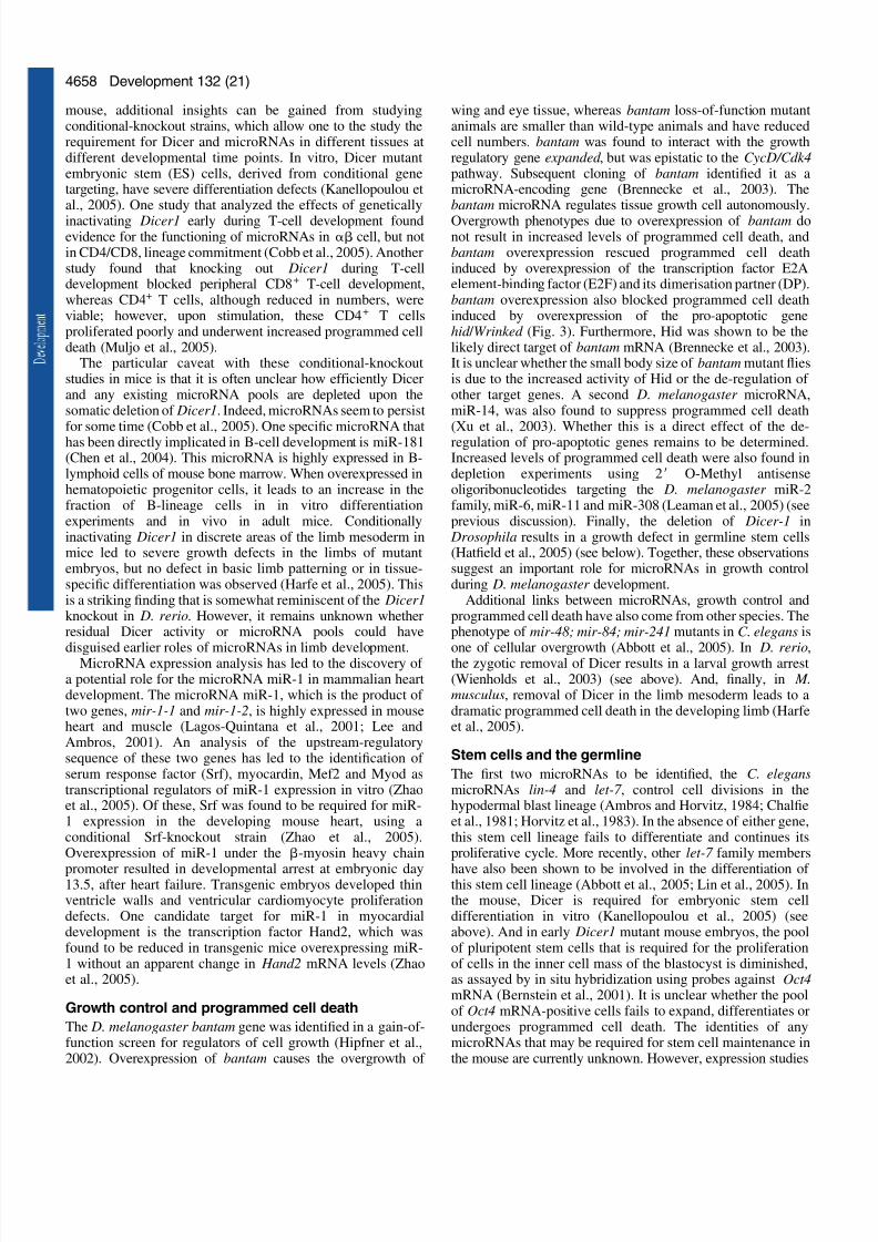
Overgrowth phenotypes due to overexpression of bantam donot result in increased levels of programmed cell death, andbantam overexpression rescued programmed cell deathinduced by overexpression of the transcription factor E2Aelement-binding factor (E2F) and its dimerisation partner (DP).bantam overexpression also blocked programmed cell deathinduced by overexpression of the pro-apoptotic genehid / Wrinked (Fig. 3). Furthermore, Hid was shown to be thelikely direct target of bantam mRNA (Brennecke et al., 2003).It is unclear whether the small body size of bantam mutant fliesis due to the increased activity of Hid or the de-regulation of other target genes. A second D. melanogaster microRNA,miR-14, was also found to suppress programmed cell death(Xu et al., 2003). Whether this is a direct effect of the de-regulation of pro-apoptotic genes remains to be determined.Increased levels of programmed cell death were also found indepletion experiments using 2Ј O-Methyl antisenseoligoribonucleotides targeting the D. melanogaster miR-2family, miR-6, miR-11 and miR-308 (Leaman et al., 2005) (seeprevious discussion). Finally, the deletion of Dicer-1 in
Drosophila results in a growth defect in germline stem cells(Hatfield et al., 2005) (see below). Together, these observationssuggest an important role for microRNAs in growth controlduring D. melanogaster development.
Additional links between microRNAs, growth control andprogrammed cell death have also come from other species. Thephenotype of mir-48; mir-84; mir-241 mutants in C. elegans is
one of cellular overgrowth (Abbott et al., 2005). In D. rerio,the zygotic removal of Dicer results in a larval growth arrest(Wienholds et al., 2003) (see above). And, finally, in M.musculus, removal of Dicer in the limb mesoderm leads to adramatic programmed cell death in the developing limb (Harfeet al., 2005).
Stem cells and the germline
The first two microRNAs to be identified, the C. elegansmicroRNAs lin-4 and let-7 , control cell divisions in thehypodermal blast lineage (Ambros and Horvitz, 1984; Chalfieet al., 1981; Horvitz et al., 1983). In the absence of either gene,this stem cell lineage fails to differentiate and continues itsproliferative cycle. More recently, other let-7 family members
have also been shown to be involved in the differentiation of this stem cell lineage (Abbott et al., 2005; Lin et al., 2005). Inthe mouse, Dicer is required for embryonic stem celldifferentiation in vitro (Kanellopoulou et al., 2005) (seeabove). And in early Dicer1 mutant mouse embryos, the poolof pluripotent stem cells that is required for the proliferationof cells in the inner cell mass of the blastocyst is diminished,as assayed by in situ hybridization using probes against Oct4mRNA (Bernstein et al., 2001). It is unclear whether the poolof Oct4 mRNA-positive cells fails to expand, differentiates orundergoes programmed cell death. The identities of anymicroRNAs that may be required for stem cell maintenance inthe mouse are currently unknown. However, expression studies
Development 132 (21)
7/30/2019 4653.full
http://slidepdf.com/reader/full/4653full 7/10
7/30/2019 4653.full
http://slidepdf.com/reader/full/4653full 8/10
4660
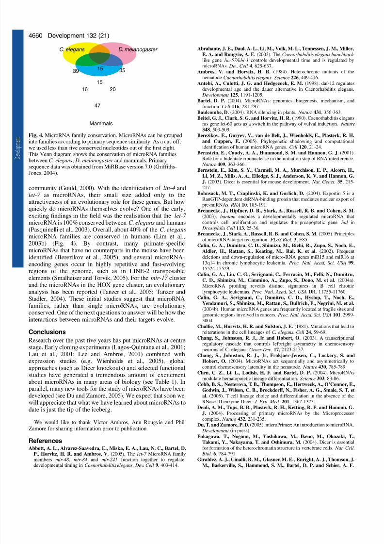
community (Gould, 2000). With the identification of lin-4 andlet-7 as microRNAs, their small size added only to theattractiveness of an evolutionary role for these genes. But howquickly do microRNAs themselves evolve? One of the early,exciting findings in the field was the realisation that the let-7 microRNA is 100% conserved between C. elegans and humans(Pasquinelli et al., 2003). Overall, about 40% of the C. elegansmicroRNA families are conserved in humans (Lim et al.,2003b) (Fig. 4). By contrast, many primate-specificmicroRNAs that have no counterparts in the mouse have beenidentified (Berezikov et al., 2005), and several microRNA-encoding genes occur in highly repetitive and fast-evolvingregions of the genome, such as in LINE-2 transposableelements (Smalheiser and Torvik, 2005). For the mir-17 cluster
and the microRNAs in the HOX gene cluster, an evolutionaryanalysis has been reported (Tanzer et al., 2005; Tanzer andStadler, 2004). These initial studies suggest that microRNAfamilies, rather than single microRNAs, are evolutionaryconserved. One of the next questions to answer will be how theinteractions between microRNAs and their targets evolve.
Conclusions
Research over the past five years has put microRNAs at centrestage. Early cloning experiments (Lagos-Quintana et al., 2001;Lau et al., 2001; Lee and Ambros, 2001) combined withexpression studies (e.g. Wienholds et al., 2005), globalapproaches (such as Dicer knockouts) and selected functionalstudies have generated a tremendous amount of excitement
about microRNAs in many areas of biology (see Table 1). Inparallel, many new tools for the study of microRNAs have beendeveloped (see Du and Zamore, 2005). We expect that soon wewill appreciate that what we have learned about microRNAs todate is just the tip of the iceberg.
We would like to thank Victor Ambros, Ann Rougvie and PhilZamore for sharing information prior to publication.
ReferencesAbbott, A. L., Alvarez-Saavedra, E., Miska, E. A., Lau, N. C., Bartel, D.
P., Horvitz, H. R. and Ambros, V. (2005). The let-7 MicroRNA familymembers mir-48, mir-84 and mir-241 function together to regulate.developmental timing in Caenorhabditis elegans. Dev. Cell 9, 403-414.
Abrahante, J. E., Daul, A. L., Li, M., Volk, M. L., Tennessen, J. M., Miller,
E. A. and Rougvie, A. E. (2003). The Caenorhabditis elegans hunchback -
like gene lin-57/hbl-1 controls developmental time and is regulated by
microRNAs. Dev. Cell 4, 625-637.
Ambros, V. and Horvitz, H. R. (1984). Heterochronic mutants of the
nematode Caenorhabditis elegans. Science 226, 409-416.
Antebi, A., Culotti, J. G. and Hedgecock, E. M. (1998). daf-12 regulates
developmental age and the dauer alternative in Caenorhabditis elegans.
Development 125, 1191-1205.Bartel, D. P. (2004). MicroRNAs: genomics, biogenesis, mechanism, and
function. Cell 116, 281-297.
Baulcombe, D. (2004). RNA silencing in plants. Nature 431, 356-363.
Beitel, G. J., Clark, S. G. and Horvitz, H. R. (1990). Caenorhabditis elegans
ras gene let-60 acts as a switch in the pathway of vulval induction. Nature
348, 503-509.
Berezikov, E., Guryev, V., van de Belt, J., Wienholds, E., Plasterk, R. H.
and Cuppen, E. (2005). Phylogenetic shadowing and computational
identification of human microRNA genes. Cell 120, 21-24.
Bernstein, E., Caudy, A. A., Hammond, S. M. and Hannon, G. J. (2001).
Role for a bidentate ribonuclease in the initiation step of RNA interference.
Nature 409, 363-366.
Bernstein, E., Kim, S. Y., Carmell, M. A., Murchison, E. P., Alcorn, H.,
Li, M. Z., Mills, A. A., Elledge, S. J., Anderson, K. V. and Hannon, G.
J. (2003). Dicer is essential for mouse development. Nat. Genet. 35, 215-
217.
Bohnsack, M. T., Czaplinski, K. and Gorlich, D. (2004). Exportin 5 is a
RanGTP-dependent dsRNA-binding protein that mediates nuclear export of
pre-miRNAs. RNA 10, 185-191.
Brennecke, J., Hipfner, D. R., Stark, A., Russell, R. B. and Cohen, S. M.
(2003). bantam encodes a developmentally regulated microRNA that
controls cell proliferation and regulates the proapoptotic gene hid in
Drosophila Cell 113, 25-36.
Brennecke, J., Stark, A., Russell, R. B. and Cohen, S. M. (2005). Principles
of microRNA-target recognition. PLoS Biol. 3, E85.
Calin, G. A., Dumitru, C. D., Shimizu, M., Bichi, R., Zupo, S., Noch, E.,
Aldler, H., Rattan, S., Keating, M., Rai, K. et al. (2002). Frequent
deletions and down-regulation of micro-RNA genes miR15 and miR16 at
13q14 in chronic lymphocytic leukemia. Proc. Natl. Acad. Sci. USA 99,
15524-15529.
Calin, G. A., Liu, C. G., Sevignani, C., Ferracin, M., Felli, N., Dumitru,
C. D., Shimizu, M., Cimmino, A., Zupo, S., Dono, M. et al. (2004a).
MicroRNA profiling reveals distinct signatures in B cell chroniclymphocytic leukemias. Proc. Natl. Acad. Sci. USA 101, 11755-11760.
Calin, G. A., Sevignani, C., Dumitru, C. D., Hyslop, T., Noch, E.,
Yendamuri, S., Shimizu, M., Rattan, S., Bullrich, F., Negrini, M. et al.
(2004b). Human microRNA genes are frequently located at fragile sites and
genomic regions involved in cancers. Proc. Natl. Acad. Sci. USA 101, 2999-
3004.
Chalfie, M., Horvitz, H. R. and Sulston, J. E. (1981). Mutations that lead to
reiterations in the cell lineages of C. elegans. Cell 24, 59-69.
Chang, S., Johnston, R. J., Jr and Hobert, O. (2003). A transcriptional
regulatory cascade that controls left/right asymmetry in chemosensory
neurons of C. elegans. Genes Dev. 17, 2123-2137.
Chang, S., Johnston, R. J., Jr, Frokjaer-Jensen, C., Lockery, S. and
Hobert, O. (2004). MicroRNAs act sequentially and asymmetrically to
control chemosensory laterality in the nematode. Nature 430, 785-789.
Chen, C. Z., Li, L., Lodish, H. F. and Bartel, D. P. (2004). MicroRNAs
modulate hematopoietic lineage differentiation. Science 303, 83-86.Cobb, B. S., Nesterova, T. B., Thompson, E., Hertweck, A., O’Connor, E.,
Godwin, J., Wilson, C. B., Brockdorff, N., Fisher, A. G., Smale, S. T. et
al. (2005). T cell lineage choice and differentiation in the absence of the
RNase III enzyme Dicer. J. Exp. Med. 201, 1367-1373.
Denli, A. M., Tops, B. B., Plasterk, R. H., Ketting, R. F. and Hannon, G.
J. (2004). Processing of primary microRNAs by the Microprocessor
complex. Nature 432, 231-235.
Du, T. and Zamore, P. D. (2005). microPrimer: An introduction to microRNA.
Development (in press).
Fukagawa, T., Nogami, M., Yoshikawa, M., Ikeno, M., Okazaki, T.,
Takami, Y., Nakayama, T. and Oshimura, M. (2004). Dicer is essential
for formation of the heterochromatin structure in vertebrate cells. Nat. Cell.
Biol. 6, 784-791.
Giraldez, A. J., Cinalli, R. M., Glasner, M. E., Enright, A. J., Thomson, J.
M., Baskerville, S., Hammond, S. M., Bartel, D. P. and Schier, A. F.
Development 132 (21)
39
C. elegans D. melanogaster
Mammals
47
15
2016
15
35
Fig. 4. MicroRNA family conservation. MicroRNAs can be groupedinto families according to primary sequence similarity. As a cut-off,we used less than five conserved nucleotides out of the first eight.This Venn diagram shows the conservation of microRNA familiesbetween C. elegans, D. melanogaster and mammals. Primarysequence data was obtained from MiRBase version 7.0 (Griffiths-Jones, 2004).
7/30/2019 4653.full
http://slidepdf.com/reader/full/4653full 9/10
4661Review
(2005). MicroRNAs regulate brain morphogenesis in zebrafish. Science 308,833-838.
Gould, S. J. (2000). Of coiled oysters and big brains: how to rescue theterminology of heterochrony, now gone astray. Evol. Dev. 2, 241-248.
Gregory, R. I., Yan, K. P., Amuthan, G., Chendrimada, T., Doratotaj, B.,
Cooch, N. and Shiekhattar, R. (2004). The Microprocessor complexmediates the genesis of microRNAs. Nature 432, 235-240.
Griffiths-Jones, S. (2004). The microRNA Registry. Nucleic Acids Res. 32,
D109-D111.Grishok, A., Pasquinelli, A. E., Conte, D., Li, N., Parrish, S., Ha, I., Baillie,
D. L., Fire, A., Ruvkun, G. and Mello, C. C. (2001). Genes and
mechanisms related to RNA interference regulate expression of the smalltemporal RNAs that control C. elegans developmental timing. Cell 106, 23-
34.
Grosshans, H., Johnson, T., Reinert, K. L., Gerstein, M. and Slack, F. J.(2005). The temporal patterning microRNA let-7 regulates severaltranscription factors at the larval to adult transition in C. elegans. Dev. Cell.
8, 321-330.
Han, J., Lee, Y., Yeom, K. H., Kim, Y. K., Jin, H. and Kim, V. N. (2004).
The Drosha-DGCR8 complex in primary microRNA processing. Genes Dev.
18, 3016-3027.
Harfe, B. D., McManus, M. T., Mansfield, J. H., Hornstein, E. and Tabin,
C. J. (2005). The RNaseIII enzyme Dicer is required for morphogenesis but
not patterning of the vertebrate limb. Proc. Natl. Acad. Sci. USA 102, 10898-10903.
Hatfield, S. D., Shcherbata, H. R., Fischer, K. A., Nakahara, K., Carthew,R. W. and Ruohola-Baker, H. (2005). Stem cell division is regulated by
the microRNA pathway. Nature 435, 974-978.
He, L., Thomson, J. M., Hemann, M. T., Hernando-Monge, E., Mu, D.,Goodson, S., Powers, S., Cordon-Cardo, C., Lowe, S. W., Hannon, G. J.
et al. (2005). A microRNA polycistron as a potential human oncogene.
Nature 435, 828-833.
Hipfner, D. R., Weigmann, K. and Cohen, S. M. (2002). The bantam generegulates Drosophila growth. Genetics 161, 1527-1537.
Hobert, O., Johnston, R. J., Jr and Chang, S. (2002). Left-right asymmetryin the nervous system: the Caenorhabditis elegans model. Nat. Rev. Neurosci. 3, 629-640.
Horvitz, H. R. and Sulston, J. E. (1980). Isolation and genetic
characterization of cell-lineage mutants of the nematode Caenorhabditiselegans. Genetics 96, 435-454.
Horvitz, H. R., Sternberg, P. W., Greenwald, I. S., Fixsen, W. and Ellis, H.M. (1983). Mutations that affect neural cell lineages and cell fates during
the development of the nematode Caenorhabditis elegans. Cold Spring Harb. Symp. Quant. Biol. 48, 453-463.
Houbaviy, H. B., Murray, M. F. and Sharp, P. A. (2003). Embryonic stemcell-specific MicroRNAs. Dev. Cell 5, 351-358.
Hutvagner, G., Simard, M. J., Mello, C. C. and Zamore, P. D. (2004).Sequence-specific inhibition of small RNA function. PLoS Biol. 2, E98.
Jing, Q., Huang, S., Guth, S., Zarubin, T., Motoyama, A., Chen, J., Di
Padova, F., Lin, S. C., Gram, H. and Han, J. (2005). Involvement of
microRNA in AU-rich element-mediated mRNA instability. Cell 120, 623-634.
John, B., Enright, A. J., Aravin, A., Tuschl, T., Sander, C. and Marks, D.
S. (2004). Human MicroRNA targets. PLoS Biol. 2, E363.
Johnson, S. M., Grosshans, H., Shingara, J., Byrom, M., Jarvis, R., Cheng,
A., Labourier, E., Reinert, K. L., Brown, D. and Slack, F. J. (2005). RASis regulated by the let-7 microRNA family. Cell 120, 635-647.
Johnston, R. J. and Hobert, O. (2003). A microRNA controlling left/right
neuronal asymmetry in Caenorhabditis elegans. Nature 426, 845-849.Kanellopoulou, C., Muljo, S. A., Kung, A. L., Ganesan, S., Drapkin, R.,
Jenuwein, T., Livingston, D. M. and Rajewsky, K. (2005). Dicer-deficient
mouse embryonic stem cells are defective in differentiation and centromericsilencing. Genes Dev. 19, 489-501.
Kidner, C. A. and Martienssen, R. A. (2005). The developmental role of microRNA in plants. Curr. Opin. Plant Biol. 8, 38-44.
Kiriakidou, M., Nelson, P. T., Kouranov, A., Fitziev, P., Bouyioukos, C.,Mourelatos, Z. and Hatzigeorgiou, A. (2004). A combined computational-
experimental approach predicts human microRNA targets. Genes Dev. 18,1165-1178.
Knight, S. W. and Bass, B. L. (2001). A role for the RNase III enzyme DCR-1 in RNA interference and germ line development in Caenorhabditis
elegans. Science 293, 2269-2271.
Knight, S. W. and Bass, B. L. (2002). The role of RNA editing by ADARs
in RNAi. Mol. Cell 10, 809-817.
Knust, E. (1997). Drosophila morphogenesis: movements behind the edge.Curr. Biol. 7, R558-R561.
Knust, E., Schrons, H., Grawe, F. and Campos-Ortega, J. A. (1992). Sevengenes of the Enhancer of split complex of Drosophila melanogaster encode
helix-loop-helix proteins. Genetics 132, 505-518.
Lagos-Quintana, M., Rauhut, R., Lendeckel, W. and Tuschl, T. (2001).Identification of novel genes coding for small expressed RNAs. Science 294,
853-858.
Lai, E. C. (2002). Micro RNAs are complementary to 3Ј UTR sequence motifsthat mediate negative post-transcriptional regulation. Nat. Genet. 30, 363-364.
Lai, E. C. (2004). Notch signaling: control of cell communication and cellfate. Development 131, 965-973.
Lai, E. C. and Posakony, J. W. (1997). The Bearded box, a novel 3Ј UTRsequence motif, mediates negative post-transcriptional regulation of
Bearded and Enhancer of split Complex gene expression. Development 124,4847-4856.
Lai, E. C., Burks, C. and Posakony, J. W. (1998). The K box, a conserved3Ј UTR sequence motif, negatively regulates accumulation of enhancer of
split complex transcripts. Development 125, 4077-4088.
Lai, E. C., Bodner, R., Kavaler, J., Freschi, G. and Posakony, J. W. (2000a).
Antagonism of notch signaling activity by members of a novel proteinfamily encoded by the bearded and enhancer of split gene complexes.
Development 127, 291-306.
Lai, E. C., Bodner, R. and Posakony, J. W. (2000b). The enhancer of split
complex of Drosophila includes four Notch-regulated members of thebearded gene family. Development 127, 3441-3455.
Lai, E. C., Tam, B. and Rubin, G. M. (2005). Pervasive regulation of Drosophila Notch target genes by GY-box-, Brd-box-, and K-box-class
microRNAs. Genes Dev. 19, 1067-1080.
Landthaler, M., Yalcin, A. and Tuschl, T. (2004). The human DiGeorgesyndrome critical region gene 8 and Its D. melanogaster homolog are
required for miRNA biogenesis. Curr. Biol. 14, 2162-2167.
Lau, N. C., Lim, L. P., Weinstein, E. G. and Bartel, D. P. (2001). An
abundant class of tiny RNAs with probable regulatory roles in
Caenorhabditis elegans. Science 294, 858-862.
Leaman, D., Chen, P. Y., Fak, J., Yalcin, A., Pearce, M., Unnerstall, U.,Marks, D. S., Sander, C., Tuschl, T. and Gaul, U. (2005). Antisense-
mediated depletion reveals essential and specific functions of microRNAsin Drosophila development. Cell 121, 1097-1108.
Lecellier, C. H., Dunoyer, P., Arar, K., Lehmann-Che, J., Eyquem, S.,Himber, C., Saib, A. and Voinnet, O. (2005). A cellular microRNA
mediates antiviral defense in human cells. Science 308, 557-560.
Lee, R. C. and Ambros, V. (2001). An extensive class of small RNAs in
Caenorhabditis elegans. Science 294, 862-864.
Lee, R. C., Feinbaum, R. L. and Ambros, V. (1993). The C. elegans
heterochronic gene lin-4 encodes small RNAs with antisensecomplementarity to lin-14. Cell 75, 843-854.
Lee, R., Feinbaum, R. and Ambros, V. (2004a). A short history of a shortRNA. Cell 116, S89-S96.
Lee, Y., Ahn, C., Han, J., Choi, H., Kim, J., Yim, J., Lee, J., Provost, P.,
Radmark, O., Kim, S. et al. (2003). The nuclear RNase III Drosha initiates
microRNA processing. Nature 425, 415-419.
Lee, Y. S., Nakahara, K., Pham, J. W., Kim, K., He, Z., Sontheimer, E. J.and Carthew, R. W. (2004b). Distinct roles for Drosophila Dicer-1 andDicer-2 in the siRNA/miRNA silencing pathways. Cell 117, 69-81.
Leviten, M. W., Lai, E. C. and Posakony, J. W. (1997). The Drosophila gene
Bearded encodes a novel small protein and shares 3Ј UTR sequence motifs
with multiple Enhancer of split complex genes. Development 124, 4039-4051.
Lewis, B. P., Shih, I. H., Jones-Rhoades, M. W., Bartel, D. P. and Burge,
C. B. (2003). Prediction of mammalian microRNA targets. Cell 115, 787-798.
Lewis, B. P., Burge, C. B. and Bartel, D. P. (2005). Conserved seed pairing,often flanked by adenosines, indicates that thousands of human genes are
microRNA targets. Cell 120, 15-20.
Lim, L. P., Glasner, M. E., Yekta, S., Burge, C. B. and Bartel, D. P. (2003a).
Vertebrate microRNA genes. Science 299, 1540.
Lim, L. P., Lau, N. C., Weinstein, E. G., Abdelhakim, A., Yekta, S.,Rhoades, M. W., Burge, C. B. and Bartel, D. P. (2003b). The microRNAsof Caenorhabditis elegans. Genes Dev. 17, 991-1008.
Lin, M., Jones-Rhoades, M. W., Lau, N. C., Bartel, D. P. and Rougvie, A.
E. (2005). Regulatory mutations upstream of mir-48, a C. elegans let-7 family
microRNA cause developmental timing defects. Dev. Cell 9, 415-422.
7/30/2019 4653.full
http://slidepdf.com/reader/full/4653full 10/10
4662
Lin, S. Y., Johnson, S. M., Abraham, M., Vella, M. C., Pasquinelli, A.,
Gamberi, C., Gottlieb, E. and Slack, F. J. (2003). The C elegans
hunchback homolog, hbl-1, controls temporal patterning and is a probablemicroRNA target. Dev. Cell 4, 639-650.
Lindsay, E. A. (2001). Chromosomal microdeletions: dissecting del22q11syndrome. Nat. Rev. Genet. 2, 858-868.
Llave, C., Kasschau, K. D., Rector, M. A. and Carrington, J. C. (2002).
Endogenous and silencing-associated small RNAs in plants. Plant Cell 14,
1605-1619.Lu, J., Getz, G., Miska, E. A., Alvarez-Saavedra, E., Lamb, J., Peck, D.,
Sweet-Cordero, A., Ebert, B. L., Mak, R. H., Ferrando, A. A. et al.(2005). MicroRNA expression profiles classify human cancers. Nature 435,834.
Lund, E., Guttinger, S., Calado, A., Dahlberg, J. E. and Kutay, U. (2003).Nuclear export of microRNA precursors. Science 303, 95-98.
Mansfield, J. H., Harfe, B. D., Nissen, R., Obenauer, J., Srineel, J.,Chaudhuri, A., Farzan-Kashani, R., Zuker, M., Pasquinelli, A. E.,
Ruvkun, G. et al. (2004). MicroRNA-responsive ‘sensor’ transgenesuncover Hox-like and other developmentally regulated patterns of vertebrate
microRNA expression. Nat. Genet. 36, 1079-1083.
McManus, M. T. (2003). MicroRNAs and cancer. Semin. Cancer Biol. 13,
253-258.
Meins, F., Jr, Si-Ammour, A. and Blevins, T. (2005). RNA silencing systems
and their relevance to plant development. Annu. Rev. Cell Dev. Biol. (inpress).
Meister, G., Landthaler, M., Dorsett, Y. and Tuschl, T. (2004). Sequence-specific inhibition of microRNA- and siRNA-induced RNA silencing. Rna
10, 544-550.
Mochizuki, K. and Gorovsky, M. A. (2005). A Dicer-like protein in
Tetrahymena has distinct functions in genome rearrangement, chromosomesegregation, and meiotic prophase. Genes Dev. 19, 77-89.
Muljo, S. A., Ansel, K. M., Kanellopoulou, C., Livingston, D. M., Rao, A.
and Rajewsky, K. (2005). Aberrant T cell differentiation in the absence of Dicer. J. Exp. Med. 202, 261-269.
O’Donnell, K. A., Wentzel, E. A., Zeller, K. I., Dang, C. V. and Mendell,J. T. (2005). c-Myc-regulated microRNAs modulate E2F1 expression.
Nature 435, 839-843.
Pasquinelli, A. E., Reinhart, B. J., Slack, F., Martindale, M. Q., Kuroda,
M. I., Maller, B., Hayward, D. C., Ball, E. E., Degnan, B., Muller, P. etal. (2000). Conservation of the sequence and temporal expression of let-7
heterochronic regulatory RNA. Nature 408, 86-89.
Pasquinelli, A. E., McCoy, A., Jimenez, E., Salo, E., Ruvkun, G.,
Martindale, M. Q. and Baguna, J. (2003). Expression of the 22 nucleotidelet-7 heterochronic RNA throughout the Metazoa: a role in life history
evolution? Evol. Dev. 5, 372-378.
Petersen, M. and Wengel, J. (2003). LNA: a versatile tool for therapeutics
and genomics. Trends Biotechnol. 21, 74-81.
Pfeffer, S., Zavolan, M., Grasser, F. A., Chien, M., Russo, J. J., Ju, J., John,B., Enright, A. J., Marks, D., Sander, C. et al. (2004). Identification of virus-encoded microRNAs. Science 304, 734-736.
Pfeffer, S., Sewer, A., Lagos-Quintana, M., Sheridan, R., Sander, C.,
Grasser, F. A., van Dyk, L. F., Ho, C. K., Shuman, S., Chien, M. et al.(2005). Identification of microRNAs of the herpesvirus family. Nat. Methods
2, 269-276.
Pillai, R. S., Bhattacharyya, S. N., Artus, C. G., Zoller, T., Cougot, N.,
Basyuk, E., Bertrand, E. and Filipowicz, W. (2005). Inhibition of translational initiation by let-7 microRNA in human cells. Science (in press).
Poy, M. N., Eliasson, L., Krutzfeldt, J., Kuwajima, S., Ma, X., Macdonald,
P. E., Pfeffer, S., Tuschl, T., Rajewsky, N., Rorsman, P. et al. (2004). Apancreatic islet-specific microRNA regulates insulin secretion. Nature 432,226-230.
Provost, P., Silverstein, R. A., Dishart, D., Walfridsson, J., Djupedal, I.,Kniola, B., Wright, A., Samuelsson, B., Radmark, O. and Ekwall, K.(2002). Dicer is required for chromosome segregation and gene silencing infission yeast cells. Proc. Natl. Acad. Sci. USA 99, 16648-16653.
Rajewsky, N. and Socci, N. D. (2004). Computational identification of microRNA targets. Dev. Biol. 267, 529-535.
Reinhart, B. J., Slack, F. J., Basson, M., Pasquinelli, A. E., Bettinger, J.C., Rougvie, A. E., Horvitz, H. R. and Ruvkun, G. (2000). The 21-
nucleotide let-7 RNA regulates developmental timing in Caenorhabditiselegans. Nature 403, 901-906.
Reinhart, B. J., Weinstein, E. G., Rhoades, M. W., Bartel, B. and Bartel,
D. P. (2002). MicroRNAs in plants. Genes Dev. 16, 1616-1626.
Rougvie, A. E. (2005). Keeping time with microRNAs. Development (in press).
Ruvkun, G., Wightman, B. and Ha, I. (2004). The 20 years it took torecognize the importance of tiny RNAs. Cell 116, S93-S96.
Shiohama, A., Sasaki, T., Noda, S., Minoshima, S. and Shimizu, N. (2003).Molecular cloning and expression analysis of a novel gene DGCR8 locatedin the DiGeorge syndrome chromosomal region. Biochem. Biophys. Res.Commun. 304, 184-190.
Slack, F. J., Basson, M., Liu, Z., Ambros, V., Horvitz, H. R. and Ruvkun,G. (2000). The lin-41 RBCC gene acts in the C. elegans heterochronic
pathway between the let-7 regulatory RNA and the LIN-29 transcriptionfactor. Mol. Cell 5, 659-669.
Smalheiser, N. R. and Torvik, V. I. (2005). Mammalian microRNAs derivedfrom genomic repeats. Trends Genet. 21, 322-326.
Stark, A., Brennecke, J., Russell, R. B. and Cohen, S. M. (2003).Identification of Drosophila microRNA targets. PLoS Biol. 1, E60.
Suh, M. R., Lee, Y., Kim, J. Y., Kim, S. K., Moon, S. H., Lee, J. Y., Cha,K. Y., Chung, H. M., Yoon, H. S., Moon, S. Y. et al. (2004). Humanembryonic stem cells express a unique set of microRNAs. Dev. Biol. 270,488-498.
Sullivan, C. S., Grundhoff, A. T., Tevethia, S., Pipas, J. M. and Ganem, D.(2005). SV40-encoded microRNAs regulate viral gene expression andreduce susceptibility to cytotoxic T cells. Nature 435, 682-686.
Sulston, J. E. and Horvitz, H. R. (1977). Post-embryonic cell lineages of thenematode, Caenorhabditis elegans. Dev. Biol. 56, 110-156.
Tanzer, A. and Stadler, P. F. (2004). Molecular evolution of a microRNAcluster. J. Mol. Biol. 339, 327-335.
Tanzer, A., Amemiya, C. T., Kim, C. B. and Stadler, P. F. (2005). Evolutionof microRNAs located within Hox gene clusters. J. Exp. Zoolog. B Mol. Dev. Evol. 304, 75-85.
Watanabe, T., Takeda, A., Mise, K., Okuno, T., Suzuki, T., Minami, N. andImai, H. (2005). Stage-specific expression of microRNAs during Xenopusdevelopment. FEBS Lett. 579, 318-324.
Wienholds, E., Koudijs, M. J., van Eeden, F. J., Cuppen, E. and Plasterk,R. H. (2003). The microRNA-producing enzyme Dicer1 is essential forzebrafish development. Nat. Genet. 35, 217-218.
Wienholds, E., Kloosterman, W. P., Miska, E., Alvarez-Saavedra, E.,Berezikov, E., de Bruijn, E., Horvitz, R. H., Kauppinen, S. and Plasterk,R. H. (2005). MicroRNA Expression in Zebrafish Embryonic Development.Science 309, 310-311.
Wightman, B., Ha, I. and Ruvkun, G. (1993). Posttranscriptional regulationof the heterochronic gene lin-14 by lin-4 mediates temporal patternformation in C. elegans. Cell 75, 855-862.
Wurmbach, E., Wech, I. and Preiss, A. (1999). The Enhancer of split
complex of Drosophila melanogaster harbors three classes of Notchresponsive genes. Mech. Dev. 80, 171-180.
Xie, X., Lu, J., Kulbokas, E. J., Golub, T. R., Mootha, V., Lindblad-Toh,K., Lander, E. S. and Kellis, M. (2005). Systematic discovery of regulatorymotifs in human promoters and 3Ј UTRs by comparison of severalmammals. Nature 434, 338-345.
Xu, P., Vernooy, S. Y., Guo, M. and Hay, B. A. (2003). The DrosophilaMicroRNA Mir-14 Suppresses Cell Death and Is Required for Normal FatMetabolism. Curr. Biol. 13, 790-795.
Yekta, S., Shih, I. H. and Bartel, D. P. (2004). MicroRNA-directed cleavageof HOXB8 mRNA. Science 304, 594-596.
Yi, R., Qin, Y., Macara, I. G. and Cullen, B. R. (2003). Exportin-5 mediatesthe nuclear export of pre-microRNAs and short hairpin RNAs. Genes Dev.17, 3011-3016.
Zhao, Y., Samal, E. and Srivastava, D. (2005). Serum response factorregulates a muscle-specific microRNA that targets Hand2 duringcardiogenesis. Nature 436, 214-220.
Development 132 (21)