Embed Size (px)
Citation preview
8/14/2019 47 Development Text
http://slidepdf.com/reader/full/47-development-text 1/73
Copyright © 2005 Pearson Education, Inc. publishing as Benjamin Cummings
PowerPoint Lectures for
Biology, Seventh Edition
Neil Campbell and Jane Reece
Lectures by Chris Romero
Chapter 47
Animal Development
8/14/2019 47 Development Text
http://slidepdf.com/reader/full/47-development-text 2/73
Copyright © 2005 Pearson Education, Inc. publishing as Benjamin Cummings
• Overview: A Body-Building Plan for Animals
• It is difficult to imagine
– That each of us began life as a single cell, a
zygote
8/14/2019 47 Development Text
http://slidepdf.com/reader/full/47-development-text 3/73
Copyright © 2005 Pearson Education, Inc. publishing as Benjamin Cummings
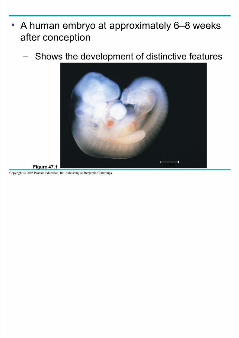
• A human embryo at approximately 6–8 weeks
after conception – Shows the development of distinctive features
Figure 47.1 1 mm
8/14/2019 47 Development Text
http://slidepdf.com/reader/full/47-development-text 4/73
Copyright © 2005 Pearson Education, Inc. publishing as Benjamin Cummings
• The question of how a zygote becomes an
animal – Has been asked for centuries
• As recently as the 18th century
– The prevailing theory was a notion called
preformation
8/14/2019 47 Development Text
http://slidepdf.com/reader/full/47-development-text 5/73
Copyright © 2005 Pearson Education, Inc. publishing as Benjamin Cummings
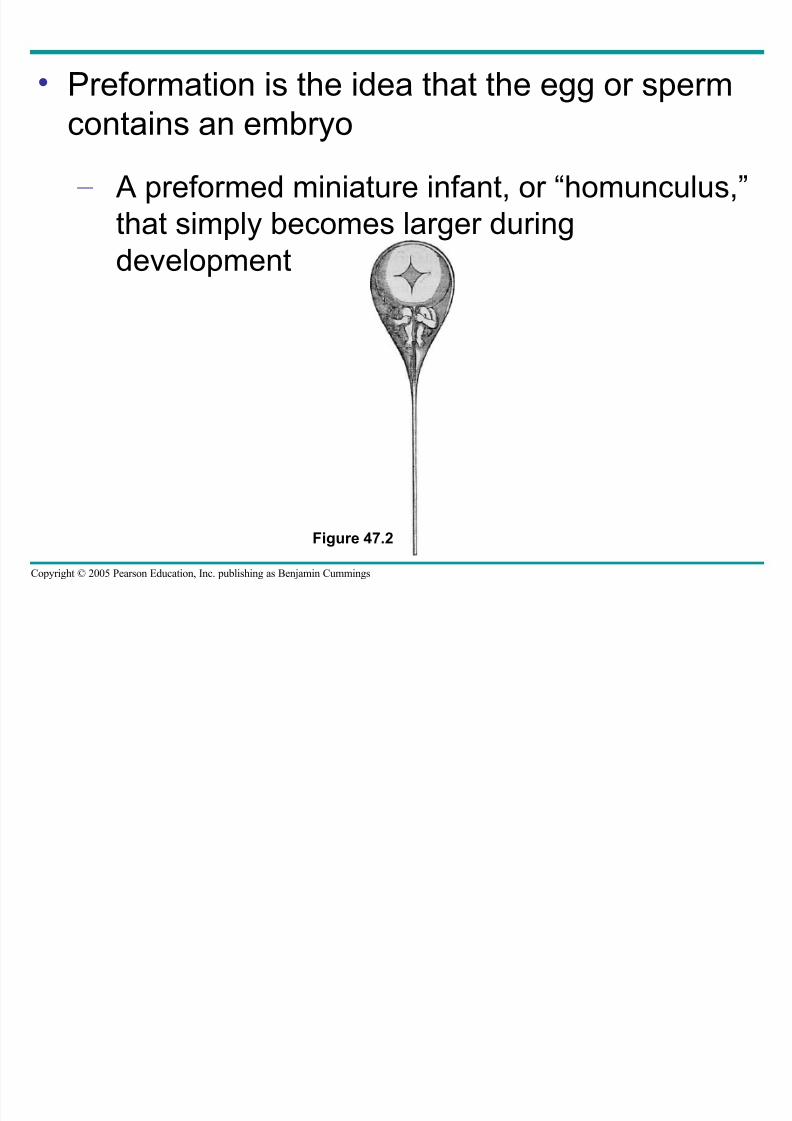
• Preformation is the idea that the egg or sperm
contains an embryo – A preformed miniature infant, or “homunculus,”
that simply becomes larger during
development
Figure 47.2
8/14/2019 47 Development Text
http://slidepdf.com/reader/full/47-development-text 6/73
Copyright © 2005 Pearson Education, Inc. publishing as Benjamin Cummings
• An organism’s development
– Is determined by the genome of the zygoteand by differences that arise between early
embryonic cells
• Cell differentiation
– Is the specialization of cells in their structure
and function
• Morphogenesis
– Is the process by which an animal takes shape
8/14/2019 47 Development Text
http://slidepdf.com/reader/full/47-development-text 7/73Copyright © 2005 Pearson Education, Inc. publishing as Benjamin Cummings
• Concept 47.1: After fertilization, embryonic
development proceeds through cleavage,gastrulation, and organogenesis
• Important events regulating development
– Occur during fertilization and each of the three
successive stages that build the animal’s body
8/14/2019 47 Development Text
http://slidepdf.com/reader/full/47-development-text 8/73Copyright © 2005 Pearson Education, Inc. publishing as Benjamin Cummings
Fertilization
• The main function of fertilization
– Is to bring the haploid nuclei of sperm and eggtogether to form a diploid zygote
• Contact of the sperm with the egg’s surface
– Initiates metabolic reactions within the egg that
trigger the onset of embryonic development
8/14/2019 47 Development Text
http://slidepdf.com/reader/full/47-development-text 9/73Copyright © 2005 Pearson Education, Inc. publishing as Benjamin Cummings
The Acrosomal Reaction
• The acrosomal reaction
– Is triggered when the sperm meets the egg
– Releases hydrolytic enzymes that digest
material surrounding the egg
8/14/2019 47 Development Text
http://slidepdf.com/reader/full/47-development-text 10/73Copyright © 2005 Pearson Education, Inc. publishing as Benjamin Cummings
• The acrosomal reaction
Sperm
nucleus
Sperm plasma
membrane
Hydrolytic enzymes
Cortical
granule
Cortical granule
membrane
EGG CYTOPLASM
Basal body
(centriole)
Sperm
head
Acrosomal
process
Actin
Acrosome
Jelly coat
Egg plasma
membrane
Vitelline layer
Fused plasma
membranes
Perivitelline
space
Fertilization
envelope
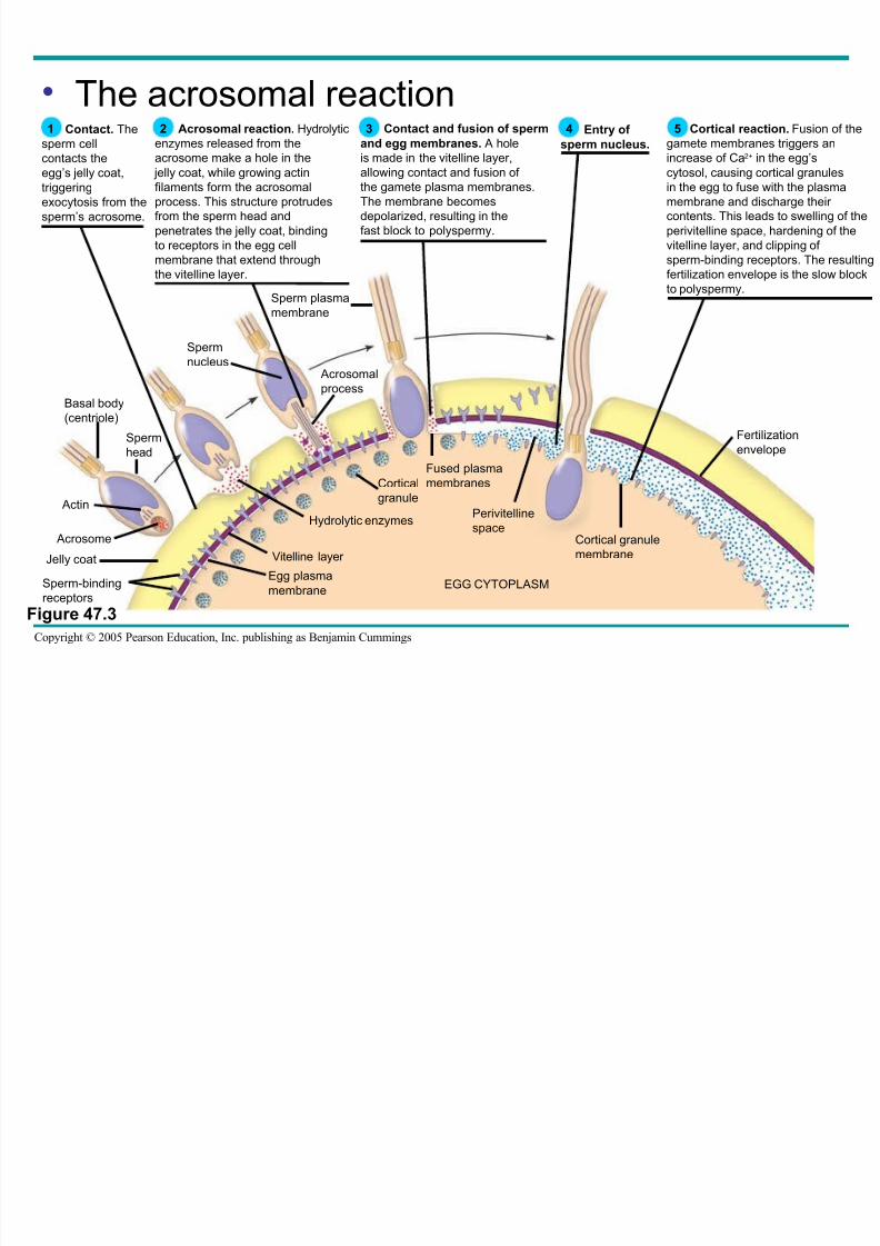
Cortical reaction. Fusion of the
gamete membranes triggers an
increase of Ca2+ in the egg’scytosol, causing cortical granules
in the egg to fuse with the plasma
membrane and discharge their
contents. This leads to swelling of the
perivitelline space, hardening of the
vitelline layer, and clipping of
sperm-binding receptors. The resulting
fertilization envelope is the slow block
to polyspermy.
5 Contact and fusion of sperm
and egg membranes. A hole
is made in the vitelline layer,allowing contact and fusion of
the gamete plasma membranes.
The membrane becomes
depolarized, resulting in the
fast block to polyspermy.
3 Acrosomal reaction. Hydrolytic
enzymes released from the
acrosome make a hole in the jelly coat, while growing actin
filaments form the acrosomal
process. This structure protrudes
from the sperm head and
penetrates the jelly coat, binding
to receptors in the egg cell
membrane that extend through
the vitelline layer.
2 Contact. The
sperm cell
contacts theegg’s jelly coat,
triggering
exocytosis from the
sperm’s acrosome.
1
Sperm-binding
receptors
Entry of
sperm nucleus.
4
Figure 47.3
8/14/2019 47 Development Text
http://slidepdf.com/reader/full/47-development-text 11/73Copyright © 2005 Pearson Education, Inc. publishing as Benjamin Cummings
• Gamete contact and/or fusion
– Depolarizes the egg cell membrane and setsup a fast block to polyspermy
8/14/2019 47 Development Text
http://slidepdf.com/reader/full/47-development-text 12/73Copyright © 2005 Pearson Education, Inc. publishing as Benjamin Cummings
The Cortical Reaction
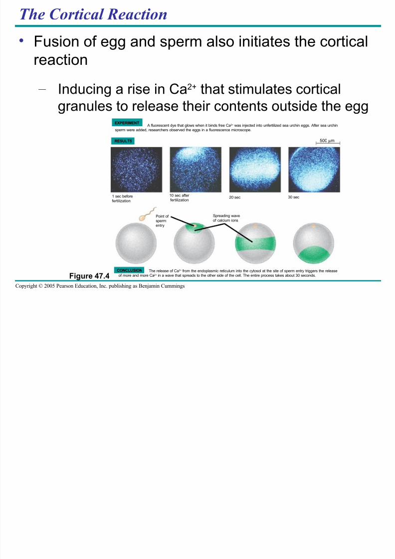
• Fusion of egg and sperm also initiates the cortical
reaction
– Inducing a rise in Ca2+ that stimulates cortical
granules to release their contents outside the egg
Figure 47.4
A fluorescent dye that glows when it binds free Ca2+ was injected into unfertilized sea urchin eggs. After sea urchinsperm were added, researchers observed the eggs in a fluorescence microscope.
EXPERIMENT
RESULTS
The release of Ca2+ from the endoplasmic reticulum into the cytosol at the site of sperm entry triggers the release
of more and more Ca2+
in a wave that spreads to the other side of the cell. The entire process takes about 30 seconds.
CONCLUSION
30 sec20 sec10 sec after
fertilization1 sec before
fertilization
Point of sperm
entry
Spreading wave
of calcium ions
500 µm
8/14/2019 47 Development Text
http://slidepdf.com/reader/full/47-development-text 13/73Copyright © 2005 Pearson Education, Inc. publishing as Benjamin Cummings
• These changes cause the formation of a
fertilization envelope – That functions as a slow block to polyspermy
8/14/2019 47 Development Text
http://slidepdf.com/reader/full/47-development-text 14/73Copyright © 2005 Pearson Education, Inc. publishing as Benjamin Cummings
Activation of the Egg
• Another outcome of the sharp rise in Ca2+ in
the egg’s cytosol – Is a substantial increase in the rates of cellular
respiration and protein synthesis by the egg
cell
• With these rapid changes in metabolism
– The egg is said to be activated
8/14/2019 47 Development Text
http://slidepdf.com/reader/full/47-development-text 15/73
Copyright © 2005 Pearson Education, Inc. publishing as Benjamin Cummings
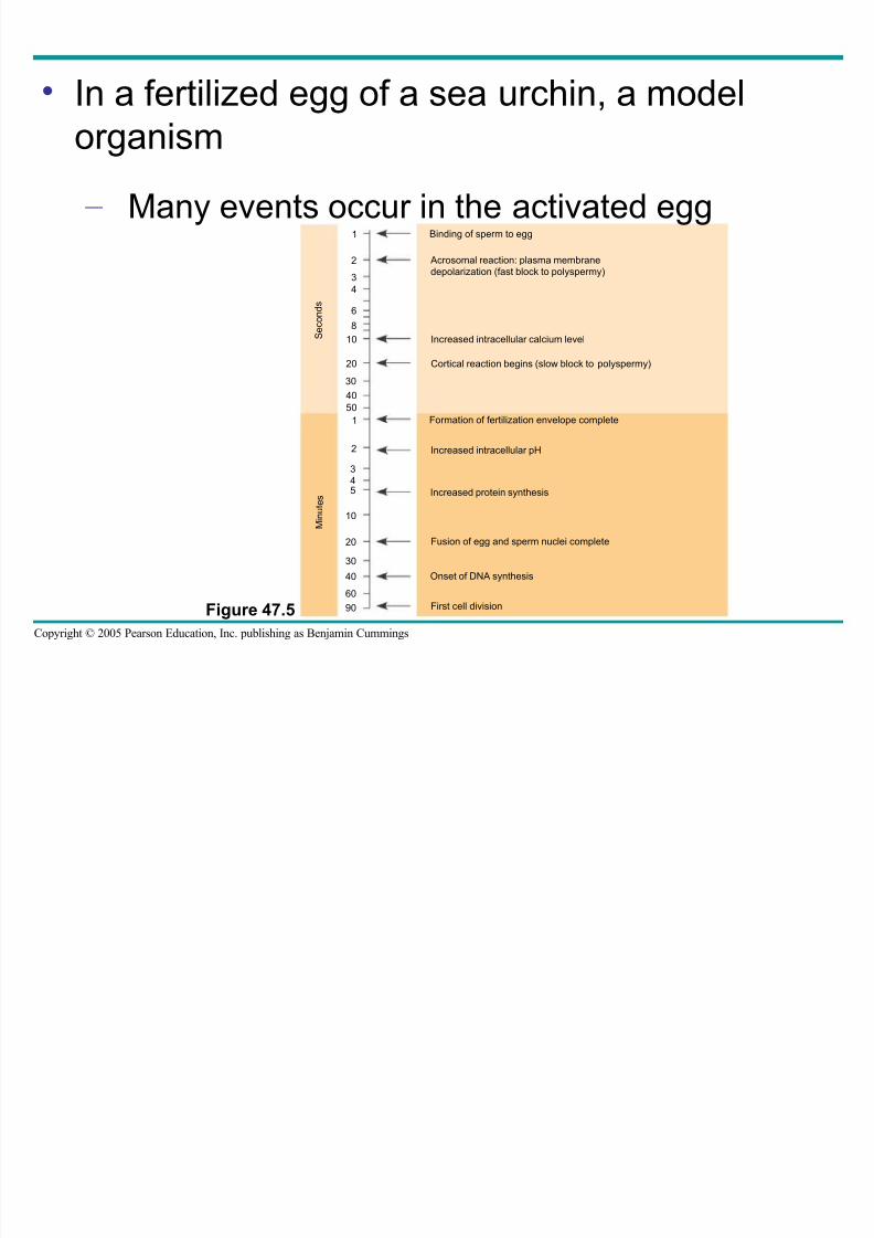
• In a fertilized egg of a sea urchin, a model
organism – Many events occur in the activated egg
Figure 47.5
Binding of sperm to egg
Acrosomal reaction: plasma membrane
depolarization (fast block to polyspermy)
Increased intracellular calcium level
Cortical reaction begins (slow block to polyspermy)
Formation of fertilization envelope complete
Increased intracellular pH
Increased protein synthesis
Fusion of egg and sperm nuclei complete
Onset of DNA synthesis
First cell division
1
2
3
4
6
8
10
20
30
40
50
1
2
3
45
10
20
30
40
60
S e c o n d s
M i n
u t e
s
90
8/14/2019 47 Development Text
http://slidepdf.com/reader/full/47-development-text 16/73
Copyright © 2005 Pearson Education, Inc. publishing as Benjamin Cummings
Fertilization in Mammals
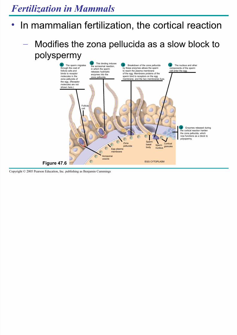
• In mammalian fertilization, the cortical reaction
– Modifies the zona pellucida as a slow block topolyspermy
Figure 47.6
Spermnucleus
Acrosomal
vesicle
Egg plasma
membrane
Zona
pellucida
Spermbasal
body
Cortical
granules
Folliclecell
EGG CYTOPLASM
The sperm migratesthrough the coat of
follicle cells andbinds to receptor
molecules in thezona pellucida of
the egg. (Receptor molecules are not
shown here.)
1This binding induces
the acrosomal reaction,in which the sperm
releases hydrolytic
enzymes into thezona pellucida.
2Breakdown of the zona pellucida
by these enzymes allows the sperm
to reach the plasma membraneof the egg. Membrane proteins of the
sperm bind to receptors on the eggmembrane, and the two membranes fuse.
3 The nucleus and other
components of the spermcell enter the egg.
4
Enzymes released duringthe cortical reaction harden
the zona pellucida, whichnow functions as a block to
polyspermy.
5
8/14/2019 47 Development Text
http://slidepdf.com/reader/full/47-development-text 17/73
Copyright © 2005 Pearson Education, Inc. publishing as Benjamin Cummings
Cleavage
• Fertilization is followed by cleavage
– A period of rapid cell division without growth
8/14/2019 47 Development Text
http://slidepdf.com/reader/full/47-development-text 18/73
Copyright © 2005 Pearson Education, Inc. publishing as Benjamin Cummings
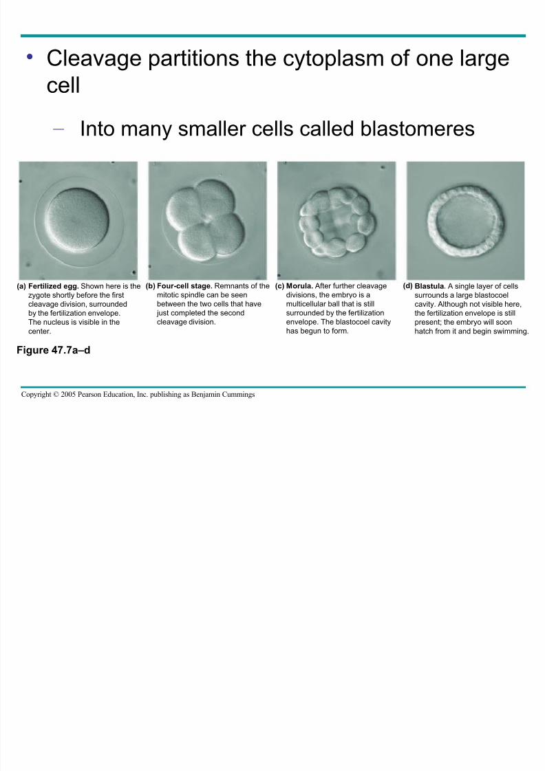
• Cleavage partitions the cytoplasm of one large
cell – Into many smaller cells called blastomeres
Figure 47.7a–d
Fertilized egg. Shown here is thezygote shortly before the first
cleavage division, surrounded
by the fertilization envelope.
The nucleus is visible in the
center.
(a) Four-cell stage. Remnants of themitotic spindle can be seen
between the two cells that have
just completed the second
cleavage division.
(b) Morula. After further cleavagedivisions, the embryo is a
multicellular ball that is still
surrounded by the fertilization
envelope. The blastocoel cavity
has begun to form.
(c) Blastula. A single layer of cellssurrounds a large blastocoel
cavity. Although not visible here,
the fertilization envelope is still
present; the embryo will soon
hatch from it and begin swimming.
(d)
8/14/2019 47 Development Text
http://slidepdf.com/reader/full/47-development-text 19/73
Copyright © 2005 Pearson Education, Inc. publishing as Benjamin Cummings
• The eggs and zygotes of many animals, except
mammals – Have a definite polarity
• The polarity is defined by the distribution of
yolk
– With the vegetal pole having the most yolk and
the animal pole having the least
8/14/2019 47 Development Text
http://slidepdf.com/reader/full/47-development-text 20/73
Copyright © 2005 Pearson Education, Inc. publishing as Benjamin Cummings
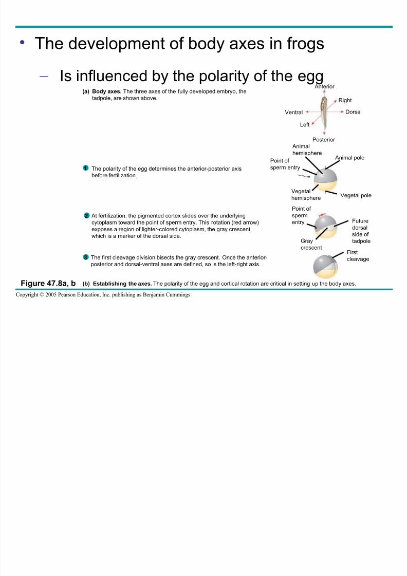
• The development of body axes in frogs
– Is influenced by the polarity of the egg
Figure 47.8a, b
Anterior
Ventral
Left
Posterior
Dorsal
Right
Body axes. The three axes of the fully developed embryo, the
tadpole, are shown above.
(a)
Animal
hemisphereAnimal pole
Point of
sperm entry
Vegetal
hemisphere Vegetal pole
Point of
spermentry Future
dorsal
side of
tadpoleGray
crescentFirst
cleavage
The polarity of the egg determines the anterior-posterior axis
before fertilization.
At fertilization, the pigmented cortex slides over the underlyingcytoplasm toward the point of sperm entry. This rotation (red arrow)
exposes a region of lighter-colored cytoplasm, the gray crescent,
which is a marker of the dorsal side.
The first cleavage division bisects the gray crescent. Once the anterior-
posterior and dorsal-ventral axes are defined, so is the left-right axis.
(b) Establishing the axes. The polarity of the egg and cortical rotation are critical in setting up the body axes.
1
2
3
8/14/2019 47 Development Text
http://slidepdf.com/reader/full/47-development-text 21/73
Copyright © 2005 Pearson Education, Inc. publishing as Benjamin Cummings
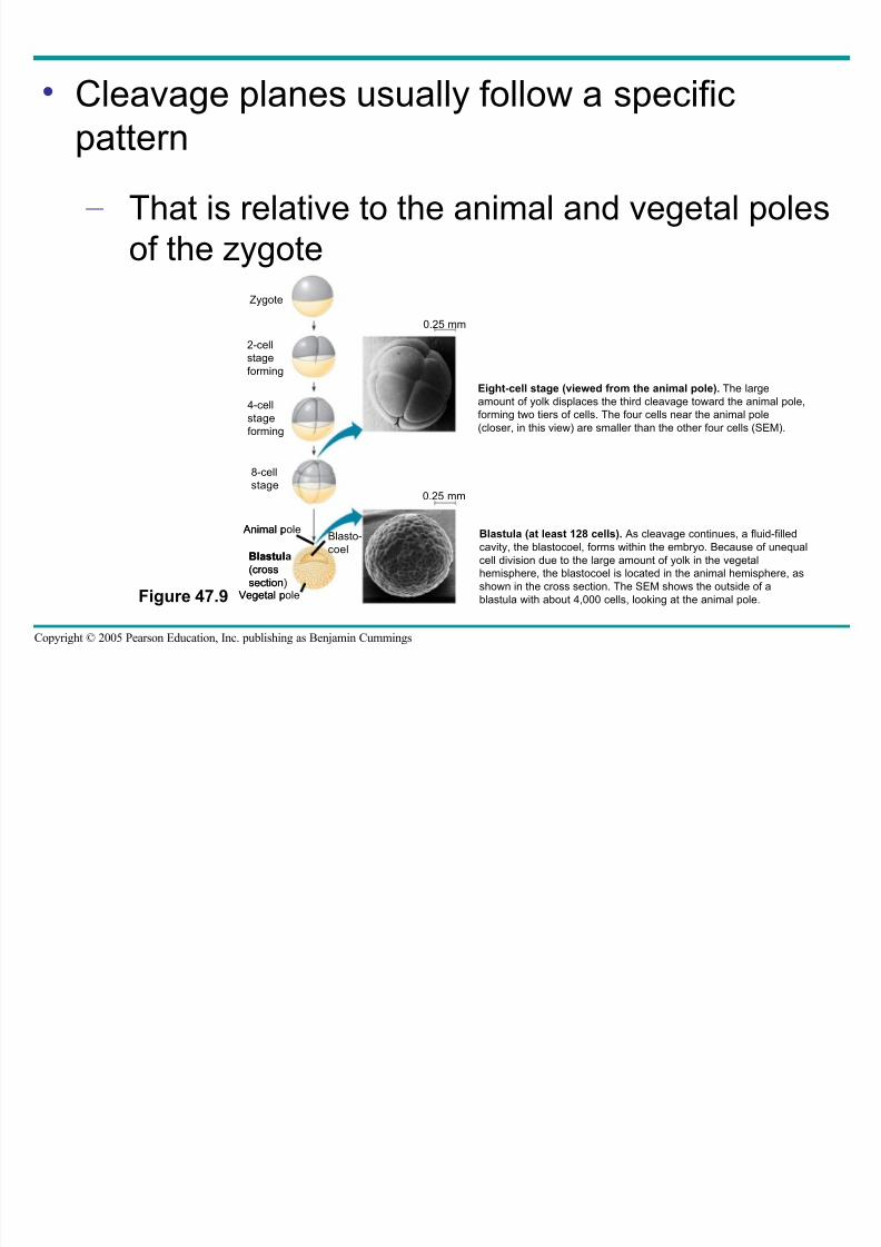
• Cleavage planes usually follow a specific
pattern – That is relative to the animal and vegetal poles
of the zygote
Figure 47.9
Zygote
2-cell
stage
forming
4-cell
stage
forming
8-cell
stage
Eight-cell stage (viewed from the animal pole). The large
amount of yolk displaces the third cleavage toward the animal pole,
forming two tiers of cells. The four cells near the animal pole
(closer, in this view) are smaller than the other four cells (SEM).
0.25 mm0.25 mm
Vegetal pole
Blastula
(cross
section)
Animal poleBlasto-
coel
Blastula (at least 128 cells). As cleavage continues, a fluid-filled
cavity, the blastocoel, forms within the embryo. Because of unequal
cell division due to the large amount of yolk in the vegetal
hemisphere, the blastocoel is located in the animal hemisphere, as
shown in the cross section. The SEM shows the outside of a
blastula with about 4,000 cells, looking at the animal pole.Vegetal pole
Blastula
(cross
section)
Animal poleBlasto-
coel
0.25 mm
0.25 mm
8/14/2019 47 Development Text
http://slidepdf.com/reader/full/47-development-text 22/73
Copyright © 2005 Pearson Education, Inc. publishing as Benjamin Cummings
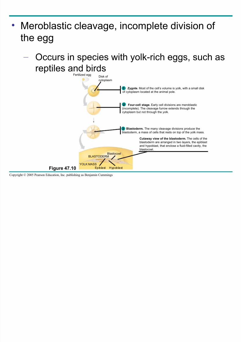
• Meroblastic cleavage, incomplete division of
the egg – Occurs in species with yolk-rich eggs, such as
reptiles and birds
Figure 47.10Epiblast Hypoblast
BLASTODERMBlastocoel
YOLK MASS
Fertilized eggDisk of
cytoplasm
Zygote. Most of the cell’s volume is yolk, with a small disk
of cytoplasm located at the animal pole.
Four-cell stage. Early cell divisions are meroblastic
(incomplete). The cleavage furrow extends through the
cytoplasm but not through the yolk.
Blastoderm. The many cleavage divisions produce the
blastoderm, a mass of cells that rests on top of the yolk mass.
Cutaway view of the blastoderm. The cells of the
blastoderm are arranged in two layers, the epiblast
and hypoblast, that enclose a fluid-filled cavity, the
blastocoel.
3
1
2
8/14/2019 47 Development Text
http://slidepdf.com/reader/full/47-development-text 23/73
Copyright © 2005 Pearson Education, Inc. publishing as Benjamin Cummings
• Holoblastic cleavage, the complete division of
the egg – Occurs in species whose eggs have little or
moderate amounts of yolk, such as sea
urchins and frogs
8/14/2019 47 Development Text
http://slidepdf.com/reader/full/47-development-text 24/73
Copyright © 2005 Pearson Education, Inc. publishing as Benjamin Cummings
Gastrulation
• The morphogenetic process called gastrulation
– Rearranges the cells of a blastula into a three-layered embryo, called a gastrula, that has a
primitive gut
8/14/2019 47 Development Text
http://slidepdf.com/reader/full/47-development-text 25/73
Copyright © 2005 Pearson Education, Inc. publishing as Benjamin Cummings
• The three layers produced by gastrulation
– Are called embryonic germ layers
• The ectoderm
– Forms the outer layer of the gastrula
• The endoderm
– Lines the embryonic digestive tract
• The mesoderm
– Partly fills the space between the endoderm and
ectoderm
8/14/2019 47 Development Text
http://slidepdf.com/reader/full/47-development-text 26/73
Copyright © 2005 Pearson Education, Inc. publishing as Benjamin Cummings
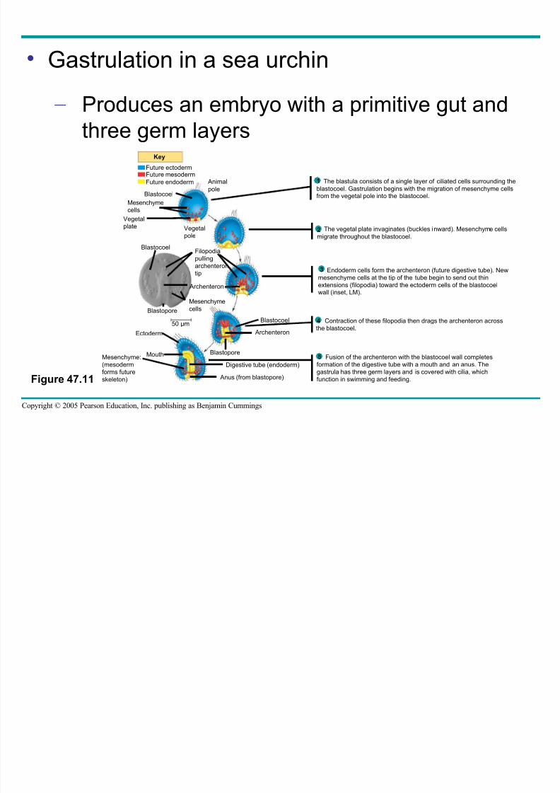
• Gastrulation in a sea urchin
– Produces an embryo with a primitive gut andthree germ layers
Figure 47.11
Digestive tube (endoderm)
Key
Future ectodermFuture mesoderm
Future endoderm
Blastocoel
Mesenchyme
cells
Vegetal
plate
Animal
pole
Vegetal
pole
Filopodia
pulling
archenteron
tip
Archenteron
Blastocoel
Blastopore
50 µm
Blastopore
Archenteron
Blastocoel
Mouth
Ectoderm
Mesenchyme:
(mesoderm
forms future
skeleton) Anus (from blastopore)
Mesenchyme
cells
The blastula consists of a single layer of ciliated cells surrounding the
blastocoel. Gastrulation begins with the migration of mesenchyme cells
from the vegetal pole into the blastocoel.
1
2 The vegetal plate invaginates (buckles inward). Mesenchyme cells
migrate throughout the blastocoel.2
Endoderm cells form the archenteron (future digestive tube). New
mesenchyme cells at the tip of the tube begin to send out thin
extensions (filopodia) toward the ectoderm cells of the blastocoelwall (inset, LM).
3
Contraction of these filopodia then drags the archenteron across
the blastocoel.
4
Fusion of the archenteron with the blastocoel wall completes
formation of the digestive tube with a mouth and an anus. The
gastrula has three germ layers and is covered with cilia, which
function in swimming and feeding.
5
8/14/2019 47 Development Text
http://slidepdf.com/reader/full/47-development-text 27/73
Copyright © 2005 Pearson Education, Inc. publishing as Benjamin Cummings
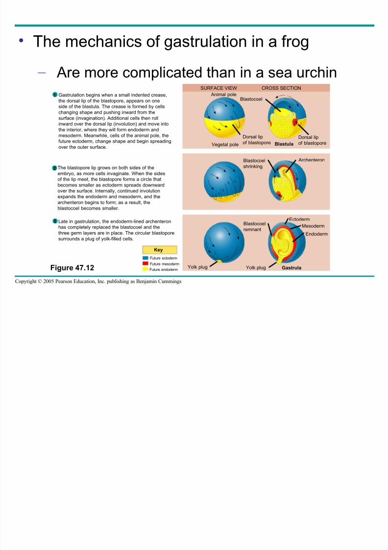
• The mechanics of gastrulation in a frog
– Are more complicated than in a sea urchin
Figure 47.12
SURFACE VIEW CROSS SECTION
Animal poleBlastocoel
Dorsal lipof blastopore
Dorsal lipof blastoporeVegetal pole Blastula
Blastocoel
shrinking
Archenteron
Blastocoel
remnant
Ectoderm
Mesoderm
Endoderm
GastrulaYolk plugYolk plug
Key
Future ectoderm
Future mesoderm
Future endoderm
Gastrulation begins when a small indented crease,
the dorsal lip of the blastopore, appears on one
side of the blastula. The crease is formed by cells
changing shape and pushing inward from the
surface (invagination). Additional cells then roll
inward over the dorsal lip (involution) and move into
the interior, where they will form endoderm and
mesoderm. Meanwhile, cells of the animal pole, thefuture ectoderm, change shape and begin spreading
over the outer surface.
The blastopore lip grows on both sides of the
embryo, as more cells invaginate. When the sides
of the lip meet, the blastopore forms a circle that
becomes smaller as ectoderm spreads downward
over the surface. Internally, continued involution
expands the endoderm and mesoderm, and the
archenteron begins to form; as a result, the
blastocoel becomes smaller.
1
2
3 Late in gastrulation, the endoderm-lined archenteron
has completely replaced the blastocoel and the
three germ layers are in place. The circular blastopore
surrounds a plug of yolk-filled cells.
8/14/2019 47 Development Text
http://slidepdf.com/reader/full/47-development-text 28/73
Copyright © 2005 Pearson Education, Inc. publishing as Benjamin Cummings
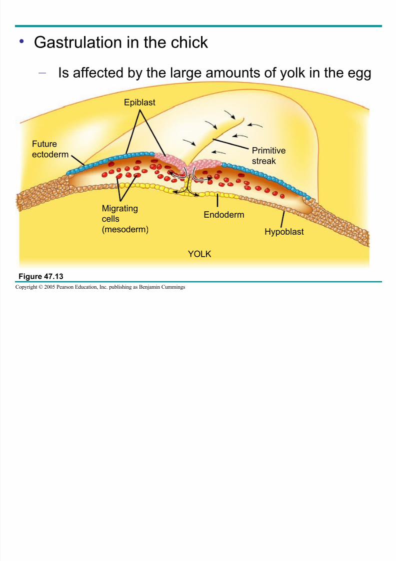
• Gastrulation in the chick
– Is affected by the large amounts of yolk in the egg
Figure 47.13
Epiblast
Future
ectoderm
Migratingcells
(mesoderm)
Endoderm
Hypoblast
YOLK
Primitive
streak
O i
8/14/2019 47 Development Text
http://slidepdf.com/reader/full/47-development-text 29/73
Copyright © 2005 Pearson Education, Inc. publishing as Benjamin Cummings
Organogenesis
• Various regions of the three embryonic germ
layers
– Develop into the rudiments of organs during
the process of organogenesis
8/14/2019 47 Development Text
http://slidepdf.com/reader/full/47-development-text 30/73
Copyright © 2005 Pearson Education, Inc. publishing as Benjamin Cummings
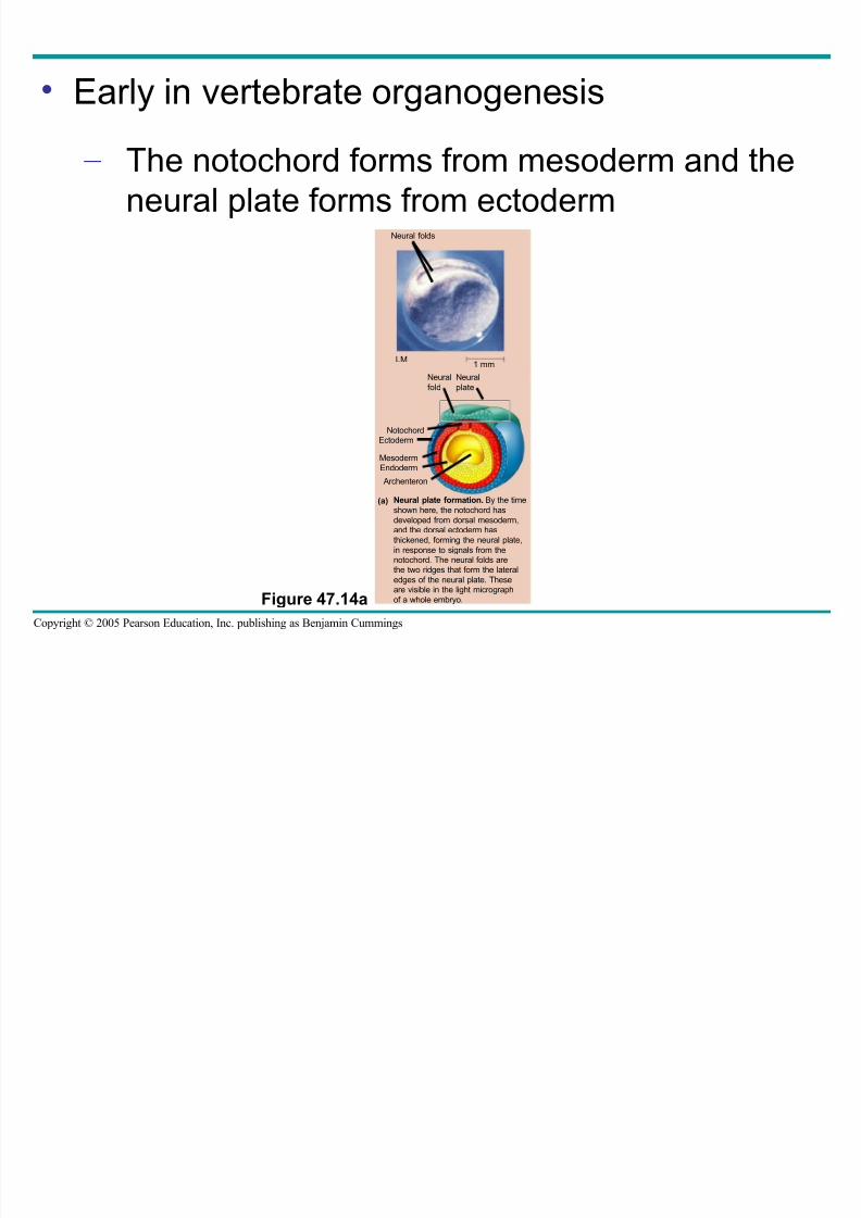
• Early in vertebrate organogenesis
– The notochord forms from mesoderm and theneural plate forms from ectoderm
Figure 47.14a
Neural plate formation. By the time
shown here, the notochord has
developed from dorsal mesoderm,and the dorsal ectoderm has
thickened, forming the neural plate,
in response to signals from thenotochord. The neural folds arethe two ridges that form the lateral
edges of the neural plate. Theseare visible in the light micrograph
of a whole embryo.
Neural folds
1 mm
Neural
fold
Neural
plate
NotochordEctoderm
MesodermEndoderm
Archenteron
(a)
LM
8/14/2019 47 Development Text
http://slidepdf.com/reader/full/47-development-text 31/73
Copyright © 2005 Pearson Education, Inc. publishing as Benjamin Cummings
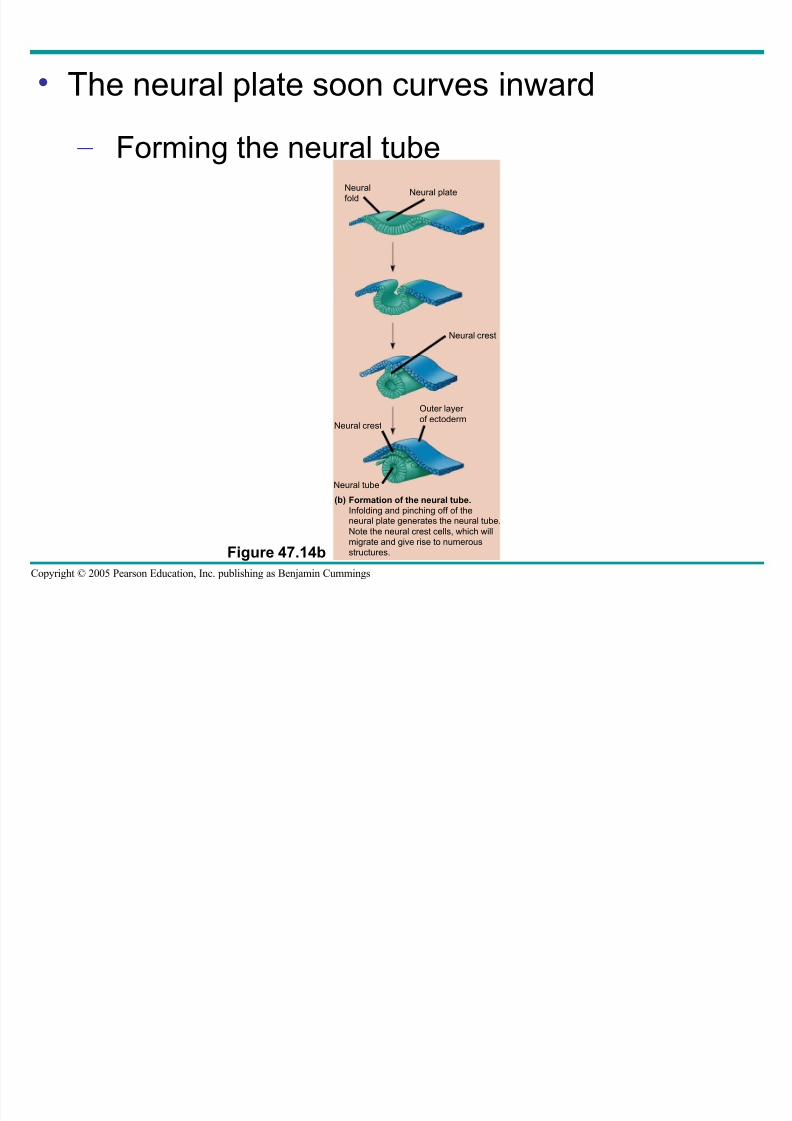
• The neural plate soon curves inward
– Forming the neural tube
Figure 47.14b
Formation of the neural tube.
Infolding and pinching off of theneural plate generates the neural tube.
Note the neural crest cells, which will
migrate and give rise to numerous
structures.
Neural
foldNeural plate
Neural crest
Outer layer
of ectodermNeural crest
Neural tube
(b)
8/14/2019 47 Development Text
http://slidepdf.com/reader/full/47-development-text 32/73
Copyright © 2005 Pearson Education, Inc. publishing as Benjamin Cummings
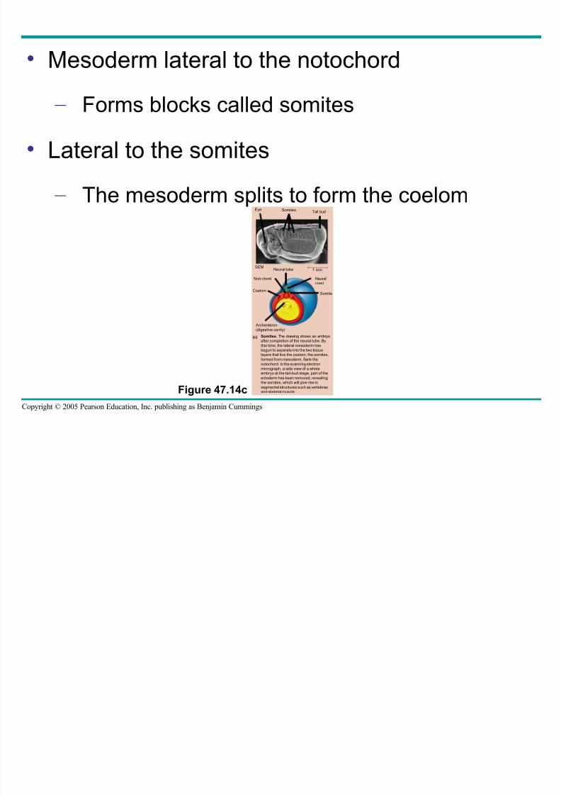
• Mesoderm lateral to the notochord
– Forms blocks called somites
• Lateral to the somites
– The mesoderm splits to form the coelom
Figure 47.14c
Somites. The drawing shows an embryoafter completion of the neural tube. By
this time, the lateral mesoderm hasbegun to separate into the two tissuelayers that line the coelom; the somites,
formed from mesoderm, flank the
notochord. In the scanning electronmicrograph, a side view of a wholeembryo at the tail-bud stage, part of the
ectoderm has been removed, revealingthe somites, which will give rise to
segmental structures such as vertebraeand skeletal muscle.
Eye Somites Tail bud
1 mmNeural tube
Notochord Neural
crest
Somite
Archenteron(digestive cavity)
Coelom
(c)
SEM
8/14/2019 47 Development Text
http://slidepdf.com/reader/full/47-development-text 33/73
Copyright © 2005 Pearson Education, Inc. publishing as Benjamin Cummings
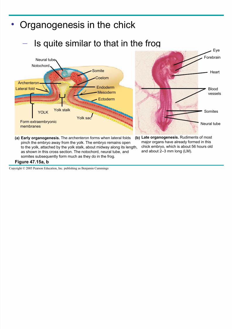
• Organogenesis in the chick
– Is quite similar to that in the frog
Figure 47.15a, b
Neural tube
Notochord
Archenteron
Lateral fold
Form extraembryonicmembranes
YOLKYolk stalk
Somite
Coelom
Endoderm
Mesoderm
Ectoderm
Yolk sac
Eye
Forebrain
Heart
Blood
vessels
Somites
Neural tube
Early organogenesis. The archenteron forms when lateral folds
pinch the embryo away from the yolk. The embryo remains open
to the yolk, attached by the yolk stalk, about midway along its length,
as shown in this cross section. The notochord, neural tube, and
somites subsequently form much as they do in the frog.
(a) Late organogenesis. Rudiments of most
major organs have already formed in this
chick embryo, which is about 56 hours old
and about 2–3 mm long (LM).
(b)
8/14/2019 47 Development Text
http://slidepdf.com/reader/full/47-development-text 34/73
Copyright © 2005 Pearson Education, Inc. publishing as Benjamin Cummings
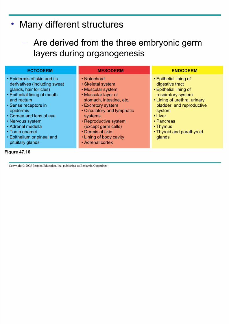
• Many different structures
– Are derived from the three embryonic germlayers during organogenesis
Figure 47.16
ECTODERM MESODERM ENDODERM
• Epidermis of skin and its
derivatives (including sweat
glands, hair follicles)
• Epithelial lining of mouth
and rectum
• Sense receptors in
epidermis
• Cornea and lens of eye
• Nervous system• Adrenal medulla
• Tooth enamel
• Epithelium or pineal and
pituitary glands
• Notochord
• Skeletal system
• Muscular system
• Muscular layer of
stomach, intestine, etc.
• Excretory system
• Circulatory and lymphatic
systems
• Reproductive system(except germ cells)
• Dermis of skin
• Lining of body cavity
• Adrenal cortex
• Epithelial lining of
digestive tract
• Epithelial lining of
respiratory system
• Lining of urethra, urinary
bladder, and reproductive
system
• Liver
• Pancreas• Thymus
• Thyroid and parathyroid
glands
De elopmental Adaptations of Amniotes
8/14/2019 47 Development Text
http://slidepdf.com/reader/full/47-development-text 35/73
Copyright © 2005 Pearson Education, Inc. publishing as Benjamin Cummings
Developmental Adaptations of Amniotes
• The embryos of birds, other reptiles, and
mammals
– Develop within a fluid-filled sac that is
contained within a shell or the uterus
• Organisms with these adaptations
– Are called amniotes
8/14/2019 47 Development Text
http://slidepdf.com/reader/full/47-development-text 36/73
Copyright © 2005 Pearson Education, Inc. publishing as Benjamin Cummings
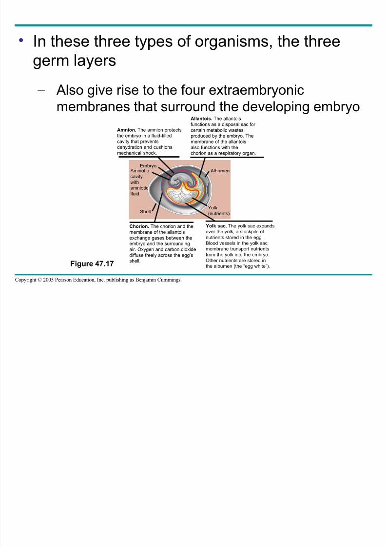
• In these three types of organisms, the three
germ layers
– Also give rise to the four extraembryonic
membranes that surround the developing embryo
Figure 47.17
Amnion. The amnion protects
the embryo in a fluid-filledcavity that prevents
dehydration and cushions
mechanical shock.
Allantois. The allantois
functions as a disposal sac for
certain metabolic wastes
produced by the embryo. Themembrane of the allantois
also functions with the
chorion as a respiratory organ.
Chorion. The chorion and the
membrane of the allantois
exchange gases between the
embryo and the surrounding
air. Oxygen and carbon dioxide
diffuse freely across the egg’s
shell.
Yolk sac. The yolk sac expands
over the yolk, a stockpile of
nutrients stored in the egg.
Blood vessels in the yolk sac
membrane transport nutrients
from the yolk into the embryo.
Other nutrients are stored in
the albumen (the “egg white”).
EmbryoAmniotic
cavity
with
amniotic
fluid
Shell
Albumen
Yolk
(nutrients)
Mammalian Development
8/14/2019 47 Development Text
http://slidepdf.com/reader/full/47-development-text 37/73
Copyright © 2005 Pearson Education, Inc. publishing as Benjamin Cummings
Mammalian Development
• The eggs of placental mammals
– Are small and store few nutrients
– Exhibit holoblastic cleavage
– Show no obvious polarity
8/14/2019 47 Development Text
http://slidepdf.com/reader/full/47-development-text 38/73
Copyright © 2005 Pearson Education, Inc. publishing as Benjamin Cummings
• Gastrulation and organogenesis
– Resemble the processes in birds and other reptiles
8/14/2019 47 Development Text
http://slidepdf.com/reader/full/47-development-text 39/73
Copyright © 2005 Pearson Education, Inc. publishing as Benjamin Cummings
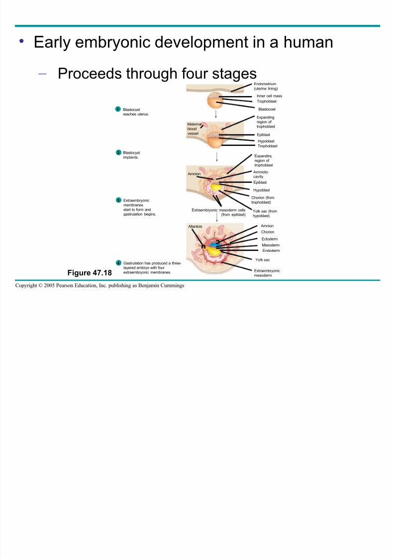
• Early embryonic development in a human
– Proceeds through four stages
Figure 47.18
Endometrium
(uterine lining)
Inner cell mass
Trophoblast
Blastocoel
Expandingregion of
trophoblast
Epiblast
HypoblastTrophoblast
Expanding
region of trophoblast
Amnioticcavity
Epiblast
Hypoblast
Chorion (from
trophoblast)
Yolk sac (from
hypoblast)
Extraembryonic mesoderm cells(from epiblast)
Amnion
Chorion
Ectoderm
Mesoderm
Endoderm
Yolk sac
Extraembryonic
mesoderm
Allantois
Amnion
Maternalblood
vessel
Blastocystreaches uterus.
1
Blastocyst
implants.2
Extraembryonic
membranes
start to form andgastrulation begins.
3
Gastrulation has produced a three-layered embryo with four
extraembryonic membranes.
4
8/14/2019 47 Development Text
http://slidepdf.com/reader/full/47-development-text 40/73
Copyright © 2005 Pearson Education, Inc. publishing as Benjamin Cummings
• At the completion of cleavage
– The blastocyst forms
• The trophoblast, the outer epithelium of the
blastocyst
– Initiates implantation in the uterus, and the
blastocyst forms a flat disk of cells
8/14/2019 47 Development Text
http://slidepdf.com/reader/full/47-development-text 41/73
Copyright © 2005 Pearson Education, Inc. publishing as Benjamin Cummings
• As implantation is completed
– Gastrulation begins
– The extraembryonic membranes begin to form
• By the end of gastrulation – The embryonic germ layers have formed
8/14/2019 47 Development Text
http://slidepdf.com/reader/full/47-development-text 42/73
Copyright © 2005 Pearson Education, Inc. publishing as Benjamin Cummings
• The extraembryonic membranes in mammals
– Are homologous to those of birds and other reptiles and have similar functions
8/14/2019 47 Development Text
http://slidepdf.com/reader/full/47-development-text 43/73
Copyright © 2005 Pearson Education, Inc. publishing as Benjamin Cummings
• Concept 47.2: Morphogenesis in animals
involves specific changes in cell shape,
position, and adhesion
• Morphogenesis is a major aspect of
development in both plants and animals – But only in animals does it involve the
movement of cells
The Cytoskeleton Cell Motility and Convergent
8/14/2019 47 Development Text
http://slidepdf.com/reader/full/47-development-text 44/73
Copyright © 2005 Pearson Education, Inc. publishing as Benjamin Cummings
The Cytoskeleton, Cell Motility, and ConvergentExtension
• Changes in the shape of a cell – Usually involve reorganization of the
cytoskeleton
8/14/2019 47 Development Text
http://slidepdf.com/reader/full/47-development-text 45/73
Copyright © 2005 Pearson Education, Inc. publishing as Benjamin Cummings
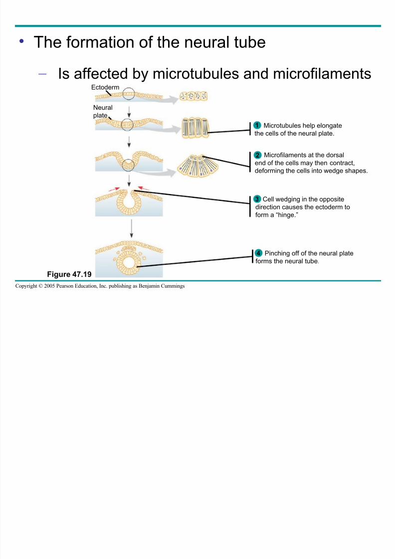
• The formation of the neural tube
– Is affected by microtubules and microfilaments
Figure 47.19
Microtubules help elongate
the cells of the neural plate.
1
Pinching off of the neural plate
forms the neural tube.
4
Ectoderm
Neural
plate
Microfilaments at the dorsal
end of the cells may then contract,
deforming the cells into wedge shapes.
Cell wedging in the opposite
direction causes the ectoderm toform a “hinge.”
2
3

8/14/2019 47 Development Text
http://slidepdf.com/reader/full/47-development-text 46/73
Copyright © 2005 Pearson Education, Inc. publishing as Benjamin Cummings
• The cytoskeleton also drives cell migration, or
cell crawling
– The active movement of cells from one place
to another
• In gastrulation, tissue invagination
– Is caused by changes in both cell shape and
cell migration
8/14/2019 47 Development Text
http://slidepdf.com/reader/full/47-development-text 47/73
Copyright © 2005 Pearson Education, Inc. publishing as Benjamin Cummings
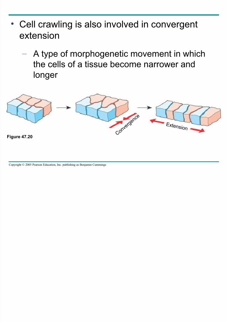
• Cell crawling is also involved in convergent
extension
– A type of morphogenetic movement in which
the cells of a tissue become narrower and
longer
Figure 47.20C o n v
e r g e n c e
E x t e n s i o n
Roles of the Extracellular Matrix and Cell
8/14/2019 47 Development Text
http://slidepdf.com/reader/full/47-development-text 48/73
Copyright © 2005 Pearson Education, Inc. publishing as Benjamin Cummings
Roles of the Extracellular Matrix and CellAdhesion Molecules
• Fibers of the extracellular matrix – May function as tracks, directing migrating
cells along particular routes
8/14/2019 47 Development Text
http://slidepdf.com/reader/full/47-development-text 49/73
Copyright © 2005 Pearson Education, Inc. publishing as Benjamin Cummings
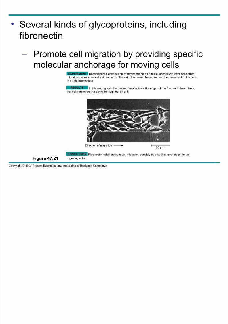
• Several kinds of glycoproteins, including
fibronectin
– Promote cell migration by providing specific
molecular anchorage for moving cells
Figure 47.21
EXPERIMENT Researchers placed a strip of fibronectin on an artificial underlayer. After positioning
migratory neural crest cells at one end of the strip, the researchers observed the movement of the cells
in a light microscope.
CONCLUSION
RESULTS In this micrograph, the dashed lines indicate the edges of the fibronectin layer. Note
that cells are migrating along the strip, not off of it.
Fibronectin helps promote cell migration, possibly by providing anchorage for the
migrating cells.
Direction of migration50 µm
8/14/2019 47 Development Text
http://slidepdf.com/reader/full/47-development-text 50/73
Copyright © 2005 Pearson Education, Inc. publishing as Benjamin Cummings
• Cell adhesion molecules
– Also contribute to cell migration and stabletissue structure
8/14/2019 47 Development Text
http://slidepdf.com/reader/full/47-development-text 51/73
Copyright © 2005 Pearson Education, Inc. publishing as Benjamin Cummings
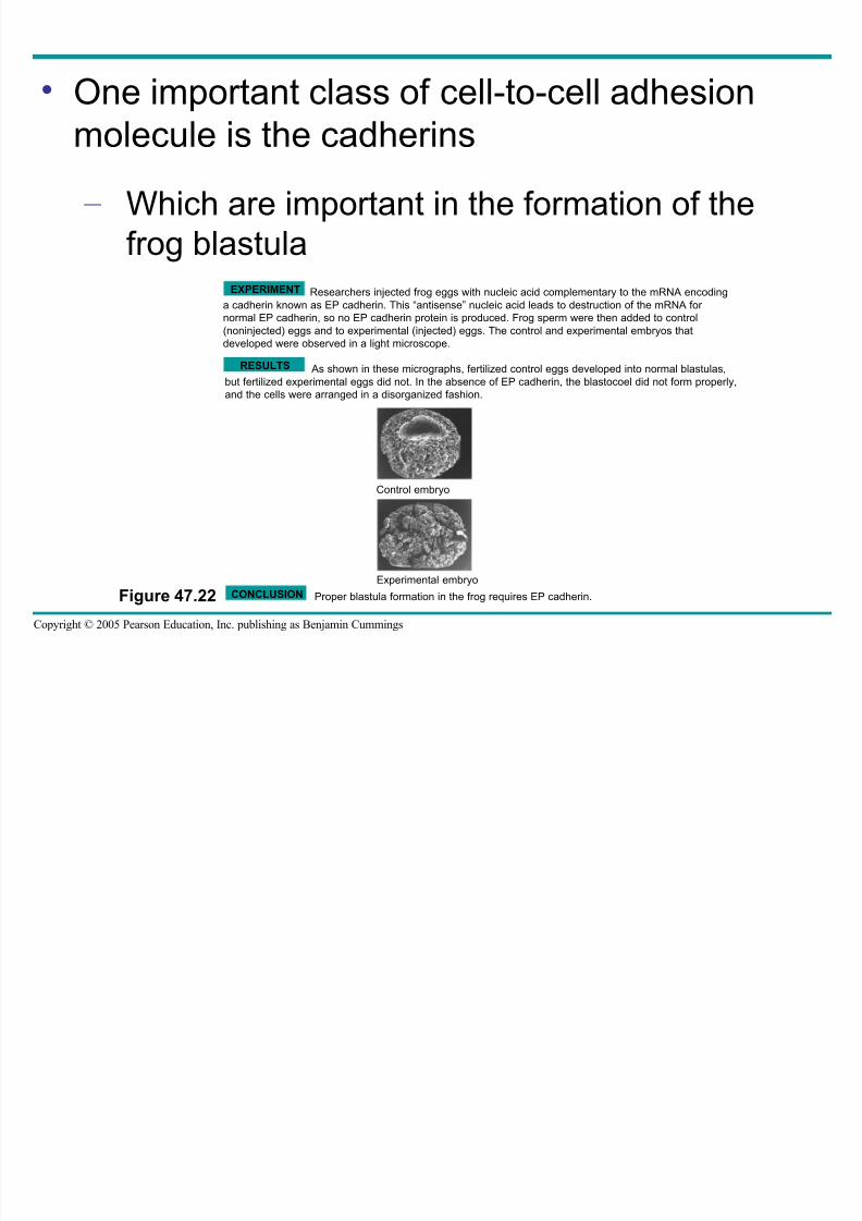
• One important class of cell-to-cell adhesion
molecule is the cadherins
– Which are important in the formation of the
frog blastula
Figure 47.22 CONCLUSION
EXPERIMENT Researchers injected frog eggs with nucleic acid complementary to the mRNA encoding
a cadherin known as EP cadherin. This “antisense” nucleic acid leads to destruction of the mRNA for normal EP cadherin, so no EP cadherin protein is produced. Frog sperm were then added to control
(noninjected) eggs and to experimental (injected) eggs. The control and experimental embryos that
developed were observed in a light microscope.
RESULTS As shown in these micrographs, fertilized control eggs developed into normal blastulas,
but fertilized experimental eggs did not. In the absence of EP cadherin, the blastocoel did not form properly,
and the cells were arranged in a disorganized fashion.
Control embryo
Experimental embryo
Proper blastula formation in the frog requires EP cadherin.
8/14/2019 47 Development Text
http://slidepdf.com/reader/full/47-development-text 52/73
Copyright © 2005 Pearson Education, Inc. publishing as Benjamin Cummings
• Concept 47.3: The developmental fate of cells
depends on their history and on inductive
signals
• Coupled with morphogenetic changes
– Development also requires the timelydifferentiation of many kinds of cells at specific
locations
• Two general principles
– Underlie differentiation during embryonic
development
8/14/2019 47 Development Text
http://slidepdf.com/reader/full/47-development-text 53/73
Fate Mapping
8/14/2019 47 Development Text
http://slidepdf.com/reader/full/47-development-text 54/73
Copyright © 2005 Pearson Education, Inc. publishing as Benjamin Cummings
Fate Mapping
• Fate maps
– Are general territorial diagrams of embryonicdevelopment
8/14/2019 47 Development Text
http://slidepdf.com/reader/full/47-development-text 55/73
Copyright © 2005 Pearson Education, Inc. publishing as Benjamin Cummings
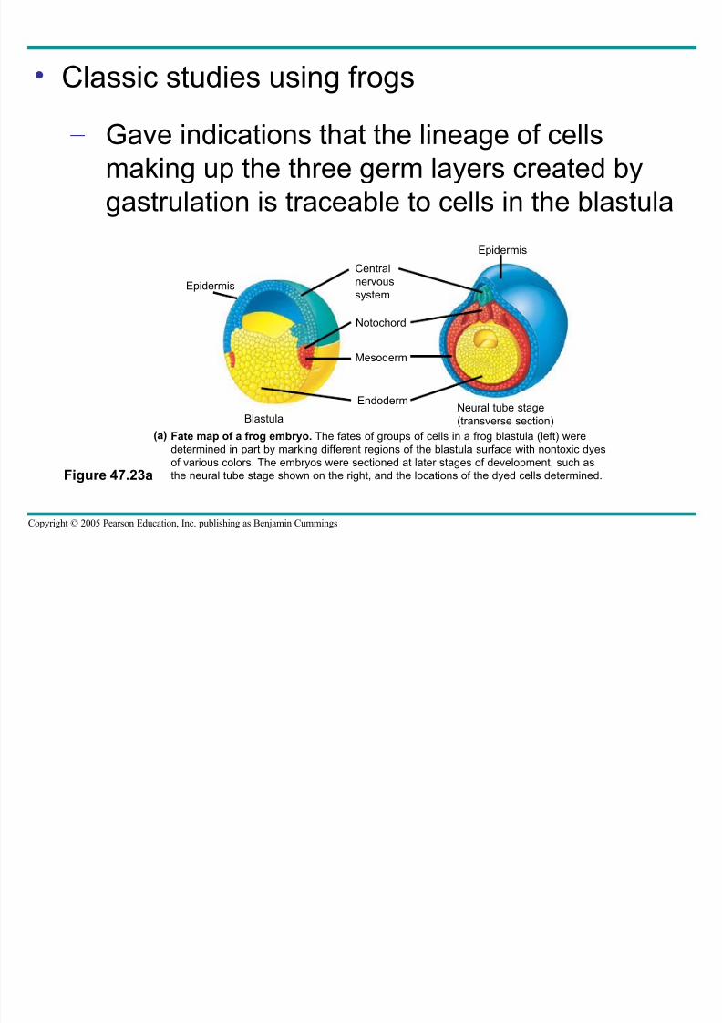
• Classic studies using frogs
– Gave indications that the lineage of cellsmaking up the three germ layers created by
gastrulation is traceable to cells in the blastula
Figure 47.23a
Fate map of a frog embryo. The fates of groups of cells in a frog blastula (left) were
determined in part by marking different regions of the blastula surface with nontoxic dyes
of various colors. The embryos were sectioned at later stages of development, such as
the neural tube stage shown on the right, and the locations of the dyed cells determined.
Neural tube stage
(transverse section)Blastula
Epidermis
Epidermis
Central
nervous
system
Notochord
Mesoderm
Endoderm
(a)
8/14/2019 47 Development Text
http://slidepdf.com/reader/full/47-development-text 56/73
Copyright © 2005 Pearson Education, Inc. publishing as Benjamin Cummings
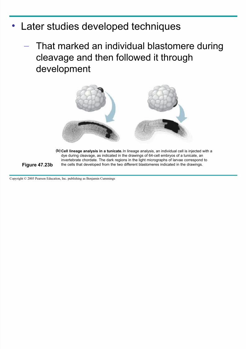
• Later studies developed techniques
– That marked an individual blastomere duringcleavage and then followed it through
development
Figure 47.23b
Cell lineage analysis in a tunicate. In lineage analysis, an individual cell is injected with a
dye during cleavage, as indicated in the drawings of 64-cell embryos of a tunicate, an
invertebrate chordate. The dark regions in the light micrographs of larvae correspond to
the cells that developed from the two different blastomeres indicated in the drawings.
(b)
Establishing Cellular Asymmetries
8/14/2019 47 Development Text
http://slidepdf.com/reader/full/47-development-text 57/73
Copyright © 2005 Pearson Education, Inc. publishing as Benjamin Cummings
g y
• To understand at the molecular level how
embryonic cells acquire their fates
– It is helpful to think first about how the basic
axes of the embryo are established
The Axes of the Basic Body Plan
8/14/2019 47 Development Text
http://slidepdf.com/reader/full/47-development-text 58/73
Copyright © 2005 Pearson Education, Inc. publishing as Benjamin Cummings
f y
• In nonamniotic vertebrates
– Basic instructions for establishing the bodyaxes are set down early, during oogenesis or
fertilization
8/14/2019 47 Development Text
http://slidepdf.com/reader/full/47-development-text 59/73
Copyright © 2005 Pearson Education, Inc. publishing as Benjamin Cummings
• In amniotes, local environmental differences
– Play the major role in establishing initialdifferences between cells and, later, the body
axes
Restriction of Cellular Potency
8/14/2019 47 Development Text
http://slidepdf.com/reader/full/47-development-text 60/73
Copyright © 2005 Pearson Education, Inc. publishing as Benjamin Cummings
f y
• In many species that have cytoplasmic
determinants
– Only the zygote is totipotent, capable of
developing into all the cell types found in the
adult
8/14/2019 47 Development Text
http://slidepdf.com/reader/full/47-development-text 61/73
Copyright © 2005 Pearson Education, Inc. publishing as Benjamin Cummings
• Unevenly distributed cytoplasmic determinants
in the egg cell
– Are important in establishing the body axes
– Set up differences in blastomeres resulting
from cleavage
8/14/2019 47 Development Text
http://slidepdf.com/reader/full/47-development-text 62/73
Copyright © 2005 Pearson Education, Inc. publishing as Benjamin Cummings
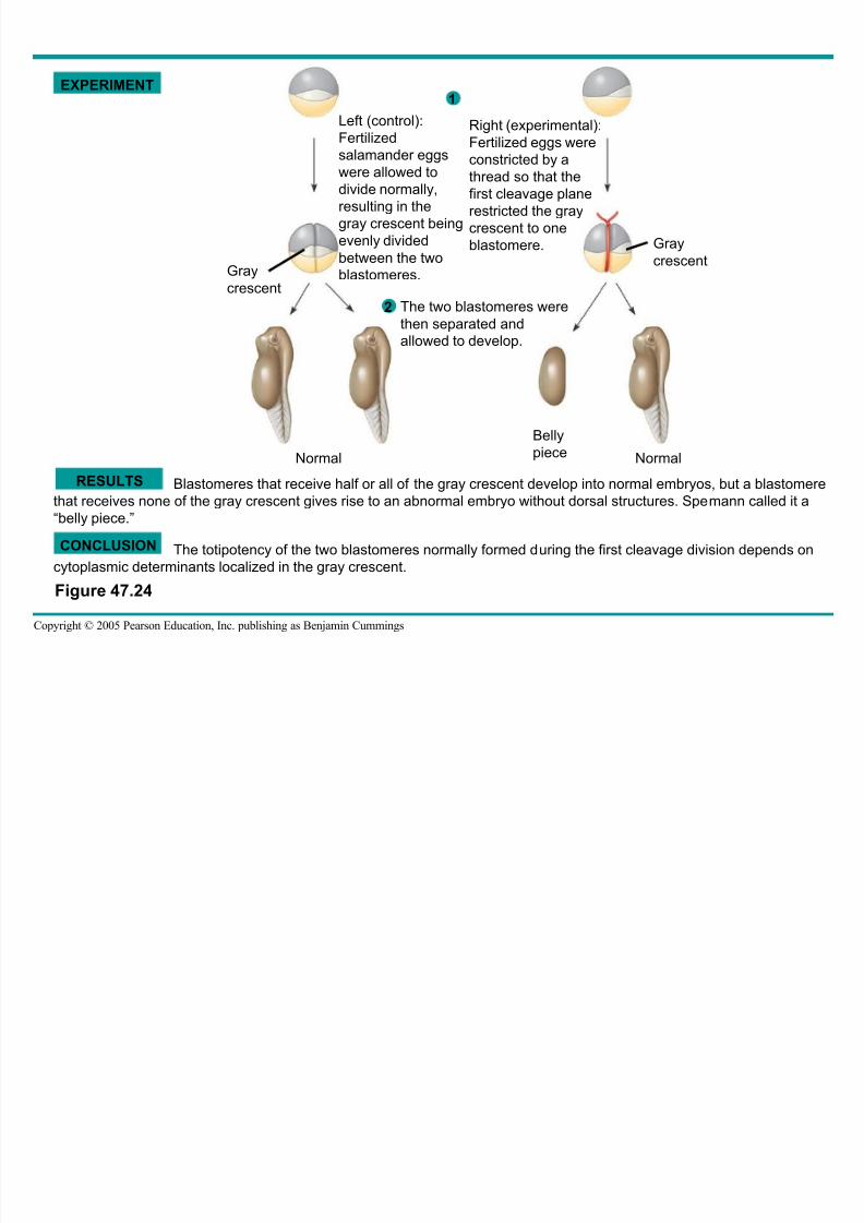
Blastomeres that receive half or all of the gray crescent develop into normal embryos, but a blastomere
that receives none of the gray crescent gives rise to an abnormal embryo without dorsal structures. Spemann called it a
“belly piece.”
EXPERIMENT
RESULTS
CONCLUSION The totipotency of the two blastomeres normally formed during the first cleavage division depends on
cytoplasmic determinants localized in the gray crescent.
Left (control):
Fertilized
salamander eggswere allowed to
divide normally,
resulting in the
gray crescent being
evenly divided
between the two
blastomeres.
Right (experimental):
Fertilized eggs were
constricted by athread so that the
first cleavage plane
restricted the gray
crescent to one
blastomere.
Gray
crescent
The two blastomeres werethen separated and
allowed to develop.
Gray
crescent
Normal
Belly
pieceNormal
1
2
Figure 47.24
8/14/2019 47 Development Text
http://slidepdf.com/reader/full/47-development-text 63/73
Copyright © 2005 Pearson Education, Inc. publishing as Benjamin Cummings
• As embryonic development proceeds
– The potency of cells becomes progressivelymore limited in all species
Cell Fate Determination and Pattern Formation by
8/14/2019 47 Development Text
http://slidepdf.com/reader/full/47-development-text 64/73
Copyright © 2005 Pearson Education, Inc. publishing as Benjamin Cummings
yInductive Signals
• Once embryonic cell division creates cells that
differ from each other
– The cells begin to influence each other’s fates
by induction
The “Organizer” of Spemann and Mangold
8/14/2019 47 Development Text
http://slidepdf.com/reader/full/47-development-text 65/73
Copyright © 2005 Pearson Education, Inc. publishing as Benjamin Cummings
• Based on the results of their most famous
experiment
– Spemann and Mangold concluded that the
dorsal lip of the blastopore functions as an
organizer of the embryo
8/14/2019 47 Development Text
http://slidepdf.com/reader/full/47-development-text 66/73
Copyright © 2005 Pearson Education, Inc. publishing as Benjamin Cummings
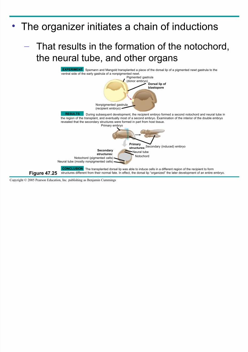
• The organizer initiates a chain of inductions
– That results in the formation of the notochord,the neural tube, and other organs
Figure 47.25
EXPERIMENT
RESULTS
CONCLUSION
Spemann and Mangold transplanted a piece of the dorsal lip of a pigmented newt gastrula to the
ventral side of the early gastrula of a nonpigmented newt.
During subsequent development, the recipient embryo formed a second notochord and neural tube in
the region of the transplant, and eventually most of a second embryo. Examination of the interior of the double embryo
revealed that the secondary structures were formed in part from host tissue.
The transplanted dorsal lip was able to induce cells in a different region of the recipient to form
structures different from their normal fate. In effect, the dorsal lip “organized” the later development of an entire embryo.
Pigmented gastrula
(donor embryo)Dorsal lip of
blastopore
Nonpigmented gastrula
(recipient embryo)
Primary embryo
Secondary (induced) embryoPrimary
structures:
Neural tube
Notochord
Secondary
structures:
Notochord (pigmented cells)Neural tube (mostly nonpigmented cells)
Formation of the Vertebrate Limb
8/14/2019 47 Development Text
http://slidepdf.com/reader/full/47-development-text 67/73
Copyright © 2005 Pearson Education, Inc. publishing as Benjamin Cummings
• Inductive signals play a major role in pattern
formation
– The development of an animal’s spatial
organization
8/14/2019 47 Development Text
http://slidepdf.com/reader/full/47-development-text 68/73
Copyright © 2005 Pearson Education, Inc. publishing as Benjamin Cummings
• The molecular cues that control pattern
formation, called positional information
– Tell a cell where it is with respect to the
animal’s body axes
– Determine how the cell and its descendentsrespond to future molecular signals
8/14/2019 47 Development Text
http://slidepdf.com/reader/full/47-development-text 69/73
Copyright © 2005 Pearson Education, Inc. publishing as Benjamin Cummings
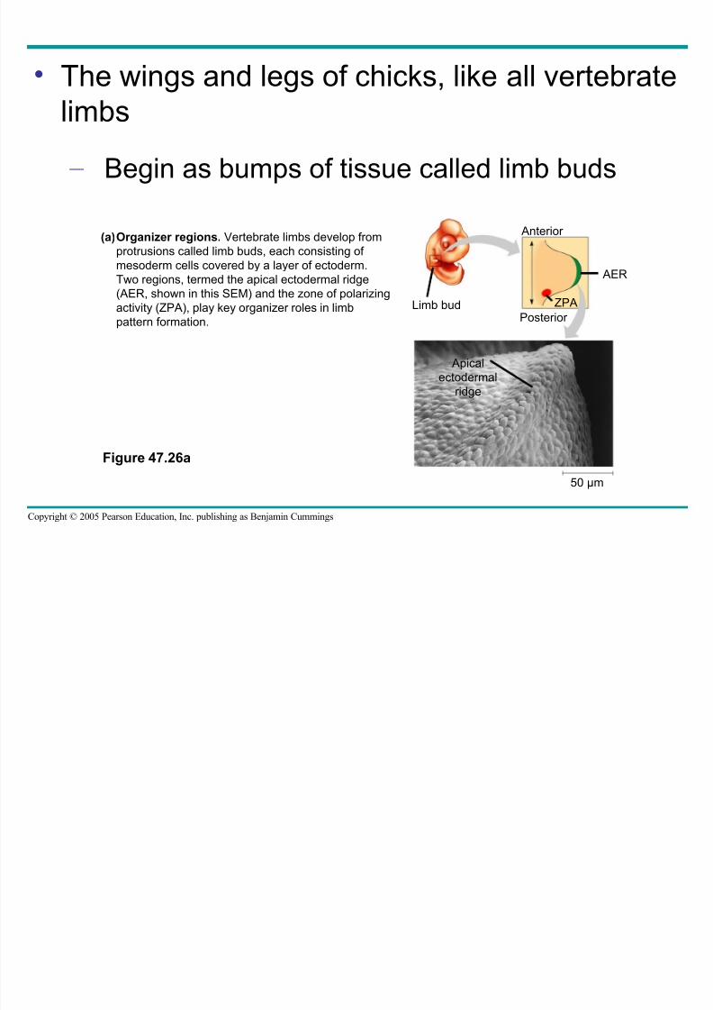
• The wings and legs of chicks, like all vertebrate
limbs
– Begin as bumps of tissue called limb buds
Figure 47.26a
Limb bud
Anterior
AER
ZPA
Posterior
Organizer regions. Vertebrate limbs develop from
protrusions called limb buds, each consisting of
mesoderm cells covered by a layer of ectoderm.
Two regions, termed the apical ectodermal ridge
(AER, shown in this SEM) and the zone of polarizing
activity (ZPA), play key organizer roles in limb
pattern formation.
(a)
Apical
ectodermalridge
50 µm
8/14/2019 47 Development Text
http://slidepdf.com/reader/full/47-development-text 70/73
Copyright © 2005 Pearson Education, Inc. publishing as Benjamin Cummings
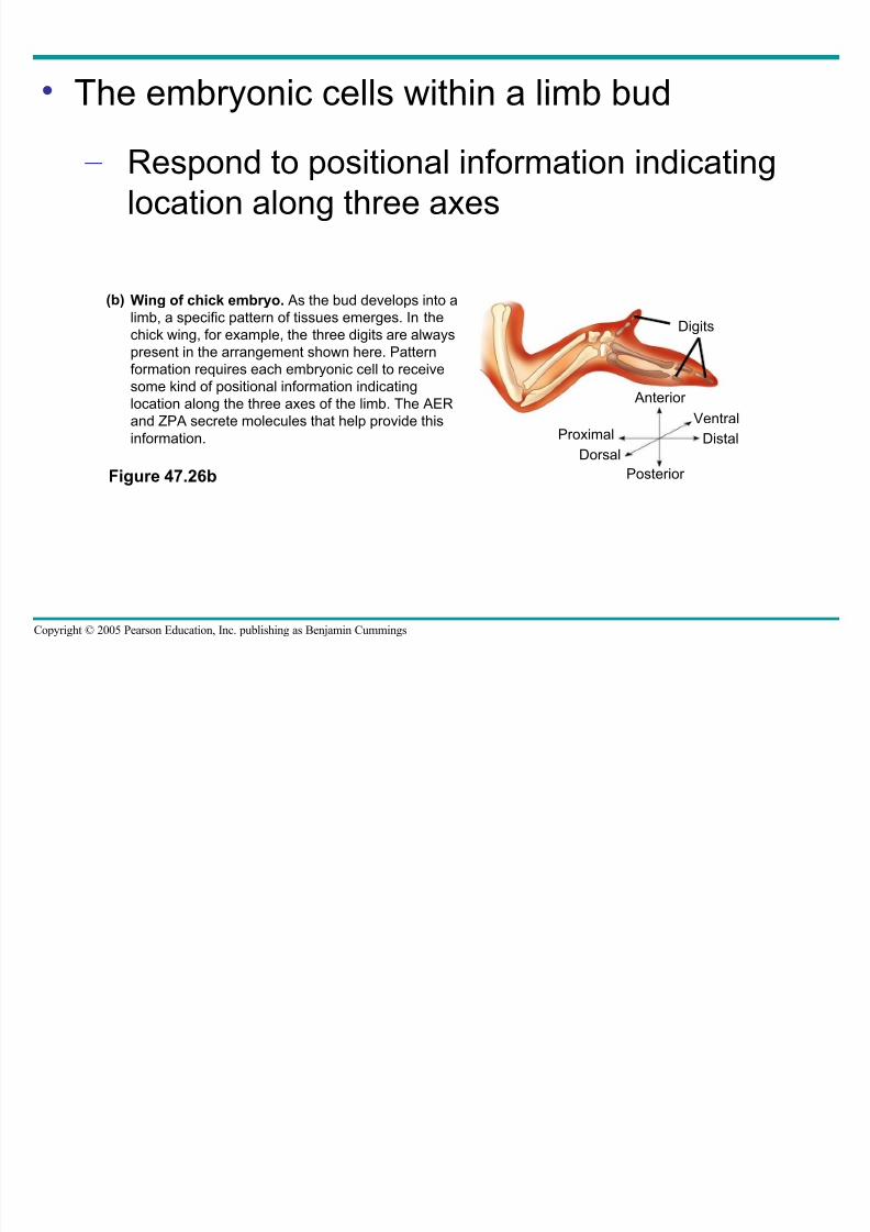
• The embryonic cells within a limb bud
– Respond to positional information indicatinglocation along three axes
Figure 47.26b
Digits
Anterior
Ventral
DistalProximal
Dorsal
Posterior
Wing of chick embryo. As the bud develops into a
limb, a specific pattern of tissues emerges. In thechick wing, for example, the three digits are always
present in the arrangement shown here. Pattern
formation requires each embryonic cell to receive
some kind of positional information indicating
location along the three axes of the limb. The AER
and ZPA secrete molecules that help provide this
information.
(b)
8/14/2019 47 Development Text
http://slidepdf.com/reader/full/47-development-text 71/73
Copyright © 2005 Pearson Education, Inc. publishing as Benjamin Cummings
• One limb-bud organizer region is the apical
ectodermal ridge (AER)
– A thickened area of ectoderm at the tip of the
bud
• The second major limb-bud organizer region isthe zone of polarizing activity (ZPA)
– A block of mesodermal tissue located
underneath the ectoderm where the posterior side of the bud is attached to the body
8/14/2019 47 Development Text
http://slidepdf.com/reader/full/47-development-text 72/73
Copyright © 2005 Pearson Education, Inc. publishing as Benjamin Cummings
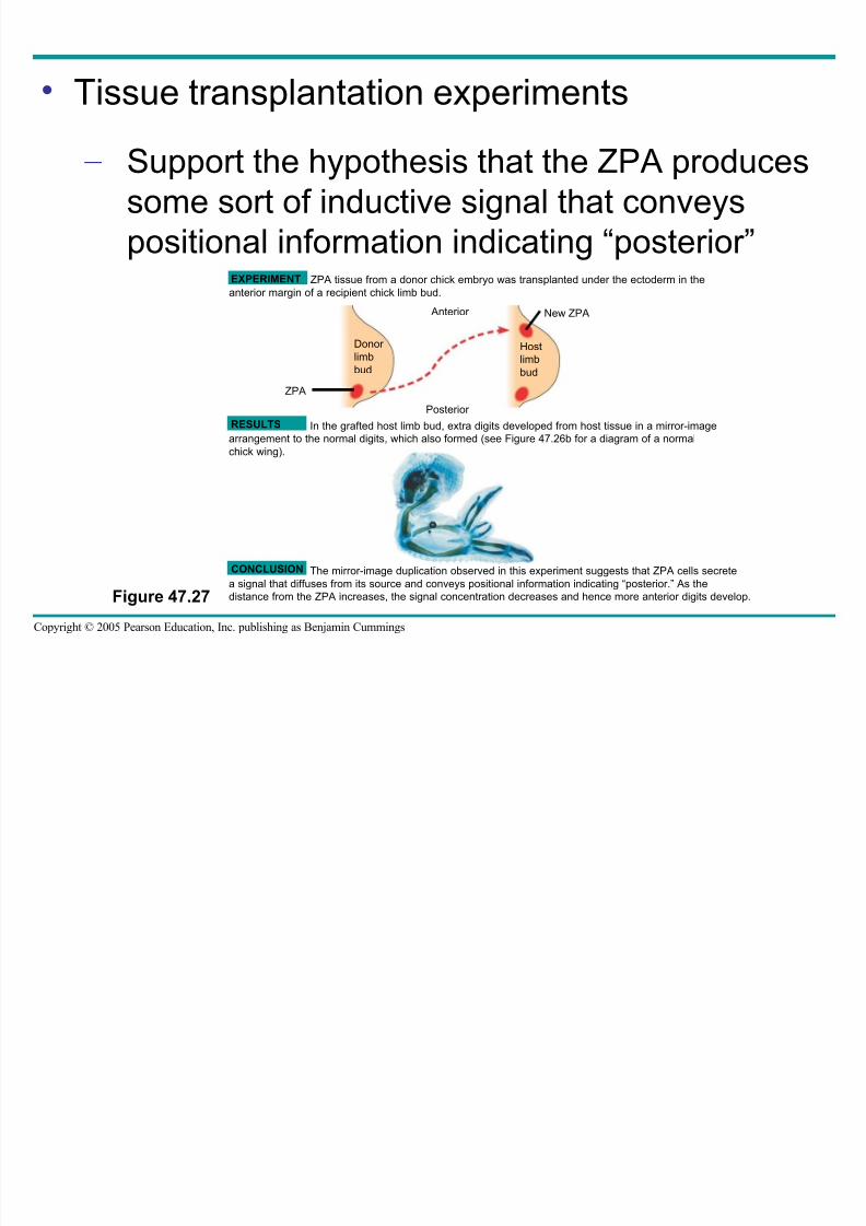
• Tissue transplantation experiments
– Support the hypothesis that the ZPA producessome sort of inductive signal that conveys
positional information indicating “posterior”
Figure 47.27
EXPERIMENT
RESULTS
CONCLUSION
ZPA tissue from a donor chick embryo was transplanted under the ectoderm in the
anterior margin of a recipient chick limb bud.
Anterior
Donor
limb
bud
Host
limb
bud
Posterior
ZPA
The mirror-image duplication observed in this experiment suggests that ZPA cells secrete
a signal that diffuses from its source and conveys positional information indicating “posterior.” As the
distance from the ZPA increases, the signal concentration decreases and hence more anterior digits develop.
New ZPA
In the grafted host limb bud, extra digits developed from host tissue in a mirror-image
arrangement to the normal digits, which also formed (see Figure 47.26b for a diagram of a normal
chick wing).
8/14/2019 47 Development Text
http://slidepdf.com/reader/full/47-development-text 73/73
• Signal molecules produced by inducing cells
– Influence gene expression in the cells thatreceive them
– Lead to differentiation and the development of
particular structures