Embed Size (px)
Citation preview
letter
260 nature genetics • volume 22 • july 1999
Regulation of anterior/posterior patterning of the axialskeleton by growth/differentiation factor 11
Alexandra C. McPherron1, Ann M. Lawler2 & Se-Jin Lee1
Departments of 1Molecular Biology and Genetics and 2Gynecology and Obstetrics, Johns Hopkins University School of Medicine, 725 N. Wolfe Street,Baltimore, Maryland 21205, USA. Correspondence should be addressed to S.-J.L. (e-mail: [email protected]).
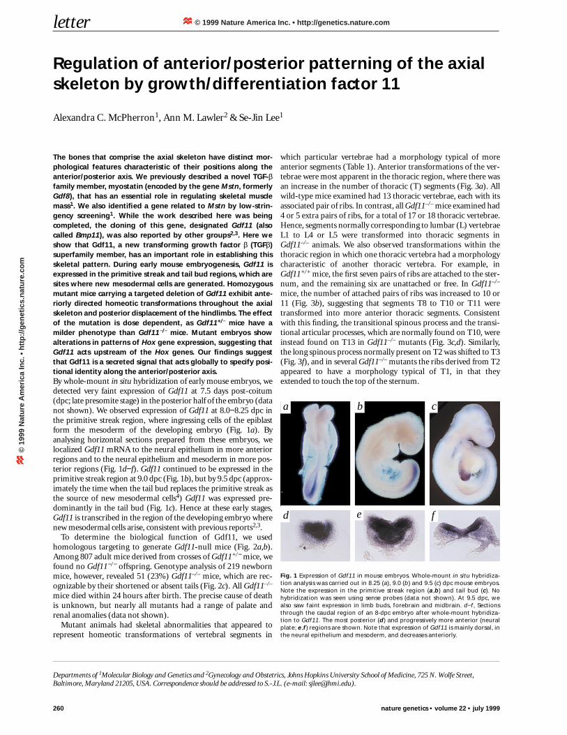
The bones that comprise the axial skeleton have distinct mor-phological features characteristic of their positions along theanterior/posterior axis. We previously described a novel TGF-βfamily member, myostatin (encoded by the gene Mstn, formerlyGdf8), that has an essential role in regulating skeletal musclemass1. We also identified a gene related to Mstn by low-strin-gency screening1. While the work described here was beingcompleted, the cloning of this gene, designated Gdf11 (alsocalled Bmp11), was also reported by other groups2,3. Here weshow that Gdf11, a new transforming growth factor β (TGFβ)superfamily member, has an important role in establishing thisskeletal pattern. During early mouse embryogenesis, Gdf11 isexpressed in the primitive streak and tail bud regions, which aresites where new mesodermal cells are generated. Homozygousmutant mice carrying a targeted deletion of Gdf11 exhibit ante-riorly directed homeotic transformations throughout the axialskeleton and posterior displacement of the hindlimbs. The effectof the mutation is dose dependent, as Gdf11+/− mice have amilder phenotype than Gdf11−/− mice. Mutant embryos showalterations in patterns of Hox gene expression, suggesting thatGdf11 acts upstream of the Hox genes. Our findings suggestthat Gdf11 is a secreted signal that acts globally to specify posi-tional identity along the anterior/posterior axis.By whole-mount in situ hybridization of early mouse embryos, wedetected very faint expression of Gdf11 at 7.5 days post-coitum(dpc; late presomite stage) in the posterior half of the embryo (datanot shown). We observed expression of Gdf11 at 8.0−8.25 dpc inthe primitive streak region, where ingressing cells of the epiblastform the mesoderm of the developing embryo (Fig. 1a). Byanalysing horizontal sections prepared from these embryos, welocalized Gdf11 mRNA to the neural epithelium in more anteriorregions and to the neural epithelium and mesoderm in more pos-terior regions (Fig. 1d−f). Gdf11 continued to be expressed in theprimitive streak region at 9.0 dpc (Fig. 1b), but by 9.5 dpc (approx-imately the time when the tail bud replaces the primitive streak asthe source of new mesodermal cells4) Gdf11 was expressed pre-dominantly in the tail bud (Fig. 1c). Hence at these early stages,Gdf11 is transcribed in the region of the developing embryo wherenew mesodermal cells arise, consistent with previous reports2,3.
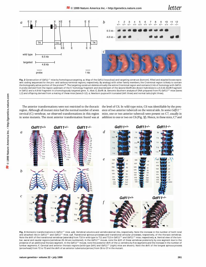
To determine the biological function of Gdf11, we usedhomologous targeting to generate Gdf11-null mice (Fig. 2a,b).Among 807 adult mice derived from crosses of Gdf11+/− mice, wefound no Gdf11−/− offspring. Genotype analysis of 219 newbornmice, however, revealed 51 (23%) Gdf11–/– mice, which are rec-ognizable by their shortened or absent tails (Fig. 2c). All Gdf11–/–
mice died within 24 hours after birth. The precise cause of deathis unknown, but nearly all mutants had a range of palate andrenal anomalies (data not shown).
Mutant animals had skeletal abnormalities that appeared torepresent homeotic transformations of vertebral segments in
which particular vertebrae had a morphology typical of moreanterior segments (Table 1). Anterior transformations of the ver-tebrae were most apparent in the thoracic region, where there wasan increase in the number of thoracic (T) segments (Fig. 3a). Allwild-type mice examined had 13 thoracic vertebrae, each with itsassociated pair of ribs. In contrast, all Gdf11–/– mice examined had4 or 5 extra pairs of ribs, for a total of 17 or 18 thoracic vertebrae.Hence, segments normally corresponding to lumbar (L) vertebraeL1 to L4 or L5 were transformed into thoracic segments inGdf11–/– animals. We also observed transformations within thethoracic region in which one thoracic vertebra had a morphologycharacteristic of another thoracic vertebra. For example, inGdf11+/+ mice, the first seven pairs of ribs are attached to the ster-num, and the remaining six are unattached or free. In Gdf11–/–
mice, the number of attached pairs of ribs was increased to 10 or11 (Fig. 3b), suggesting that segments T8 to T10 or T11 weretransformed into more anterior thoracic segments. Consistentwith this finding, the transitional spinous process and the transi-tional articular processes, which are normally found on T10, wereinstead found on T13 in Gdf11–/– mutants (Fig. 3c,d). Similarly,the long spinous process normally present on T2 was shifted to T3(Fig. 3f), and in several Gdf11–/– mutants the ribs derived from T2appeared to have a morphology typical of T1, in that theyextended to touch the top of the sternum.
Fig. 1 Expression of Gdf11 in mouse embryos. Whole-mount in situ hybridiza-tion analysis was carried out in 8.25 (a), 9.0 (b) and 9.5 (c) dpc mouse embryos.Note the expression in the primitive streak region (a,b) and tail bud (c). Nohybridization was seen using sense probes (data not shown). At 9.5 dpc, wealso saw faint expression in limb buds, forebrain and midbrain. d−f, Sectionsthrough the caudal region of an 8-dpc embryo after whole-mount hybridiza-tion to Gdf11. The most posterior (d) and progressively more anterior (neuralplate; e,f) regions are shown. Note that expression of Gdf11 is mainly dorsal, inthe neural epithelium and mesoderm, and decreases anteriorly.
fed
a b c
© 1999 Nature America Inc. • http://genetics.nature.com©
199
9 N
atu
re A
mer
ica
Inc.
• h
ttp
://g
enet
ics.
nat
ure
.co
m
letter
nature genetics • volume 22 • july 1999 261
The anterior transformations were not restricted to the thoracicregion. Although all mutant mice had the normal number of sevencervical (C) vertebrae, we observed transformations in this regionin some mutants. The most anterior transformation found was at
the level of C6. In wild-type mice, C6 was identifiable by the pres-ence of two anterior tuberculi on the ventral side. In some Gdf11–/–
mice, one or two anterior tuberculi were present on C7, usually inaddition to one or two on C6 (Fig. 3f). Hence, in these mice, C7 and
Fig. 2 Construction of Gdf11–/– mice by homologous targeting. a, Map of the Gdf11 locus (top) and targeting construct (bottom). Filled and stippled boxes repre-sent coding sequences for the pro- and carboxy-terminal regions, respectively. By analogy with other family members, the C-terminal region is likely to containthe biologically active portion of the protein16. The targeting construct deletes virtually the entire C-terminal region and contains 11 kb of homology with Gdf11.A probe derived from the region upstream of the 5´ homology fragment and downstream of the second EcoRI site shown hybridizes to a 6.5-kb EcoRI fragmentin Gdf11 and a 4.8-kb fragment in a homologously targeted gene. X, XbaI; E, EcoRI. b, Genomic Southern analysis of DNA prepared from F1 Gdf11+/– mice (lanes1,2) and offspring derived from a mating of these mice (lanes 3−12). c, Newborn pups with truncated (left three) and normal tails (right three).
a b
c
Fig. 3 Homeotic transformations in Gdf11–/– mice. a,b, Vertebral columns and vertebrosternal ribs, respectively. Note the increase in the number of both totaland attached ribs in Gdf11+/– and Gdf11–/– mice. c,d, Transitional spinous processes and transitional articular processes, respectively, of the thoracic vertebrae.Note the shift of the transitional vertebrae (asterisks) from T10 in wild-type to T11 and T13 in Gdf11+/– and Gdf11–/– mice, respectively. e, Ventral view of the lum-bar, sacral and caudal regions (vertebrae 26−34 are numbered). In the Gdf11+/– mouse, note the shift of these vertebrae posteriorly by one segment due to thepresence of an additional thoracic segment. In the Gdf11–/– mouse, note the posterior shift of the L1 vertebra by five segments and the increase in the number oflumbar segments. f, Cervical and anterior thoracic regions (wild-type (left) and Gdf11–/– (right) mice are shown). Note the shift of the longest spinous process(arrowhead) from T2 to T3 and the shift of an anterior tuberculus (arrow) from C6 to C7 in the mutant.
a
b
c
d
e
f
© 1999 Nature America Inc. • http://genetics.nature.com©
199
9 N
atu
re A
mer
ica
Inc.
• h
ttp
://g
enet
ics.
nat
ure
.co
m
letter
262 nature genetics • volume 22 • july 1999
C6 appeared to have been at least partially transformed to have amorphology resembling that of C6 and C5, respectively. Transfor-mations of the axial skeleton also extended into the lumbar region.Whereas Gdf11+/+ animals normally have only 6 lumbar vertebrae,Gdf11–/– mice had 7–9 (Fig. 3e), at least 6 of which must havederived from segments that would normally correspond to sacraland caudal vertebrae. Hence, Gdf11–/– mice had 6−8 additional pre-sacral vertebrae, which was also evident from the posterior dis-placement of the hindlimbs relative to the forelimbs (Fig. 4a). In thesacral and caudal regions, the exact nature of the abnormalities wasnot as readily identifiable, as the vertebrae were severely malformedwith extensive fusions of cartilage. The total number of vertebrae inthis region, however, was reduced from the normal number (∼ 34).
Gdf11+/− mice also showed abnormalities in the axial skeleton,although the phenotype was milder than in Gdf11−/− mice.Gdf11+/– mice showed the presence of an additional thoracic seg-ment with an associated pair of ribs (Fig. 3a), which was almostalways attached to the sternum (Fig. 3b). Hence, T8 appeared tohave been transformed to a more anterior thoracic segment, andL1 appeared to have been transformed to a posterior thoracicsegment. To varying degrees, we observed other abnormalitiesindicative of anterior transformations in Gdf11+/– mice. Theseincluded a shift of the long spinous process characteristic of T2by one segment to T3, a shift of the transitional articular andspinous processes from T10 to T11, an asymmetric shift of ananterior tuberculus on C6 to C7, and transformation of T2 to T1,
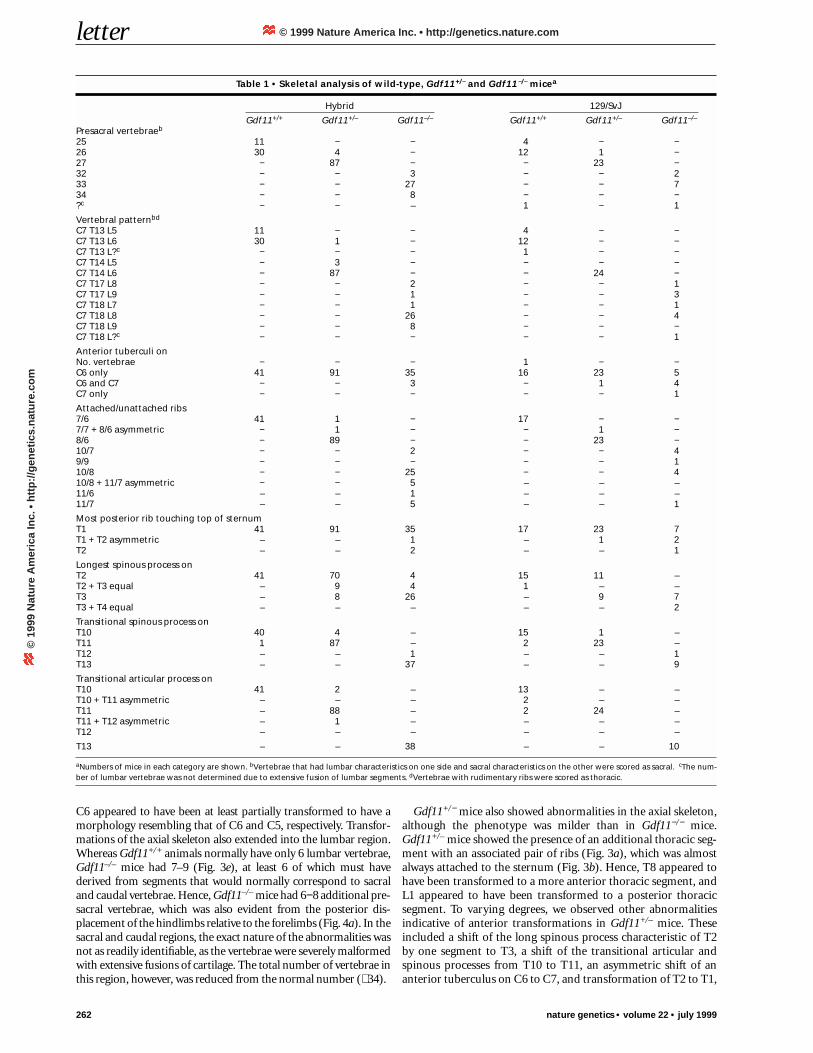
Table 1 • Skeletal analysis of wild-type, Gdf11+/− and Gdf11−/− micea
Hybrid 129/SvJ
Gdf11+/+ Gdf11+/– Gdf11–/– Gdf11+/+ Gdf11+/– Gdf11–/–
Presacral vertebraeb
25 11 − − 4 − −26 30 4 − 12 1 −27 − 87 − − 23 −32 − − 3 − − 233 − − 27 − − 734 − − 8 − − −?c − − – 1 − 1
Vertebral patternbd
C7 T13 L5 11 − − 4 − −C7 T13 L6 30 1 − 12 − −C7 T13 L?c − − − 1 − −C7 T14 L5 − 3 − − − −C7 T14 L6 − 87 − − 24 −C7 T17 L8 − − 2 − − 1C7 T17 L9 − − 1 − − 3C7 T18 L7 − − 1 − − 1C7 T18 L8 − − 26 − − 4C7 T18 L9 − − 8 − − −C7 T18 L?c − − − − − 1
Anterior tuberculi onNo. vertebrae − − − 1 − −C6 only 41 91 35 16 23 5C6 and C7 − − 3 − 1 4C7 only − − − − − 1
Attached/unattached ribs7/6 41 1 − 17 − −7/7 + 8/6 asymmetric − 1 − − 1 −8/6 − 89 − − 23 −10/7 − − 2 − − 49/9 − − − − − 110/8 − − 25 − − 410/8 + 11/7 asymmetric − − 5 – – –11/6 – – 1 – – –11/7 – – 5 – – 1
Most posterior rib touching top of sternumT1 41 91 35 17 23 7T1 + T2 asymmetric – – 1 – 1 2T2 – – 2 – – 1
Longest spinous process onT2 41 70 4 15 11 –T2 + T3 equal – 9 4 1 – –T3 – 8 26 – 9 7T3 + T4 equal – – – – – 2
Transitional spinous process onT10 40 4 – 15 1 –T11 1 87 – 2 23 –T12 – – 1 – – 1T13 – – 37 – – 9
Transitional articular process onT10 41 2 – 13 – –T10 + T11 asymmetric – – – 2 – –T11 – 88 – 2 24 –T11 + T12 asymmetric – 1 – – – –T12 – – – – – –
T13 – – 38 – – 10
aNumbers of mice in each category are shown. bVertebrae that had lumbar characteristics on one side and sacral characteristics on the other were scored as sacral. cThe num-ber of lumbar vertebrae was not determined due to extensive fusion of lumbar segments. dVertebrae with rudimentary ribs were scored as thoracic.
© 1999 Nature America Inc. • http://genetics.nature.com©
199
9 N
atu
re A
mer
ica
Inc.
• h
ttp
://g
enet
ics.
nat
ure
.co
m
letter
nature genetics • volume 22 • july 1999 263
in which the rib associated with T2 touched the top of the ster-num (Fig. 3c,d and Table 1).
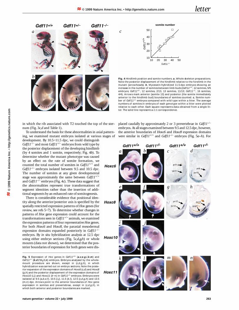
To understand the basis for these abnormalities in axial pattern-ing, we examined mutant embryos isolated at various stages ofdevelopment. By 10.5−11.5 dpc, we could distinguishGdf11–/– and most Gdf11+/– embryos from wild type bythe posterior displacement of the developing hindlimb(by 4 somites and 1 somite, respectively; Fig. 4b). Todetermine whether the mutant phenotype was causedby an effect on the rate of somite formation, wecounted the total number of somites in Gdf11+/+ andGdf11–/– embryos isolated between 9.5 and 10.5 dpc.The number of somites at any given developmentalstage was approximately the same between Gdf11+/+
and Gdf11–/– embryos (Fig. 4c). These data suggest thatthe abnormalities represent true transformations ofsegment identities rather than the insertion of addi-tional segments by an enhanced rate of somitogenesis.
There is considerable evidence that positional iden-tity along the anterior/posterior axis is specified by thespatially restricted expression patterns of Hox genes (forreview, see refs 5−7). To determine whether changes inpatterns of Hox gene expression could account for thetransformations seen in Gdf11−/− animals, we examinedthe expression patterns of four representative Hox genes.For both Hoxc6 and Hoxc8, the paraxial mesodermalexpression domains expanded posteriorly in Gdf11–/–
embryos. By in situ hybridization analysis at 12.5 dpcusing either embryo sections (Fig. 5c,d,g,h) or wholemounts (data not shown), we determined that the pos-terior boundaries of expression for both genes were dis-
placed caudally by approximately 2 or 3 prevertebrae in Gdf11–/–
embryos. At all stages examined between 9.5 and 12.5 dpc, however,the anterior boundaries of Hoxc6 and Hoxc8 expression domainswere similar in Gdf11+/+ and Gdf11–/– embryos (Fig. 5a–h). For
Fig. 4 Hindlimb position and somite numbers. a, Whole skeleton preparations.Note the posterior displacement of the hindlimb relative to the forelimb in themutant (arrowheads). b, Myostatin-hybridized 11.5-dpc embryos showing anincrease in the number of somites between limb buds (Gdf11+/+, 12 somites, 5/5embryos; Gdf11+/–, 12 somites, 2/13, 13 somites, 11/13; Gdf11–/–, 16 somites,4/4). Arrows mark anterior (somite 13) and posterior (the somite immediatelyanterior to the hindlimb bud) boundaries of somites counted. c, Somite num-ber of Gdf11–/– embryos compared with wild type within a litter. The averagenumbers of somites in embryos of each genotype within a litter were plottedrelative to each other. Each square represents data obtained from a single lit-ter. The solid line represents a 1:1 correspondence.
Somite number
10 20 30 40 5010
20
30
40
50
Gdf11-/-
Gdf
11+/
+
a c
Fig. 5 Expression of Hox genes in Gdf11+/+ (a,c,e,g,i,k,m) andGdf11–/– (b,d,f,h,j,l,n) embryos. Embryos analysed by the whole-mount procedure are shown, except in (c,d,g,h), in whichhybridization was carried out on embryo sections. Note the poste-rior expansion of the expression domains of Hoxc6 (c,d) and Hoxc8(g,h) and the posterior displacement of the expression domains ofHoxc10 (i,j) and Hoxc11 (k−n) in Gdf11–/– embryos. Embryos wereisolated at 9.5 (a,b,e,f), 10.5 (i,j), 11.5 (k,l), 12.5 (c,d,g,h) and 13.5(m,n) dpc. Arrows point to the anterior boundaries of Hox geneexpression in somites and prevertebrae, except in (c,d,g,h), inwhich both anterior and posterior boundaries are marked.
a b c d
hgfe
i j
k l m n
b
somite number
Gdf11–/–
© 1999 Nature America Inc. • http://genetics.nature.com©
199
9 N
atu
re A
mer
ica
Inc.
• h
ttp
://g
enet
ics.
nat
ure
.co
m
letter
264 nature genetics • volume 22 • july 1999
Hoxc10 and Hoxc11, the Gdf11 mutation caused the entire expres-sion domains to be shifted posteriorly. The anterior boundary ofthe Hoxc10 expression domain appeared similar in Gdf11+/+ andGdf11–/– embryos relative to the hindlimbs (Fig. 5i,j), which weredisplaced posteriorly in Gdf11–/– embryos (Fig. 4b). The analysis ofHoxc11 was complicated by the fact that prevertebral expressionwas delayed in mutants (note the faint signal in mutant (Fig. 5l) rel-ative to wild-type (Fig. 5k) embryos at 11.5 dpc), consistent withthe observation that the hindlimbs developed later in Gdf11–/– thanGdf11+/+ embryos (data not shown). Based on an examination ofembryos between 11.5 and 13.5 dpc, however, the prevertebralexpression of Hoxc11 appeared to be shifted posteriorly relative tonot only the forelimbs but also the hindlimbs (Fig. 5m,n). Thesealterations in Hox gene expression provide further evidence that theskeletal abnormalities seen in Gdf11–/– animals represent homeotictransformations and that Gdf11 acts upstream of the Hox genes toregulate axial patterning.
The expression pattern of Gdf11 and the phenotype of Gdf11–/–
mice suggest that Gdf11 acts either directly or indirectly on meso-dermal precursor cells to specify positional identity along the ante-rior/posterior axis. The intermediate phenotype observed inGdf11+/– mice also suggests that the ability of Gdf11 to influenceaxial patterning is concentration dependent. This is consistent witha variety of different models for Gdf11 action, including a mor-phogen model. Whatever the mechanism of action of Gdf11, it istempting to speculate that either changing local concentrations ofGdf11 or differences in the competence of target cells to respond toGdf11 may be partly responsible for progressively more posterioridentities being conferred on cells generated at progressively laterstages of development. An elucidation of the mechanism of actionof Gdf11 will require the identification of Gdf11 receptors and ananalysis of their distribution. In this regard, additional thoracic ver-tebrae and a range of kidney defects have been observed to a milderdegree in mice lacking the activin type IIB receptor8 (ActRIIB). Thesimilarities in these phenotypes raise the possibility that ActRIIBmay be a receptor for Gdf11, although differences in the pheno-types suggest that ActRIIB cannot be the sole receptor for Gdf11and that Gdf11 cannot be the sole ligand for ActRIIB in vivo.
To our knowledge, Gdf11 is the first secreted protein factor thathas been shown to function globally to regulate anterior/posterioraxial patterning. In fact, the homeotic transformations observed inGdf11 mutant mice are more extensive than those seen thus fareither by genetic manipulation of presumed patterning genes (forreview, see refs 5−7) or by administration of retinoic acid (forreview, see ref. 9). It will be important to investigate whether Gdf11and retinoic acid interact to regulate Hox gene expression and
anterior/posterior patterning and whether Gdf11 regulates the pat-terning of tissues other than those described here.
MethodsCloning of Gdf11 and construction of Gdf11–/– mice. Mouse 129/SvJ andhuman genomic libraries were made in λ FIX II, and a human spleencDNA library was made in λ ZAP II (Stratagene) according to instructionsfrom the manufacturer. We obtained Gdf11 sequences by low-stringencyscreening of these libraries using a Mstn probe. We carried out libraryscreening, RNA isolation and poly(A) selection as described10. We deducedthe structure of Gdf11 from restriction mapping and partial sequencing ofgenomic phage clones. Vectors for preparing the targeting construct werekindly provided by P. Soriano and K. Thomas. R1 ES cells from strain129/SvJ (kindly provided by A. Nagy, R. Nagy and W. Abramow-Newerly)were transfected with the targeting construct, selected with gancyclovir (2µM) and G418 (250 µg/ml), and analysed by Southern-blot analysis asdescribed1. We injected homologously targeted clones into C57BL/6J blas-tocysts and transferred them into pseudopregnant females.
Skeleton preparations. Newborn mice and 18.5-dpc fetuses were skinnedand eviscerated, fixed in 80% ethanol, dehydrated in 95% ethanol for 1 dand acetone for 3 d. We then stained skeletons in 10% acetic acid in ethanolcontaining 0.003% Alizarin red and 0.0045% Alcian blue for ∼ 36 h. Afterstaining, skeletons were cleared in 1% potassium hydroxide and trans-ferred to 20%, 50%, 80% and 100% glycerol over several days.
Analysis of embryos. For somite counting, embryos were fixed in 4%paraformaldehyde, dehydrated to methanol and cleared in 1:1 benzyl alco-hol:benzyl benzoate. We carried out whole-mount in situ hybridizationanalysis using digoxigenin-labelled probes as described11, except blockingand antibody incubation steps were as in Knecht et al.12. For analysis ofGdf11 expression patterns, we used probes corresponding to the pro-region and 3´ UTR to avoid cross-hybridization to Mstn. After hybridiza-tion some embryos were frozen in OCT, and cryostat sections (40 µm)were taken. We carried out in situ hybridization using digoxigenin-labelledHoxc6 and Hoxc8 riboprobes on frozen sections (20 µm) of 12.5 dpcembryos as described13. Antibody incubation was carried out at 4 oCovernight as described14. Hoxc6, Hoxc8 and Hoxc11 probes were kindlyprovided by M. Capecchi and A. Boulet. We cloned the Hoxc10 3´ UTR byPCR using primers based on the published sequence15.
AcknowledgementsWe thank P. Dunlap for assistance with maintenance of mice; A. Boulet andM. Capecchi for providing Hox clones; and D. Nathans, M. Capecchi,P. Beachy and E. Hsiao for discussions. This work was supported by researchgrant R01H035887 from the NIH (S.-J.L.). The early phase of this work wassupported by grants from the Edward Mallinckrodt, Jr. Foundation andMetaMorphix, Inc. (S.-J.L).
Received 13 May; accepted 25 May 1998.
1. McPherron, A.C., Lawler, A.M. & Lee, S.-J. Regulation of skeletal muscle mass inmice by a new TGF-β superfamily member. Nature 387, 83–90 (1997).
2. Gamer, L.W. et al. A novel BMP expressed in developing mouse limb, spinal cord,and tail bud is a potent mesoderm inducer in Xenopus embryos. Dev. Biol. 80,222–232 (1999).
3. Nakashima, M., Toyono, T., Akamine, A. & Joyner, A. Expression ofgrowth/differentiation factor 11, a new member of the BMP/TGF-β superfamilyduring mouse embryogenesis. Mech. Dev. 80, 185–189 (1999).
4. Tam, P.P.L. & Tan, S.-S. The somitogenetic potential of cells in the primitive streakand the tail bud of the organogenesis-stage mouse embryo. Development 115,703–715 (1992).
5. Capecchi, M.R. Hox genes and mammalian development. Cold Spring Harb. Symp.Quant. Biol. 62, 273–281 (1997).
6. Favier, B. & Dollé, P. Developmental functions of mammalian Hox genes. Mol.Hum. Reprod. 3, 115–131 (1997).
7. Mark, M., Rijli, F.M. & Chambon, P. Homeobox genes in embryogenesis andpathogenesis. Pediatr. Res. 42, 421–429 (1997).
8. Oh, S.P. & Li, E. The signaling pathway mediated by the type IIB activin receptorcontrols axial patterning and lateral asymmetry in the mouse. Genes Dev. 11,1812–1826 (1997).
9. Durston, A.J., van der Wees, J., Pijnappel, W.W.M., Schilthuis, J.G. & Godsave, S.F.Retinoid signalling and axial patterning during early vertebrate embryogenesis.
Cell. Mol. Life Sci. 53, 339–349 (1997).10. Lee, S.-J. Identification of a novel member of the transforming growth factor-β
superfamily. Mol. Endocrinol. 4, 1034–1040 (1990).11. Wilkinson, D.G. Whole mount in situ hybridization of vertebrate embryos. in In
Situ Hybridization: A Practical Approach (ed. Wilkinson, D.G.) 75−83 (IRL Press,Oxford, 1992).
12. Knecht, A.K., Good, P.J., Dawid, I.B. & Harland, R.M. Dorsal-ventral patterningand differentiation of noggin-induced neural tissue in the absence of mesoderm.Development 121, 1927–1935 (1995).
13. Wilkinson, D.G., Bailes, J.A. & McMahon, A.P. Expression of the proto-oncogeneint-1 is restricted to specific cells in the developing mouse embryo. Cell 50, 79–88(1987).
14. Schaeren-Wiemers, N. & Gerfin-Moser, A. A single protocol to detect transcriptsof various types and expression levels in neural tissue and cultured cells: in situhybridization using digoxigenin-labelled cRNA probes. Histochemistry 100,431–440 (1993).
15. Peterson, R.L., Jacobs, D.F. & Awgulewitsch, A. Hox-3.6: isolation andcharacterization of a new murine homeobox gene located in the 5´ region of theHox-3 cluster. Mech. Dev. 37, 151–166 (1992).
16. McPherron, A.C. & Lee, S.-J. The transforming growth factor β superfamily. inGrowth Factors and Cytokines in Health and Disease (eds LeRoith, D. & Bondy, C.)357–393 (JAI Press, Greenwich, 1996).
© 1999 Nature America Inc. • http://genetics.nature.com©
199
9 N
atu
re A
mer
ica
Inc.
• h
ttp
://g
enet
ics.
nat
ure
.co
m


![Integrating the Healthcare Enterprise€¦ · Document Source Document ConsumerOn Entry [ITI Document Registry Document Repository Provide&Register Document Set – b [ITI-41] →](https://img.pdfslide.net/doc/110x75/5f08a1eb7e708231d422f7c5/integrating-the-healthcare-enterprise-document-source-document-consumeron-entry.jpg)


