Embed Size (px)
Citation preview
Nature © Macmillan Publishers Ltd 1998
8
letters to nature
NATURE | VOL 391 | 15 JANUARY 1998 295
20 mM Tris-Cl (pH 7.4) containing 2 mM MgSO4 was added. Samples werefiltered through Whatman GF/F filters. Filter-retained radioactivity was mea-sured by liquid scintillation counting. Nonspecific binding (usually ,35%total) was defined as the amount of [3H]vinblastine bound in the presence of10 mM unlabelled vinblastine and was subtracted from all values. When used,modulators were included at a final concentration of 5 mM.
In drug-dissociation kinetic experiments15,17, plasma membranes wereequilibrated for 120 min at 22 8C in the dark, in the presence of 92 nM[3H]vinblastine. Subsequently, the plasma membranes were taken to 12 8Cafter which non-labelled vinblastine was added to a final concentration of10 mM in the presence or absence of either 3 mM nicardipine or 3 mMCP100 ¼ 356. Samples were filtered at the indicated times as described. InFig. 4d, dissociation of [3H]vinblastine was plotted as the natural logarithm ofthe ratio of the amount of drug bound at time t (Bt) over the amount atequilibrium (Be), as a function of time t.
Received 28 May; accepted 24 September 1997.
1. Van Veen, H. W. & Konings, W. N. Drug efflux proteins in multidrug resistant bacteria. Biol. Chem.378, 769–777 (1997).
2. Nikaido, H. Prevention of drug access to bacterial targets: permeability barriers and active efflux.Science 264, 382–388 (1994).
3. Van Veen, H. W. et al. Multidrug resistance mediated by a bacterial homolog of the human multidrugtransporter MDR1. Proc. Natl Acad. Sci. USA 93, 10668–10672 (1996).
4. Bolhuis, H. et al. Multidrug resistance in Lactococcus lactis: evidence for ATP-dependent drugextrusion from the inner leaflet of the cytoplasmic membrane. EMBO J. 15, 4239–4245 (1996).
5. Higgins, C. F. ABC transporters: from microorganisms to man. Annu. Rev. Cell Biol. 8, 67–113 (1992).6. Chen, C. et al. Internal duplication and homology with bacterial transport proteins in the mdr1 (P-
glycoprotein) gene from multidrug resistance human cells. Cell 47, 381–389 (1986).7. Endicott, J. A. & Ling, V. The biochemistry of P-glycoprotein mediated multidrug resistance. Annu.
Rev. Biochem. 58, 137–171 (1989).8. Gottesman, M. M., Hrycyna, C. A., Schoenlein, P. V., Germann, U. A. & Pastan, I. Genetic analysis of
the multidrug transporter. Annu. Rev. Genet. 29, 607–649 (1995).9. Kozak, M. Point mutations define a sequence flanking the AUG initiator codon that modulates
translation by eukaryotic ribosomes. Cell 44, 283–292 (1986).10. Gill, D. R. et al. Separation of drug transport and chloride channel functions of the human multidrug
resistance P-glycoprotein. Cell 71, 23–32 (1992).11. Goodfellow, H. R. et al. Protein kinase C-mediated phosphorylation does not regulate drug transport
by the human multidrug resistance P-glycoprotein. J. Biol. Chem. 271, 13668–13674 (1996).12. Cole, S. P. C. et al. Overexpression of a transporter gene in a multidrug-resistant human lung cancer
cell line. Science 258, 1650–1654 (1992).13. Vaughn, J. L., Goodwin, R. H., Tompkins, G. J. & McCawley, P. The establishment of two cell lines
from the insect Spodoptera frugiperda (Lepidoptera: Noctuidae). In Vitro 13, 213–217 (1977).14. Tamai, I. & Safa, A. R. Azidopine non-competitively interacts with vinblastine and cyclosporin A
binding to P-glycoprotein in multidrug resistant cells. J. Biol. Chem. 266, 16796–16801 (1991).15. Ferry, D. R., Russell, M. A. & Cullen, M. H. P-glycoprotein possesses a 1,4-dihydropyridine-selective
drug acceptor site which is allosterically coupled to a vinca-alkaloid-selective binding site. Biochem.Biophys. Res. Commun. 188, 440–445 (1992).
16. Ferry, D. R. & Kerr, D. J. in New Molecular Targets For Cancer Chemotherapy (eds Kerr, D. J. &Workman, P.) 177–193 (CRC Press, London, 1994).
17. Malkhandi, J. et al. Dexniguldipine-HCl is a potent allosteric inhibitor of [3H]vinblastine binding toP-glycoprotein of CCRF ADR 5000 cells. Eur. J. Pharmacol. 288, 105–114 (1994).
18. Shao, Y.-M., Ayesh, S. & Stein, W. D. Mutually co-operative interactions between modulators of P-glycoprotein. Biochim. Biophys. Acta 1360, 30–38 (1997).
19. Martin, C., Berridge, G., Higgins, C. F. & Callaghan, R. The multidrug resistance reversal agentSR33557 modulates vinca alkaloid binding to P-glycoprotein by an allosteric interaction. Br. J.Pharmacol. 122, 765–771 (1997).
20. Kostenis, E. & Mohr, K. Two-point kinetic experiments to quantify allosteric effects on radioliganddissociation. Trends Pharmacol. Sci. 17, 280–283 (1996).
21. Schinkel, A. H. et al. Disruption of the mouse mdr1a P-glycoprotein gene leads to a deficiency in theblood-brain barrier and to increased sensitivity to drugs. Cell 77, 491–502 (1994).
22. Van Helvoort, A. et al. MDR1 P-glycoprotein is a lipid translocase of broad specificity, while MDR3 P-glycoprotein specifically translocates phosphatidylcholine. Cell 87, 507–517 (1996).
23. Ruetz, S., Brault, M., Dalton, W. & Gros, P. Functional interactions between synthetic alkylphospholipids and the ABC transporters P-glycoprotein, Ste-6, MRP, and Pgh-1. Biochemistry 36,8180–8188 (1997).
24. Higgins, C. F. & Gottesman, M. M. Is the multidrug transporter a flippase? Trends Biochem. Sci. 17,18–21 (1992).
25. Lever, J. E. Active amino acid transport in plasma membrane vesicles from Simian virus 40-transformed mouse fibroblasts. Characteristics of electrochemical Na+ gradient stimulated uptake.J. Biol. Chem. 252, 1990–1997 (1977).
26. Cano-Cauci, D. F. & Riordan, J. R. Action of calcium antagonists on multidrug resistant cells. Biochem.Pharmacol. 36, 2115–2123 (1987).
27. Callaghan, R. & Riordan, J. R. Synthetic and natural opiates interact with P-glycoprotein in multidrugresistant cells. J. Biol. Chem. 268, 16059–16064 (1993).
28. Shapiro, A. B. & Ling, V. Reconstitution of drug transport by purified P-glycoprotein. J. Biol. Chem.270, 16167–16175 (1995).
29. Chifflet, S., Chiesa, U. T. R. & Tolosa, S. A method for the determination of inorganic phosphate in thepresence of labile organic phosphate and high concentrations of protein: application to lens ATPases.Analyt. Biochem. 168, 1–4 (1988).
Acknowledgements. We are grateful to Sahofi Recherche for supplying SR33557. We thank M. Muller foranti-MRP1 antibody K5, D. Gill for plasmid pKSMDR1, and J. Taylor, K. Linton, E. Blott, G. Berridge,C. Martin, G. Begley, R. Horvath, C. Nastrucci, S. Hyde, D. Gill and P. A. McNaughton for discussions.This research was funded by the Biotechnology program of the Commission of the European Com-munities, the Dutch Cancer Society, the Cancer Research Campaign (UK), and the Imperial CancerResearch Fund. H.W.V.V. was the recipient of a short-term EMBO fellowship, and is a fellow of the RoyalNetherlands Academy of Arts and Sciences. C.F.H. is a Howard Hughes international Research Scholar.
Correspondence and requests for materials to H.W.V.V. (e-mail: [email protected]).
Thecandidate tumoursuppressor p33ING1
cooperateswithp53incell growthcontrolIgor Garkavtsev*¶k, Irina A. Grigorian†¶,Valeria S. Ossovskaya‡#, Mikhail V. Chernov§,Peter M. Chumakov‡ & Andrei V. Gudkov†
* Department of Medical Biochemistry and Southern Alberta Cancer ResearchCenter, University of Calgary, Calgary, Alberta, Canada T2N 4N1† Department of Molecular Genetics, College of Medicine, University of Illinois atChicago, Chicago, Illinois 60607, USA‡ Engelhardt Institute of Molecular Biology, Moscow, Russia§ Cleveland Clinic Foundation, Cleveland, Ohio 44195, USA¶ These authors contributed equally to this work.. . . . . . . . . . . . . . . . . . . . . . . . . . . . . . . . . . . . . . . . . . . . . . . . . . . . . . . . . . . . . . . . . . . . . . . . . . . . . . . . . . . . . . . . . . . . . . . . . . . . . . . . . . . . . . . . . . . . . . . . .
The candidate tumour-suppressor gene ING1 has been identifiedby using the genetic suppressor element (GSE) methodology1.ING1 encodes a nuclear protein, p33ING1, overexpression of whichinhibits growth of different cell lines. The properties of p33ING1
suggest its involvement in the negative regulation of cell pro-liferation and in the control of cellular ageing, anchorage depen-dence and apoptosis1–3. These cellular functions depend largely onthe activity of p53, a tumour-suppressor gene that determines thecellular response to various types of stress4. Here we report thatthe biological effects of ING1 and p53 are interrelated and requirethe activity of both genes: neither of the two genes can, on its own,cause growth inhibition when the other one is suppressed.Furthermore, activation of transcription from the p21/WAF1promoter, a key mechanism of p53-mediated growth control,depends on the expression of ING1. A physical associationbetween p33ING1 and p53 proteins has been detected by immuno-precipitation. These results indicate that p33ING1 is a componentof the p53 signalling pathway that cooperates with p53 in thenegative regulation of cell proliferation by modulating p53-dependent transcriptional activation.
Expression of ING1 is upregulated in senescent humanfibroblasts2, and ectopic expression of ING1 cDNA leads to arrestin the G1 phase of the cell cycle or induces apoptosis in several celltypes1,3. Inhibition of ING1 expression by antisense RNA promotesanchorage-independent growth in mouse breast epithelial cells,increases the frequency of focus formation in NIH 3T3 cells, andprolongs the lifespan of diploid human fibroblasts in culture1. ING1is mapped to human chromosome 13q34 (refs 5, 6), and loss ofheterozygosity in this chromosomal locus has been reported insquamous-cell carcinomas of the head and neck7. Limited analysisof tumour cell lines revealed mutation of ING1 in the neuroblas-toma cell line and reduced expression in several breast carcinomacell lines1.
Association of ING1 with growth suppression, replicative senes-cence, anchorage dependence, and apoptosis raised the question ofits relationship with p53, a tumour-suppressor gene involved in thecontrol of all these cellular functions4. To test this possibility, wecompared the effects of overexpression or suppression of p33ING1 incells differing in their p53 status. Modulation of endogenous p33ING1
expression was achieved by retroviral transduction of either full-length ING1 cDNA or an antisense-oriented ING1 cDNA fragment(anti-ING1 GSE) acting as a potent inhibitor of p33ING1 expression1.We found that the suppression of colony formation by ING1
Present address: kDepartment of Functional Genomics, Genome Therapeutics Corporation, Waltham,Massachusetts 02154, USA; # Department of Pathology, University of California, San Francisco,California 94143, USA.
Nature © Macmillan Publishers Ltd 1998
8
letters to nature
296 NATURE | VOL 391 | 15 JANUARY 1998
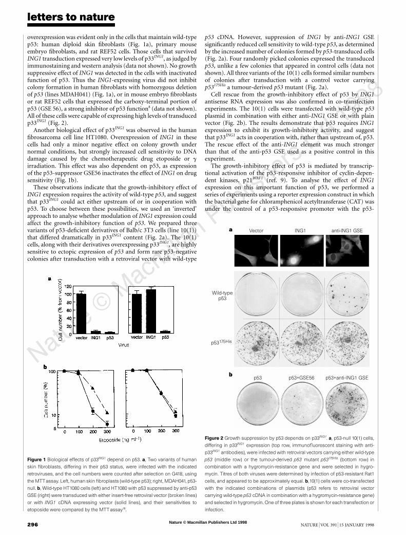
overexpression was evident only in the cells that maintain wild-typep53: human diploid skin fibroblasts (Fig. 1a), primary mouseembryo fibroblasts, and rat REF52 cells. Those cells that survivedING1 transduction expressed very low levels of p33ING1, as judged byimmunostaining and western analysis (data not shown). No growthsuppressive effect of ING1 was detected in the cells with inactivatedfunction of p53. Thus the ING1-expressing virus did not inhibitcolony formation in human fibroblasts with homozygous deletionof p53 (lines MDAH041) (Fig. 1a), or in mouse embryo fibroblastsor rat REF52 cells that expressed the carboxy-terminal portion ofp53 (GSE 56), a strong inhibitor of p53 function8 (data not shown).All of these cells were capable of expressing high levels of transducedp33ING1 (Fig. 2).
Another biological effect of p33ING1 was observed in the humanfibrosarcoma cell line HT1080. Overexpression of ING1 in thesecells had only a minor negative effect on colony growth undernormal conditions, but strongly increased cell sensitivity to DNAdamage caused by the chemotherapeutic drug etoposide or girradiation. This effect was also dependent on p53, as expressionof the p53-suppressor GSE56 inactivates the effect of ING1 on drugsensitivity (Fig. 1b).
These observations indicate that the growth-inhibitory effect ofING1 expression requires the activity of wild-type p53, and suggestthat p33ING1 could act either upstream of or in cooperation withp53. To choose between these possibilities, we used an ‘inverted’approach to analyse whether modulation of ING1 expression couldaffect the growth-inhibitory function of p53. We prepared threevariants of p53-deficient derivatives of Balb/c 3T3 cells (line 10(1))that differed dramatically in p33ING1 content (Fig. 2a). The 10(1)cells, along with their derivatives overexpressing p33ING1, are highlysensitive to ectopic expression of p53 and form rare p53-negativecolonies after transduction with a retroviral vector with wild-type
p53 cDNA. However, suppression of ING1 by anti-ING1 GSEsignificantly reduced cell sensitivity to wild-type p53, as determinedby the increased number of colonies formed by p53-transduced cells(Fig. 2a). Four randomly picked colonies expressed the transducedp53, unlike a few colonies that appeared in control cells (data notshown). All three variants of the 10(1) cells formed similar numbersof colonies after transduction with a control vector carryingp53175His a tumour-derived p53 mutant (Fig. 2a).
Cell rescue from the growth-inhibitory effect of p53 by ING1antisense RNA expression was also confirmed in co-transfectionexperiments. The 10(1) cells were transfected with wild-type p53plasmid in combination with either anti-ING1 GSE or with plainvector (Fig. 2b). The results demonstrate that p53 requires ING1expression to exhibit its growth-inhibitory activity, and suggestthat p33ING1 acts in cooperation with, rather than upstream of, p53.The rescue effect of the anti-ING1 element was much strongerthan that of the anti-p53 GSE used as a positive control in thisexperiment.
The growth-inhibitory effect of p53 is mediated by transcrip-tional activation of the p53-responsive inhibitor of cyclin-depen-dent kinases, p21WAF1 (ref. 9). To analyse the effect of ING1expression on this important function of p53, we performed aseries of experiments using a reporter expression construct in whichthe bacterial gene for chloramphenicol acetyltransferase (CAT) wasunder the control of a p53-responsive promoter with the p53-
Figure 1 Biological effects of p33ING1 depend on p53. a, Two variants of human
skin fibroblasts, differing in their p53 status, were infected with the indicated
retroviruses, and the cell numbers were counted after selection on G418, using
the MTTassay. Left, human skin fibroplasts (wild-type p53); right, MDAH041, p53-
null. b, Wild-type HT1080 cells (left) and HT1080 with p53 suppressed by anti-p53
GSE (right) were transduced with either insert-free retroviral vector (broken lines)
or with ING1 cDNA expressing vector (solid lines), and their sensitivities to
etoposide were compared by the MTTassay18.
Vector ING1 anti-ING1 GSE
Wild-typep53
p53175His
p53+anti-ING1 GSEp53+GSE56p53
a
b
Figure 2 Growth suppression by p53 depends on p33ING1. a, p53-null 10(1) cells,
differing in p33ING1 expression (top row, immunofluorescent staining with anti-
p33ING1 antibodies), were infected with retroviral vectors carrying either wild-type
p53 (middle row) or the tumour-derived p53 mutant p53175His (bottom row) in
combination with a hygromycin-resistance gene and were selected in hygro-
mycin. Titres of both viruses were determined by infection of p53-resistant Rat1
cells, and appeared to be approximately equal. b,10(1) cells were co-transfected
with the indicated combinations of plasmids (p53 refers to retroviral vector
carrying wild-type p53 cDNA in combination with a hygromycin-resistance gene)
and selected in hygromycin. One of three plates is shown for each transfection or
infection.
Nature © Macmillan Publishers Ltd 1998
8
letters to nature
NATURE | VOL 391 | 15 JANUARY 1998 297
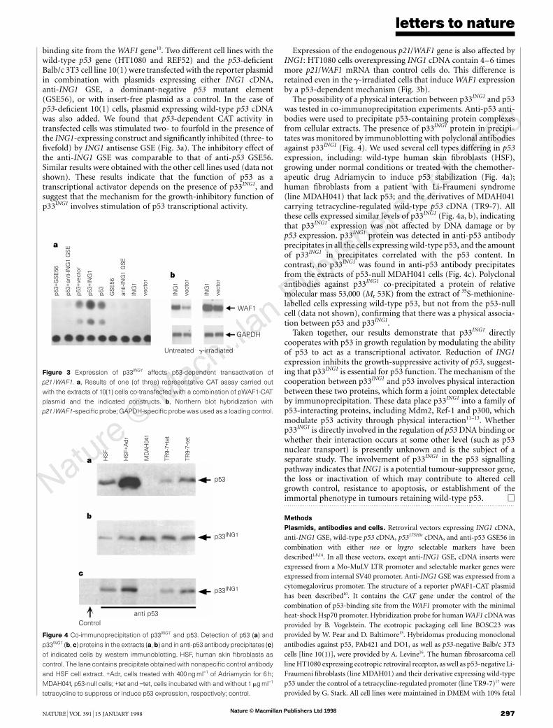
binding site from the WAF1 gene10. Two different cell lines with thewild-type p53 gene (HT1080 and REF52) and the p53-deficientBalb/c 3T3 cell line 10(1) were transfected with the reporter plasmidin combination with plasmids expressing either ING1 cDNA,anti-ING1 GSE, a dominant-negative p53 mutant element(GSE56), or with insert-free plasmid as a control. In the case ofp53-deficient 10(1) cells, plasmid expressing wild-type p53 cDNAwas also added. We found that p53-dependent CAT activity intransfected cells was stimulated two- to fourfold in the presence ofthe ING1-expressing construct and significantly inhibited (three- tofivefold) by ING1 antisense GSE (Fig. 3a). The inhibitory effect ofthe anti-ING1 GSE was comparable to that of anti-p53 GSE56.Similar results were obtained with the other cell lines used (data notshown). These results indicate that the function of p53 as atranscriptional activator depends on the presence of p33ING1, andsuggest that the mechanism for the growth-inhibitory function ofp33ING1 involves stimulation of p53 transcriptional activity.
Expression of the endogenous p21/WAF1 gene is also affected byING1: HT1080 cells overexpressing ING1 cDNA contain 4–6 timesmore p21/WAF1 mRNA than control cells do. This difference isretained even in the g-irradiated cells that induce WAF1 expressionby a p53-dependent mechanism (Fig. 3b).
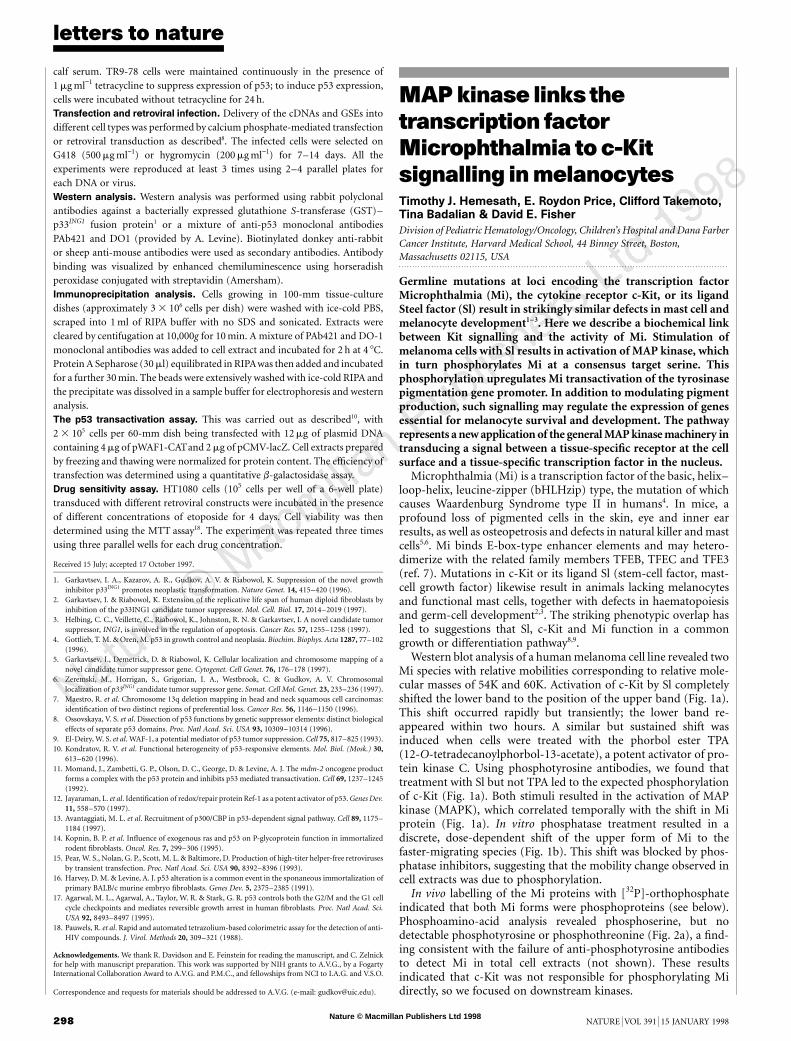
The possibility of a physical interaction between p33ING1 and p53was tested in co-immunoprecipitation experiments. Anti-p53 anti-bodies were used to precipitate p53-containing protein complexesfrom cellular extracts. The presence of p33ING1 protein in precipi-tates was monitored by immunoblotting with polyclonal antibodiesagainst p33ING1 (Fig. 4). We used several cell types differing in p53expression, including: wild-type human skin fibroblasts (HSF),growing under normal conditions or treated with the chemother-apeutic drug Adriamycin to induce p53 stabilization (Fig. 4a);human fibroblasts from a patient with Li-Fraumeni syndrome(line MDAH041) that lack p53; and the derivatives of MDAH041carrying tetracycline-regulated wild-type p53 cDNA (TR9-7). Allthese cells expressed similar levels of p33ING1 (Fig. 4a, b), indicatingthat p33ING1 expression was not affected by DNA damage or byp53 expression. p33ING1 protein was detected in anti-p53 antibodyprecipitates in all the cells expressing wild-type p53, and the amountof p33ING1 in precipitates correlated with the p53 content. Incontrast, no p33ING1 was found in anti-p53 antibody precipitatesfrom the extracts of p53-null MDAH041 cells (Fig. 4c). Polyclonalantibodies against p33ING1 co-precipitated a protein of relativemolecular mass 53,000 (Mr 53K) from the extract of 35S-methionine-labelled cells expressing wild-type p53, but not from the p53-nullcell (data not shown), confirming that there was a physical associa-tion between p53 and p33ING1.
Taken together, our results demonstrate that p33ING1 directlycooperates with p53 in growth regulation by modulating the abilityof p53 to act as a transcriptional activator. Reduction of ING1expression inhibits the growth-suppressive activity of p53, suggest-ing that p33ING1 is essential for p53 function. The mechanism of thecooperation between p33ING1 and p53 involves physical interactionbetween these two proteins, which form a joint complex detectableby immunoprecipitation. These data place p33ING1 into a family ofp53-interacting proteins, including Mdm2, Ref-1 and p300, whichmodulate p53 activity through physical interaction11–13. Whetherp33ING1 is directly involved in the regulation of p53 DNA binding orwhether their interaction occurs at some other level (such as p53nuclear transport) is presently unknown and is the subject of aseparate study. The involvement of p33ING1 in the p53 signallingpathway indicates that ING1 is a potential tumour-suppressor gene,the loss or inactivation of which may contribute to altered cellgrowth control, resistance to apoptosis, or establishment of theimmortal phenotype in tumours retaining wild-type p53. M. . . . . . . . . . . . . . . . . . . . . . . . . . . . . . . . . . . . . . . . . . . . . . . . . . . . . . . . . . . . . . . . . . . . . . . . . . . . . . . . . . . . . . . . . . . . . . . . . . . . . . . . . . . . . . . . . . . . . . . . .
Methods
Plasmids, antibodies and cells. Retroviral vectors expressing ING1 cDNA,anti-ING1 GSE, wild-type p53 cDNA, p53175His cDNA, and anti-p53 GSE56 incombination with either neo or hygro selectable markers have beendescribed1,8,14. In all these vectors, except anti-ING1 GSE, cDNA inserts wereexpressed from a Mo-MuLV LTR promoter and selectable marker genes wereexpressed from internal SV40 promoter. Anti-ING1 GSE was expressed from acytomegalovirus promoter. The structure of a reporter pWAF1-CAT plasmidhas been described10. It contains the CAT gene under the control of thecombination of p53-binding site from the WAF1 promoter with the minimalheat-shock Hsp70 promoter. Hybridization probe for human WAF1 cDNA wasprovided by B. Vogelstein. The ecotropic packaging cell line BOSC23 wasprovided by W. Pear and D. Baltimore15. Hybridomas producing monoclonalantibodies against p53, PAb421 and DO1, as well as p53-negative Balb/c 3T3cells [line 10(1)], were provided by A. Levine16. The human fibrosarcoma cellline HT1080 expressing ecotropic retroviral receptor, as well as p53-negative Li-Fraumeni fibroblasts (line MDAH01) and their derivative expressing wild-typep53 under the control of a tetracycline-regulated promoter (line TR9-7)17 wereprovided by G. Stark. All cell lines were maintained in DMEM with 10% fetal
WAF1
GAPDH
Untreated γ-irradiated
p53+ve
cto
r
p53+G
SE56
p53+IN
G1
p53+anti-IN
G1 G
SE
p53
GSE56
anti-IN
G1 G
SE
ING
1
vecto
r
ING
1
vecto
r
ING
1
vecto
r
a
b
Figure 3 Expression of p33ING1 affects p53-dependent transactivation of
p21/WAF1. a, Results of one (of three) representative CAT assay carried out
with the extracts of 10(1) cells co-transfected with a combination of pWAF1-CAT
plasmid and the indicated constructs. b, Northern blot hybridization with
p21/WAF1-specific probe; GAPDH-specific probe was used as a loading control.
p53
p33ING1
p33ING1
anti p53
Control
TR9-7
–tet
TR9-7
+te
t
MD
AH
041
HSF+A
dr
HSF
a
b
c
Figure 4 Co-immunoprecipitation of p33ING1 and p53. Detection of p53 (a) and
p33ING1 (b, c) proteins in the extracts (a, b) and in anti-p53 antibody precipitates (c)
of indicated cells by western immunoblotting. HSF, human skin fibroblasts as
control. The lane contains precipitate obtained with nonspecific control antibody
and HSF cell extract. +Adr, cells treated with 400 ngml−1 of Adriamycin for 6 h;
MDAH041, p53-null cells; +tet and −tet, cells incubated with and without 1 mgml−1
tetracycline to suppress or induce p53 expression, respectively; control.
Nature © Macmillan Publishers Ltd 1998
8
letters to nature
298 NATURE | VOL 391 | 15 JANUARY 1998
calf serum. TR9-78 cells were maintained continuously in the presence of1 mg ml−1 tetracycline to suppress expression of p53; to induce p53 expression,cells were incubated without tetracycline for 24 h.Transfection and retroviral infection. Delivery of the cDNAs and GSEs intodifferent cell types was performed by calcium phosphate-mediated transfectionor retroviral transduction as described8. The infected cells were selected onG418 (500 mg ml−1) or hygromycin (200 mg ml−1) for 7–14 days. All theexperiments were reproduced at least 3 times using 2–4 parallel plates foreach DNA or virus.Western analysis. Western analysis was performed using rabbit polyclonalantibodies against a bacterially expressed glutathione S-transferase (GST)–p33ING1 fusion protein1 or a mixture of anti-p53 monoclonal antibodiesPAb421 and DO1 (provided by A. Levine). Biotinylated donkey anti-rabbitor sheep anti-mouse antibodies were used as secondary antibodies. Antibodybinding was visualized by enhanced chemiluminescence using horseradishperoxidase conjugated with streptavidin (Amersham).Immunoprecipitation analysis. Cells growing in 100-mm tissue-culturedishes (approximately 3 3 106 cells per dish) were washed with ice-cold PBS,scraped into 1 ml of RIPA buffer with no SDS and sonicated. Extracts werecleared by centifugation at 10,000g for 10 min. A mixture of PAb421 and DO-1monoclonal antibodies was added to cell extract and incubated for 2 h at 4 8C.Protein A Sepharose (30 ml) equilibrated in RIPAwas then added and incubatedfor a further 30 min. The beads were extensively washed with ice-cold RIPA andthe precipitate was dissolved in a sample buffer for electrophoresis and westernanalysis.The p53 transactivation assay. This was carried out as described10, with2 3 105 cells per 60-mm dish being transfected with 12 mg of plasmid DNAcontaining 4 mg of pWAF1-CATand 2 mg of pCMV-lacZ. Cell extracts preparedby freezing and thawing were normalized for protein content. The efficiency oftransfection was determined using a quantitative b-galactosidase assay.Drug sensitivity assay. HT1080 cells (105 cells per well of a 6-well plate)transduced with different retroviral constructs were incubated in the presenceof different concentrations of etoposide for 4 days. Cell viability was thendetermined using the MTT assay18. The experiment was repeated three timesusing three parallel wells for each drug concentration.
Received 15 July; accepted 17 October 1997.
1. Garkavtsev, I. A., Kazarov, A. R., Gudkov, A. V. & Riabowol, K. Suppression of the novel growthinhibitor p33ING1 promotes neoplastic transformation. Nature Genet. 14, 415–420 (1996).
2. Garkavtsev, I. & Riabowol, K. Extension of the replicative life span of human diploid fibroblasts byinhibition of the p33ING1 candidate tumor suppressor. Mol. Cell. Biol. 17, 2014–2019 (1997).
3. Helbing, C. C., Veillette, C., Riabowol, K., Johnston, R. N. & Garkavtsev, I. A novel candidate tumorsuppressor, ING1, is involved in the regulation of apoptosis. Cancer Res. 57, 1255–1258 (1997).
4. Gottlieb, T. M. & Oren, M. p53 in growth control and neoplasia. Biochim. Biophys. Acta 1287, 77–102(1996).
5. Garkavtsev, I., Demetrick, D. & Riabowol, K. Cellular localization and chromosome mapping of anovel candidate tumor suppressor gene. Cytogenet. Cell Genet. 76, 176–178 (1997).
6. Zeremski, M., Horrigan, S., Grigorian, I. A., Westbrook, C. & Gudkov, A. V. Chromosomallocalization of p33ING1 candidate tumor suppressor gene. Somat. Cell Mol. Genet. 23, 233–236 (1997).
7. Maestro, R. et al. Chromosome 13q deletion mapping in head and neck squamous cell carcinomas:identification of two distinct regions of preferential loss. Cancer Res. 56, 1146–1150 (1996).
8. Ossovskaya, V. S. et al. Dissection of p53 functions by genetic suppressor elements: distinct biologicaleffects of separate p53 domains. Proc. Natl Acad. Sci. USA 93, 10309–10314 (1996).
9. El-Deiry, W. S. et al. WAF-1, a potential mediator of p53 tumor suppression. Cell 75, 817–825 (1993).10. Kondratov, R. V. et al. Functional heterogeneity of p53-responsive elements. Mol. Biol. (Mosk.) 30,
613–620 (1996).11. Momand, J., Zambetti, G. P., Olson, D. C., George, D. & Levine, A. J. The mdm-2 oncogene product
forms a complex with the p53 protein and inhibits p53 mediated transactivation. Cell 69, 1237–1245(1992).
12. Jayaraman, L. et al. Identification of redox/repair protein Ref-1 as a potent activator of p53. Genes Dev.11, 558–570 (1997).
13. Avantaggiati, M. L. et al. Recruitment of p300/CBP in p53-dependent signal pathway. Cell 89, 1175–1184 (1997).
14. Kopnin, B. P. et al. Influence of exogenous ras and p53 on P-glycoprotein function in immortalizedrodent fibroblasts. Oncol. Res. 7, 299–306 (1995).
15. Pear, W. S., Nolan, G. P., Scott, M. L. & Baltimore, D. Production of high-titer helper-free retrovirusesby transient transfection. Proc. Natl Acad. Sci. USA 90, 8392–8396 (1993).
16. Harvey, D. M. & Levine, A. J. p53 alteration is a common event in the sponaneous immortalization ofprimary BALB/c murine embryo fibroblasts. Genes Dev. 5, 2375–2385 (1991).
17. Agarwal, M. L., Agarwal, A., Taylor, W. R. & Stark, G. R. p53 controls both the G2/M and the G1 cellcycle checkpoints and mediates reversible growth arrest in human fibroblasts. Proc. Natl Acad. Sci.USA 92, 8493–8497 (1995).
18. Pauwels, R. et al. Rapid and automated tetrazolium-based colorimetric assay for the detection of anti-HIV compounds. J. Virol. Methods 20, 309–321 (1988).
Acknowledgements. We thank R. Davidson and E. Feinstein for reading the manuscript, and C. Zelnickfor help with manuscript preparation. This work was supported by NIH grants to A.V.G., by a FogartyInternational Collaboration Award to A.V.G. and P.M.C., and fellowships from NCI to I.A.G. and V.S.O.
Correspondence and requests for materials should be addressed to A.V.G. (e-mail: [email protected]).
MAPkinase links thetranscription factorMicrophthalmia toc-Kitsignalling inmelanocytesTimothy J. Hemesath, E. Roydon Price, Clifford Takemoto,Tina Badalian & David E. Fisher
Division of Pediatric Hematology/Oncology, Children’s Hospital and Dana FarberCancer Institute, Harvard Medical School, 44 Binney Street, Boston,Massachusetts 02115, USA. . . . . . . . . . . . . . . . . . . . . . . . . . . . . . . . . . . . . . . . . . . . . . . . . . . . . . . . . . . . . . . . . . . . . . . . . . . . . . . . . . . . . . . . . . . . . . . . . . . . . . . . . . . . . . . . . . . . . . . . .
Germline mutations at loci encoding the transcription factorMicrophthalmia (Mi), the cytokine receptor c-Kit, or its ligandSteel factor (Sl) result in strikingly similar defects in mast cell andmelanocyte development1–3. Here we describe a biochemical linkbetween Kit signalling and the activity of Mi. Stimulation ofmelanoma cells with Sl results in activation of MAP kinase, whichin turn phosphorylates Mi at a consensus target serine. Thisphosphorylation upregulates Mi transactivation of the tyrosinasepigmentation gene promoter. In addition to modulating pigmentproduction, such signalling may regulate the expression of genesessential for melanocyte survival and development. The pathwayrepresents a new application of the general MAP kinase machinery intransducing a signal between a tissue-specific receptor at the cellsurface and a tissue-specific transcription factor in the nucleus.
Microphthalmia (Mi) is a transcription factor of the basic, helix–loop-helix, leucine-zipper (bHLHzip) type, the mutation of whichcauses Waardenburg Syndrome type II in humans4. In mice, aprofound loss of pigmented cells in the skin, eye and inner earresults, as well as osteopetrosis and defects in natural killer and mastcells5,6. Mi binds E-box-type enhancer elements and may hetero-dimerize with the related family members TFEB, TFEC and TFE3(ref. 7). Mutations in c-Kit or its ligand Sl (stem-cell factor, mast-cell growth factor) likewise result in animals lacking melanocytesand functional mast cells, together with defects in haematopoiesisand germ-cell development2,3. The striking phenotypic overlap hasled to suggestions that Sl, c-Kit and Mi function in a commongrowth or differentiation pathway8,9.
Western blot analysis of a human melanoma cell line revealed twoMi species with relative mobilities corresponding to relative mole-cular masses of 54K and 60K. Activation of c-Kit by Sl completelyshifted the lower band to the position of the upper band (Fig. 1a).This shift occurred rapidly but transiently; the lower band re-appeared within two hours. A similar but sustained shift wasinduced when cells were treated with the phorbol ester TPA(12-O-tetradecanoylphorbol-13-acetate), a potent activator of pro-tein kinase C. Using phosphotyrosine antibodies, we found thattreatment with Sl but not TPA led to the expected phosphorylationof c-Kit (Fig. 1a). Both stimuli resulted in the activation of MAPkinase (MAPK), which correlated temporally with the shift in Miprotein (Fig. 1a). In vitro phosphatase treatment resulted in adiscrete, dose-dependent shift of the upper form of Mi to thefaster-migrating species (Fig. 1b). This shift was blocked by phos-phatase inhibitors, suggesting that the mobility change observed incell extracts was due to phosphorylation.
In vivo labelling of the Mi proteins with [32P]-orthophosphateindicated that both Mi forms were phosphoproteins (see below).Phosphoamino-acid analysis revealed phosphoserine, but nodetectable phosphotyrosine or phosphothreonine (Fig. 2a), a find-ing consistent with the failure of anti-phosphotyrosine antibodiesto detect Mi in total cell extracts (not shown). These resultsindicated that c-Kit was not responsible for phosphorylating Midirectly, so we focused on downstream kinases.