Embed Size (px)
Citation preview

articles
Negative regulation of the Apaf-1 apoptosome by Hsp70
Ayman Saleh*†‡, Srinivasa M. Srinivasula*‡, Levent Balkir†, Paul D. Robbins†§ and Emad S. Alnemri*§¶*Center for Apoptosis Research and Department of Microbiology and Immunology, Kimmel Cancer Institute,
Thomas Jefferson University, Philadelphia, Pennsylvania 19107, USA†Department of Molecular Genetics and Biochemistry, University of Pittsburgh, Pittsburgh, Pennsylvania 15261, USA
‡These authors contributed equally to this work§These authors contributed equally to this work
¶e-mail: [email protected]
Release of cytochrome c from mitochondria by apoptotic signals induces ATP/dATP-dependent formation of the oligomeric Apaf-1–caspase-9 apoptosome. Here we show that the documented anti-apoptotic effect of the principal heat-shock protein, Hsp70, is mediated through its direct association with the caspase-recruitment domain (CARD) of Apaf-1 and through inhibition of apoptosome formation. The interaction between Hsp70 and Apaf-1 prevents oligomerization of Apaf-1 and association of Apaf-1 with procaspase-9. On the basis of these results, we propose that resistance to apoptosis exhibited by stressed cells and some tumours, which constitutively express high levels of Hsp70, may be due in part to modulation of Apaf-1 function by Hsp70.
poptosis is essential for cell development and tissue homeos-tasis in eukaryotic organisms1–3. The molecular machinerythat drives the apoptotic programme consists of a family of
cysteine proteases, the caspases that cleave their substrates after spe-cific aspartic acid residues (reviewed in refs 4–8). In normal cells,caspases are constitutively expressed as inactive single polypeptidechains, known as procaspases, and their activation requires specificproteolytic cleavage5–8. Active caspases can typically amplify apop-totic events by their ability to cleave their own precursor forms aswell as those of other caspases9,10.
Mitochondria have a crucial function in initiating the cascade ofcaspase activation in response to different apoptotic signals11. Dis-ruption of the outer mitochondrial membrane by apoptotic stimuliresults in the release of cytochrome c into the cytoplasm12. Cyto-chrome c binds to the cytosolic apoptotic-protease-activating factor1 (Apaf-1), thereby promoting Apaf-1-mediated activation of cas-pase-9 in an ATP/dATP-dependent manner13–15. Active caspase-9can subsequently amplify the caspase cascade by its ability to proc-ess its own proenzyme as well as the effector caspase-3 (refs 13–15).Association of procaspase-9 with Apaf-1 is an essential step in its
A
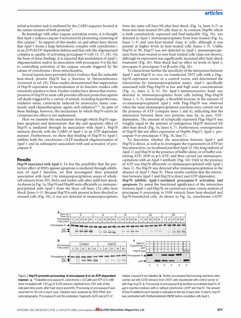
Figure 1 Inducible Hsp70 associates with Apaf-1. a, Whole-cell extracts (400 µg) obtained from 293, HeLa and Jurkat cells directly after heat-shock (HS) treatment (lanes 5–7) or 12 h after heat shock (lanes 1–3), or from non-heat-treated 293 cells (NHS), were incubated with 4 µg anti-Apaf-1 antibody coupled to cyanogenbromide-activated sepharose. Immunoprecipitates were fractionated on a 12% SDS–polyacrylamide gel and immunoblotted with antibodies against Hsp70, Hsp90 and Apaf-1. b, S100 extracts (120 µg) obtained from 293 cells before heat shock (No HS) or at the indicated time points (0–24 h) after heat shock, or from 293 cells transfected with Flag–hsp70 (transfected), were analysed by western blotting with antibodies against Hsp90, Hsp70, Hsp27, Apaf-1, procaspase-9 and procaspase-3. The same membrane was reprobed for β-actin and served as a
loading control. c, S100 extracts (350 µg) from 293T cells transfected with a control plasmid or with Flag–hsp70 were incubated with or without ATP and then immunoprecipitated with an anti-Flag antibody after adjusting [NaCl] to 50 or 500 mM. Immunoprecipitates were analysed by western blotting with an anti-Apaf-1 antibody. The heavy chain of the Flag monoclonal antibody is labelled and served as a loading control. IgG, immunoglobulin G. d, Purified Apaf-1 and Hsp70 (4 µg each) were mixed and incubated with or without ATP, ADP or γ-S-ATP and then immunoprecipitated with an anti-Apaf-1 antibody. Immunoprecipitates (IP) and supernatants (S) were fractionated by SDS–PAGE and immunoprobed with anti-Hsp70 (upper panel) and anti-Apaf-1 (lower panel) antibodies.
Hsp70
Hsp90
Apaf-1
293
HeL
a
Jurk
at
293
HeL
a
Jurk
at
293
HS (12 h) HS (0 h)NHS
Apaf-1L Hsp70 NTP
ATP ADP ATP
Apaf-1L
Hsp70
γS-ATP
Flag–Hsp70
ATP +++ + +
50 mM[NaCl] 500 mM
Apaf-1
IgG
HS recovery time (h)NoHS
Trans-fected
Hsp90
Hsp70
Hsp27
Apaf-1
pcasp-3
pcasp-9
β-actin
a b c
d
1 2 3 4 5 6
1 2 3 4 5 6
7
1 2 3 4 5 6 7
1 2 3 4 5 6 7 8 9 10
0 4 8 12 24 ––– –
–– –
IP S IP S IP S IP S IP S–
–+ + + ++ + + +
+
© 2000 Macmillan Magazines Ltd476 NATURE CELL BIOLOGY | VOL 2 | AUGUST 2000 | www.nature.com/ncb
articles
initial activation and is mediated by the CARD sequence located atthe amino termini of both proteins16.
By homology with other caspase-activation events, it is thoughtthat Apaf-1 induces caspase-9 activation by promoting clustering ofthis caspase14. In support of this model, we and others have shownthat Apaf-1 forms a large heteromeric complex with cytochrome cin an ATP/dATP-dependent fashion and that only the oligomerizedcomplex is capable of activating procaspase-9 (refs 15, 17, 18). Onthe basis of these findings, it is expected that modulation of Apaf-1oligomerization and/or its association with procaspase-9 is the keyto controlling activation of the caspase cascade triggered by therelease of cytochrome c from mitochondria.
Several reports have provided direct evidence that the inducibleheat-shock protein Hsp70 has a function in thermotolerance(reviewed in ref. 19). These studies demonstrated that suppressionof Hsp70 expression or neutralization of its function renders cellsextremely sensitive to heat. Further studies have shown that overex-pression of Hsp70 in most cells provides efficient protection againstcell death triggered by a variety of stimuli, including hyperthermia,oxidative stress, cytotoxicity induced by monocytes, many com-monly used chemotheraputic agents and radiation20–23. In spite ofthese findings, however, the mechanism by which Hsp70 exerts itscytoprotective effect is not understood.
Here we examine the mechanisms through which Hsp70 regu-lates apoptosis and demonstrate that the anti-apoptotic effect ofHsp70 is mediated through its association with Apaf-1. Hsp70interacts directly with the CARD of Apaf-1 in an ATP-dependentmanner. Furthermore, we show that binding of Hsp70 to Apaf-1inhibits both the cytochrome c/ATP-mediated oligomerization ofApaf-1 and its subsequent association with and activation of pro-caspase-9.
ResultsHsp70 associates with Apaf-1. To test the possibility that the pro-tective effect of HSPs against apoptosis is mediated through inhibi-tion of Apaf-1 function, we first investigated their potentialassociation with Apaf-1 by immunoprecipitation assays of whole-cell extracts from 293, HeLa and Jurkat cells after mild heat shock.As shown in Fig. 1a, Hsp70 and Hsp90 were efficiently co-immuno-precipitated with Apaf-1 from the three cell lines 12 h after heatshock (lanes 1–3). Because Hsp70 is only present in heat-shocked orstressed cells (Fig. 1b), it was not detected in immunoprecipitates
from the same cell lines 0 h after heat shock (Fig. 1a, lanes 5–7) orfrom non-heat-treated 293 cells (lane 4). In contrast, Hsp90, whichis both constitutively expressed and heat-inducible (Fig. 1b), wasdetected in Apaf-1 immunoprecipitates from heat-treated (Fig. 1a,lanes 1–3) and non-heat-treated (lane 4) cells, although it waspresent at higher levels in heat-treated cells (lanes 1–3). UnlikeHsp70 or 90, Hsp27 was not detected in Apaf-1 immunoprecipi-tates from heat-treated or non-heat-treated cells (data not shown),although its expression was significantly increased after heat-shocktreatment (Fig. 1b). Heat shock had no effect on levels of Apaf-1,procapase-9, procaspase-3 or β-actin (Fig. 1b).
To characterize further the specificity of the interaction betweenApaf-1 and Hsp70 in vivo, we transfected 293T cells with a Flag–hsp70 expression vector or a control vector, and determined theinteractions by immunoprecipitation assays. Apaf-1 specificallyassociated with Flag–Hsp70 at low and high ionic concentrations(Fig. 1c, lanes 2, 4, 6). No Apaf-1-immunoreactive band wasdetected in immunoprecipitates from cells lacking Flag–Hsp70(lanes 1, 3, 5), Interestingly, a threefold increase in the amount ofco-immunoprecipitated Apaf-1 with Flag–Hsp70 was observedwhen the same immunoprecipitation reactions were carried out inthe presence of ATP (compare lanes 2 and 4), indicating that theinteraction between these two proteins may be, in part, ATP-dependent. The amount of ectopically expressed Flag–Hsp70 wasroughly equal to the amount of endogenous Hsp70 detected 8 hafter heat shock (Fig. 1b, lanes 4, 7). Furthermore, overexpressionof Hsp70 did not affect expression of Hsp90, Hsp27, Apaf-1, pro-caspase-9 or procaspase-3 (Fig. 1b, lane 7).
To determine whether the association between Apaf-1 andHsp70 is direct, as well as to investigate the requirement of ATP forthis interaction, we incubated purified Apaf-1L (the long isoform ofApaf-1) and Hsp70 in the presence of buffer alone, or of buffer con-taining ATP, ADP or γ-S-ATP, and then carried out immunopre-cipitations with an Apaf-1 antibody (Fig. 1d). Only in the presenceof ATP was Hsp70 efficiently co-immunoprecipitated with Apaf-1(lane 1). No Hsp70 was detected after immunoprecipitation in theabsence of Apaf-1 (lane 9). These results confirm that the interac-tion between Apaf-1 and Hsp70 is direct and ATP-dependent.Hsp70 inhibits Apaf-1-mediated procaspase-9 activation andapoptosis. To assess the functional significance of the interactionbetween Apaf-1 and Hsp70, we carried out a time-course analysis ofprocaspase-9 processing in S100 extracts from heat-shocked andhsp70-transfected cells. As shown in Fig. 2a, cytochrome c/ATP-
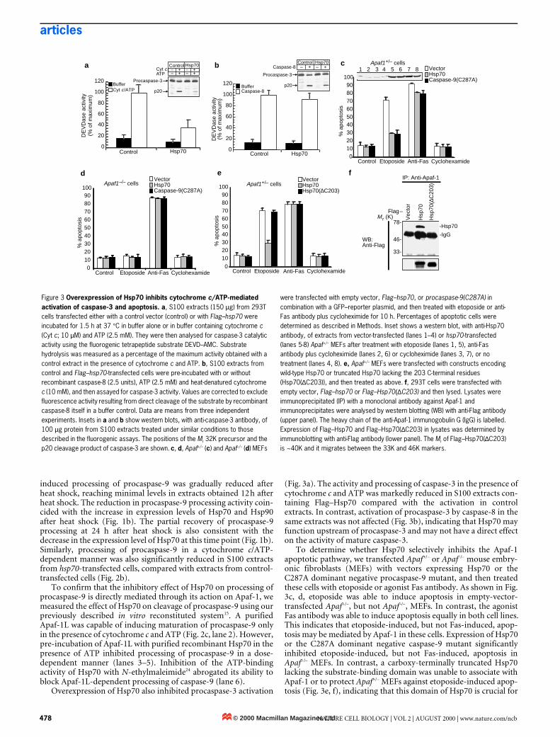
Figure 2 Hsp70 prevents processing of procaspase-9 in an ATP-dependent manner. a, 35S-labelled procaspase-9, cytochrome c (10 µM) and ATP (2.5 mM) were incubated with 150 µg of S100 extracts obtained from 293 cells at the indicated time points after heat shock (post-HS). Processing of procaspase-9 was observed for 60 min in each case. Samples were analysed by SDS–PAGE and autoradiography. Procaspase-9 and the proteolytic fragments (p35 and p37) of
mature caspase-9 are labelled. b, Similar procaspase-9-processing reactions were carried out with S100 extracts from 293T cells transfected with control vector or with Flag–hsp70. c, Processing of procaspase-9 by purified recombinant Apaf-1L (4 µg) in reaction mixtures with or without cytochrome c/ATP and Hsp70. The amount of Hsp70 added to each sample is indicated on the top of each lane. In lane 6, Hsp70 was pretreated with N-ethylmaleimide (NEM) before incubation with Apaf-1.
cApaf-1L
Cytochrome c ATP
Hsp70 (µg)
p35
Post-HSTime (h)
ProcessingTime (min)
Procaspase-9
p37p35
a
ControlProcessingTime (min)
p37p35
b
0 4 8 12 24
0 10 20 30 60
0 10 20 30 60 0 10 20 30 60
0 10 20 30 60 0 10 20 30 60 0 10 20 30 60 0 10 20 30 60
Hsp70 + +++
++
++
++
++
+ + + +––1 0 1 2 4 NEM
1 2 3 4 5 6
Procaspase-9 Procaspase-9
© 2000 Macmillan Magazines LtdNATURE CELL BIOLOGY | VOL 2 | AUGUST 2000 | www.nature.com/ncb 477
articles
induced processing of procaspase-9 was gradually reduced afterheat shock, reaching minimal levels in extracts obtained 12 h afterheat shock. The reduction in procaspase-9 processing activity coin-cided with the increase in expression levels of Hsp70 and Hsp90after heat shock (Fig. 1b). The partial recovery of procaspase-9processing at 24 h after heat shock is also consistent with thedecrease in the expression level of Hsp70 at this time point (Fig. 1b).Similarly, processing of procaspase-9 in a cytochrome c/ATP-dependent manner was also significantly reduced in S100 extractsfrom hsp70-transfected cells, compared with extracts from control-transfected cells (Fig. 2b).
To confirm that the inhibitory effect of Hsp70 on processing ofprocaspase-9 is directly mediated through its action on Apaf-1, wemeasured the effect of Hsp70 on cleavage of procaspase-9 using ourpreviously described in vitro reconstituted system15. A purifiedApaf-1L was capable of inducing maturation of procaspase-9 onlyin the presence of cytochrome c and ATP (Fig. 2c, lane 2). However,pre-incubation of Apaf-1L with purified recombinant Hsp70 in thepresence of ATP inhibited processing of procaspase-9 in a dose-dependent manner (lanes 3–5). Inhibition of the ATP-bindingactivity of Hsp70 with N-ethylmaleimide24 abrogated its ability toblock Apaf-1L-dependent processing of caspase-9 (lane 6).
Overexpression of Hsp70 also inhibited procaspase-3 activation
(Fig. 3a). The activity and processing of caspase-3 in the presence ofcytochrome c and ATP was markedly reduced in S100 extracts con-taining Flag–Hsp70 compared with the activation in controlextracts. In contrast, activation of procaspase-3 by caspase-8 in thesame extracts was not affected (Fig. 3b), indicating that Hsp70 mayfunction upstream of procaspase-3 and may not have a direct effecton the activity of mature caspase-3.
To determine whether Hsp70 selectively inhibits the Apaf-1apoptotic pathway, we transfected Apaf+/– or Apaf–/– mouse embry-onic fibroblasts (MEFs) with vectors expressing Hsp70 or theC287A dominant negative procaspase-9 mutant, and then treatedthese cells with etoposide or agonist Fas antibody. As shown in Fig.3c, d, etoposide was able to induce apoptosis in empty-vector-transfected Apaf+/–, but not Apaf–/–, MEFs. In contrast, the agonistFas antibody was able to induce apoptosis equally in both cell lines.This indicates that etoposide-induced, but not Fas-induced, apop-tosis may be mediated by Apaf-1 in these cells. Expression of Hsp70or the C287A dominant negative caspase-9 mutant significantlyinhibited etoposide-induced, but not Fas-induced, apoptosis inApaf+/– MEFs. In contrast, a carboxy-terminally truncated Hsp70lacking the substrate-binding domain was unable to associate withApaf-1 or to protect Apaf+/– MEFs against etoposide-induced apop-tosis (Fig. 3e, f), indicating that this domain of Hsp70 is crucial for
Figure 3 Overexpression of Hsp70 inhibits cytochrome c/ATP-mediated activation of caspase-3 and apoptosis. a, S100 extracts (150 µg) from 293T cells transfected either with a control vector (control) or with Flag–hsp70 were incubated for 1.5 h at 37 °C in buffer alone or in buffer containing cytochrome c (Cyt c; 10 µM) and ATP (2.5 mM). They were then analysed for caspase-3 catalytic activity using the fluorogenic tetrapeptide substrate DEVD–AMC. Substrate hydrolysis was measured as a percentage of the maximum activity obtained with a control extract in the presence of cytochrome c and ATP. b, S100 extracts from control and Flag–hsp70-transfected cells were pre-incubated with or without recombinant caspase-8 (2.5 units), ATP (2.5 mM) and heat-denatured cytochrome c (10 mM), and then assayed for caspase-3 activity. Values are corrected to exclude fluorescence activity resulting from direct cleavage of the substrate by recombinant caspase-8 itself in a buffer control. Data are means from three independent experiments. Insets in a and b show western blots, with anti-caspase-3 antibody, of 100 µg protein from S100 extracts treated under similar conditions to those described in the fluorogenic assays. The positions of the Mr 32K precursor and the p20 cleavage product of caspase-3 are shown. c, d, Apaf+/– (c) and Apaf–/– (d) MEFs
were transfected with empty vector, Flag–hsp70, or procaspase-9(C287A) in combination with a GFP–reporter plasmid, and then treated with etoposide or anti-Fas antibody plus cycloheximide for 10 h. Percentages of apoptotic cells were determined as described in Methods. Inset shows a western blot, with anti-Hsp70 antibody, of extracts from vector-transfected (lanes 1–4) or hsp70-transfected (lanes 5-8) Apaf+/– MEFs after treatment with etoposide (lanes 1, 5), anti-Fas antibody plus cycloheximide (lanes 2, 6) or cycloheximide (lanes 3, 7), or no treatment (lanes 4, 8). e, Apaf+/– MEFs were transfected with constructs encoding wild-type Hsp70 or truncated Hsp70 lacking the 203 C-terminal residues (Hsp70(∆C203)), and then treated as above. f, 293T cells were transfected with empty vector, Flag–hsp70 or Flag–Hsp70(∆C203) and then lysed. Lysates were immunoprecipitated (IP) with a monoclonal antibody against Apaf-1 and immunoprecipitates were analysed by western blotting (WB) with anti-Flag antibody (upper panel). The heavy chain of the anti-Apaf-1 immunogobulin G (IgG) is labelled. Expression of Flag–Hsp70 and Flag–Hsp70(∆C203) in lysates was determined by immunoblotting with anti-Flag antibody (lower panel). The Mr of Flag–Hsp70(∆C203) is ~40K and it migrates between the 33K and 46K markers.
Control Hsp70Cyt cATP
+ ++ +
p20
0
20
40
60
80
100
120
Control Hsp70
BufferCyt c/ATP
DE
VD
ase
activ
ity(%
of m
axim
um)
Control Hsp70+ +Caspase-8
p20
0
20
40
60
80
100
120
Control Hsp70
BufferCaspase-8
DE
VD
ase
activ
ity(%
of m
axim
um)
0102030405060708090
100
Apaf1–/– cells
Apaf1+/– cells
Control Etoposide Anti-Fas
% a
popt
osis
Cyclohexamide
0102030405060708090
100
Control Etoposide Anti-Fas
% a
popt
osis
Cyclohexamide0
102030405060708090
100
Control Etoposide Anti-Fas
% a
popt
osis
Cyclohexamide
1 2 3 4 5 6 7 8
-IgG
-Hsp70
IP: Anti-Apaf-1
Flag–
Vec
tor
78-
46-
33-
WB:Anti-Flag
fe
c
d
a b––
–– – – Vector
Hsp70Caspase-9(C287A)
VectorHsp70Caspase-9(C287A)
VectorHsp70Hsp70(∆C203)
Apaf1+/– cells
Procaspase-3Procaspase-3
Hsp
70(∆
C20
3)
Hsp
70
Mr (K)
© 2000 Macmillan Magazines Ltd478 NATURE CELL BIOLOGY | VOL 2 | AUGUST 2000 | www.nature.com/ncb
articles
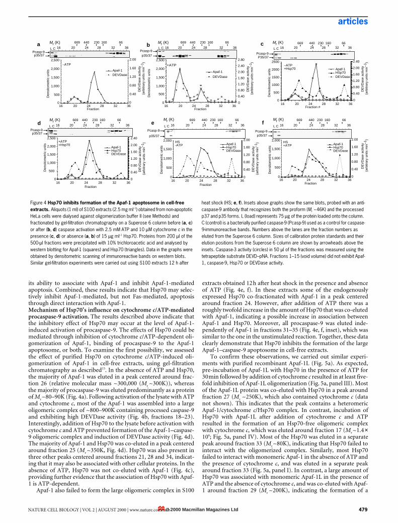
its ability to associate with Apaf-1 and inhibit Apaf-1-mediatedapoptosis. Combined, these results indicate that Hsp70 may selec-tively inhibit Apaf-1-mediated, but not Fas-mediated, apoptosisthrough direct interaction with Apaf-1.Mechanism of Hsp70’s influence on cytochrome c/ATP-mediatedprocaspase-9 activation. The results described above indicate thatthe inhibitory effect of Hsp70 may occur at the level of Apaf-1-induced activation of procaspase-9. The effects of Hsp70 could bemediated through inhibition of cytochrome c/ATP-dependent oli-gomerization of Apaf-1, binding of procaspase-9 to the Apaf-1apoptosome, or both. To examine the first possibility, we assessedthe effect of purified Hsp70 on cytochrome c/ATP-induced oli-gomerization of Apaf-1 in cell-free extracts, using gel-filtrationchromatography as described15. In the absence of ATP and Hsp70,the majority of Apaf-1 was eluted in a peak centered around frac-tion 26 (relative molecular mass ~300,000 (Mr ~300K)), whereasthe majority of procaspase-9 was eluted predominantly as a proteinof Mr ~80–90K (Fig. 4a). Following activation of the lysate with ATPand cytochrome c, most of the Apaf-1 was assembled into a largeoligomeric complex of ~800–900K containing processed caspase-9and exhibiting high DEVDase activity (Fig. 4b, fractions 18–23).Interestingly, addition of Hsp70 to the lysate before activation withcytochrome c and ATP prevented formation of the Apaf-1–caspase-9 oligomeric complex and induction of DEVDase activity (Fig. 4d).The majority of Apaf-1 and Hsp70 was co-eluted in a peak centeredaround fraction 25 (Mr ~350K, Fig. 4d). Hsp70 was also present inthree other peaks centered around fractions 21, 28 and 34, indicat-ing that it may also be associated with other cellular proteins. In theabsence of ATP, Hsp70 was not co-eluted with Apaf-1 (Fig. 4c),providing further evidence that the association of Hsp70 with Apaf-1 is ATP-dependent.
Apaf-1 also failed to form the large oligomeric complex in S100
extracts obtained 12 h after heat shock in the presence and absenceof ATP (Fig. 4e, f). In these extracts some of the endogenouslyexpressed Hsp70 co-fractionated with Apaf-1 in a peak centeredaround fraction 24. However, after addition of ATP there was aroughly twofold increase in the amount of Hsp70 that was co-elutedwith Apaf-1, indicating a possible increase in association betweenApaf-1 and Hsp70. Moreover, all procaspase-9 was eluted inde-pendently of Apaf-1 in fractions 31–35 (Fig. 4e, f, inset), which wassimilar to the one in the unstimulated reaction. Together, these dataclearly demonstrate that Hsp70 inhibits the formation of the largeApaf-1–caspase-9 apoptosome in cell-free extracts.
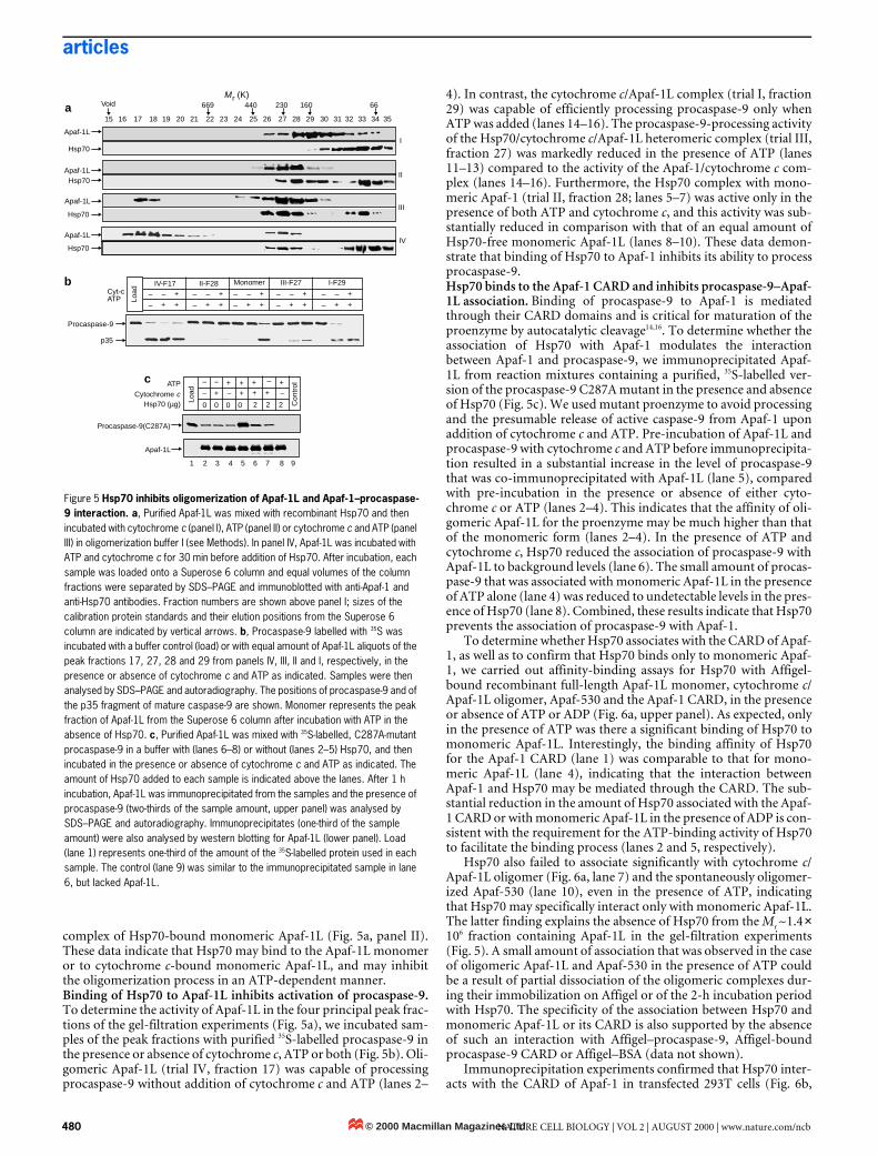
To confirm these observations, we carried out similar experi-ments with purified recombinant Apaf-1L (Fig. 5a). As expected,pre-incubation of Apaf-1L with Hsp70 in the presence of ATP for30 min followed by addition of cytochrome c resulted in at least five-fold inhibition of Apaf-1L oligomerization (Fig. 5a, panel III). Mostof the Apaf-1L protein was co-eluted with Hsp70 in a peak aroundfraction 27 (Mr ~250K), which also contained cytochrome c (datanot shown). This indicates that the peak contains a heteromericApaf-1/cytochrome c/Hsp70 complex. In contrast, incubation ofHsp70 with Apaf-1L after addition of cytochrome c and ATPresulted in the formation of an Hsp70-free oligomeric complexwith cytochrome c, which was eluted around fraction 17 (Mr ~1.4 ×10⁶; Fig. 5a, panel IV). Most of the Hsp70 was eluted in a separatepeak around fraction 33 (Mr ~80K), indicating that Hsp70 failed tointeract with the oligomerized complex. Similarly, most Hsp70failed to interact with monomeric Apaf-1 in the absence of ATP andthe presence of cytochrome c, and was eluted in a separate peakaround fraction 33 (Fig. 5a, panel I). In contrast, a large amount ofHsp70 was associated with monomeric Apaf-1L in the presence ofATP and the absence of cytochrome c, and was co-eluted with Apaf-1 around fraction 29 (Mr ~200K), indicating the formation of a
Figure 4 Hsp70 inhibits formation of the Apaf-1 apoptosome in cell-free extracts. Aliquots (1 ml) of S100 extracts (2.5 mg ml–1) obtained from non-apoptotic HeLa cells were dialysed against oligomerization buffer II (see Methods) and fractionated by gel-filtration chromatography on a Superose 6 column before (a, c) or after (b, d) caspase activation with 2.5 mM ATP and 10 µM cytochrome c in the presence (c, d) or absence (a, b) of 15 µg ml–1 Hsp70. Proteins from 200 µl of the 500-µl fractions were precipitated with 10% trichloroacetic acid and analysed by western blotting for Apaf-1 (squares) and Hsp70 (triangles). Data in the graphs were obtained by densitometric scanning of immunoreactive bands on western blots. Similar gel-filtration experiments were carried out using S100 extracts 12 h after
heat shock (HS; e, f). Insets above graphs show the same blots, probed with an anti-caspase-9 antibody that recognizes both the proform (Mr ~46K) and the processed p37 and p35 forms. L (load) represents 75 µg of the protein loaded onto the column. C (control) is a bacterially purified caspase-9 (Pcasp-9) used as a control for caspase-9-immunoreactive bands. Numbers above the lanes are the fraction numbers as eluted from the Superose 6 column. Sizes of calibration protein standards and their elution positions from the Superose 6 column are shown by arrowheads above the insets. Caspase-3 activity (circles) in 50 µl of the fractions was measured using the tetrapeptide substrate DEVD–pNA. Fractions 1–15 (void volume) did not exhibit Apaf-1, caspase-9, Hsp70 or DEVDase activity.
669 440 230 160 66
Fraction
0
500
1,000
1,500
2,000
2,500
16 20 24 28 32 36
0.40
0.80
1.20
1.60
2.00p35/37
Pcasp-9l I l l l l l l l l l l l l l l l l l l l
Mr (K)
Mr (K)
Mr (K) Mr (K)
Mr (K) Mr (K)
Den
sito
met
ric u
nits Apaf-1
DEVDase
L Ca
–ATP
l I l l l l l l l l l l l l l l l l l l l
0.40
0.80
1.20
1.60
2.00
2.40
2.80
Apaf-1
DEVDase
Apaf-1HSp70DEVDase
b
+ATP
l I l l l l l l l l l l l l l l l l l l l
Apaf-1Hsp70DEVDase
... ...
Den
sito
met
ric u
nits
Fraction
0
500
1,000
1,500
2,000
2,500
16 20 24 28 32 360
0.40
0.80
1.20
1.60
2.00
2.40
d
+ATP+Hsp70
l I l l l l l l l l l l l l l l l l l l l
Den
sito
met
ric u
nits
0
500
1,000
1,500
2,000
0.40
0.80
1.20
1.60
2.00
e
Apaf-1Hsp70DEVDase
... ...
HS-ATP
0
500
1,000
1,500
2,000
16 20 24 28 32 360
0.40
0.80
1.20
1.60
2.00
l I l l l l l l l l l l l l l l l l l l l
Fraction
Den
sito
met
ric u
nits
f
Apaf-1Hsp70DEVDase
... ...
HS+ATP
0
500
1000
1500
2000
2500
0.40
0.80
1.20
1.60
2.00
2.40
Den
sito
met
ric u
nits
... ...
l I l l l l l l l l l l l l l l l l l l l
c
–ATP+Hsp70
16 20 24 28 32 36
669 440 230 160 66
L C 16 20 24 28 32 36
500
1,000
1,500
2,000
2,500
Den
sito
met
ric u
nits
Fraction16 20 24 28 32 36
p35/37Pcasp-9
P35/37Pcasp-9
L C 16 20 24 28 32 36
Fraction #16 20 24 28 32 36
669 440 230 160 66
L C 16 20 24 28 32 36
p35/37Pcasp-9
p35/37
Pcasp-9
669 440 230 160 66
L C 16 20 24 28 32 36
Fraction16 20 24 28 32 36
Pcasp-9p35/37
669 440 230 160 66
L C 16 20 24 28 32 36
00
D
EV
Das
e ac
tivity
(arb
itrar
y un
its m
in–1
)
D
EV
Das
e ac
tivity
(arb
itrar
y un
its m
in–1
)
D
EV
Das
e ac
tivity
(arb
itrar
y un
its m
in–1
)
D
EV
Das
e ac
tivity
(arb
itrar
y un
its m
in–1
)
D
EV
Das
e ac
tivity
(arb
itrar
y un
its m
in–1
)
D
EV
Das
e ac
tivity
(arb
itrar
y un
its m
in–1
)
00
0
669 440 230 160 66
© 2000 Macmillan Magazines LtdNATURE CELL BIOLOGY | VOL 2 | AUGUST 2000 | www.nature.com/ncb 479
articles
complex of Hsp70-bound monomeric Apaf-1L (Fig. 5a, panel II).These data indicate that Hsp70 may bind to the Apaf-1L monomeror to cytochrome c-bound monomeric Apaf-1L, and may inhibitthe oligomerization process in an ATP-dependent manner.Binding of Hsp70 to Apaf-1L inhibits activation of procaspase-9.To determine the activity of Apaf-1L in the four principal peak frac-tions of the gel-filtration experiments (Fig. 5a), we incubated sam-ples of the peak fractions with purified 35S-labelled procaspase-9 inthe presence or absence of cytochrome c, ATP or both (Fig. 5b). Oli-gomeric Apaf-1L (trial IV, fraction 17) was capable of processingprocaspase-9 without addition of cytochrome c and ATP (lanes 2–
4). In contrast, the cytochrome c/Apaf-1L complex (trial I, fraction29) was capable of efficiently processing procaspase-9 only whenATP was added (lanes 14–16). The procaspase-9-processing activityof the Hsp70/cytochrome c/Apaf-1L heteromeric complex (trial III,fraction 27) was markedly reduced in the presence of ATP (lanes11–13) compared to the activity of the Apaf-1/cytochrome c com-plex (lanes 14–16). Furthermore, the Hsp70 complex with mono-meric Apaf-1 (trial II, fraction 28; lanes 5–7) was active only in thepresence of both ATP and cytochrome c, and this activity was sub-stantially reduced in comparison with that of an equal amount ofHsp70-free monomeric Apaf-1L (lanes 8–10). These data demon-strate that binding of Hsp70 to Apaf-1 inhibits its ability to processprocaspase-9.Hsp70 binds to the Apaf-1 CARD and inhibits procaspase-9–Apaf-1L association. Binding of procaspase-9 to Apaf-1 is mediatedthrough their CARD domains and is critical for maturation of theproenzyme by autocatalytic cleavage14,16. To determine whether theassociation of Hsp70 with Apaf-1 modulates the interactionbetween Apaf-1 and procaspase-9, we immunoprecipitated Apaf-1L from reaction mixtures containing a purified, 35S-labelled ver-sion of the procaspase-9 C287A mutant in the presence and absenceof Hsp70 (Fig. 5c). We used mutant proenzyme to avoid processingand the presumable release of active caspase-9 from Apaf-1 uponaddition of cytochrome c and ATP. Pre-incubation of Apaf-1L andprocaspase-9 with cytochrome c and ATP before immunoprecipita-tion resulted in a substantial increase in the level of procaspase-9that was co-immunoprecipitated with Apaf-1L (lane 5), comparedwith pre-incubation in the presence or absence of either cyto-chrome c or ATP (lanes 2–4). This indicates that the affinity of oli-gomeric Apaf-1L for the proenzyme may be much higher than thatof the monomeric form (lanes 2–4). In the presence of ATP andcytochrome c, Hsp70 reduced the association of procaspase-9 withApaf-1L to background levels (lane 6). The small amount of procas-pase-9 that was associated with monomeric Apaf-1L in the presenceof ATP alone (lane 4) was reduced to undetectable levels in the pres-ence of Hsp70 (lane 8). Combined, these results indicate that Hsp70prevents the association of procaspase-9 with Apaf-1.
To determine whether Hsp70 associates with the CARD of Apaf-1, as well as to confirm that Hsp70 binds only to monomeric Apaf-1, we carried out affinity-binding assays for Hsp70 with Affigel-bound recombinant full-length Apaf-1L monomer, cytochrome c/Apaf-1L oligomer, Apaf-530 and the Apaf-1 CARD, in the presenceor absence of ATP or ADP (Fig. 6a, upper panel). As expected, onlyin the presence of ATP was there a significant binding of Hsp70 tomonomeric Apaf-1L. Interestingly, the binding affinity of Hsp70for the Apaf-1 CARD (lane 1) was comparable to that for mono-meric Apaf-1L (lane 4), indicating that the interaction betweenApaf-1 and Hsp70 may be mediated through the CARD. The sub-stantial reduction in the amount of Hsp70 associated with the Apaf-1 CARD or with monomeric Apaf-1L in the presence of ADP is con-sistent with the requirement for the ATP-binding activity of Hsp70to facilitate the binding process (lanes 2 and 5, respectively).
Hsp70 also failed to associate significantly with cytochrome c/Apaf-1L oligomer (Fig. 6a, lane 7) and the spontaneously oligomer-ized Apaf-530 (lane 10), even in the presence of ATP, indicatingthat Hsp70 may specifically interact only with monomeric Apaf-1L.The latter finding explains the absence of Hsp70 from the Mr ~1.4 ×10⁶ fraction containing Apaf-1L in the gel-filtration experiments(Fig. 5). A small amount of association that was observed in the caseof oligomeric Apaf-1L and Apaf-530 in the presence of ATP couldbe a result of partial dissociation of the oligomeric complexes dur-ing their immobilization on Affigel or of the 2-h incubation periodwith Hsp70. The specificity of the association between Hsp70 andmonomeric Apaf-1L or its CARD is also supported by the absenceof such an interaction with Affigel–procaspase-9, Affigel-boundprocaspase-9 CARD or Affigel–BSA (data not shown).
Immunoprecipitation experiments confirmed that Hsp70 inter-acts with the CARD of Apaf-1 in transfected 293T cells (Fig. 6b,
Figure 5 Hsp70 inhibits oligomerization of Apaf-1L and Apaf-1–procaspase-9 interaction. a, Purified Apaf-1L was mixed with recombinant Hsp70 and then incubated with cytochrome c (panel I), ATP (panel II) or cytochrome c and ATP (panel III) in oligomerization buffer I (see Methods). In panel IV, Apaf-1L was incubated with ATP and cytochrome c for 30 min before addition of Hsp70. After incubation, each sample was loaded onto a Superose 6 column and equal volumes of the column fractions were separated by SDS–PAGE and immunoblotted with anti-Apaf-1 and anti-Hsp70 antibodies. Fraction numbers are shown above panel I; sizes of the calibration protein standards and their elution positions from the Superose 6 column are indicated by vertical arrows. b, Procaspase-9 labelled with 35S was incubated with a buffer control (load) or with equal amount of Apaf-1L aliquots of the peak fractions 17, 27, 28 and 29 from panels IV, III, II and I, respectively, in the presence or absence of cytochrome c and ATP as indicated. Samples were then analysed by SDS–PAGE and autoradiography. The positions of procaspase-9 and of the p35 fragment of mature caspase-9 are shown. Monomer represents the peak fraction of Apaf-1L from the Superose 6 column after incubation with ATP in the absence of Hsp70. c, Purified Apaf-1L was mixed with 35S-labelled, C287A-mutant procaspase-9 in a buffer with (lanes 6–8) or without (lanes 2–5) Hsp70, and then incubated in the presence or absence of cytochrome c and ATP as indicated. The amount of Hsp70 added to each sample is indicated above the lanes. After 1 h incubation, Apaf-1L was immunoprecipitated from the samples and the presence of procaspase-9 (two-thirds of the sample amount, upper panel) was analysed by SDS–PAGE and autoradiography. Immunoprecipitates (one-third of the sample amount) were also analysed by western blotting for Apaf-1L (lower panel). Load (lane 1) represents one-third of the amount of the 35S-labelled protein used in each sample. The control (lane 9) was similar to the immunoprecipitated sample in lane 6, but lacked Apaf-1L.
15
I
II
III
669 440 230 160 66Mr (K)
Voida
+ + + + +
Apaf-1L
Procaspase-9(C287A)
Cytochrome cHsp70 (µg)
c + + + ATP
Cyt-cATP
Procaspase-9
p35
b Monomer
IV
16 17 18 19 20 21 22 23 24 25 26 27 28 29 30 31 32 33 34 35
Apaf-1L
Hsp70
Apaf-1LHsp70
Apaf-1L
Hsp70
Apaf-1L
Hsp70
Load
IV-F17 II-F28 III-F27 I-F29
Load
Con
trol– – –
–– –
0 0 0 0 2 2 2
1 2 3 4 5 6 7 8 9
–
–
– +
+ +–
–
– +
+ +–
–
– +
+ +–
–
– +
+ +–
–
– +
+ +
© 2000 Macmillan Magazines Ltd480 NATURE CELL BIOLOGY | VOL 2 | AUGUST 2000 | www.nature.com/ncb
articles
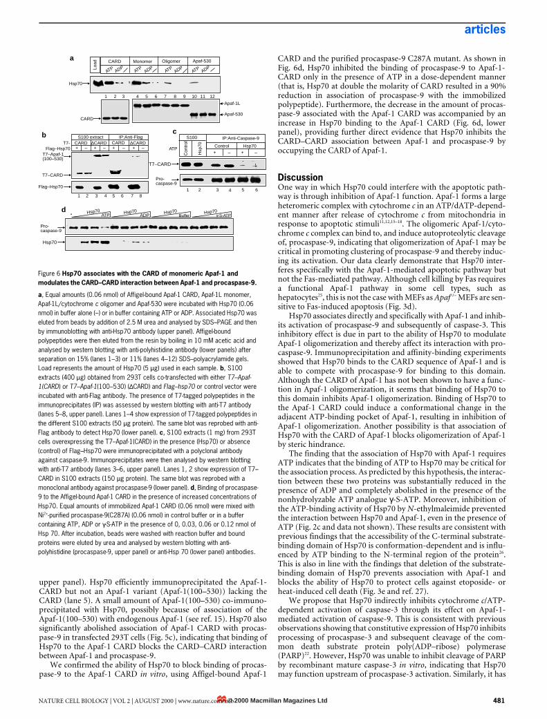
upper panel). Hsp70 efficiently immunoprecipitated the Apaf-1-CARD but not an Apaf-1 variant (Apaf-1(100–530)) lacking theCARD (lane 5). A small amount of Apaf-1(100–530) co-immuno-precipitated with Hsp70, possibly because of association of theApaf-1(100–530) with endogenous Apaf-1 (see ref. 15). Hsp70 alsosignificantly abolished association of Apaf-1 CARD with procas-pase-9 in transfected 293T cells (Fig. 5c), indicating that binding ofHsp70 to the Apaf-1 CARD blocks the CARD–CARD interactionbetween Apaf-1 and procaspase-9.
We confirmed the ability of Hsp70 to block binding of procas-pase-9 to the Apaf-1 CARD in vitro, using Affigel-bound Apaf-1
CARD and the purified procaspase-9 C287A mutant. As shown inFig. 6d, Hsp70 inhibited the binding of procaspase-9 to Apaf-1-CARD only in the presence of ATP in a dose-dependent manner(that is, Hsp70 at double the molarity of CARD resulted in a 90%reduction in association of procaspase-9 with the immobilizedpolypeptide). Furthermore, the decrease in the amount of procas-pase-9 associated with the Apaf-1 CARD was accompanied by anincrease in Hsp70 binding to the Apaf-1 CARD (Fig. 6d, lowerpanel), providing further direct evidence that Hsp70 inhibits theCARD–CARD association between Apaf-1 and procaspase-9 byoccupying the CARD of Apaf-1.
DiscussionOne way in which Hsp70 could interfere with the apoptotic path-way is through inhibition of Apaf-1 function. Apaf-1 forms a largeheteromeric complex with cytochrome c in an ATP/dATP-depend-ent manner after release of cytochrome c from mitochondria inresponse to apoptotic stimuli11,12,15–18. The oligomeric Apaf-1/cyto-chrome c complex can bind to, and induce autoproteolytic cleavageof, procaspase-9, indicating that oligomerization of Apaf-1 may becritical in promoting clustering of procaspase-9 and thereby induc-ing its activation. Our data clearly demonstrate that Hsp70 inter-feres specifically with the Apaf-1-mediated apoptotic pathway butnot the Fas-mediated pathway. Although cell killing by Fas requiresa functional Apaf-1 pathway in some cell types, such ashepatocytes25, this is not the case with MEFs as Apaf–/– MEFs are sen-sitive to Fas-induced apoptosis (Fig. 3d).
Hsp70 associates directly and specifically with Apaf-1 and inhib-its activation of procaspase-9 and subsequently of caspase-3. Thisinhibitory effect is due in part to the ability of Hsp70 to modulateApaf-1 oligomerization and thereby affect its interaction with pro-caspase-9. Immunoprecipitation and affinity-binding experimentsshowed that Hsp70 binds to the CARD sequence of Apaf-1 and isable to compete with procaspase-9 for binding to this domain.Although the CARD of Apaf-1 has not been shown to have a func-tion in Apaf-1 oligomerization, it seems that binding of Hsp70 tothis domain inhibits Apaf-1 oligomerization. Binding of Hsp70 tothe Apaf-1 CARD could induce a conformational change in theadjacent ATP-binding pocket of Apaf-1, resulting in inhibition ofApaf-1 oligomerization. Another possibility is that association ofHsp70 with the CARD of Apaf-1 blocks oligomerization of Apaf-1by steric hindrance.
The finding that the association of Hsp70 with Apaf-1 requiresATP indicates that the binding of ATP to Hsp70 may be critical forthe association process. As predicted by this hypothesis, the interac-tion between these two proteins was substantially reduced in thepresence of ADP and completely abolished in the presence of thenonhydrolyzable ATP analogue γ-S-ATP. Moreover, inhibition ofthe ATP-binding activity of Hsp70 by N-ethylmaleimide preventedthe interaction between Hsp70 and Apaf-1, even in the presence ofATP (Fig. 2c and data not shown). These results are consistent withprevious findings that the accessibility of the C-terminal substrate-binding domain of Hsp70 is conformation-dependent and is influ-enced by ATP binding to the N-terminal region of the protein26.This is also in line with the findings that deletion of the substrate-binding domain of Hsp70 prevents association with Apaf-1 andblocks the ability of Hsp70 to protect cells against etoposide- orheat-induced cell death (Fig. 3e and ref. 27).
We propose that Hsp70 indirectly inhibits cytochrome c/ATP-dependent activation of caspase-3 through its effect on Apaf-1-mediated activation of caspase-9. This is consistent with previousobservations showing that constitutive expression of Hsp70 inhibitsprocessing of procaspase-3 and subsequent cleavage of the com-mon death substrate protein poly(ADP–ribose) polymerase(PARP)22. However, Hsp70 was unable to inhibit cleavage of PARPby recombinant mature caspase-3 in vitro, indicating that Hsp70may function upstream of procaspase-3 activation. Similarly, it has
Figure 6 Hsp70 associates with the CARD of monomeric Apaf-1 and modulates the CARD–CARD interaction between Apaf-1 and procaspase-9.
a, Equal amounts (0.06 nmol) of Affigel-bound Apaf-1 CARD, Apaf-1L monomer, Apaf-1L/cytochrome c oligomer and Apaf-530 were incubated with Hsp70 (0.06 nmol) in buffer alone (–) or in buffer containing ATP or ADP. Associated Hsp70 was eluted from beads by addition of 2.5 M urea and analysed by SDS–PAGE and then by immunoblotting with anti-Hsp70 antibody (upper panel). Affigel-bound polypeptides were then eluted from the resin by boiling in 10 mM acetic acid and analysed by western blotting with anti-polyhistidine antibody (lower panels) after separation on 15% (lanes 1–3) or 11% (lanes 4–12) SDS–polyacrylamide gels. Load represents the amount of Hsp70 (5 µg) used in each sample. b, S100 extracts (400 µg) obtained from 293T cells co-transfected with either T7–Apaf-1(CARD) or T7–Apaf-1(100–530) (∆CARD) and Flag–hsp70 or control vector were incubated with anti-Flag antibody. The presence of T7-tagged polypeptides in the immunoprecipitates (IP) was assessed by western blotting with anti-T7 antibody (lanes 5–8, upper panel). Lanes 1–4 show expression of T7-tagged polypeptides in the different S100 extracts (50 µg protein). The same blot was reprobed with anti-Flag antibody to detect Hsp70 (lower panel). c, S100 extracts (1 mg) from 293T cells overexpressing the T7–Apaf-1(CARD) in the presence (Hsp70) or absence (control) of Flag–Hsp70 were immunoprecipitated with a polyclonal antibody against caspase-9. Immunoprecipitates were then analysed by western blotting with anti-T7 antibody (lanes 3–6, upper panel). Lanes 1, 2 show expression of T7–CARD in S100 extracts (150 µg protein). The same blot was reprobed with a monoclonal antibody against procaspase-9 (lower panel). d, Binding of procaspase-9 to the Affigel-bound Apaf-1 CARD in the presence of increased concentrations of Hsp70. Equal amounts of immobilized Apaf-1 CARD (0.06 nmol) were mixed with Ni2+-purified procaspase-9(C287A) (0.06 nmol) in control buffer or in a buffer containing ATP, ADP or γ-S-ATP in the presence of 0, 0.03, 0.06 or 0.12 nmol of Hsp 70. After incubation, beads were washed with reaction buffer and bound proteins were eluted by urea and analysed by western blotting with anti-polyhistidine (procaspase-9, upper panel) or anti-Hsp 70 (lower panel) antibodies.
S100 IP:Anti-Caspase-9
Control Hsp70ATP
+ – –+
T7–CARD
b IP:Anti-FlagS100 extractT7-
Flag–Hsp70
T7–CARD
Flag–Hsp70∆CARDCARD CARD ∆CARD
T7–Apaf-1(100–530)
a
c
ATP ADPATP
ATPATP
ADPADP
ADP
Hsp70
CARD
Apaf-1L
Apaf-530
d Hsp70 Hsp70 Hsp70 Hsp70ATP ADP Buffer γ-S-ATP
Pro-caspase-9
Hsp70
Load MonomerCARD Oligomer Apaf-530
1 2 3 4 5 6 7 8 9 10 11 12
+ – + – + – + –
1 2 3 4 5 6 7 8
Con
trol
Hsp
70
1 2 3 4 5 6
Pro-caspase-9
© 2000 Macmillan Magazines LtdNATURE CELL BIOLOGY | VOL 2 | AUGUST 2000 | www.nature.com/ncb 481
articles
been demonstrated that overexpression of Hsp70 blocks PARPcleavage in some cells after heat shock and treatment with tumournecrosis factor-α (TNF-α)28.
It has also been proposed that Hsp70 prevents apoptosis byother mechanisms. The anti-apoptotic effect of Hsp70 againstTNFα and staurosporine is associated with inhibition of late cas-pase-dependent apoptotic events28 (that is, after the release of cyto-chrome c, activation of caspase-3 and cleavage of death substrates),such as changes in the cytosolic phospholipase A2, which releasesarachidonic acid from membrane phospholipids29, and changes innuclear morphology. Other data indicate that Hsp70 could inhibitthe ATP depletion associated with cell death30. This raises the possi-bility that Hsp70 may protect mitochondrial membrane from dam-age and therefore prevent the efflux of cytochrome c to thecytoplasm31. However, further studies are needed to clarify both theimportance of energy deprivation in the death process and theactual mechanism of action by which Hsp70 may influence it.
The ability of Hsp70 to prevent stress-induced apoptosis couldlimit the efficacy of cancer therapy. Expression of Hsp70 has beenfound to be an indicator of poor therapeutic outcome in breastcancer32. A function of Hsp70 in tumorigenesis has been proposedon the basis of the observation that many transformed cells exhibitelevated levels of Hsp70 (refs 32–35). Increased expression ofHsp70 can protect some cells from monocyte-induced and TNFα-induced cell death, and could therefore allow precancerous cells toescape immune surveillance32,33. This effect is probably a conse-quence of the ability of Hsp70 to prevent apoptosis, as targeted neu-tralization of Hsp70 expression in some cancerous cell linespromotes cell death34–37. The ability of Hsp70 to interfere with themitochondrial pathway of apoptosis could provide a growth advan-tage to tumour cells and account for their resistance to cytotoxictreatments.
In summary, our results show that Hsp70 can prevent apoptosisby affecting signalling events upstream of procaspase-3 activationthrough inhibition of Apaf-1 function. The effect of Hsp70 onApaf-1 probably accounts for its ability to provide resistance to thedocumented stress-induced apoptosis. Characterization of mole-cules that modulate the interaction between Apaf-1 and Hsp70could be a first step towards effective antitumour therapy. h
MethodsPlasmid construction and transfection.Complementary DNAs encoding Apaf-1 and caspase-9 variants were generated by polymerase chain
reaction (PCR) and cloned in expression plasmids as described14. Inducible Hsp70 cDNA was cloned
with an N-terminal Flag tag in pcDNA3. Transfections were carried out using standard procedures as
described14.
Purification of recombinant proteins.Apaf-1L was expressed in Sf-9 cells with the baculovirus system. Recombinant monomeric Apaf-1 and
oligomeric Apaf-1 complex were purified to homogeneity as described15. Apaf-530, the CARD of Apaf-1
and the procaspase-9 C287A mutant were expressed in bacteria as C-terminally His6-tagged proteins and
purified to homogeneity on Ni2+/nitrilotriacetic acid (NTA) and a Superose12 fast protein liquid
chromatography (FPLC) column as described14. Purified recombinant Hsp70 (inducible form) was from
Stressgen Biotechnologies (Victoria BC, Canada).
Heat-shock treatment.293, Jurkat and HeLa cells were suspended at 1 × 10⁶ cells per ml in closed polyethylene tubes (15 ml),
heat shocked by incubation in a water bath for 1 h at 42 °C and allowed to recover for 12 h at 37 °C for
maximum production of HSPs, as described38.
Oligomerization of Apaf-1L protein.All oligomerization reactions of Apaf-1L were carried out by incubating 4 µg Apaf-1 (~34 pmol) with 6
µg of purified recombinant Hsp70 (~80 pmol) in the presence or absence of cytochrome c (5 µg, ~0.4
nmol) and ATP (2.5 mM) at room temperature for 1 h in a final volume of 100 µl of oligomerization
buffer I (25 mM HEPES pH 7.4, 50 mM NaCl, 10 mM KCl, 5 mM MgCl2, 100 µg ml–1 BSA, 5% glycerol,
and 0.1 mM dithiothreitol). After incubation, a further 100 µl of oligomerization buffer I was added to
each sample and the reaction mixture was directly loaded onto a Superose 6 FPLC column at a flow rate
of 0.2 ml min–1. Aliquots containing 50 µl of the 500-µl fractions were fractionated by SDS–PAGE and
analysed for the presence of Apaf-1L and Hsp70 by western blotting with their respective antibodies.
Fractionation of S100 extracts.S100 extracts were prepared from HeLa cells as described13 in a buffer containing 40 mM PIPES/KOH
pH 7.0, 10 mM KCl, 2 mM EDTA, 3 mM dithiothreitol and protease inhibitors (20 µg ml–1 leupeptin, 10
µg ml–1 pepstatin, 10 µg ml–1 aprotinin and 2 mM PMSF). Extracts were then dialysed against the
extraction buffer lacking KCl, but containing 5% glycerol and 0.1% (w/v) CHAPS (oligomerization
buffer II). The dialysis step was essential to deplete ATP from the extracts. The protein concentration was
adjusted to 2.5 mg ml–1 and stimulation of Apaf-1 oligomerization and activation of the caspases was
carried out by addition of 2 mM MgCl2, 10 µM cytochrome c and 2.5 mM ATP. Purified recombinant
Hsp70 was added at 6 µg per mg protein of S100 extracts. This amount of Hsp70 is physiologically
relevant to the endogenous expression of Hsp70 in HeLa cells at 12 h after heat shock, as measured by
comparing the densities of known amounts of purified Hsp70 with different dilutions of stress-induced
S100 extracts in western blots. In each reaction, 2.5 mg of S100 extract (1 ml) was loaded onto a Superose
6 column pre-equilibrated with oligomerization buffer II, and proteins were eluted from the column at
a flow rate of 0.07 ml min–1. From each of the 0.5-ml fractions, 0.2 ml were precipitated with 10%
trichloroacetic acid and analysed by SDS–PAGE; this was followed by western blotting for Apaf-1,
caspase-9 and Hsp70 using appropriate antibodies.
Caspase-3 activity was assessed in each of the column fractions by measuring the cleavage activity of
the colorimetric substrate DEVD–para-nitroaniline (DEVD–pNA), using the ApoAlert CPP32 Assay Kit
(Clontech). Cleavage activity in 100 µl of each fraction was observed for 5–15 min. One arbitrary unit of
caspase-3 activity is defined as the amount of caspase-3 required to produce 1 pmol pNA per min at 30
°C, at a saturating substrate concentration.
Western blotting and immunoprecipitation.Western blots and immunoprecipitations were carried out as described14,15 using available commercial
and non-commercial antibodies.
Affinity binding of Hsp70 to Apaf-1L and its CARD.Purified versions (0.2 nmol) of monomeric Apaf-1L, oligomeric Apaf-1L, Apaf-530 and the CARD of
Apaf-1 were individually coupled to 100 µl Affigel-10 (BioRad), according to the manufacturer’s
protocol. Each Affigel-coupled polypeptide (30 µl) was mixed with 4 µg of recombinant Hsp70 (~0.06
nmol) in oligomerization buffer I alone and in a buffer containing 2.5 mM ATP or ADP. Samples were
incubated for 2 h at room temperature with continuous agitation. Beads were then washed with 5 × 1.5
ml of the oligomerization buffer to remove unbound proteins. Associated proteins were eluted from the
Affigel-bound polypeptides with 2.5 M urea, and the presence of Hsp70 was detected by immunoblotting
with its specific antibody. To verify that equal amounts of proteins were used in each of the affinity
reactions, Affigel-bound polypeptides were eluted by boiling the beads in 10 mM acetic acid and then
western blotting with an antibody against the polyhistidine tag. The same procedure was used to test the
effect of increased concentrations of Hsp70 (0.03, 0.06 and 0.12 nmol) on the affinity binding of a
recombinant procaspase-9 (0.06 nmol) to the Affigel-bound CARD of Apaf-1.
Apoptosis assays.Apaf+/– or Apaf–/– cells (0.5 × 10⁵ cells per well) in 12-well plates were transfected with 0.3 µg pEGFP–N1
reporter plasmid (Clontech), 1.2 µg of empty vector plasmid or plasmids encoding Flag–Hsp70 or the
procaspase-9 C287A mutant, using the LipofectAMINE method. Cells were treated with etoposide (100
µM) or agonist Fas antibody (50 ng ml–1) plus cycloheximide (1 µg ml–1) for 10 h and then stained with
Annexin V–PE (Pharmingen). Normal and apoptotic GFP-expressing cells were counted using
fluorescence microscopy. The percentage of apoptotic cells in each experiment was expressed as the mean
percentage of GFP-expressing cells exhibiting staining for Annexin V.
RECEIVED 17 JANUARY 2000; REVISED 13 MARCH 2000; ACCEPTED 20 APRIL 2000; PUBLISHED 6 JULY 2000.
1. Kerr, J. F., Wyllie, A. H. & Currie, A. R. Apoptosis: a basic biological phenomenon with wide- ranging implications in tissue kinetics. Br. J. Cancer 4, 239–257 (1972).
2. Vaux, D. L., Haecker, G. & Strasser, A. An evolutionary perspective on apoptosis. Cell 76, 777–779
(1994).3. Steller, H. Mechanisms and genes of cellular suicide. Science 267, 1445–1449 (1995).
4. Alnemri, E. S. Mammalian cell death proteases: a family of highly conserved aspartate specific
cysteine proteases. J. Cell Biochem. 64, 33–42 (1997).
5. Cohen, G. M. Caspases: the executioners of apoptosis. Biochem. J. 326, 1–16 (1997).
6. Salvesen, G. S. & Dixit, V. M. Caspases: intracellular signaling by proteolysis. Cell 14, 443–446 (1997).
7. Cryns, V. & Yuan, J. Proteases to die for. Genes Dev. 12, 1551–1570 (1998).
8. Thornberry, N. A. & Lazebnik, Y. Caspases: enemies within. Science 281, 1312–1316 (1998).
9. Fernandes-Alnemri, T. et al. In vitro activation of CPP32 and Mch3 by Mch4, a novel human
apoptotic cysteine protease containing two FADD-like domains. Proc. Natl Acad. Sci. USA 93, 7464–
7469 (1996).
10. Stennicke, H. R. et al. Pro-caspase-3 is a major physiologic target of caspase-8. J. Biol. Chem. 273,
27084–27090 (1998).
11. Green, D. R. & Reed, J. C. Mitochondria and apoptosis. Science 281, 1309–1312 (1998).
12. Goldstein, J. C., Waterhouse, N. J., Juin, P., Evan, G. I. & Green, D. R. The coordinate release of
cytochrome c during apoptosis is rapid, complete and kinetically invariant. Nature Cell Biol. 2, 156–162 (2000).
13. Li, P. et al. Cytochrome c and dATP-dependent formation of Apaf-1/caspase-9 complex initiates an
apoptotic protease cascade. Cell 91, 479–489 (1997).
14. Srinivasula, S. M., Ahmad, M., Fernandes-Alnemri, T. & Alnemri, E. S. Autoactivation of procaspase-9 by Apaf-1-mediated oligomerization. Mol. Cell 1, 949–957 (1998).
15. Saleh, A., Srinivasula, S. M., Acharya, S., Fishel, R. & Alnemri, E. S. Cytochrome c and dATP-
mediated oligomerization of Apaf-1 is a prerequisite for procaspase-9 activation. J.Biol.Chem. 274, 17941–17945 (1999).
16. Qin, H. et al. Structural basis of procaspase-9 recruitment by the apoptotic protease-activating factor
1. Nature 399, 549–557 (1999).
17. Zou, H., Li, Y., Liu, X. & Wang, X. An APAF-1 cytochrome c multimeric complex is a functional
apoptosome that activates procaspase-9. J. Biol. Chem. 274, 11549–11556 (1999).
18. Cain, K., Brown, D. G., Langlais, C. & Cohen, G. M. Caspase activation involves the formation of the
aposome, a large (approximately 700 kDa) caspase-activating complex. J. Biol. Chem. 274, 22686–22692 (1999).
© 2000 Macmillan Magazines Ltd482 NATURE CELL BIOLOGY | VOL 2 | AUGUST 2000 | www.nature.com/ncb
articles
19. Li, G. C., Mivechi, N. F. & Weitzel, G. Heat shock proteins, thermotolerance, and their relevance to
clinical hyperthermia. Int. J. Hyperthermia 11, 459–488 (1995).
20. Jaattela, M., Wissing, D., Bauer, P. A. & Li, G. C. Major heat shock protein hsp70 protects tumor cells
from tumor necrosis factor cytotoxicity. EMBO J. 11, 3507–3512 (1992).
21. Jaattela, M. & Wissing, D. Heat-shock proteins protect cells from monocyte cytotoxicity: possible
mechanism of self-protection. J. Exp. Med. 177, 231–236 (1993).
22. Mosser, D. D., Caron, A. W., Bourget, L., Denis-Larose, C. & Massie, B. Role of the human heat
shock protein hsp70 in protection against stress-induced apoptosis. Mol. Cell Biol. 17, 5317–5327
(1997).
23. Jaattela, M., Wissing, D., Kokholm, K., Kallunki, T. & Egeblad, M. Hsp70 exerts its anti-apoptotic
function downstream of caspase-3-like proteases. EMBO J. 17, 6124–6134 (1998).
24. Hermawan, A. & Chirico, W. J. N-Ethylmaleimide-modified Hsp70 inhibits protein folding. Arch.
Biochem. Biophys. 369, 157–162 (1999).
25. Yin, X. M. et al. Bid-deficient mice are resistant to Fas-induced hepatocellular apoptosis. Nature 400,
886–891 (1999).
26. Schmid, D., Baici, A., Gehring, H. & Christen, P. Kinetics of molecular chaperone action. Science 263,
971–973 (1994).
27. Li, G. C., Li, L., Liu, R. Y., Rehman, M. & Lee, W. M. F. Heat shock protein hsp70 protects cells from
thermal stress even after deletion of its ATP-binding domain. Proc. Natl Acad. Sci. USA 89, 2036–2040
(1992).
28. Buzzard, K. A., Giaccia, A. J., Killender, M. & Anderson, R. L. Heat shock protein 72 modulates
pathways of stress-induced apoptosis. J. Biol. Chem. 273, 17147–17153 (1998).
29. Wissing, D., Mouritzen, H., Egeblad, M., Poirier, G. G. & Jaattela, M. Involvement of caspase-
dependent activation of cytosolic phospholipase A2 in tumor necrosis factor-induced apoptosis. Proc.
Natl Acad. Sci. USA 94, 5073–5077 (1997).
30. Wong, H. R., Menendez, I. Y., Ryan, M. A. & Denenberg, A. G. Increased expression of heat shock
protein-70 protects A549 cells against hyperoxia. Am. J. Physiol. 275, L836–L841 (1998).
31. Polla, B. S. et al. Mitochondria are selective targets for the protective effects of heat shock against
oxidative injury. Proc. Natl Acad. Sci. USA 93, 6458–6463 (1996).
32. Ciocca, D. R. et al. Heat shock protein hsp70 in patients with axillary lymph node-negative breast
cancer: prognostic implications. J. Natl Cancer Inst. 85, 570–574 (1993).
33. Jaattela, M. Escaping cell death: survival proteins in cancer. Exp. Cell. Res. 248, 30–43 (1999).
34. Dunning, A. M. et al. A systematic review of genetic polymorphisms and breast cancer risk. Cancer
Epidemiol. Biomarkers Prev. 8, 843–854 (1999).
35. Wei, Y. Q., Zhao, X., Kariya, Y., Teshigawara, K. & Uchida A. Inhibition of proliferation and
induction of apoptosis by abrogation of heat-shock protein (HSP) 70 expression in tumor cells.
Cancer Immunol. Immunother. 40, 73–78 (1995).
36. Robertson, J. D., Datta, K., Biswal, S. S. & Kehrer, J. P. Heat-shock protein 70 antisense oligomers
enhance proteasome inhibitor-induced apoptosis. Biochem. J. 344, 477–485 (1999).
37. Kaur, J., Kaur, J. & Ralhan, R. Induction of apoptosis by abrogation of HSP70 expression in human
oral cancer cells. Int. J. Cancer 85, 1–5 (2000).
38. Creagh, E. M. & Cotter, T. G. Selective protection by HSP70 against cytotoxic drug-, but not Fas-
induced T-cell apoptosis. Immunology 97, 36–44.
ACKNOWLEDGEMENTS
We thank the members of Robbins’ laboratory, especially M. Serrano, B. Baldwin, T. Kenniston and J.
Mai, for technical support. We also thank Y. Lazebnik and S. H. Kaufmann for Apaf-1 and caspase-9
antibodies, respectively, and R. Morimoto for hsp70 cDNA. This work was supported by NIH grants
AG14357 and AG13487 (to E.S.A.) and CA55227 (to P.D.R.).
Correspondence and requests for materials should be addressed to E.S.A.
© 2000 Macmillan Magazines LtdNATURE CELL BIOLOGY | VOL 2 | AUGUST 2000 | www.nature.com/ncb 483


![Integrating the Healthcare Enterprise€¦ · Document Source Document ConsumerOn Entry [ITI Document Registry Document Repository Provide&Register Document Set – b [ITI-41] →](https://img.pdfslide.net/doc/110x75/5f08a1eb7e708231d422f7c5/integrating-the-healthcare-enterprise-document-source-document-consumeron-entry.jpg)


