Embed Size (px)
Citation preview
letters to nature
NATURE | VOL 406 | 31 AUGUST 2000 | www.nature.com 1005
18. Kirk, K., Horner, H. A. & Kirk, J. Glucose uptake in Plasmodium falciparum-infected erythrocytes is an
equilibrative not an active process. Mol. Biochem. Parasitol. 25, 195±205 (1996).
19. Carter, N. S. et al. Isolation and functional characterization of the PfNT1 nucleoside transporter gene
from Plasmodium falciparum. J. Biol. Chem. 275, 10683±10691 (2000).
20. Hibbs, A. R., Stenzel, D. J. & Saul, A. Macromolecular transport in malaria±does the duct exist? Eur. J.
Cell Biol. 72, 182±188 (1997).
21. Crandall, I. & Sherman, I. W. Plasmodium falciparum (human malaria)-induced modi®cations in
human erythrocyte band 3 protein. Parasitology 102, 335±340 (1991).
22. Bull, P. C. et al. Parasite antigens on the infected red cell surface are targets for naturally acquired
immunity to malaria. Nature Med. 4, 358±360 (1998).
23. Elford, B. C., Cowan, G. M. & Ferguson, D. J. Parasite-regulated membrane transport processes and
metabolic control in malaria-infected erythrocytes. Biochem. J. 308, 361±374 (1995).
24. Hamill, O. P., Marty, A., Neher, E., Sakmann, B. & Sigworth, F. J. Improved patch-clamp techniques
for high-resolution current recording from cells and cell-free membrane patches. P¯ugers Arch. 391,
85±100 (1981).
25. Trager, W. & Jensen, J. B. Human malaria parasites in continuous culture. Science 193, 673±675
(1976).
26. Goldman, D. E. Potential, impedance, and recti®cation in membranes. J. Gen. Physiol. 27, 36±60
(1943).
27. Lauer, S. A., Rathod, P. K., Ghori, N. & Haldar, K. A membrane network for nutrient import in red
cells infected with the malaria parasite. Science 276, 1122±1125 (1997).
Acknowledgements
We thank L. Miller for steady help and advice, and S. Brazer, F. Cohen, A. Sher andT. Wellems for reviewing the manuscript.
Correspondence and requests for materials should be addressed to S.A.D.(e-mail [email protected]).
.................................................................Effects of oncogenic mutations inSmoothened and Patched can bereversed by cyclopamineJussi Taipale*, James K. Chen*, Michael K. Cooper²*, Baolin Wang*,Randall K. Mann*, Ljiljana Milenkovic³, Matthew P. Scott³& Philip A. Beachy*
* Department of Molecular Biology and Genetics, and ² Department of Neurology,Howard Hughes Medical Institute, The Johns Hopkins University School of
Medicine, Baltimore, Maryland 21205, USA³ Departments of Developmental Biology and Genetics, Howard HughesMedical Institute, Stanford University School of Medicine, Stanford, California
94305-5427, USA
..............................................................................................................................................
Basal cell carcinoma, medulloblastoma, rhabdomyosarcoma andother human tumours are associated with mutations that activatethe proto-oncogene Smoothened (SMO) or that inactivate thetumour suppressor Patched (PTCH). Smoothened and Patchedmediate the cellular response to the Hedgehog (Hh) secretedprotein signal, and oncogenic mutations affecting these proteinscause excess activity of the Hh response pathway1,2. Here we showthat the plant-derived teratogen cyclopamine, which inhibits theHh response3,4, is a potential `mechanism-based' therapeutic agentfor treatment of these tumours. We show that cyclopamine orsynthetic derivatives with improved potency block activation ofthe Hh response pathway and abnormal cell growth associatedwith both types of oncogenic mutation. Our results also indicatethat cyclopamine may act by in¯uencing the balance betweenactive and inactive forms of Smoothened.
Whereas embryonic loss of Sonic hedgehog (Shh) signalling canresult in cyclopia and other developmental defects5, inappropriateactivation of the Shh response pathway is associated with severaltypes of human tumour6±13. Current approaches to treatment ofsuch neoplastic disorders are limited by the cytotoxic effects oftherapeutic agents on proliferating tissues. Alternative `mechanism-based' approaches speci®cally targeting abnormally active signallingpathways in de®ned types of cancer14 might avoid such toxicity,
particularly if the pathways in question functioned primarily inembryonic development and were not required for survival inadults. Cyclopamine, a plant steroidal alkaloid, induces cyclopiain vertebrate embryos15 and has been shown to act by inhibiting thecellular response to the Shh signal3,4. To evaluate the therapeuticpotential of cyclopamine for the treatment of Hh-pathway-asso-ciated disorders we investigated the mechanism by which cyclopa-mine acts.
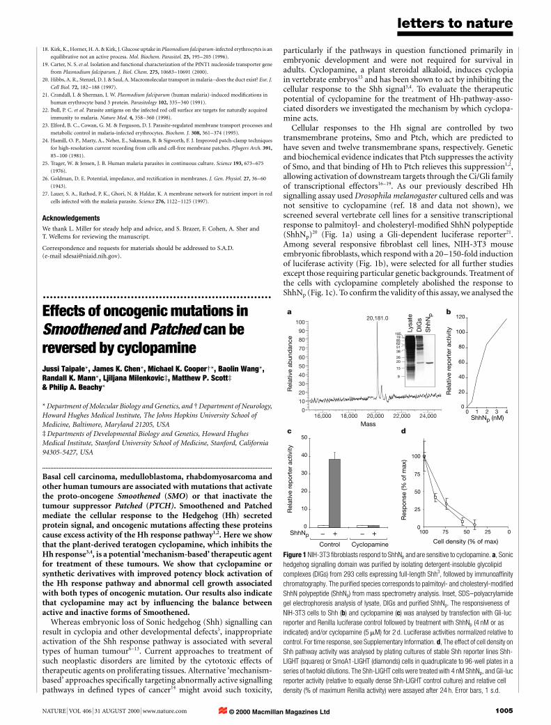
Cellular responses to the Hh signal are controlled by twotransmembrane proteins, Smo and Ptch, which are predicted tohave seven and twelve transmembrane spans, respectively. Geneticand biochemical evidence indicates that Ptch suppresses the activityof Smo, and that binding of Hh to Ptch relieves this suppression1,2,allowing activation of downstream targets through the Ci/Gli familyof transcriptional effectors16±19. As our previously described Hhsignalling assay used Drosophila melanogaster cultured cells and wasnot sensitive to cyclopamine (ref. 18 and data not shown), wescreened several vertebrate cell lines for a sensitive transcriptionalresponse to palmitoyl- and cholesteryl-modi®ed ShhN polypeptide(ShhNp)20 (Fig. 1a) using a Gli-dependent luciferase reporter21.Among several responsive ®broblast cell lines, NIH-3T3 mouseembryonic ®broblasts, which respond with a 20±150-fold inductionof luciferase activity (Fig. 1b), were selected for all further studiesexcept those requiring particular genetic backgrounds. Treatment ofthe cells with cyclopamine completely abolished the response toShhNp (Fig. 1c). To con®rm the validity of this assay, we analysed the
Lysa
te
DIG
sS
hhN
p
1851985625138
2620
15
9
0
20
40
60
80
100
120
0 1 2 3 4ShhNp (nM)
0
10
20
30
40
50
Control Cyclopamine
ShhNp + +––0
25
50
75
100
0255075100
Cell density (% of max)
Res
pon
se (%
of m
ax)
Rel
ativ
e re
por
ter
activ
ity
Rel
ativ
e re
por
ter
activ
ityR
elat
ive
abun
dan
ce
100
90
80
70
60
50
40
30
20
10
016,000 18,000 20,000 22,000 24,000
20,181.0a
c d
b
Mass
Figure 1 NIH-3T3 ®broblasts respond to ShhNp and are sensitive to cyclopamine. a, Sonic
hedgehog signalling domain was puri®ed by isolating detergent-insoluble glycolipid
complexes (DIGs) from 293 cells expressing full-length Shh3, followed by immunoaf®nity
chromatography. The puri®ed species corresponds to palmitoyl- and cholesteryl-modi®ed
ShhN polypeptide (ShhNp) from mass spectrometry analysis. Inset, SDS±polyacrylamide
gel electrophoresis analysis of lysate, DIGs and puri®ed ShhNp. The responsiveness of
NIH-3T3 cells to Shh (b) and cyclopamine (c) was analysed by transfection with Gli-luc
reporter and Renilla luciferase control followed by treatment with ShhNp (4 nM or as
indicated) and/or cyclopamine (5 mM) for 2 d. Luciferase activities normalized relative to
control. For time response, see Supplementary Information. d, The effect of cell density on
Shh pathway activity was analysed by plating cultures of stable Shh reporter lines Shh-
LIGHT (squares) or SmoA1-LIGHT (diamonds) cells in quadruplicate to 96-well plates in a
series of twofold dilutions. The Shh-LIGHT cells were treated with 4 nM ShhNp, and Gli-luc
reporter activity (relative to equally dense Shh-LIGHT control culture) and relative cell
density (% of maximum Renilla activity) were assayed after 24 h. Error bars, 1 s.d.
© 2000 Macmillan Magazines Ltd

letters to nature
1006 NATURE | VOL 406 | 31 AUGUST 2000 | www.nature.com
effects of overexpression of known pathway components, treatmentwith known pathway inhibitors, or both (Table 1). The main®ndings of Drosophila and mouse genetic analyses were con®rmed,indicating that NIH-3T3 cells provide a faithful and physiologicallymeaningful model for analysis of the Shh signalling pathway.Interestingly, for a full response to ShhNp cells had to be assayedafter reaching saturation density (Fig. 1d).
The steroidal nature of cyclopamine and its ability to disruptcholesterol synthesis or transport3,22,23 indicated that it might affectthe action of Ptch, which contains an apparent sterol-sensingdomain1,22. Having established the general characteristics of Shhresponse and cyclopamine inhibition in mouse embryonic ®bro-blasts, we assayed ®broblasts derived from Ptch-/- mouse embryosfor cyclopamine sensitivity. Mice lacking functional Ptch showwidespread transcriptional activation of targets of Shh signalling,including Ptch itself 24. As b-galactosidase is expressed under thecontrol of the Ptch promoter in these cells, its expression can be usedto assay the state of Shh pathway activity. Addition of cyclopamineto Ptch-/- cells signi®cantly suppressed b-galactosidase expression(Fig. 2a) and the activity of the Gli-luc reporter (see SupplementaryInformation), indicating that cyclopamine can inhibit Shh pathwayactivity in the absence of Ptch function. In contrast, cyclopaminefailed to prevent pathway activation induced by Gli2 overexpression(Table 1).
These results suggest that Hh-pathway-related tumours asso-ciated with loss of Ptch function might respond to treatment withcyclopamine, and that the target of cyclopamine action is likely to bea pathway component that functions between Ptch and the Gliproteins. It is unlikely that Ptch2 is the target of cyclopamine actionin Ptch-/- cells, as Ptch is the main regulator of Shh pathwayactivation in embryos24 and Ptch2 activity appears not to beexpressed in Ptch-/- cells (data not shown).
To investigate further the mechanism of cyclopamine action, wetransfected NIH-3T3 cells transiently with both luciferase reporterand Smo complementary DNA, and found that overexpression ofSmo in the absence of Shh induces reporter expression abouttenfold. This Shh-independent activation of the response pathwaycan be suppressed by 5 mM cyclopamine (Fig. 2b), consistent with atarget of cyclopamine action downstream of Ptch and with amechanism that does not involve direct interference with Shhbinding. Cyclopamine at this concentration had little effect on
reporter expression induced by the oncogenic Smo mutantsW539L13 (SmoA1; Fig. 2b) and S537N11 (SmoA2; data notshown); cells from Ptch mutant embryos gave similar results (seeSupplementary Information).
These results indicate that cyclopamine may act upon Smo, andthat activating mutations render Smo proteins resistant. An alter-native interpretation would be that activated Smo proteins producea high abundance of a downstream component and that a highcyclopamine level is required to suppress the increased concentra-tion of this component. This alternative model, however, wouldpredict that intermediate or low levels of pathway activation byoncogenic Smo proteins expressed at low levels should be subject tocyclopamine inhibition; on the contrary, we observe that cyclopa-mine resistance is sustained under these conditions (Fig. 2b). Inaddition, cyclopamine resistance is not observed in cells expressinghigh levels of wild-type Smo activated by maximal Shh stimulation(not shown), again suggesting that cyclopamine does not act upon acomponent downstream of Smo.
The oncogenic SmoA1 protein has been reported to resistsuppression by Ptch25, indicating that oncogenic Smo proteinsmay not be subject to normal regulation. We found, however, thatthis resistance is partial. The activating effects of SmoA1 or SmoA2can be completely inhibited by transfection of a 9-to-1 ratio of aPtch construct or of Ptch-CTD, which encodes a carboxy-terminallydeleted protein expressed at a higher level26 (Fig. 2c and data notshown). We also found that SmoA1, thus inhibited by Ptch-CTD,responds well to stimulation by ShhNp. Under these circumstances,
b
Forskolin(µM)
Control Cyclopamine(µM)
1 3 10 5 1000
10
20
30
40
50
a
β-ga
lact
osid
ase
activ
ity p
er µ
g p
rote
in
Rel
ativ
e re
por
ter
activ
ity
– + – + – + – + – + – +Cyclop-amine
Control SmoA1 Smo
20
40
60
c
ShhNp – + – + – +
Control Cyclop-amine
Forskolin
60
40
20
SmoA1 and Ptch-CTDtransfected NIH-3T3 cells
50 5 0.5 50
Control+ShhNp
Rel
ativ
e re
por
ter
activ
ity
NIH-3T3 cellsPtch–/– cells
Figure 2 Cyclopamine acts downstream of Ptch by inhibiting Smo activity. a, Cyclopamine
and forskolin can inhibit Hh pathway activity in Ptch-/-cells (measured as expression of b-
gal activity from the Ptch locus after 3 d of treatment). b, Sensitivity of wild-type and
oncogenic Smo to cyclopamine was determined by transfecting NIH-3T3 cells with Gli-luc
reporter and the w/w ratio of the expression vector indicated, followed by treatment with
5 mM cyclopamine for 2 d. Error bars, 1 s.d. The leftmost four bars are as in Fig. 1c.
c, Activation of pathway by oncogenic Smo is inhibited by high amounts of Ptch (compare
with b); ShhNp restores pathway activity even in the presence of cyclopamine. NIH-3T3
cells were transfected with reporter, Ptch-CTD and SmoA1 (Ptch:Smo DNA ratio 9), and
treated with ShhNp (2 nM), cyclopamine (5 mM) and/or forskolin (100 mM) as indicated for
2 d. Data from a representative experiment are shown.
Table 1 Luciferase activity from a Gli-dependent reporter as induced bycombinations of Shh pathway inducing and suppressing treatments
Inducer Suppressor
None Ptc Cyclopamine Forskolin PKAa Gli3-N
None - - - - - -ShhNp +++ + - - - -Smo + - - - - NDActivated Smo +++ +* +* - - -Gli2 ++ ++ ++ ++ + -.............................................................................................................................................................................
The indicated constructs were transfected alone or in combination (1:1 ratio) to NIH-3T3 cells,followed by treatment with ShhNp (4 nM), cyclopamine (5 mM) or forskolin (100 mM) for 2 days. Gli3-N, Gli3 truncated at residue 700, generating a repressor form. PKAa, constitutively active proteinkinase A catalytic subunit.* Higher dose can completely suppress (see Figs 2, 3).
© 2000 Macmillan Magazines Ltd

letters to nature
NATURE | VOL 406 | 31 AUGUST 2000 | www.nature.com 1007
induction of the Gli-responsive reporter is resistant to 5 mMcyclopamine (Fig. 2c), which would normally abolish Shh signal-ling. These results indicate that activated Smo molecules in thepresence of suf®cient Ptch can contribute to an essentially normal,albeit cyclopamine-resistant, response to the Shh signal.
The ®nding that oncogenic Smo is regulated by high levels of Ptchindicates that it might also be subject to regulation by high levels ofcyclopamine. To circumvent the cytotoxic effects of cyclopamineconcentrations greater than 10 mM, we tested several chemicallysynthesized cyclopamine derivatives. As seen in Fig. 3a, the cyclop-
amine derivative 3-keto, N-aminoethyl aminocaproyl dihydrocin-namoyl cyclopamine (KAAD-cyclopamine; see Methods), had 10±20-fold higher potency than cyclopamine in inhibition of b-galactosidase expression in p2Ptch-/-cells, with similar or lowertoxicity (Fig. 3a). This compound also had greater potency insuppression of ShhNp-induced pathway activity (Fig. 3b). Impor-tantly, it completely suppressed SmoA1-induced reporter activity ata concentration around tenfold higher than that required forsuppression of pathway activation induced by ShhNp (Fig. 3b).
As activation of the Hh response pathway is associated with
0 0.01
0.03 0.1 0.3
2.5
1.25
0.62
0.31
0.16
0
0
0.2
0.4
0.6
0.8
1
1.2
0.01 0.1 1 10
CyclopaminePtch–/–
KAAD cyclopaminePtch–/–
Nor
mal
ized
β-g
alac
tosi
das
e ac
tivity
0
2
4
6
8
0.001 0.01 0.1 1
Fold
incr
ease
in c
ell n
umb
er
0
0.2
0.4
0.6
0.8
1
1.2
0.01 0.1 1 10
ShhNp
SmoA1 (W539L)
Nor
mal
ized
rep
orte
r ac
tivity
100.1 1
Num
ber
of c
olon
ies
10
0
KAAD cyclopamine (µM) KAAD cyclopamine (µM)
KAAD cyclopamine (µM)Inhibitor (µM)
a
c
b
d
HO
Figure 3 Reversal of pathway activation and abnormal cell growth caused by oncogenic
mutations in Ptch and Smo. a, Con¯uent cultures of p2Ptch-/- cells were treated with
cyclopamine (®lled squares) or KAAD-cyclopamine (open squares) for 2 d.
b-galactosidase activity was determined relative to cell mass (treated duplicate plate; Cell
Titer 96AQ; Promega). Signi®cant toxicity was not observed. b, Shh-LIGHT2 (diamonds)
and SmoA1-LIGHT (circles) cells were treated with 4 nM ShhNp (Shh-LIGHT2) and KAAD
cyclopamine (both lines) for 2 d. c, p2Ptch-/- cells were cultured in 2% calf serum in the
presence of KAAD cyclopamine for 7 d (triplicate wells) and cell number was determined
with a Coulter counter. Inset, micrographs of treated cells. d, SmoA1-LIGHT cells were
plated in soft agar with the indicated concentrations of KAAD-cyclopamine. Left,
ethidium-bromide-stained colonies after 17 d. Right, number of colonies (.150 mm) per
®eld after 10 d. Error bars, 1 s.d. Relative reporter activities in a, b are normalized to
maximum.
1
6.8
45.0
1.00 0.66
0
10
20
30
40
50
Protein levels(Renilla activity)
Transfection Control Smo-R SmoA1-R
Smo-RSmo Renilla
A1 (W539L)*
Rel
ativ
e re
por
ter
activ
ity
Inactive Smo
23
4
5176
C
N
G460 S537 G533 W539Y (11.3) N (19.4) L (15.0) L (32.8)
T (15.1) H (7.3)T (6.8)Y (6.5)S (4.7)A (2.9)
Cytoplasmic
23
4
51
CRD
Extracellular
7
N
Active Smo
6
Patchedcyclopamine
Activating mutations Ptc Smo Gli
Shh Ptc Smo Gli
Ptc Smo Gli
Ptc SmoA Gli
Nor
mal
Tum
our
KAAD cyclopamine
a b c
Figure 4 Mechanism of cyclopamine reversal of oncogenic Hh pathway activation. a, NIH-
3T3 cells were transfected with Gli-luc reporter and a construct encoding the indicated
Smo±Renilla luciferase fusion protein. Shh pathway activity and Smo protein levels were
measured as ®re¯y and Renilla luciferase activities relative to b-galactosidase
transfection control, respectively. Expression levels of epitope-tagged Smo and SmoA1
also were similar (data not shown). b, A two-state model of Smo function and cyclopamine
action. The states may re¯ect conformational change (as shown), distinct subcellular
localizations or states of covalent modi®cation. Below, summary of oncogenic11,13 (bold)
and other activating mutations in Smo. The potency of each mutant protein in pathway
activation relative to wild-type Smo (value set at 1; ShhN cotransfection, 21.1) is in
parentheses. Relative potencies were determined at 10% w/w effector plasmid. c,
Cyclopamine inhibition of the Hh response pathway. Under physiological conditions the
inactive state of Smo (red) is maintained by active Ptch (green), leading to lack of Gli-
mediated transcription (red). Shh ligand activates the pathway by binding to and
converting Ptch to its inactive state (red), which in turn permits activity of Smo (green) and
results in Gli-mediated transcription (green). In tumours, Gli-mediated transcription is
ligand-independent owing to loss of Ptch function (Ptch) or constitutive activation of Smo
(SmoA). Cyclopamine inhibits the Hh response pathway and abnormal growth by inhibiting
Smo activity.
© 2000 Macmillan Magazines Ltd

letters to nature
1008 NATURE | VOL 406 | 31 AUGUST 2000 | www.nature.com
neoplastic transformation in various types of tumour, we investi-gated the growth properties of response-activated cells in low serumor soft agar27, conditions generally considered to reveal neoplastictransformation. As seen in Fig. 3c, p2Ptch-/- cell growth in low serumwas markedly inhibited by addition of KAAD-cyclopamine, with50% of maximal inhibition at ,50 nM. We also found that SmoA1-LIGHT cells, a cell line expressing SmoA1 clonally derived fromNIH-3T3 cells, can form colonies in soft agar medium. Addition ofKAAD-cyclopamine markedly inhibited colony growth, although,500 nM was required for 50% inhibition (Fig. 3d). This concen-tration was higher than that required for 50% maximal inhibition ofp2Ptch-/- cell growth, consistent with the higher amount of KAAD-cyclopamine required to block activation of the Hh response path-way by oncogenic Smo.
The activity of other signalling pathways headed by seven-transmembrane-domain receptors is determined by the balancebetween active and inactive forms of the receptor28,29. Consistentwith such a model for Smo, the level of Renilla-luciferase- orepitope-tagged Smo in transfected cells is not signi®cantly affectedby the presence of an oncogenic mutation, despite greatly elevatedreporter activity (Fig. 4a). These results indicate that oncogenic Smomay have a higher intrinsic ability to activate the pathway, and thatwild-type Smo may exist in a balance between active and inactiveforms28,29. Whereas cyclopamine and Ptch activities would shift thisbalance towards the inactive state, oncogenic mutations might exertthe opposite effect, and this could account for the higher levels ofPtch and cyclopamine activity required to suppress oncogenic Smo.To test this model, we identi®ed eight additional mutations in anextensive mutagenesis screen for activated Smo proteins (Fig. 4b).All the activating Smo mutants were cyclopamine resistant, and thelevel of resistance positively correlated with increased potency (seeSupplementary Information), consistent with a model in whichcyclopamine affects the balance between activity states of Smo (Fig.4b).
The transition of G-protein-coupled receptors with seven trans-membrane (TM) domains from the inactive to the active state isthought to involve a conformational shift in which the cytoplasmicends of the TM6 and TM7 helices tilt outwards, exposing a bindingpocket for a downstream signalling molecule, the Ga subunit29. Ourmutational analysis of Smo is consistent with such a conformationalshift, as eight of the Smo-activating mutations introduce bulkierside chains at G533 and S537. Assuming an a-helical conformationfor TM7, these substitutions all protrude from the same face of thehelix. Furthermore, as multiple distinct substitutions at each ofthese two residues result in activation (Fig. 4b), Smo would appearto be activated by alteration of helix-packing interactions ratherthan by creation or disruption of a single critical interaction. Aconformational shift of the TM7 helix with respect to other TMhelices is suggestive of the type of conformational transition postu-lated for activation of G-protein-coupled receptors and raises thepossibility that conformation-based transduction has been conservedin the evolution of seven-transmembrane-domain receptors.
Activation of the Hh response pathway has been linked to severaltypes of human tumour. For example, patients with basal cell nevussyndrome (also termed Gorlin syndrome), an autosomal dominantdisorder associated with heterozygous loss-of-function mutationsin PTCH, display increased incidence of many tumours, mostnotably basal cell carcinoma (BCC), medulloblastoma, rhabdomyo-sarcoma and ®brosarcoma6±8. In addition, loss-of-function muta-tions in PTCH or activating mutations in SMO are found in around40% of sporadic BCC and 25% of primitive neuroectodermaltumours6,9±13. Our results indicate that cyclopamine inhibits theShh pathway by antagonizing Smo and that the activation of the Hhresponse pathway by either type of oncogenic mutation is blockedby cyclopamine or its derivatives (Fig. 4c). Levels of cyclopamine orthe related compound jervine required to phenocopy the embryonicmalformations in Shh-/- embryos are tolerated by pregnant females
of various species, indicating that it might be possible to use thesecompounds or their derivatives without severe toxicity in non-pregnant adults to reverse activation of the Shh response pathwayfor therapeutic purposes. M
MethodsSynthesis of KAAD-cyclopamine and puri®cation of ShhNp
KAAD-cyclopamine was synthesized from cyclopamine by a route to be describedelsewhere (J.K.C. and P.A.B., manuscript in preparation). Cholesterol- and palmitate-modi®ed mouse Sonic hedgehog signalling domain ShhNp was puri®ed as described inSupplementary Information (see also Fig. 1a).
cDNA cloning, mutagenesis and constructs
A mouse full-length Smo cDNA was isolated using standard methods on the basis ofhomology to rat and human Smo sequences. Of two described activating mutations inhuman Smo13,25, one (SmoM2; corresponds to W539L in mSmo and referred to here asSmoA1) activated mouse Smo and the other (SmoM1; corresponding to R565Q) did not.For mutagenesis screens, Ala substitutions were introduced at more than 20 conserved orhydrophilic residues within Smo transmembrane segments; in addition, selected residueswere changed to 16 different amino acids (all except W, M, K and E) using a degenerateoligonucleotide and screened by transfection of a pool of clones. We secondarily screened53 (G533) or 37 (G460) individual clones to identify mutations at these positions thatactivate Smo. Expression constructs used: pRK5 for full-length mouse Ptch, C-terminallytruncated Ptch-CTD, Gli3(1±700) repressor19 and constitutively active PKA18 cDNAs, andpGE (transient transfections) or pcDNA3.1+hygro (stable lines; Invitrogen) for thevarious Smo cDNAs. pGE was derived from pEGFP-C1 (Clontech) by removal of theenhanced green ¯uorescent protein cDNA. Renilla luciferase was fused to the C terminusof Smo. These fusion protein constructs had similar activity to corresponding untaggedconstructs in the NIH-3T3 assay.
Cell lines
Primary ®broblasts lacking functional Ptch (Ptch-/-) and a clonal subline (p2Ptch-/-)were derived from Ptch-/- mouse embryos. The NIH-3T3 cell clones Shh-LIGHTand Shh-LIGHT2 stably incorporating the Gli-luc reporter and TK-Renilla vectors wereestablished by co-transfection with vector encoding G418 resistance (pSV-Neo). SmoA1-LIGHT cells are a clonal subline of Shh-LIGHT expressing oncogenic Smo (veri®ed byimmunoblotting).
Transfection and reporter assays
Con¯uent cultures of NIH-3T3 cells were plated at 1:6 dilution to 24- or 96-well plates. Onthe next day, we transfected the cells with Renilla luciferase (pRL-TK or pRL-SV40;Clontech) or b-galactosidase transfection control (10% w/w DNA), Gli-luc reporter(40%) and the constructs indicated (50%) using Fugene 6 (Roche) transfection reagent(250 ng (24-well plate) or 100 ng (96-well plate) DNA per well, 3:1 ratio (v/w) of reagent toDNA). After the cells had reached saturation density (1±2 days), they were changed to low-serum medium (0.5% bovine calf serum) and treated with the reagents indicated for 1±2days. All reporter assays were normalized for transfection ef®ciency using Renillaluciferase or b-galactosidase transfection control (luminometric detection, Promega dualluc; Tropix galacto-light).
Received 12 May; accepted 6 July 2000.
1. Goodrich, L. V. & Scott, M. P. Hedgehog and patched in neural development and disease. Neuron 21,
1243±1257 (1998).
2. Ingham, P. W. Transducing hedgehog: the story so far. EMBO J. 17, 3505±3511 (1998).
3. Cooper, M. K., Porter, J. A., Young, K. E. & Beachy, P. A. Plant-derived and synthetic teratogens inhibit
the ability of target tissues to respond to Sonic hedgehog signaling. Science 280, 1603±1607 (1998).
4. Incardona, J. P., Gaf®eld, W., Kapur, R. P. & Roelink, H. The teratogenic Veratrum alkaloid
cyclopamine inhibits sonic hedgehog signal transduction. Development 125, 3553±3562 (1998).
5. Chiang, C. et al. Cyclopia and defective axial patterning in mice lacking Sonic hedgehog gene function.
Nature 383, 407±413 (1996).
6. Hahn, H., Wojnowski, L., Miller, G. & Zimmer, A. The patched signaling pathway in tumorigenesis
and development: lessons from animal models. J. Mol. Med. 77, 459±468 (1999).
7. Hahn, H. et al. Mutations of the human homolog of Drosophila patched in the nevoid basal cell
carcinoma syndrome. Cell 85, 841±851 (1996).
8. Johnson, R. L. et al. Human homolog of patched, a candidate gene for the basal cell nevus syndrome.
Science 272, 1668±1671 (1996).
9. Gailani, M. et al. The role of the human homologue of Drosophila patched in sporadic basal cell
carcinomas. Nature Genet. 14, 78±81 (1996).
10. Pietsch, T. et al. Medulloblastomas of the desmoplastic variant carry mutations of the human
homologue of Drosophila patched. Cancer Res. 57, 2085±2088 (1997).
11. Reifenberger, J. et al. Missense mutations in SMOH in sporadic basal cell carcinomas of the skin and
primitive neuroectodermal tumors of the central nervous system. Cancer Res. 58, 1798±1803 (1998).
12. Raffel, C. et al. Sporadic medulloblastomas contain PTCH mutations. Cancer Res. 57, 842±845 (1997).
13. Xie, J. et al. Activating Smoothened mutations in sporadic basal-cell carcinoma. Nature 391, 90±92
(1998).
14. Gibbs, J. B. Mechanism-based target identi®cation and drug discovery in cancer research. Science 287,
1969±1973 (2000).
15. Keeler, R. F. & Binns, W. Teratogenic compounds of Veratrum californicum (Durand). V. Comparison
of cyclopian effects of steroidal alkaloids from the plant and structurally related compounds from
other sources. Teratology 1, 5±10 (1968).
© 2000 Macmillan Magazines Ltd

letters to nature
NATURE | VOL 406 | 31 AUGUST 2000 | www.nature.com 1009
16. Aza-Blanc, P., RemõÂrez-Weber, F.-A., Laget, M.-P., Schwartz, C. & Kornberg, T. B. Proteolysis that is
inhibited by Hedgehog targets cubitus interruptus protein to the nucleus and converts it to a repressor.
Cell 89, 1043±1053 (1997).
17. Methot, N. & Basler, K. Hedgehog controls limb development by regulating the activities of distinct
transcriptional activator and repressor forms of Cubitus interruptus. Cell 96, 819±831 (1999).
18. Chen, C. H. et al. Nuclear traf®cking of Cubitus interruptus in the transcriptional regulation of
Hedgehog target gene expression. Cell 98, 305±316 (1999).
19. Wang, B., Fallon, J. & Beachy, P. Hedgehog-regulated processing of Gli3 produces an anterior/
posterior repressor gradient in the developing vertebrate limb. Cell 100, 423±434 (2000).
20. Pepinsky, R. B. et al. Identi®cation of a palmitic acid-modi®ed form of human Sonic hedgehog. J. Biol.
Chem. 273, 14037±14045 (1998).
21. Sasaki, H., Hui, C.-C., Nakafuku, M. & Kondoh, H. A binding site for Gli proteins is essential for
HNF3b ¯oor plate enhancer activity in transgenics and can respond to Shh in vitro. Development 124,
1313±1322 (1997).
22. Beachy, P. A. et al. Multiple roles of cholesterol in hedgehog protein biogenesis and signaling. Cold
Spring Harb. Symp. Quant. Biol. 62, 191±204 (1997).
23. Lange, Y., Ye, J., Rigney, M. & Steck, T. L. Regulation of endoplasmic reticulum cholesterol by plasma
membrane cholesterol. J. Lipid. Res. 40, 2264±2270 (1999).
24. Goodrich, L. V., Milenkovic, L., Higgins, K. M. & Scott, M. P. Altered neural cell fates and
medulloblastoma in mouse patched mutants. Science 277, 1109±1113 (1997).
25. Murone, M., Rosenthal, A. & de Sauvage, F. J. Sonic hedgehog signaling by the patched-smoothened
receptor complex. Curr. Biol. 9, 76±84 (1999).
26. Fuse, N. et al. Sonic hedgehog protein signals not as a hydrolytic enzyme but as an apparent ligand for
patched. Proc. Natl Acad. Sci. USA 96, 10992±10999 (1999).
27. Shin, S. I., Freedman, V. H., Risser, R. & Pollack, R. Tumorigenicity of virus-transformed cells in nude
mice is correlated speci®cally with anchorage independent growth in vitro. Proc. Natl Acad. Sci. USA
72, 4435±4439 (1975).
28. Bond, R. A. et al. Physiological effects of inverse agonists in transgenic mice with myocardial
overexpression of the beta 2-adrenoceptor. Nature 374, 272±276 (1995).
29. Bourne, H. R. How receptors talk to trimeric G proteins. Curr. Opin. Cell Biol. 9, 134±142 (1997).
Supplementary information is available on Nature's World-Wide Web site(http://www.nature.com) or as paper copy from the London editorial of®ce of Nature.
Acknowledgements
We thank P. A. Cole, D. Leahy, J. Nathans, R. Reed and G. Seydoux for critical review ofthe manuscript, W. Gaf®eld for Veratrum extracts and K. Young for technical assistance.J.T. was supported by the Finnish Academy and the European Molecular BiologyOrganization. J.K.C. is a recipient of a postdoctoral fellowship from the Damon Runyon-Walter Winchell Cancer Research Fund and M.K.C. was supported by a postdoctoralfellowship from the Howard Hughes Medical Institute and a career development awardfrom the Burroughs Wellcome Fund. P.A.B. and M.P.S. are investigators of the HowardHughes Medical Institute.
Correspondence and requests for materials should be addressed to P.A.B.(e-mail: [email protected]).
.................................................................The APC tumour suppressorhas a nuclear export functionRina Rosin-Arbesfeld, Fiona Townsley & Mariann Bienz
Laboratory of Molecular Biology, Hills Road, Cambridge CB2 2QH, UK
..............................................................................................................................................
The adenomatous polpyposis coli (APC) protein is mutated inmost colorectal tumours1. Nearly all APC mutations are trunca-tions, and many of these terminate in the mutation cluster regionlocated halfway through the protein2±4. In cancer cells expressingmutant APC, b-catenin is stabilized5,6 and translocates into thenucleus to act as a transcriptional co-activator of T-cell factor7.During normal development, APC also promotes the destabiliza-tion of b-catenin and Drosophila Armadillo8±11. It does so bybinding to the Axin complex which earmarks b-catenin/Arma-dillo for degradation by the proteasome pathway12. APC has aregulatory role in this process13,14, which is poorly understood.Here we show that APC contains highly conserved nuclear exportsignals 39 adjacent to the mutation cluster region that enable it toexit from the nucleus. This ability is lost in APC mutant cancercells, and we provide evidence that b-catenin accumulates in thenucleus as a result. Thus, the ability of APC to exit from thenucleus appears to be critical for its tumour suppressor function.
Adenomatous polpyposis coli proteins are found in severalsubcellular compartments of mammalian and Drosophila cellsincluding the cytoplasm, nucleus and adhesive cadherin/cateninjunctions9,10,15. To identify the targeting domains for these compart-ments, we tagged various fragments of the ubiquitously expressedDrosophila APC, called E-APC/dAPC2, with green ¯uorescentprotein (GFP) and expressed these in transgenic ¯y embryos andin monkey COS cells. The subcellular distribution of GFP±E-APC isindistinguishable from that of endogenous E-APC in embryos9
(data not shown). In COS cells transfected with GFP±E-APC, wesee green ¯uorescence in the cytoplasm, concentrated at the plasmamembrane (to be described elsewhere), but also some in the nucleus(Fig. 1a). Unexpectedly, an amino-terminal fragment of E-APC(ARDcore; Fig. 2a) accumulates in the nucleus (Fig. 1b). Evidently,E-APC is capable of entering the nucleus by means of its Nterminus. We have not studied this further, but we note that thisN terminus spans the highly conserved Armadillo repeat domain(ARD). b-catenin contains a closely related ARD that mediates itsnuclear import independently of the Ran/Importin machinery16,17.
In contrast, carboxy-terminal fragments of E-APC (Cterm1 and2; Fig. 2a) are ef®ciently excluded from the nucleus (Fig. 1c), moreso than the full-length protein. Thus, the C terminus of E-APCeither contains a cytoplasmic anchoring domain or an ef®cientnuclear export signal (NES). To distinguish between these possibi-lities, we tested our GFP constructs by treating transfected cells withleptomycin B (LMB), a highly speci®c drug that inhibits nuclearexport by directly blocking the nuclear export receptor CRM1(refs 18±20). Indeed, this results in even distribution ofCterm1 and Cterm2 throughout cytoplasm and nucleus (Fig. 1d).
Figure 1 Nuclear export sequences within Drosophila and human APC. a±d, COS cells
transfected with GFP-tagged constructs as indicated, without (a±c) or with (d) LMB.
Cterm1 was excluded from nuclei at least as ef®ciently as Cterm2 (c); this exclusion, like
that of Cterm2 (d), was blocked by LMB. e, f, Face-on views of 4±5-hour-old Drosophila
embryos, stained with anti-E-APC (green) outlining the apical cellular junctions (arrows),
and anti-lamin (red) outlining the nuclear envelopes. Note the nuclear accumulation of E-
APC after LMB treatment (f). g±n, COS cells transfected with coreNES constructs.
g, k, ARDcore; h, l, coreNES from E-APC 20R4; i, m, coreNESala from E-APC 20R4;
j, coreNES from human APC 20R3; n, coreNESala from human APC 20R3. All coreNES
constructs described in the text behaved essentially the same. Cells in k±m were treated
with LMB; the staining patterns of the human constructs (j, n) after LMB treatment are
similar to those in k±m.
© 2000 Macmillan Magazines Ltd


![Integrating the Healthcare Enterprise€¦ · Document Source Document ConsumerOn Entry [ITI Document Registry Document Repository Provide&Register Document Set – b [ITI-41] →](https://img.pdfslide.net/doc/110x75/5f08a1eb7e708231d422f7c5/integrating-the-healthcare-enterprise-document-source-document-consumeron-entry.jpg)