Embed Size (px)
Citation preview
Nature © Macmillan Publishers Ltd 1997
letters to nature
NATURE | VOL 388 | 21 AUGUST 1997 787
13. Reid, P. A. & Watts, C. Cycling of cell-surface MHC glycoproteins through primaquine-sensitiveintracellular compartments. Nature 346, 655–657 (1990).
14. Neefjes, J. J., Stollorz, V., Peters, P. J., Geuze, H. J. & Ploegh, H. L. The biosynthetic pathway of MHCclass II but not class I molecules intersects the endocytic route. Cell 61, 171–183 (1990).
15. Roosnek, E., Demotz, S., Corradin, G. & Lanzavecchia, A. Kinetics of MHC–antigen complexformation on antigen-presenting cells. Immunol. 140, 4079–4082 (1988).
16. Lanzavecchia, A., Reid, P. A. & Watts, C. Irreversible association of peptides with class II MHCmolecules in living cells. Nature 357, 249–252 (1992).
17. Nelson, C. A., Petzold, S. J. & Unanue, E. R. Peptides determine the lifespan of MHC class II moleculesin the antigen-presenting cell. Nature 371, 250–252 (1994).
18. Amigorena, S., Drake, J. R., Webster, P. & Mellman, I. Transient accumulation of new class II MHCmolecules in a novel endocytic compartment in B lymphocytes. Nature 369, 113–120 (1994).
19. Germain, R. N. & Hendrix, L. R. MHC class II structure, occupancy and surface expressiondetermined by post-endoplasmic reticulum antigen binding. Nature 353, 134–139 (1991).
20. Pierre, P. et al. Developmental regulation of MHC class II transport in mouse dendritic cells. Naturethis issue. EDS to complete on page
Acknowledgements. We thank D. Scheidegger and M. Dessing for technical assistance, E. Long fordiscussion, and C. Watts, F. Sallusto, K. Karjalainen and M. Colonna for critically reading the manuscript.The Basel Institute for Immunology was founded and is supported by F. Hoffmann-La Roche, Basel,Switzerland.
Correspondence and requests for material should be addressed to M.C. (e-mail: [email protected]).
Developmental regulationofMHCclass II transportinmousedendritic cellsPhilippe Pierre*, Shannon J. Turley*, Evelina Gatti,Michael Hull, Joseph Meltzer†, Asra Mirza†,Kayo Inaba†, Ralph M. Steinman† & Ira Mellman
Department of Cell Biology, Yale University School of Medicine, 333 Cedar Street,PO Box 208002, New Haven, Connecticut 06520, USA† The Rockefeller University, 1230 York Avenue, New York, New York 10021, USA* These authors contributed equally to this work.. . . . . . . . . . . . . . . . . . . . . . . . . . . . . . . . . . . . . . . . . . . . . . . . . . . . . . . . . . . . . . . . . . . . . . . . . . . . . . . . . . . . . . . . . . . . . . . . . . . . . . . . . . . . . . . . . . . . . . . . .
Dendritic cells (DCs) have the unique capacity to initiate primaryand secondary immune responses1–3. They acquire antigens inperipheral tissues and migrate to lymphoid organs where theypresent processed peptides to T cells. DCs must therefore exist indistinct functional states, an idea that is supported by observa-tions that they downregulate endocytosis and upregulate surfacemolecules of the class II major histocompatibility complex (MHC)upon maturation4–7. Here we investigate the features of DCmaturation by reconstituting the terminal differentiation ofmouse DCs in vitro and in situ. We find that early DCs, corre-sponding to those found in peripheral tissues, exhibit a phenotypein which most class II molecules are intracellular and localized tolysosomes. Upon maturation, these cells give rise to a newintermediate phenotype in which intracellular class II moleculesare found in peripheral non-lysosomal vesicles, similar to thespecialized CIIV population seen in B cells. The intermediate cellsthen differentiate into late DCs which express almost all of theirclass II molecules on the plasma membrane. These variations inclass II compartmentalization are accompanied by dramaticalterations in the intracellular transport of the new class IImolecules and in antigen presentation. We found that althoughearly DCs could not present antigen immediately after uptake,efficient presentation of the previously internalized antigenoccurred after maturation, 24–48 hours later. By regulatingclass II transport and compartmentalization, DCs are able todelay antigen display, a property crucial to their role in immunesurveillance.
Mouse bone marrow is a major source of DCs when cultivatedwith granulocyte–macrophage colony-stimulating factor (GM-CSF)8. Immunofluorescence microscopy of these cultures revealedthree distinct developmental stages. Cells were identified as DCs bythe expected repertoire of antigens and expression of MHC class II,cell shape, and adherence. As reported previously8, DCs were found
by immunofluorescence or FACS to be negative or weakly positivefor the granulocyte marker GR1, negative for the macrophagemarker SER-4, but strongly positive for CD11c and MHC class II.Contaminating SER-4 or GR1-positive cells were negative for classII and judged not to be DCs.
After 4–5 days, DCs were found in proliferating clusters looselyattached to adherent stromal cells8. By confocal microscopy, most ofthe cells present in or migrating out from the clusters showed littleMHC class II on their surface, but contained abundant intracellularclass II (Fig. 1a). The class II-positive vesicles represented lysosomes(MIICs) and late endosomes, being positive for lgp-B/lamp-2 andH2-M (Fig. 1a). Thus, they were characteristic of MIIC as defined inhuman lymphoblasts and human DCs9–12. As the MIIC-containingcells were present in proliferating clusters, we defined them as ‘early’DCs.
With increasing time in culture, two additional cell populationswere detected. The first of these (‘intermediate’ DCs) was presenttransiently and comprised non-adherent cells that had little surfaceMHC class II (Fig. 1b). They were strikingly unlike the early cells,however, because most of their intracellular class II was in a vesiclepopulation that was devoid of lysosomal markers (Fig. 1b, arrows),and thus reminiscent of non-lysosomal, class II-positive CIIVisolated from A20 B cells13. At later times in particular, the vesiclesaccumulated directly beneath the plasma membrane (Fig. 1b, right),whereas the lysosomes became concentrated in the perinuclearregion.
The third major DC population accumulated with time until by8–10 days it represented almost all of the non-adherent class II-positive cells. These ‘late’ cells had a more classical DC phenotype,with long processes that stained for class II (green) (Fig. 1c). Littleclass II remained intracellularly, with most of the now largely classII-depleted H2-M/lamp-positive lysosomes visualized as poorlyresolved clusters of red-staining vesicles in the perinuclear region.Further characterization indicated that markers such as DEC-205and 2A1 were absent from early cells but expressed at moderate andhigh levels on intermediate and late cells, respectively8,14 (results notshown). Early cells, but not late cells, were capable of efficient fluidendocytosis (W. Garrett and I.M., unpublished results), as foundpreviously for human cells4.
To determine whether early cells pass through the intermediatephenotype before reaching maturity, we produced highly purifiedpopulations of early cells by gently dislodging and isolating pro-liferating clusters on serum columns, followed by depletion ofcontaminating cells by fluorescent-activated cell sorting (FACS)8.This approach yielded ,95% pure populations of early DCs. Asquantified in Fig. 1d , after 5–8 h in culture, the early cells had nearlydisappeared and .60% of the population of exhibited the inter-mediate phenotype (for example, see Fig. 1b); 20–30% exhibitedthe late or mature phenotype (as shown in Fig. 1c). After 24 h,,90% of the cells were found to be of the late phenotype. As thenumber of cells remained constant throughout, these resultsstrongly suggest that there is a sequential relationship in thematuration pathway. The rapid maturation kinetics observedusing purified cluster-derived cells probably reflect their greaterdevelopmental synchrony.
To ensure that the developmental sequence was not peculiar tobone marrow cultures, we investigated whether tissue DCs hadsimilar properties. We first examined epidermal Langerhans cells15.In epidermal explants, class II was present in these cells in apunctuate pattern which co-localized with lysosomal marker H2-M, reminiscent of early bone marrow DCs (Fig. 2, upper rightpanels). If explants were incubated in culture medium and theLangerhans cells allowed to mature in situ, within 4 h the degree ofclass II and H2-M co-localization decreased, with class II stainingremaining punctuate but becoming progressively less coincidentwith H2-M, which became concentrated at the cell body (Fig. 2,right panels). This pattern was consistent with the intermediate
Nature © Macmillan Publishers Ltd 1997
letters to nature
788 NATURE | VOL 388 | 21 AUGUST 1997
phenotype. By 8 h, class II staining appeared homogeneous over theentirety of the cell, suggesting that it was present on the plasmamembrane (fig. 2, lower right panels), consistent with cells of thelate phenotype.
These results were confirmed by monitoring the development ofLangerhans cells isolated from the explants16, which were found tofollow the same morphological transitions observed using the bonemarrow cultures (Fig. 2, left panels). Similar results were obtainedfor splenic DCs derived from red pulp (not shown). Thus, DCs inperipheral tissues adopt the early phenotype but can be induced byisolation, cytokine exposure or physical disruption to undergo thesame type of maturational pattern as DCs in bone marrow culture.
The shift of MHC class II from lysosomal to non-lysosomalstructures was confirmed by immuno-electron microscopy and bycell fractionation in Percoll density gradients; MHC class II abdimers in lysosomal and non-lysosomal fractions alike were foundin the SDS-stable conformation, suggesting that they were loadedwith peptide (not shown). Of greater interest was to determine
whether the sequential appearance of class II in lysosomal then non-lysosomal structures reflected a shift of intracellular class II fromlysosome-like MIIC to endosome-like class II vesicles (CIIV),similar those found in murine B cells13. CIIVs represent a distinctvesicle population and are resolved from conventional endocyticorganelles by free-flow electrophoresis17. We fractionated early andlate DC populations by free-flow electrophoresis and found, asexpected, that each population had a typical fractionation pattern:plasma membrane markers (LFA-1 detected by western blot) wererestricted to unshifted fractions containing the major peak of cellproteins, whereas endosome- and lysosome-containing fractions(detected by b-hexosaminidase) were deflected towards the anode(results not shown).
When the free-flow electrophoresis fractions were probed bywestern blotting (Fig. 3a), early DCs exhibited class II in unshiftedfractions (containing endoplasmic reticulum, Golgi and plasmamembranes), as well as in fractions that co-migrated with lysosomalmarkers. The pattern was similar for cultures containing intermedi-
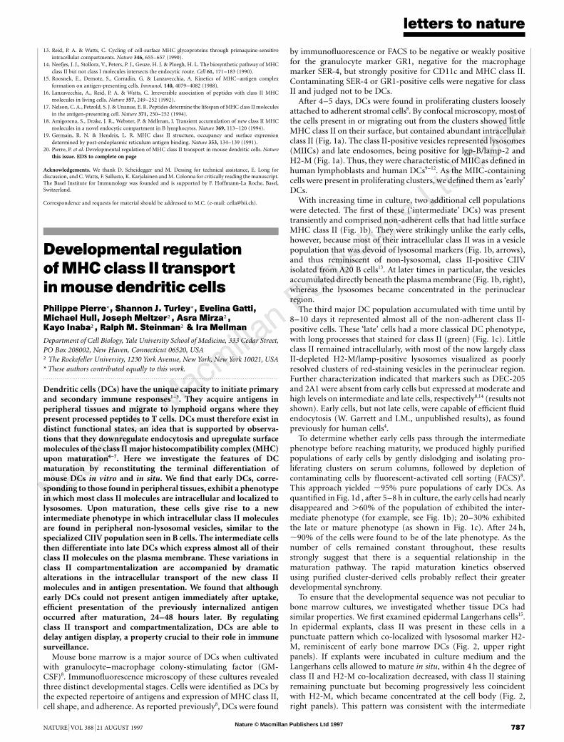
Figure 1 MHC class II distribution defines three distinct stages in the developmental maturation of bone-marrow-derived dendritic cells in vitro. DCs from bone marrow
cultures were collected, plated on glass coverslips, and processed for immunofluorescence and confocal microscopy. In each case, MHC class II molecules were
visualized using an FITC-conjugated second antibody and displayed as green staining. a, Early-DC phenotype. Optically merged confocal image of cells derived from
proliferating clusters and stained for H2-M (red, left) or lysosomal membrane protein lgp-B/lamp-2 (LGP, red; right) and class II (green). Cells were isolated on day 4 of
culture in the presence of GM-CSF. Class II co-localizes with both H2-M and lgp/lamp (yellow). Scale bars,10 mm. b, Intermediate-DC phenotype. Optically merged
confocal image of non-adherent cells stained for lgp-B/lamp-2 (red) and class II (green) after 6–7 days of culture in the presence of GM-CSF. Arrows indicate the
presence of class II-containing vesicles depleted of lysosomal markers. Cells in the images on the left in the panels are the first to appear in culture and still contain
some co-localized class II and lysosomal markers in the perinuclear region. Cells in the images on the right appear later and show a typical alignment of the non-
lysosomal CIIV-like structures just beneath the plasma membrane. The CIIV-like structures were all unlikely to be macropinosomes as they were negative for surface
markers MHC class I and LFA-1, both of which gave circumferential staining (not shown). Class II-negative cells staining for lgp/lamp in the field represent granulocyte
or monocyte contaminants of the culture. c, Late DC phenotype. Optically merged confocal image of non-adherentDC stained for lgp-B/lamp-2 (left, red) or H2-M (right
red) and class II (green) on day 8 of culture. All cells havea dendritic, sea-urchin-like appearance,with most class IImolecules now on the cell surface. Lysosomes often
appeared as a poorly resolved cluster concentrated in the perinuclear cytoplasm and devoid of MHC class II. d, Sequential appearance of early, intermediate and late
DC phenotypes in highly purified cultures. Proliferating clusters were isolated on 50%-serum columns and early DCs further purified by depletion of GR1-positive
granulocyte by FACS. Nearly homogeneous populations of early cells (95% pure) were returned to culture in the presence of GM-CSF and assayed by
immunofluorescence microscopy at after 2, 5, 8 and 24h. The total number of cells was constant at each time point, indicating that there was neither cell loss nor
proliferation. Using the phenotypic criteria defined for a–c, the cell distribution in each population was quantified. After 5 h, only 12% of the cells were of the early
phenotype, whereas .60% were of the intermediate phenotype; late cells had increased to represent .20% of the population. By 24 h, few early or intermediate cells
remained and cultures consisted mainly of late cells.
Nature © Macmillan Publishers Ltd 1997
letters to nature
NATURE | VOL 388 | 21 AUGUST 1997 789
ate and late cells but, in addition, a distinct peak of class II wasdetected in strongly anodally shifted membranes whose electro-phoretic migration was identical to CIIV. Although it needs to beconfirmed that these anodally shifted class II-containing mem-branes are functionally analogous to B-cell CIIV, it is interestingthat a similar population of vesicles can be detected in developingDCs with non-lysosomal class II-containing vesicles.
To determine whether DC maturation is accompanied by alteredtransport of MHC class II molecules, we did pulse-chase experi-ments using cluster-derived early DCs or cultures containing inter-mediate and late cells. We first showed by metabolic labelling thatthe relative rates of class II synthesis were similar in the two cellpopulations, with late cells synthesizing ,20% more class II perhour than early cells (results not shown).
Next, early and late DCs were pulse-labelled for 20 min and thearrival of class II molecules at the plasma membrane monitored bycell-surface biotinylation18,19. As shown in Fig. 3b, cell-surfacedelivery was strikingly inefficient in early DCs: although Ii chain-free ab dimers could be detected in total lysates within 60 min ofchase, only a small fraction of these were inserted into the cellsurface after 240 min (Fig. 3b, top panels). In contrast, late DCs weremuch better at delivering labelled class II molecules to the surface(Fig. 3b, middle panels): by 4 h, a large fraction of labelled abdimers had reached the cell surface. This efficient delivery in lateDCs was comparable to that in murine A20 cells.
The results from several pulse-chase experiments were quantified
by phosphorimaging to determine the percentage of total precipi-table class II recovered after biotinylation. The results revealed thatlate DCs were at least 10-fold more efficient at transporting abdimers to the surface than were early cluster-derived cells (Fig. 3c).After correcting for the inherent inefficiency of the biotinylationprocedure (see Methods)13, we estimate that late DCs inserted atleast 50% of their newly synthesized class II molecules into theplasma membrane, as compared to ,5% for early cells and almost100% for A20 B cells.
As early DCs localize most of their class II to MIIC, newlysynthesized ab dimers are probably targeted to lysosomes andtherefore have a different fate from those that reach the surface oflate cells. In both early and late cells, peptide loading (as judged bystability in SDS) was the same (Fig. 3d), but class II molecules inearly cells were degraded more rapidly than in late cells (in early cellsthe half-life was ,12 h, as compared to 36–40 h in late cell popula-tions; Fig. 3e). Nevertheless, some of the class II synthesized in earlycells probably did survive to reach the plasma membrane. In pulse-chase experiments done over 24 h, about 15% of the class II labelledduring an initial 20 min pulse became accessible to surface biotiny-lation after 18 h (results not shown).
Our results are consistent with the belief that DCs accumulateantigen in the periphery and deliver it to lymphoid organs, and theyindicate that peripheral early cells may sequester class II–peptidecomplexes intracellularly to prevent ‘premature’ presentation to Tcells in the periphery. To test this, we incubated cluster-derived early
LCs in freshly prepared epidermal sheets
Maturation ex vivo Maturation in situ
0 h
2 h
14 h
Intermediate
Early
Late
0 h
4 h
8 h
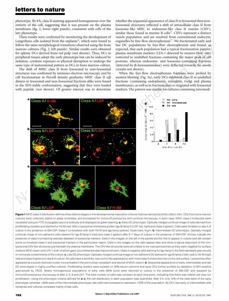
Figure 2 Developmental maturation of Langerhans cells (LCs) in culture and in situ. Epidermal sheets were cultured in situ or used for the isolation of individual LCs
following collagenase treatment15,16. Both explants and single cells were stained for MHC class II (using the mAb M5/114) and H2-M (using an affinity-purified rabbit
antibody against the H2-M cytoplasmic domain) and visualized by confocal microscopy. Top, a low-magnification view of MHC class II-positive LCs in an intact
epidermal sheet. Class II staininggives a punctate appearance in cells with typically dendritic LC morphology; cells appear to form a network throughout the epidermis.
Right, higher-magnification views of LCs in epidermal sheets cultured in situ for 0, 4 or 8 h, and then stained for class II (green) and H2-M to mark lysosomes (red). Co-
localization was strong directly after explant between class II and H2-M. Co-localization decreased with time, with H2-M staining become concentrated in the cell body
while class II staining extended throughout each cell, presumably at the plasma membrane. Left, maturation of single LCs cultured for 0, 2 or 14h ex vivo, that is, after
isolation from an epidermal sheet. Cells were stained for class II (green) and H2-M (red) and had the morphological and organizational features of early, intermediate
and late DCs from bone marrow cultures.
Nature © Macmillan Publishers Ltd 1997
letters to nature
790 NATURE | VOL 388 | 21 AUGUST 1997
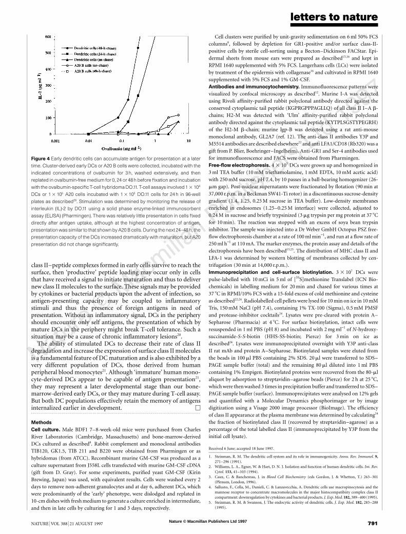
cells with various concentrations of ovalbumin for 3 h, washedthem, and then cultured them for 0, 24 or 48 h without antigen. Thecells were fixed to arrest development and assayed for immunogeniccomplexes. There was little presentation in the early cells fixeddirectly after the 3 h antigen uptake, even at very high antigenconcentrations (Fig. 4). However, after incubation for 24–48 h inthe absence of antigen, presentation activity increased dramatically,with virtually all of the cells achieving the mature phenotype by 48 h.Thus, immature DCs, similar to cells found in peripheral tissues,can accumulate antigen for later presentation.
A different picture emerged when the same experiment was donewith the A20 B-cell line. Although widely used as an efficientantigen-presenting cell (APC), exposure of A20 cells to ovalbuminfor 3 h resulted in relatively little presentation when fixed cells wereassayed either before or after the chase (Fig. 4). Although our
concentrations of antigen were high, most studies with A20 cells orDCs have used 1–3-day incubations with antigen (often receptor-bound) and incubation of unfixed APCs with T cells, rather than thepulse-chase–fixation protocol used here. In control experiments,presentation was maximal for both DCs and A20 cells at .100-foldlower antigen concentrations if cells were incubated with ovalbuminfor 48 h and unfixed cells were used for T-cell assays.
Early DCs in the periphery should thus accumulate antigen andnewly synthesized class II molecules in lysosomes or MIIC, whereprocessing and peptide loading can occur and the resulting immu-nogenic complexes can be retained. Although many of these com-plexes are degraded, some may survive until the DCs startmaturating and class II–peptide complexes can be transferred tothe plasma membrane, perhaps via the CIIV-like structures seen inthe intermediate DC population. Alternatively, if only a few of the
Early DC
Late DC
Cathode 48 60 70 78 Anode
Class IIb
Class IIb
PM/ER Endo/Lyso CIIV
24012060300C 24012060300CTime of chase (min)
ab
ab
Early DC
Late DC
24012060300C 24012060300CTime of chase (min)
ab
ab
Total Surface
Total Surface
a
b
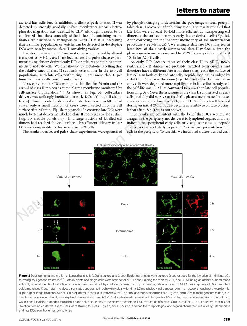
Figure3 Intracellular transportof newly synthesized MHC class II is tightly regulated during the maturationof dendritic cells.a, Free-flowelectrophoresis.Western blots
for MHC class II b-chain corresponding to the separation by free-flow electrophoresis of intracellular membranes in early (top) or intermediate and late DCs (bottom).
The separation profiles of early and late cells were virtually identical. Lysosomes (b-hexosaminidase; peak at around fraction 60) were anodally shifted relative to the
plasma membrane (LFA-1; peak at around fraction 50) by at least 10 fractions (not shown). In early DCs, class II molecules were detected in the fractions corresponding
to the plasma membrane (PM) (which also contained endoplasmic reticulum (ER); not shown) as well as in fractions containing endosomes and lysosomes. In
intermediate and late DC populations, class II was detected in the PM/ER fractions, as well as in the endosome and lysosome fractions. A distinct peak of class II was
anodally deflected relative to the major lysosome peak; this electrophoretic mobility corresponded to that of CIIV13. As already observed by immunofluorescence, the
amount of class II present inPM-containing fractionsof lateDCswas muchhigher than forearlyDCs.b,c, Detection of class II in total cell lysatesandon the cell surface.
DCs and A20 B cells were pulse-labelled for 20min and chased for the indicated times before cell-surface biotinylation. MHC class II molecules were
immunoprecipitated using Y3P mAb and the surface appearance was determined by streptavidin–agarose precipitation of the biotinylated class II molecules. In
total cell lysates, labelled class II became accessible to precipitation after 30min, as expected because Y3Ponly detects class II molecules after Ii chain dissociation25.
In early DCs, no class II was detected at the cell surface even after 240min of chase. In late DCs, significant amounts of labelled class II became biotinylated at the
surface after a 120-min lag, as in A20 B cells that transport nearly 100% of their class II to the surface13,24. Quantification of two experiments is shown in c. The ratio of
surface class II to total class IIwas corrected for biotinylationefficiency (seeMethods) andexpressed in arbitraryunits (sample variations,20%). After 4 h, lateDCshad
10-fold more class II at the surface than did early DCs, despite the fact that class II synthesis in late DCs was ,20% higher than in the early cells. The total amount of
class II did not change over this time, as detected by western blot. d, SDS-stable dimer formation was comparable in early and late DCs. Formation of SDS-stable ab
dimers in early versus late DCs was determined in total cell lysates using unboiled samples for SDS–PAGE. The 65K SDS-stable dimer was quantified by
phosphorimaging and plotted in arbitrary units. e, Turnover of MHC class II molecules in early and late DCs. DC cultures were labelled with 35S-Translabel for 1 h
and then chased for the indicated times. Total class II was immunoprecipitated using M5/114 mAb, boiled samples were analysed by SDS–PAGE, and a- or b-chain
quantified by phosphorimaging. Class II half-lives were determined by linear regression to be ,12 h for early cells and 35–40 h for late cells.
Nature © Macmillan Publishers Ltd 1997
letters to nature
NATURE | VOL 388 | 21 AUGUST 1997 791
class II–peptide complexes formed in early cells survive to reach thesurface, then ‘productive’ peptide loading may occur only in cellsthat have received a signal to initiate maturation and thus to delivernew class II molecules to the surface. These signals may be providedby cytokines or bacterial products upon the advent of infection, soantigen-presenting capacity may be coupled to inflammatorystimuli and thus the presence of foreign antigens in need ofpresentation. Without an inflammatory signal, DCs in the peripheryshould encounter only self antigens, the presentation of which bymature DCs in the periphery might break T-cell tolerance. Such asituation may be a cause of chronic inflammatory lesions20.
The ability of stimulated DCs to decrease their rate of class IIdegradation and increase the expression of surface class II moleculesis a fundamental feature of DC maturation and is also exhibited by avery different population of DCs, those derived from humanperipheral blood monocytes21. Although ‘immature’ human mono-cyte-derived DCs appear to be capable of antigen presentation21,they may represent a later developmental stage than our bone-marrow-derived early DCs, or they may mature during T-cell assay.But both DC populations effectively retain the memory of antigensinternalized earlier in development. M. . . . . . . . . . . . . . . . . . . . . . . . . . . . . . . . . . . . . . . . . . . . . . . . . . . . . . . . . . . . . . . . . . . . . . . . . . . . . . . . . . . . . . . . . . . . . . . . . . . . . . . . . . . . . . . . . . . . . . . . .
Methods
Cell culture. Male BDF1 7–8-week-old mice were purchased from CharlesRiver Laboratories (Cambridge, Massachusetts) and bone-marrow-derivedDCs cultured as described8. Rabbit complement and monoclonal antibodiesTIB120, GK1.5, TIB 211 and B220 were obtained from Pharmingen or ashybridomas (from ATCC). Recombinant murine GM-CSF was produced as aculture supernatant from J558L cells transfected with murine GM-CSF cDNA(gift from D. Gray). For some experiments, purified yeast GM-CSF (KirinBrewing, Japan) was used, with equivalent results. Cells were washed every 2days to remove non-adherent granulocytes and at day 6, adherent DCs, whichwere predominantly of the ‘early’ phenotype, were dislodged and replated in10-cm dishes with fresh medium to generate a culture enriched in intermediate,and then in late cells by culturing for 1 and 3 days, respectively.
Cell clusters were purified by unit-gravity sedimentation on 6 ml 50% FCScolumns8, followed by depletion for GR1-positive and/or surface class-II-positive cells by sterile cell-sorting using a Becton–Dickinson FACStar. Epi-dermal sheets from mouse ears were prepared as described15,16 and kept inRPMI 1640 supplemented with 5% FCS. Langerhans cells (LCs) were isolatedby treatment of the epidermis with collagenase16 and cultivated in RPMI 1640supplemented with 5% FCS and 1% GM-CSF.Antibodies and immunocytochemistry. Immunofluorescence patterns werevisualized by confocal microscopy as described12. Murine I-A was detectedusing Rivoli affinity-purified rabbit polyclonal antibody directed against theconserved cytoplasmic tail peptide (KGPRGPPPAGLLQ) of all class II I–A b-chains; H2-M was detected with ‘Ulm’ affinity-purified rabbit polyclonalantibody directed against the cytoplasmic tail peptide (KYTPLSGSTYPEGRH)of the H2-M b-chain; murine lgp-B was detected using a rat anti-mousemonoclonal antibody, GL2A7 (ref. 12). The anti-class II antibodies Y3P andM5514 antibodies are described elsewhere22 and anti LFA1/CD18 (Rb320) was agift from P. Blier, Boehringer–Ingelheim). Anti-GR1 and Ser-4 antibodies usedfor immunofluorescence and FACS were obtained from Pharmingen.Free-flow electrophoresis. 4 3 107 DCs were grown up and homogenized in3 ml TEA buffer (10 mM triethamolamine, 1 mM EDTA, 10 mM acetic acid)with 250 mM sucrose, pH 7.4, by 10 passes in a ball-bearing homogenizer (26-mm gap). Post-nuclear supernatants were fractionated by flotation (90 min at37,000 r.p.m. in a Beckman SW41-Ti rotor) in a discontinuous sucrose-densitygradient (1.4, 1.25, 0.25 M sucrose in TEA buffer). Low-density membranesenriched in endosomes (1.25–0.25 M interface) were collected, adjusted to0.24 M in sucrose and briefly trypsinized (3 mg trypsin per mg protein at 37 8Cfor 10 min). The reaction was stopped with an excess of soya bean trypsininhibitor. The sample was injected into a Dr Weber GmbH Octopus PSZ free-flow electrophoresis chamber at a rate of 100 ml min−1, and run at a flow rate of250 ml h−1 at 110 mA. The marker enzymes, the protein assay and details of theelectrophoresis have been described13,23. The distribution of MHC class II andLFA-1 was determined by western blotting of membranes collected by cen-trifugation (30 min at 14,000 r.p.m.).Immunoprecipitation and cell-surface biotinylation. 3 3 107 DCs werepulse-labelled with 10 mCi in 5 ml of [35S]methionine Translabel (ICN Bio-chemicals) in labelling medium for 20 min and chased for various times at37 8C in RPMI/10% FCS with a 15-fold excess of cold methionine and cysteineas described22,24. Radiolabelled cell pellets were lysed for 10 min on ice in 10 mMTris, 150 mM NaCl (pH 7.4), containing 1% TX-100 (Sigma), 0.5 mM PMSFand protease-inhibitor cocktails24. Lysates were pre-cleared with protein A–Sepharose (Pharmacia) at 4 8C. For surface biotinylation, intact cells wereresuspended in 1 ml PBS (pH 8) and incubated with 2 mg ml−1 of N-hydroxy-succinamide-S-S-biotin (HHS-SS-biotin; Pierce) for 3 min on ice asdescribed24. Lysates were immunoprecipitated overnight with Y3P anti-classII rat mAb and protein A–Sepharose. Biotinylated samples were eluted fromthe beads in 100 ml PBS containing 2% SDS. 20 ml were transferred to SDS–PAGE sample buffer (total) and the remaining 80 ml diluted into 1 ml PBScontaining 1% Empigen. Biotinylated proteins were recovered from the 80-mlaliquot by adsorption to streptavidin–agarose beads (Pierce) for 2 h at 25 8C,which were then washed 3 times in precipitation buffer and transferred to SDS–PAGE sample buffer (surface). Immunoprecipitates were analysed on 12% gelsand quantified with a Molecular Dynamics phosphorimager or by imagedigitization using a Visage 2000 image processor (BioImage). The efficiencyof class II appearance at the plasma membrane was determined by calculating24
the fraction of biotinylated class II (recovered by streptavidin–agarose) as apercentage of the total labelled class II (immunoprecipitated by Y3P from theinitial cell lysate).
Received 6 June; accepted 18 June 1997.
1. Steinman, R. M. The dendritic cell system and its role in immunogenicity. Annu. Rev. Immunol. 9,271–296 (1991).
2. Williams, L. A., Egner, W. & Hart, D. N. J. Isolation and function of human dendritic cells. Int. Rev.Cytol. 153, 41–103 (1994).
3. Caux, C. & Banchereau, J. in Blood Cell Biochemistry (eds Gordon, J. & Whetton, T.) 263–301(Plenum, London, 1996).
4. Sallusto, F., Cella, M., Danieli, C. & Lanzavecchia, A. Dendritic cells use macropinocytosis and themannose receptor to concentrate macromolecules in the major histocompatibiity complex class IIcompartment: downregulation by cytokines and bacterial products. J. Exp. Med. 182, 389–400 (1995).
5. Steinman, R. M. & Swanson, J. The endocytic activity of dendritic cells. J. Exp. Med. 182, 283–288(1995).
Figure 4 Early dendritic cells can accumulate antigen for presentation at a later
time. Cluster-derived early DCs or A20 B cells were collected, incubated with the
indicated concentrations of ovalbumin for 3 h, washed extensively, and then
replated in ovalbumin-free medium for 0, 24 or 48 h before fixation and incubation
with the ovalbumin-specific T-cell hybridomaDO.11. T-cell assays involved 1 3 104
DCs or 1 3 105 A20 cells incubated with 1 3 105 DO.11 cells for 24 h in 96-well
plates as described26. Stimulation was determined by monitoring the release of
interleukin (IL)-2 by DO.11 using a solid phase enzyme-linked immunosorbent
assay (ELISA) (Pharmingen). There was relatively little presentation in cells fixed
directly after antigen uptake, although at the highest concentration of antigen,
presentationwas similar to that shown byA20 B cells. During the next 24–48h, the
presentation capacity of the DCs increased dramatically with maturation, but A20
presentation did not change significantly.
Nature © Macmillan Publishers Ltd 1997
letters to nature
792 NATURE | VOL 388 | 21 AUGUST 1997
6. Austyn, J. M. New insights into the mobilization and phagocytic activity of dendritic cells. J. Exp. Med.183, 1287–1292 (1996).
7. De Smedt, T. et al. Regulation of dendritic cell numbers and maturation by lipopolysaccharide in vivo.J. Exp. Med. 184, 1413–1424 (1996).
8. Inaba, K. et al. Generation of large numbers of dendritic cells from mouse bone marrow culturessupplemented with granulocyte/macrophage colony-stimulating factor. J. Exp. Med. 76, 1693–1702(1992).
9. Peters, P. J., Neefjes, J. J., Oorschot, V., Ploegh, H. L. & Geuze, H. J. Segregation of MHC class IImolecules from MHC class I molecules in the Golgi complex for transport to lysosomalcompartments. Nature 349, 669–676 (1991).
10. Kleijmeer, M. J. et al. MHC class II compartments and the kinetics of antigen presentation in activatedmouse spleen dendritic cells. J. Immunol. 154, 5715–5724 (1995).
11. Nijman, H. W. et al. Antigen capture and major histocompatibility class II compartments of freshlyisolated and cultured human blood dendritic cells. J. Exp. Med. 182, 163–174 (1995).
12. Pierre, P. et al. HLA-DM is expressed in conventional and unconventional MHC class II-containingcompartments. Immunity 4, 229–239 (1996).
13. Amigorena, S., Drake, J. R., Webster, P. & Mellman, I. Transient accumulation of new class II moleculesin a novel endocytic compartment in B lymphocytes. Nature 369, 113–120 (1994).
14. Jiang, W. et al. The receptor DEC-205 expressed by dendritic cells and thymic epithelial cells isinvolved in antigen processing. Nature 375, 151–155 (1995).
15. Schuler, G. & Steinman, R. M. Murine epidermal Langerhans cells mature into potent immunosti-mulatory dendritic cells in vitro. J. Exp. Med. 161, 526–546 (1985).
16. Larsen, C. P. et al. Migration and maturation of Langerhans cells in skin transplant and explants. J.Exp. Med. 172, 1483–1494 (1990).
17. Marsh, M. et al. Rapid analytical and preparative isolation of functional endosomes by free flowelectrophoresis. J. Cell Biol. 104, 875–886 (1987).
18. Hunziker, W., Harter, C., Matter, K. & Mellman, I. Basolateral sorting in MDCK cells requires adistinct cytoplasmic domain determinant. Cell 66, 907–920 (1991).
19. Harter, C. & Mellman, I. Transport of the lysosomal membrane glycoprotein lgp120 (lgp-A) tolysosomes does not require appearance on the plasma membrane. J. Cell Biol. 117, 311–325 (1992).
20. Thomas, R. & Lipsky, P. E. Could endogenous self-peptides presented by dendritic cells initiaterheumatoid arthritis? Immunol. Today 17, 559–564 (1997).
21. Cella, M., Engering, A., Pinet, V., Pieters, J. & Lanzavecchia, A. Inflammatory stimuli induceaccumulation of MHC class II complexes on dendritic cells. Nature 388, 782–787 (1997).
22. Germain, R. N. & Hendrix, L. H. MHC class II structure, occupancy and surface expressiondetermined by post-endoplasmic reticulum antigen binding. Nature 353, 134–139 (1991).
23. Schmid, S. L., Fuchs, R., Male, P. & Mellman, I. Two distinct subpopulations of endosomes involved inmembrane recycling and transport to lysosomes. Cell 52, 73–83 (1988).
24. Amigorena, S. et al. Invariant chain cleavage and peptide loading in post-endosomal MHC class IIvesicles. J. Exp. Med. 181, 1729–1741 (1995).
25. Larsen, C. P. et al. Regulation of immunostimulatory function and costimulatory molecule (B7-1 andB7-2) expresison on murine dendritic cells. J. Immunol. 152, 5208–5219 (1994).
26. Buus, S., Colon, S., Smith, C., Freed, J. H., Miles, C. & Grey, H. M. Interaction between a ‘‘processed’’ovalbumin peptide and Ia molecules. Proc. Natl Acad. Sci. USA 83, 3968–3971 (1986).
Acknowledgements. We thank our colleagues and R. Flavell for helpful discussions, P. Webster forelectron microscopy, and P. Male for confocal microscopy. P.P. was supported by a long-term EMBOfellowship, S.J.T. by an NIH predoctoral training grant, and R.M.S. and I.M. by NIH research grants.
Correspondence and requests for materials should be addressed to I.M. (e-mail: [email protected]).
DistinctactionsofcisandtransATPwithin thedouble ringof thechaperoninGroELHays S. Rye*†, Steven G. Burston†, Wayne A. Fenton†,Joseph M. Beechem‡, Zhaohui Xu*§, Paul B. Sigler*§& Arthur L. Horwich*†
* Howard Hughes Medical Institute, † Department of Genetics, School ofMedicine, and § Department of Molecular Biophysics and Biochemistry,Yale University, New Haven, Connecticut 06510, USA‡ Department of Molecular Physiology and Biophysics, Vanderbilt University,Nashville, Tennessee 37232, USA. . . . . . . . . . . . . . . . . . . . . . . . . . . . . . . . . . . . . . . . . . . . . . . . . . . . . . . . . . . . . . . . . . . . . . . . . . . . . . . . . . . . . . . . . . . . . . . . . . . . . . . . . . . . . . . . . . . . . . . . .
The chaperonin GroEL is a double-ring structure with a centralcavity in each ring that provides an environment for the efficientfolding of proteins1–3 when capped by the co-chaperone GroES inthe presence of adenine nucleotides4–8. Productive folding of thesubstrate rhodanese has been observed in cis ternary complexes,where GroES and polypeptide are bound to the same ring, formedwith either ATP, ADP or non-hydrolysable ATP analogues2,9,suggesting that the specific requirement for ATP is confined toan action in the trans ring that evicts GroES and polypeptide fromthe cis side9. We show here, however, that for the folding of malatedehydrogenase and Rubisco there is also an absolute requirementfor ATP in the cis ring, as ADP and AMP-PNP are unable to
promote folding. We investigated the specific roles of binding andhydrolysis of ATP in the cis and trans rings using mutant forms ofGroEL that bind ATP but are defective in its hydrolysis. Binding ofATP and GroES in cis initiated productive folding inside a highlystable GroEL–ATP–GroES complex. To discharge GroES andpolypeptide, ATP hydrolysis in the cis ring was required to forma GroEL–ADP–GroES complex with decreased stability, primingthe cis complex for release by ATP binding (without hydrolysis) inthe trans ring. These observations offer an explanation of whyGroEL functions as a double-ring complex.
The monomeric protein rhodanese has recently been shown toreach native form inside cis ternary GroEL–GroES complexes thatwere formed in ATP, AMP-PNP or ADP, albeit at different rates(ATP . AMP-PNP . ADP)2,9. The single-ring GroEL mutant SR1has been used to produce obligate and stable cis complexes forstudy1,2,9. Because it has no second ring, the SR1 mutant does notreceive the signal from trans-sided ATP that normally evicts GroESand substrate6. After GroES and any of the three nucleotides wereadded to rhodanese–SR1 binary complexes, productive folding wasshown to occur in the cis cavity. We observed that rhodanese thatwas refolded inside SR1–GroES formed in the presence of ATPcould be released efficiently to the medium by brief incubation at4 8C, a treatment previously shown to lead to rapid dissociation ofGroES from an ADP complex with GroEL6. Similarly, folding ofornithine transcarbamylase (OTC) from binary complexes with SR1occurred in the presence of GroES, with almost identical kineticswith either ATP or ADP, when GroES was released by incubation at4 8C, thereby allowing OTC trimerization (data not shown). Thesedata suggest that the previous observation of OTC folding in thepresence of ATP in a single turnover from a cis ternary complex1
resulted from ADP-driven folding during preparation of the com-plex, with subsequent release when ATP was added. In experimentsusing SR1 and the green fluorescent protein (GFP), which alsorefolds inside cis complexes formed with any of the three nucleo-tides, brief treatment at 4 8C also leads to efficient release of GFP(Fig. 1a).
When the same tests were performed with two stringent substrateproteins, Rubisco from Rhodospirillum rubrum10,11 and mitochon-drial malate dehydrogenase (MDH) from pig heart12–14, we observedthat only ATP could promote reactivation from wild-type or SR1 cisternary complexes (Fig. 1b, c). Release of GroES from SR1 bytreatment at 4 8C was essential for production of enzymatic activity,because both Rubisco and MDH are homodimers, requiringassembly of the refolded monomeric subunits to reach activeform. Direct incubation at 4 8C of metastable intermediates ofRubisco that were produced after dilution from denaturant (intothe same chloride-free buffer used in all of the Rubisco studies)6 didnot result in enzymatic activity (data not shown). Remarkably, thekinetics of reactivation by the SR1–GroES ternary complexes inATP were similar to, if not faster than, those achieved in similarreactions with wild-type GroEL, indicating that a stable folding-active state is produced at SR1. Addition of ‘trap’ molecules (such as337/349)15, which are able to bind but not release non-nativesubstrate proteins, at the time of cold release of GroES had noeffect on the kinetics of reactivation (kinetics not shown, but see Fig.1f, traces 1, 4). These data indicate that, as with rhodanese, foldingof both Rubisco and MDH proceeded to a committed state in theATP cis ternary complexes; that is, the substrates reached conforma-tions no longer recognizable by chaperonin.
We wished to follow directly the folding of substrate within thevarious cis complexes, thereby obviating any requirement for therelease of GroES and peptide to assay enzymatic activity. Wetherefore examined by stopped-flow changes in the fluorescencetotal intensity and anisotropy of tryptophan residues of Rubisco incomplexes formed following the addition of GroES and nucleotidesto Rubisco–chaperonin binary complexes (Fig. 1d, e). This takesadvantage of the absence of tryptophan from both GroEL and


![Integrating the Healthcare Enterprise€¦ · Document Source Document ConsumerOn Entry [ITI Document Registry Document Repository Provide&Register Document Set – b [ITI-41] →](https://img.pdfslide.net/doc/110x75/5f08a1eb7e708231d422f7c5/integrating-the-healthcare-enterprise-document-source-document-consumeron-entry.jpg)


