Embed Size (px)
Citation preview
© 1999 Macmillan Magazines Ltd
letters to nature
290 NATURE | VOL 401 | 16 SEPTEMBER 1999 | www.nature.com
novel protein interactions and pathways of complex assembly. J. Cell Biol. 125, 1327±1340
(1994).
Acknowledgements
We thank P. Tsao for generating the b2-ARt cell line and for advice and assistance with ¯owcytometric experiments; U. Klein for the b2-AR tail GST fusion protein; J. Benovic forGRK-2 and -5 cDNAs; and R. Kelly, P. Walter, F. Brodsky, P. Peluso and J. Wilhelm forvaluable discussion. T.T.C. is supported by an NIH Institutional Training Grant. Thesestudies were supported by the NIH.
Correspondence and requests for materials should be addressed to M.v.Z. (e-mail:[email protected]. edu).
.................................................................EPS8 and E3B1 transducesignals from Ras to RacGiorgio Scita*², Johan Nordstrom²³, Roberta Carbone*,Pierluigi Tenca*, Giuseppina Giardina*, Silvio Gutkind§,Mattias BjarnegaÊ rd³, Christer Betsholtz³ & Pier Paolo Di Fiore*k
* Department of Experimental Oncology, European Institute of Oncology,
Via Ripamonti, 435, 20141 Milan, Italy³ Department of Medical Biochemistry, University of Goteborg,
Medicinaregatan 13, Box 440, SE 405 30 Goteborg, Swedenk Istituto di Microbiologia, University of Bari, 70124, Bari, Italy
§ Molecular Signaling Unit, Laboratory of Cellular Development and Oncology,
National Institute of Health, Bethesda, Maryland 20892, USA² These authors contributed equally to this work
..............................................................................................................................................
The small guanine nucleotide (GTP)-binding protein Rac regu-lates mitogen-induced cytoskeletal changes and c-Jun amino-terminal kinase (JNK), and its activity is required for Ras-mediated cell transformation1. Epistatic analysis placed Rac as akey downstream target in Ras signalling2; however, the biochem-ical mechanism regulating the cross-talk among these small GTP-binding proteins remains to be elucidated. Eps8 (relative mole-cular mass 97,000) is a substrate of receptors with tyrosine kinaseactivity3 which binds, through its SH3 domain, to a proteindesignated E3b1/Abi-1 (refs 4, 5). Here we show that Eps8 andE3b1/Abi-1 participate in the transduction of signals from Ras toRac, by regulating Rac-speci®c guanine nucleotide exchangefactor (GEF) activities. We also show that Eps8, E3b1 and Sos-1form a tri-complex in vivo that exhibits Rac-speci®c GEF activityin vitro. We propose a model in which Eps8 mediates the transferof signals between Ras and Rac, by forming a complex with E3b1and Sos-1.
Rac proteins belong to the sub-family of Rho-GTPases thatbecome activated when bound GDP is exchanged for GTP.Within this family, Rho regulates the assembly of stress ®bres,Rac controls membrane ruf¯ing and lamellipodia and Cdc42mediates the formation of ®lopodia. Rho-GTPases are organizedin hierarchical cascades, in which activated Ras or Cdc42 canindependently activate Rac1,2. Phosphatidylinositol 3-phosphatekinase (PI(3)K) is the immediate downstream effector of Ras6,and might activate Rac through the products of its catalytic activity,phosphoinositide phosphates (PtdInsPs), which can bind andmodulate the activity of GEFs for Rac, like Vav and h-Sos-1 (refs7, 8). Initial results indicated that overexpression of Eps8 increasedJNK activity, leaving mitogen-activated protein kinase (MAPK)unperturbed, leading to the testable hypothesis that Eps8 might beinvolved in the regulation of Rac (G.S. and P.P.D.F., unpublishedobservations).
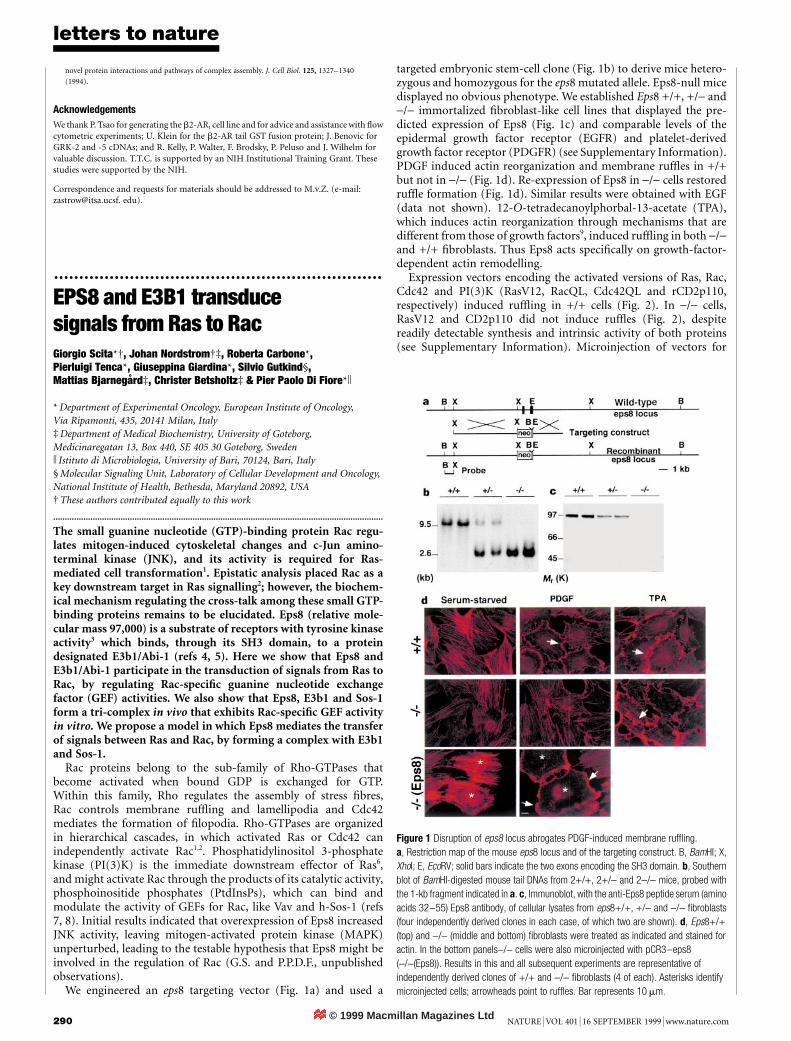
We engineered an eps8 targeting vector (Fig. 1a) and used a
targeted embryonic stem-cell clone (Fig. 1b) to derive mice hetero-zygous and homozygous for the eps8 mutated allele. Eps8-null micedisplayed no obvious phenotype. We established Eps8 +/+, +/- and-/- immortalized ®broblast-like cell lines that displayed the pre-dicted expression of Eps8 (Fig. 1c) and comparable levels of theepidermal growth factor receptor (EGFR) and platelet-derivedgrowth factor receptor (PDGFR) (see Supplementary Information).PDGF induced actin reorganization and membrane ruf¯es in +/+but not in -/- (Fig. 1d). Re-expression of Eps8 in -/- cells restoredruf¯e formation (Fig. 1d). Similar results were obtained with EGF(data not shown). 12-O-tetradecanoylphorbal-13-acetate (TPA),which induces actin reorganization through mechanisms that aredifferent from those of growth factors9, induced ruf¯ing in both -/-and +/+ ®broblasts. Thus Eps8 acts speci®cally on growth-factor-dependent actin remodelling.
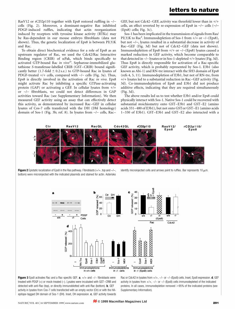
Expression vectors encoding the activated versions of Ras, Rac,Cdc42 and PI(3)K (RasV12, RacQL, Cdc42QL and rCD2p110,respectively) induced ruf¯ing in +/+ cells (Fig. 2). In -/- cells,RasV12 and CD2p110 did not induce ruf¯es (Fig. 2), despitereadily detectable synthesis and intrinsic activity of both proteins(see Supplementary Information). Microinjection of vectors for
Figure 1 Disruption of eps8 locus abrogates PDGF-induced membrane ruf¯ing.
a, Restriction map of the mouse eps8 locus and of the targeting construct. B, BamHI; X,
XhoI; E, EcoRV; solid bars indicate the two exons encoding the SH3 domain. b, Southern
blot of BamHI-digested mouse tail DNAs from 2+/+, 2+/- and 2-/- mice, probed with
the 1-kb fragment indicated in a. c, Immunoblot, with the anti-Eps8 peptide serum (amino
acids 32±55) Eps8 antibody, of cellular lysates from eps8+/+, +/- and -/- ®broblasts
(four independently derived clones in each case, of which two are shown). d, Eps8+/+
(top) and -/- (middle and bottom) ®broblasts were treated as indicated and stained for
actin. In the bottom panels-/- cells were also microinjected with pCR3±eps8
(-/-(Eps8)). Results in this and all subsequent experiments are representative of
independently derived clones of +/+ and -/- ®broblasts (4 of each). Asterisks identify
microinjected cells; arrowheads point to ruf¯es. Bar represents 10 mm.
© 1999 Macmillan Magazines Ltd
letters to nature
NATURE | VOL 401 | 16 SEPTEMBER 1999 | www.nature.com 291
RasV12 or rCD2p110 together with Eps8 restored ruf¯ing in -/-cells (Fig. 2). Moreover, a dominant-negative Ras inhibitedPDGF-induced ruf¯es, indicating that actin reorganizationinduced by receptors with tyrosine kinase activity (RTKs) maybe Ras-dependent in our mouse embryo ®broblasts (data notshown). Thus, the genetic localization of Eps8 is between PI(3)Kand Rac.
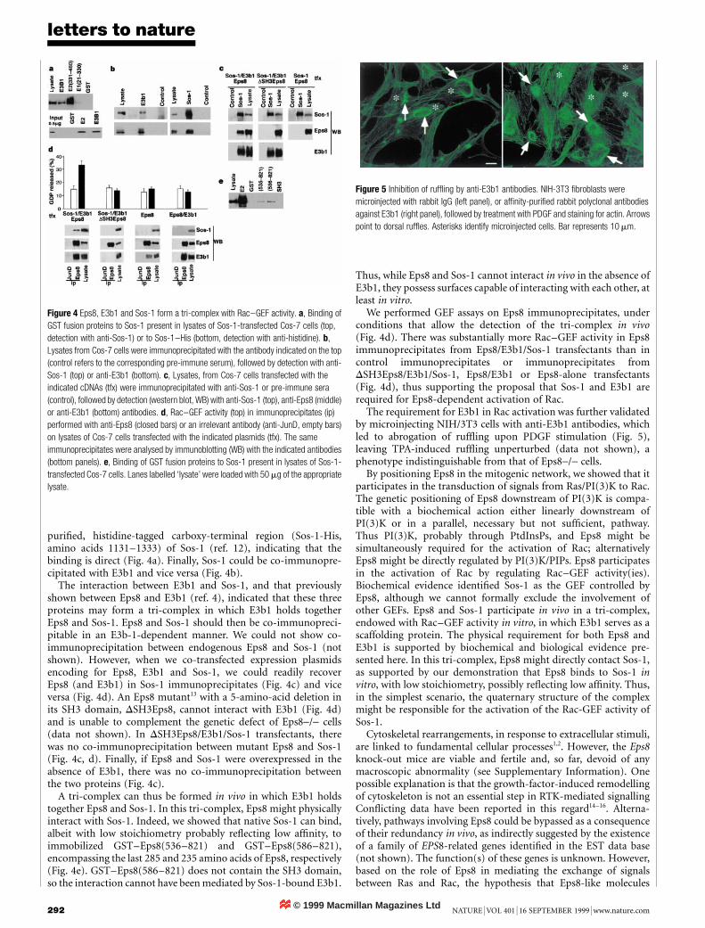
To obtain direct biochemical evidence for a role of Eps8 as anupstream regulator of Rac, we used the Cdc42/Rac InteractiveBinding region (CRIB) of aPak, which binds speci®cally toactivated GTP-bound Rac in vivo10. Sepharose-immobilized glu-tathione S-transferase-labelled CRIB (GST±CRIB) bound signi®-cantly better (1:7-fold 6 0:2 s:e:) to GTP-bound Rac in lysates ofPDGF-treated +/+ cells, compared with -/- cells (Fig. 3a). Thus,Eps8 is directly involved in the activation of Rac in vivo. Eps8might activate Rac by inhibiting a speci®c GTPase-activatingprotein (GAP) or activating a GEF. In cellular lysates from +/+or -/- ®broblasts, we could not detect differences in GAPactivities toward Rac (see Supplementary Information). We thenmeasured GEF activity using an assay that can effectively detectthis activity, as demonstrated by increased Rac±GEF in cellularlysates of Cos-7 cells transfected with the DH (Dbl homology)domain of Sos-1 (Fig. 3b; ref. 8). In lysates from -/- cells, Rac±
GEF, but not Cdc42±GEF, activity was threefold lower than in +/+cells, an effect reverted by re-expression of Eps8 in -/- cells (-/-(Eps8) cells; Fig. 3c).
Sos-1 has been implicated in the transmission of signals from Ras/PI(3)K to Rac8. Immunodepletion of Sos-1 from +/+ or -/-(Eps8),but not -/-, lysates resulted in a substantial decrease in activity ofRac±GEF (Fig. 3d) but not of Cdc42±GEF (data not shown).Immunodepletion of Eps8 from +/+ or -/-(Eps8) lysates caused amarked reduction in GEF activity, which become comparable tothat detected in -/- lysates or in Sos-1-depleted +/+ lysates (Fig. 3d).Thus Eps8 is directly responsible for activation of a Rac-speci®cGEF activity, which is probably represented by Sos-1. E3b1 (alsoknown as Abi-1) and RN-tre interact with the SH3 domain of Eps8(refs 4, 5, 11). Immunodepletion of E3b1, but not of RN-tre, from+/+ lysates led to a substantial reduction in Rac±GEF activity (Fig.3d). Co-immunodepletion of Eps8 and E3b1 did not produceadditive effects, indicating that they are required simultaneously(Fig. 3d).
The above results led us to test whether E3b1 and/or Eps8 couldphysically interact with Sos-1. Native Sos-1 could be recovered withsubstantial stoichiometry onto GST±E3b1 and GST±E2 (aminoacids 331±480 of E3b1), but not onto GSTor GST±E1 (amino acids1±330 of E3b1). GST±E3b1 and GST±E2 also interacted with a
Figure 3 Eps8 activates Rac and a Rac-speci®c GEF. a, +/+ and -/- ®broblasts were
treated with PDGF (+) or mock-treated (-). Lysates were incubated with GST±CRIB and
detected with anti-Rac (top), or directly immunoblotted with anti-Rac (bottom). b, GEF
activity in lysates from Cos-7 cells transfected with an empty vector (Ctr) or with the HA-
epitope-tagged DH domain of Sos-1 (DH). Inset, DH expression. c, GEF activity towards
Rac or Cdc42 in lysates from +/+, -/- or -/-(Eps8) cells. Inset, Eps8 expression. d, GEF
activity in lysates from +/+, -/- or -/-(Eps8) cells immunodepleted of the indicated
proteins. In all cases, immunodepletion removed .95% of the indicated proteins (see
Supplementary Information).
Figure 2 Epistatic localization of Eps8 in the Ras pathway. Fibroblasts (+/+, top and -/-,
bottom) were microinjected with the indicated plasmids and stained for actin. Asterisks
identify microinjected cells and arrows point to ruf¯es. Bar represents 10 mm.
© 1999 Macmillan Magazines Ltd
letters to nature
292 NATURE | VOL 401 | 16 SEPTEMBER 1999 | www.nature.com
puri®ed, histidine-tagged carboxy-terminal region (Sos-1-His,amino acids 1131±1333) of Sos-1 (ref. 12), indicating that thebinding is direct (Fig. 4a). Finally, Sos-1 could be co-immunopre-cipitated with E3b1 and vice versa (Fig. 4b).
The interaction between E3b1 and Sos-1, and that previouslyshown between Eps8 and E3b1 (ref. 4), indicated that these threeproteins may form a tri-complex in which E3b1 holds togetherEps8 and Sos-1. Eps8 and Sos-1 should then be co-immunopreci-pitable in an E3b-1-dependent manner. We could not show co-immunoprecipitation between endogenous Eps8 and Sos-1 (notshown). However, when we co-transfected expression plasmidsencoding for Eps8, E3b1 and Sos-1, we could readily recoverEps8 (and E3b1) in Sos-1 immunoprecipitates (Fig. 4c) and viceversa (Fig. 4d). An Eps8 mutant13 with a 5-amino-acid deletion inits SH3 domain, DSH3Eps8, cannot interact with E3b1 (Fig. 4d)and is unable to complement the genetic defect of Eps8-/- cells(data not shown). In DSH3Eps8/E3b1/Sos-1 transfectants, therewas no co-immunoprecipitation between mutant Eps8 and Sos-1(Fig. 4c, d). Finally, if Eps8 and Sos-1 were overexpressed in theabsence of E3b1, there was no co-immunoprecipitation betweenthe two proteins (Fig. 4c).
A tri-complex can thus be formed in vivo in which E3b1 holdstogether Eps8 and Sos-1. In this tri-complex, Eps8 might physicallyinteract with Sos-1. Indeed, we showed that native Sos-1 can bind,albeit with low stoichiometry probably re¯ecting low af®nity, toimmobilized GST±Eps8(536±821) and GST±Eps8(586±821),encompassing the last 285 and 235 amino acids of Eps8, respectively(Fig. 4e). GST±Eps8(586±821) does not contain the SH3 domain,so the interaction cannot have been mediated by Sos-1-bound E3b1.
Thus, while Eps8 and Sos-1 cannot interact in vivo in the absence ofE3b1, they possess surfaces capable of interacting with each other, atleast in vitro.
We performed GEF assays on Eps8 immunoprecipitates, underconditions that allow the detection of the tri-complex in vivo(Fig. 4d). There was substantially more Rac±GEF activity in Eps8immunoprecipitates from Eps8/E3b1/Sos-1 transfectants than incontrol immunoprecipitates or immunoprecipitates fromDSH3Eps8/E3b1/Sos-1, Eps8/E3b1 or Eps8-alone transfectants(Fig. 4d), thus supporting the proposal that Sos-1 and E3b1 arerequired for Eps8-dependent activation of Rac.
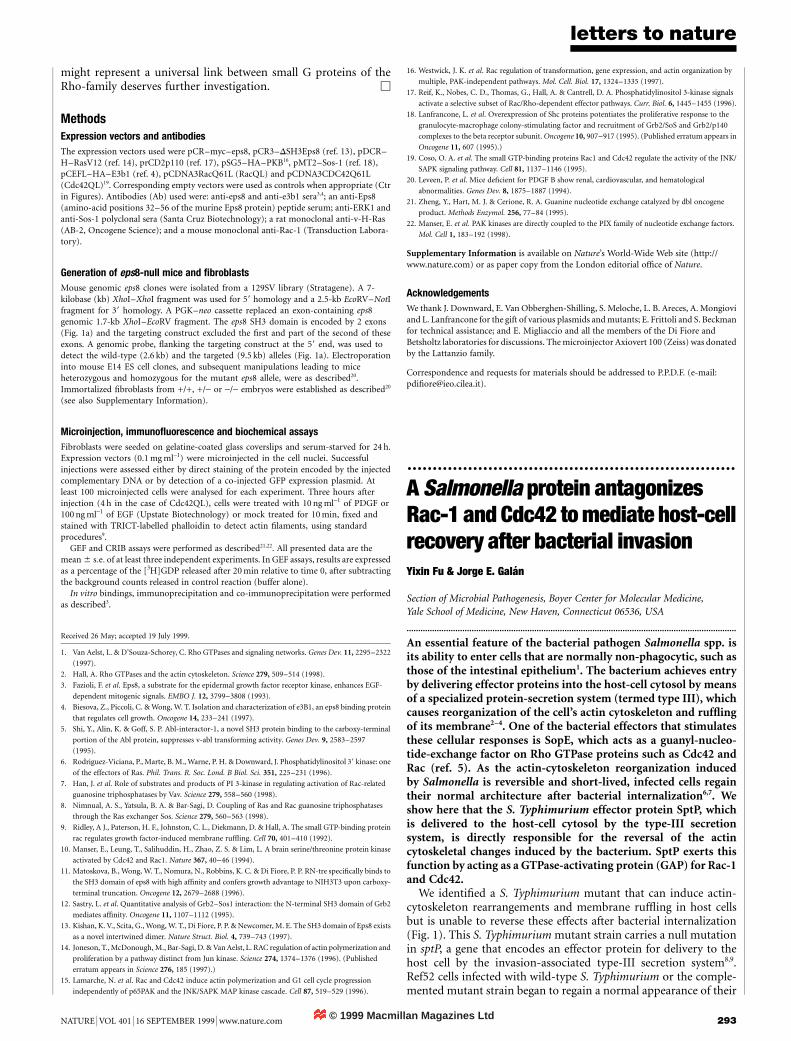
The requirement for E3b1 in Rac activation was further validatedby microinjecting NIH/3T3 cells with anti-E3b1 antibodies, whichled to abrogation of ruf¯ing upon PDGF stimulation (Fig. 5),leaving TPA-induced ruf¯ing unperturbed (data not shown), aphenotype indistinguishable from that of Eps8-/- cells.
By positioning Eps8 in the mitogenic network, we showed that itparticipates in the transduction of signals from Ras/PI(3)K to Rac.The genetic positioning of Eps8 downstream of PI(3)K is compa-tible with a biochemical action either linearly downstream ofPI(3)K or in a parallel, necessary but not suf®cient, pathway.Thus PI(3)K, probably through PtdInsPs, and Eps8 might besimultaneously required for the activation of Rac; alternativelyEps8 might be directly regulated by PI(3)K/PIPs. Eps8 participatesin the activation of Rac by regulating Rac±GEF activity(ies).Biochemical evidence identi®ed Sos-1 as the GEF controlled byEps8, although we cannot formally exclude the involvement ofother GEFs. Eps8 and Sos-1 participate in vivo in a tri-complex,endowed with Rac±GEF activity in vitro, in which E3b1 serves as ascaffolding protein. The physical requirement for both Eps8 andE3b1 is supported by biochemical and biological evidence pre-sented here. In this tri-complex, Eps8 might directly contact Sos-1,as supported by our demonstration that Eps8 binds to Sos-1 invitro, with low stoichiometry, possibly re¯ecting low af®nity. Thus,in the simplest scenario, the quaternary structure of the complexmight be responsible for the activation of the Rac-GEF activity ofSos-1.
Cytoskeletal rearrangements, in response to extracellular stimuli,are linked to fundamental cellular processes1,2. However, the Eps8knock-out mice are viable and fertile and, so far, devoid of anymacroscopic abnormality (see Supplementary Information). Onepossible explanation is that the growth-factor-induced remodellingof cytoskeleton is not an essential step in RTK-mediated signallingCon¯icting data have been reported in this regard14±16. Alterna-tively, pathways involving Eps8 could be bypassed as a consequenceof their redundancy in vivo, as indirectly suggested by the existenceof a family of EPS8-related genes identi®ed in the EST data base(not shown). The function(s) of these genes is unknown. However,based on the role of Eps8 in mediating the exchange of signalsbetween Ras and Rac, the hypothesis that Eps8-like molecules
Figure 4 Eps8, E3b1 and Sos-1 form a tri-complex with Rac±GEF activity. a, Binding of
GST fusion proteins to Sos-1 present in lysates of Sos-1-transfected Cos-7 cells (top,
detection with anti-Sos-1) or to Sos-1±His (bottom, detection with anti-histidine). b,
Lysates from Cos-7 cells were immunoprecipitated with the antibody indicated on the top
(control refers to the corresponding pre-immune serum), followed by detection with anti-
Sos-1 (top) or anti-E3b1 (bottom). c, Lysates, from Cos-7 cells transfected with the
indicated cDNAs (tfx) were immunoprecipitated with anti-Sos-1 or pre-immune sera
(control), followed by detection (western blot, WB) with anti-Sos-1 (top), anti-Eps8 (middle)
or anti-E3b1 (bottom) antibodies. d, Rac±GEF activity (top) in immunoprecipitates (ip)
performed with anti-Eps8 (closed bars) or an irrelevant antibody (anti-JunD, empty bars)
on lysates of Cos-7 cells transfected with the indicated plasmids (tfx). The same
immunoprecipitates were analysed by immunoblotting (WB) with the indicated antibodies
(bottom panels). e, Binding of GST fusion proteins to Sos-1 present in lysates of Sos-1-
transfected Cos-7 cells. Lanes labelled `lysate' were loaded with 50 mg of the appropriate
lysate.
Figure 5 Inhibition of ruf¯ing by anti-E3b1 antibodies. NIH-3T3 ®broblasts were
microinjected with rabbit IgG (left panel), or af®nity-puri®ed rabbit polyclonal antibodies
against E3b1 (right panel), followed by treatment with PDGF and staining for actin. Arrows
point to dorsal ruf¯es. Asterisks identify microinjected cells. Bar represents 10 mm.
© 1999 Macmillan Magazines Ltd
letters to nature
NATURE | VOL 401 | 16 SEPTEMBER 1999 | www.nature.com 293
might represent a universal link between small G proteins of theRho-family deserves further investigation. M
MethodsExpression vectors and antibodies
The expression vectors used were pCR±myc±eps8, pCR3±DSH3Eps8 (ref. 13), pDCR±H±RasV12 (ref. 14), prCD2p110 (ref. 17), pSG5±HA±PKB16, pMT2±Sos-1 (ref. 18),pCEFL±HA±E3b1 (ref. 4), pCDNA3RacQ61L (RacQL) and pCDNA3CDC42Q61L(Cdc42QL)19. Corresponding empty vectors were used as controls when appropriate (Ctrin Figures). Antibodies (Ab) used were: anti-eps8 and anti-e3b1 sera3,4; an anti-Eps8(amino-acid positions 32±56 of the murine Eps8 protein) peptide serum; anti-ERK1 andanti-Sos-1 polyclonal sera (Santa Cruz Biotechnology); a rat monoclonal anti-v-H-Ras(AB-2, Oncogene Science); and a mouse monoclonal anti-Rac-1 (Transduction Labora-tory).
Generation of eps8-null mice and ®broblasts
Mouse genomic eps8 clones were isolated from a 129SV library (Stratagene). A 7-kilobase (kb) XhoI±XhoI fragment was used for 59 homology and a 2.5-kb EcoRV±NotIfragment for 39 homology. A PGK±neo cassette replaced an exon-containing eps8genomic 1.7-kb XhoI±EcoRV fragment. The eps8 SH3 domain is encoded by 2 exons(Fig. 1a) and the targeting construct excluded the ®rst and part of the second of theseexons. A genomic probe, ¯anking the targeting construct at the 59 end, was used todetect the wild-type (2.6 kb) and the targeted (9.5 kb) alleles (Fig. 1a). Electroporationinto mouse E14 ES cell clones, and subsequent manipulations leading to miceheterozygous and homozygous for the mutant eps8 allele, were as described20.Immortalized ®broblasts from +/+, +/- or -/- embryos were established as described20
(see also Supplementary Information).
Microinjection, immuno¯uorescence and biochemical assays
Fibroblasts were seeded on gelatine-coated glass coverslips and serum-starved for 24 h.Expression vectors (0.1 mg ml-1) were microinjected in the cell nuclei. Successfulinjections were assessed either by direct staining of the protein encoded by the injectedcomplementary DNA or by detection of a co-injected GFP expression plasmid. Atleast 100 microinjected cells were analysed for each experiment. Three hours afterinjection (4 h in the case of Cdc42QL), cells were treated with 10 ng ml-1 of PDGF or100 ng ml-1 of EGF (Upstate Biotechnology) or mock treated for 10 min, ®xed andstained with TRICT-labelled phalloidin to detect actin ®laments, using standardprocedures9.
GEF and CRIB assays were performed as described21,22. All presented data are themean 6 s:e: of at least three independent experiments. In GEF assays, results are expressedas a percentage of the [3H]GDP released after 20 min relative to time 0, after subtractingthe background counts released in control reaction (buffer alone).
In vitro bindings, immunoprecipitation and co-immunoprecipitation were performedas described3.
Received 26 May; accepted 19 July 1999.
1. Van Aelst, L. & D'Souza-Schorey, C. Rho GTPases and signaling networks. Genes Dev. 11, 2295±2322
(1997).
2. Hall, A. Rho GTPases and the actin cytoskeleton. Science 279, 509±514 (1998).
3. Fazioli, F. et al. Eps8, a substrate for the epidermal growth factor receptor kinase, enhances EGF-
dependent mitogenic signals. EMBO J. 12, 3799±3808 (1993).
4. Biesova, Z., Piccoli, C. & Wong, W. T. Isolation and characterization of e3B1, an eps8 binding protein
that regulates cell growth. Oncogene 14, 233±241 (1997).
5. Shi, Y., Alin, K. & Goff, S. P. Abl-interactor-1, a novel SH3 protein binding to the carboxy-terminal
portion of the Abl protein, suppresses v-abl transforming activity. Genes Dev. 9, 2583±2597
(1995).
6. Rodriguez-Viciana, P., Marte, B. M., Warne, P. H. & Downward, J. Phosphatidylinositol 39 kinase: one
of the effectors of Ras. Phil. Trans. R. Soc. Lond. B Biol. Sci. 351, 225±231 (1996).
7. Han, J. et al. Role of substrates and products of PI 3-kinase in regulating activation of Rac-related
guanosine triphosphatases by Vav. Science 279, 558±560 (1998).
8. Nimnual, A. S., Yatsula, B. A. & Bar-Sagi, D. Coupling of Ras and Rac guanosine triphosphatases
through the Ras exchanger Sos. Science 279, 560±563 (1998).
9. Ridley, A J., Paterson, H. F., Johnston, C. L., Diekmann, D. & Hall, A. The small GTP-binding protein
rac regulates growth factor-induced membrane ruf¯ing. Cell 70, 401±410 (1992).
10. Manser, E., Leung, T., Salihuddin, H., Zhao, Z. S. & Lim, L. A brain serine/threonine protein kinase
activated by Cdc42 and Rac1. Nature 367, 40±46 (1994).
11. Matoskova, B., Wong, W. T., Nomura, N., Robbins, K. C. & Di Fiore, P. P. RN-tre speci®cally binds to
the SH3 domain of eps8 with high af®nity and confers growth advantage to NIH3T3 upon carboxy-
terminal truncation. Oncogene 12, 2679±2688 (1996).
12. Sastry, L. et al. Quantitative analysis of Grb2±Sos1 interaction: the N-terminal SH3 domain of Grb2
mediates af®nity. Oncogene 11, 1107±1112 (1995).
13. Kishan, K. V., Scita, G., Wong, W. T., Di Fiore, P. P. & Newcomer, M. E. The SH3 domain of Eps8 exists
as a novel intertwined dimer. Nature Struct. Biol. 4, 739±743 (1997).
14. Joneson, T., McDonough, M., Bar-Sagi, D. & Van Aelst, L. RAC regulation of actin polymerization and
proliferation by a pathway distinct from Jun kinase. Science 274, 1374±1376 (1996). (Published
erratum appears in Science 276, 185 (1997).)
15. Lamarche, N. et al. Rac and Cdc42 induce actin polymerization and G1 cell cycle progression
independently of p65PAK and the JNK/SAPK MAP kinase cascade. Cell 87, 519±529 (1996).
16. Westwick, J. K. et al. Rac regulation of transformation, gene expression, and actin organization by
multiple, PAK-independent pathways. Mol. Cell. Biol. 17, 1324±1335 (1997).
17. Reif, K., Nobes, C. D., Thomas, G., Hall, A. & Cantrell, D. A. Phosphatidylinositol 3-kinase signals
activate a selective subset of Rac/Rho-dependent effector pathways. Curr. Biol. 6, 1445±1455 (1996).
18. Lanfrancone, L. et al. Overexpression of Shc proteins potentiates the proliferative response to the
granulocyte-macrophage colony-stimulating factor and recruitment of Grb2/SoS and Grb2/p140
complexes to the beta receptor subunit. Oncogene 10, 907±917 (1995). (Published erratum appears in
Oncogene 11, 607 (1995).)
19. Coso, O. A. et al. The small GTP-binding proteins Rac1 and Cdc42 regulate the activity of the JNK/
SAPK signaling pathway. Cell 81, 1137±1146 (1995).
20. Leveen, P. et al. Mice de®cient for PDGF B show renal, cardiovascular, and hematological
abnormalities. Genes Dev. 8, 1875±1887 (1994).
21. Zheng, Y., Hart, M. J. & Cerione, R. A. Guanine nucleotide exchange catalyzed by dbl oncogene
product. Methods Enzymol. 256, 77±84 (1995).
22. Manser, E. et al. PAK kinases are directly coupled to the PIX family of nucleotide exchange factors.
Mol. Cell 1, 183±192 (1998).
Supplementary Information is available on Nature's World-Wide Web site (http://www.nature.com) or as paper copy from the London editorial of®ce of Nature.
Acknowledgements
We thank J. Downward, E. Van Obberghen-Shilling, S. Meloche, L. B. Areces, A. Mongioviand L. Lanfrancone for the gift of various plasmids and mutants; E. Frittoli and S. Beckmanfor technical assistance; and E. Migliaccio and all the members of the Di Fiore andBetsholtz laboratories for discussions. The microinjector Axiovert 100 (Zeiss) was donatedby the Lattanzio family.
Correspondence and requests for materials should be addressed to P.P.D.F. (e-mail:pdi®[email protected]).
.................................................................A Salmonella protein antagonizesRac-1 and Cdc42 to mediate host-cellrecovery after bacterial invasionYixin Fu & Jorge E. GalaÂn
Section of Microbial Pathogenesis, Boyer Center for Molecular Medicine,Yale School of Medicine, New Haven, Connecticut 06536, USA
..............................................................................................................................................
An essential feature of the bacterial pathogen Salmonella spp. isits ability to enter cells that are normally non-phagocytic, such asthose of the intestinal epithelium1. The bacterium achieves entryby delivering effector proteins into the host-cell cytosol by meansof a specialized protein-secretion system (termed type III), whichcauses reorganization of the cell's actin cytoskeleton and ruf¯ingof its membrane2±4. One of the bacterial effectors that stimulatesthese cellular responses is SopE, which acts as a guanyl-nucleo-tide-exchange factor on Rho GTPase proteins such as Cdc42 andRac (ref. 5). As the actin-cytoskeleton reorganization inducedby Salmonella is reversible and short-lived, infected cells regaintheir normal architecture after bacterial internalization6,7. Weshow here that the S. Typhimurium effector protein SptP, whichis delivered to the host-cell cytosol by the type-III secretionsystem, is directly responsible for the reversal of the actincytoskeletal changes induced by the bacterium. SptP exerts thisfunction by acting as a GTPase-activating protein (GAP) for Rac-1and Cdc42.
We identi®ed a S. Typhimurium mutant that can induce actin-cytoskeleton rearrangements and membrane ruf¯ing in host cellsbut is unable to reverse these effects after bacterial internalization(Fig. 1). This S. Typhimurium mutant strain carries a null mutationin sptP, a gene that encodes an effector protein for delivery to thehost cell by the invasion-associated type-III secretion system8,9.Ref52 cells infected with wild-type S. Typhimurium or the comple-mented mutant strain began to regain a normal appearance of their