Embed Size (px)
Citation preview
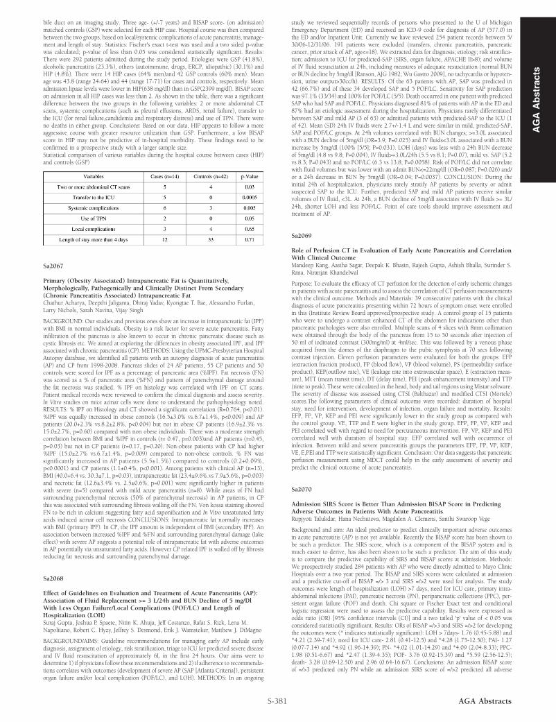
bile duct on an imaging study. Three age- (+/-7 years) and BISAP score- (on admission)matched controls (GSP) were selected for each HIP case. Hospital course was then comparedbetween the two groups, based on local/systemic complications of acute pancreatitis, manage-ment and length of stay. Statistics: Fischer's exact t-test was used and a two sided p-valuewas calculated; p-value of less than 0.05 was considered statistically significant. Results:There were 292 patients admitted during the study period. Etiologies were GSP (41.8%),alcoholic pancreatitis (23.3%), others (autoimmune, drugs, ERCP, idiopathic) (30.1%) andHIP (4.8%). There were 14 HIP cases (64% men)and 42 GSP controls (60% men). Meanage was 43.8 (range 24-64) and 44 (range 17-71) for cases and controls, respectively. Meanadmission lipase levels were lower in HIP(638 mg/dl) than in GSP(2399 mg/dl). BISAP scoreon admission in all HIP cases was less than 2. As shown in the table, there was a significantdifference between the two groups in the following variables: 2 or more abdominal CTscans, systemic complications (such as pleural effusions, ARDS, renal failure), transfer tothe ICU (for renal failure,candidemia and respiratory distress) and use of TPN. There wereno deaths in either group. Conclusions: Based on our data, HIP appears to follow a moreaggressive course with greater resource utilization than GSP. Furthermore, a low BISAPscore in HIP may not be predictive of in-hospital morbidity. These findings need to beconfirmed in a prospective study with a larger sample size.Statistical comparison of various variables during the hospital course between cases (HIP)and controls (GSP)
Sa2067
Primary (Obesity Associated) Intrapancreatic Fat is Quantitatively,Morphologically, Pathogenically and Clinically Distinct From Secondary(Chronic Pancreatitis Associated) Intrapancreatic FatChathur Acharya, Deepthi Jaligama, Dhiraj Yadav, Kyongtae T. Bae, Alessandro Furlan,Larry Nichols, Sarah Navina, Vijay Singh
BACKGROUND: Our studies and previous ones show an increase in intrapancreatic fat (IPF)with BMI in normal individuals. Obesity is a risk factor for severe acute pancreatitis. Fattyinfiltration of the pancreas is also known to occur in chronic pancreatic disease such ascystic fibrosis etc. We aimed at exploring the differences in obesity associated IPF, and IPFassociated with chronic pancreatitis (CP).METHODS: Using the UPMC-Presbyterian HospitalAutopsy database, we identified all patients with an autopsy diagnosis of acute pancreatitis(AP) and CP from 1998-2008. Pancreas slides of 24 AP patients, 55 CP patients and 50controls were scored for IPF as a percentage of pancreatic area (%IPF). Fat necrosis (FN)was scored as a % of pancreatic area (%FN) and pattern of parenchymal damage aroundthe fat necrosis was studied. % IPF on histology was correlated with IPF on CT scans.Patient medical records were reviewed to confirm the clinical diagnosis and assess severity.In Vitro studies on mice acinar cells were done to understand the pathophysiology noted.RESULTS: % IPF on Histology and CT showed a significant correlation (R=0.764, p<0.01).%IPF was equally increased in obese controls (16.5±3.0% vs.6.7±1.4%, p<0.009) and APpatients (20.0+2.3% vs.8.2±2.8%, p<0.004) but not in obese CP patients (16.9±2.3% vs.15.0±2.7%, p=0.60) compared with non obese individuals. There was a moderate strengthcorrelation between BMI and %IPF in controls (r= 0.47, p=0.003)and AP patients (r=0.45,p=0.03) but not in CP patients (r=0.17, p=0.20). Non-obese patients with CP had higher%IPF (15.0±2.7% vs.6.7±1.4%, p=0.009) compared to non-obese controls. % FN wassignificantly increased in AP patients (5.5±1.5%) compared to controls (0.2+0.09%,p<0.0001) and CP patients (1.1±0.4%, p<0.001). Among patients with clinical AP (n=13),BMI (40.0+6.4 vs. 30.3±7.1, p=0.03), intrapancreatic fat (23.4±9.6%.vs 7.9±5.6%, p=0.003)and necrotic fat (12.6±3.4% vs. 2.5±0.6%, p=0.001) were significantly higher in patientswith severe (n=5) compared with mild acute pancreatitis (n=8). While areas of FN hadsurrounding parenchymal necrosis (50% of parenchymal necrosis) in AP patients, in CPthis was associated with surrounding fibrosis walling off the FN. Von kossa staining showedFN to be rich in calcium suggesting fatty acid saponification and In Vitro unsaturated fattyacids induced acinar cell necrosis CONCLUSIONS: Intrapancreatic fat normally increaseswith BMI (primary IPF). In CP, the IPF amount is independent of BMI (secondary IPF). Anassociation between increased %IPF and %FN and surrounding parenchymal damage (lakeeffect) with severe AP suggests a potential role of intrapancreatic fat with adverse outcomesin AP potentially via unsaturated fatty acids. However CP related IPF is walled off by fibrosisreducing fat necrosis and surrounding parenchymal damage.
Sa2068
Effect of Guidelines on Evaluation and Treatment of Acute Pancreatitis (AP):Association of Fluid Replacement >= 3 L/24h and BUN Decline of 5 mg/DlWith Less Organ Failure/Local Complications (POF/LC) and Length ofHospitalization (LOH)Suraj Gupta, Joshua P. Spaete, Nitin K. Ahuja, Jeff Costanzo, Rafat S. Rizk, Lena M.Napolitano, Robert C. Hyzy, Jeffrey S. Desmond, Erik J. Wamsteker, Matthew J. DiMagno
BACKGROUND/AIMS: Guideline recommendations for managing early AP include earlydiagnosis, assignment of etiology, risk stratification, triage to ICU for predicted severe diseaseand IV fluid resuscitation of approximately 6L in the first 24 hours. Our aims were todetermine 1) if physicians follow these recommendations and 2) if adherence to recommenda-tions correlates with outcomes (development of severe AP (SAP [Atlanta Criteria]), persistentorgan failure and/or local complication (POF/LC), and LOH). METHODS: In an ongoing
S-381 AGA Abstracts
study we reviewed sequentially records of persons who presented to the U of MichiganEmergency Department (ED) and received an ICD-9 code for diagnosis of AP (577.0) inthe ED and/or Inpatient Unit. Currently we have reviewed 254 patient records between 5/30/06-12/31/06. 191 patients were excluded (transfers, chronic pancreatitis, pancreaticcancer, prior attack of AP, age<=18). We extracted data for diagnosis; etiology; risk stratifica-tion; admission to ICU for predicted-SAP (SIRS, organ failure, APACHE II>8); and volumeof IV fluid resuscitation at 24h, including measures of adequate resuscitation (normal BUNor BUN decline by 5mg/dl [Ranson, AJG 1982; Wu Gastro 2009], no tachycardia or hypoten-sion, urine output>30cc/h). RESULTS: Of the 63 patients with AP, SAP was predicted in42 (66.7%) and of these 34 developed SAP and 5 POF/LC. Sensitivity for SAP predictionwas 97.1% (33/34) and 100% for POF/LC (5/5). Death occurred in one patient with predictedSAP who had SAP and POF/LC. Physicians diagnosed 81% of patients with AP in the ED and87% had an etiologic assessment during the hospitalization. Physicians rarely differentiatedbetween SAP and mild AP (3 of 63) or admitted patients with predicted-SAP to the ICU (1of 42). Mean (SD) 24h IV fluids were 2.7+/-1.4 L and were similar in mild, predicted-SAP,SAP and POF/LC groups. At 24h volumes correlated with BUN changes; >=3.0L associatedwith a BUN decline of 5mg/dl (OR=3.9; P=0.025) and IV fluids<3.0L associated with a BUNincrease by 5mg/dl (100% [5/5]; P=0.031). LOH (days) was less with a 24h BUN decreaseof 5mg/dl (4.8 vs 9.8; P=0.004), IV fluid>=3.0L/24h (5.5 vs 8.1; P=0.07), mild vs. SAP (5.2vs 8.3; P=0.043) and no POF/LC (6.3 vs 13.8; P=0.0058). Risk of POF/LC did not correlatewith fluid volumes but was lower with an admit BUN<=22mg/dl (OR=0.087; P=0.026) and/or a 24h decrease in BUN by 5mg/dl (OR=0.04; P=0.0037). CONCLUSION: During theinitial 24h of hospitalization, physicians rarely stratify AP patients by severity or admitsuspected SAP to the ICU. Further, predicted SAP and mild AP patients receive similarvolumes of IV fluid, <3L. At 24h, a BUN decline of 5mg/dl associates with IV fluids >= 3L/24h, shorter LOH and less POF/LC. Point of care tools should improve assessment andtreatment of AP.
Sa2069
Role of Perfusion CT in Evaluation of Early Acute Pancreatitis and CorrelationWith Clinical OutcomeMandeep Kang, Aastha Sagar, Deepak K. Bhasin, Rajesh Gupta, Ashish Bhalla, Surinder S.Rana, Niranjan Khandelwal
Purpose: To evaluate the efficacy of CT perfusion for the detection of early ischemic changesin patients with acute pancreatitis and to assess the correlation of CT perfusion measurementswith the clinical outcome. Methods and Materials: 39 consecutive patients with the clinicaldiagnosis of acute pancreatitis presenting within 72 hours of symptom onset were enrolledin this (Institute Review Board approved)prospective study. A control group of 15 patientswho were to undergo a contrast enhanced CT of the abdomen for indications other thanpancreatic pathologies were also enrolled. Multiple scans of 4 slices with 8mm collimationwere obtained through the body of the pancreas from 15 to 50 seconds after injection of50 ml of iodinated contrast (300mg/ml) at 4ml/sec. This was followed by a venous phaseacquired from the domes of the diaphragm to the pubic symphysis at 70 secs followingcontrast injection. Eleven perfusion parameters were evaluated for both the groups: EFP(extraction fraction product), FP (blood flow), VP (blood volume), PS (permeability surfaceproduct), KEP(outflow rate), VE (leakage rate into extravascular space), E (extraction meas-ure), MTT (mean transit time), DT (delay time), PEI (peak enhancement intensity) and TTP(time to peak). These were calculated in the head, body and tail regions using Mistar software.The severity of disease was assessed using CTSI (Balthazar) and modified CTSI (Mortele)scores.The following parameters of clinical outcome were recorded: duration of hospitalstay, need for intervention, development of infection, organ failure and mortality. Results:EFP, FP, VP, KEP and PEI were significantly lower in the study group as compared withthe control group. VE, TTP and E were higher in the study group. EFP, FP, VP, KEP andPEI correlated well with regard to need for percutaneous intervention. FP, VP, KEP and PEIcorrelated well with duration of hospital stay. EFP correlated well with occurrence ofinfection. Between mild and severe pancreatitis groups the parameters EFP, FP, VP, KEP,VE, E,PEI and TTP were statistically significant. Conclusion: Our data suggests that pancreaticperfusion measurement using MDCT could help in the early assessment of severity andpredict the clinical outcome of acute pancreatitis.
Sa2070
Admission SIRS Score is Better Than Admission BISAP Score in PredictingAdverse Outcomes in Patients With Acute PancreatitisRupjyoti Talukdar, Hana Nechutova, Magdalen A. Clemens, Santhi Swaroop Vege
Background and aim: An ideal predictor to predict clinically important adverse outcomesin acute pancreatitis (AP) is not yet available. Recently the BISAP score has been shown tobe such a predictor. The SIRS score, which is a component of the BISAP system and ismuch easier to derive, has also been shown to be such a predictor. The aim of this studyis to compare the predictive capability of SIRS and BISAP scores at admission. Methods:We prospectively studied 284 patients with AP who were directly admitted to Mayo ClinicHospitals over a two year period. The BISAP and SIRS scores were calculated at admissionand a predictive cut-off of BISAP =/> 3 and SIRS =/>2 were used for analysis. The studyoutcomes were length of hospitalization (LOH) >7 days, need for ICU care, primary intra-abdominal infections (PAI), pancreatic necrosis (PN), peripancreatic collections (PPC), per-sistent organ failure (POF) and death. Chi square or Fischer Exact test and conditionallogistic regression were used to assess the predictive capability. Results were expressed asodds ratio (OR) [95% confidence intervals (CI)] and a two tailed 'p' value of < 0.05 wasconsidered statistically significant. Results: ORs of BISAP =/>3 and SIRS =/>2 for developingthe outcomes were (* indicates statistically significant): LOH > 7days- 1.76 (0.45-5.88) and*4.21 (2.39-7.41); need for ICU care- 2.81 (0.41-12.5) and *4.28 (1.75-12.50); PAI- 1.27(0.07-7.14) and *4.92 (1.96-14.39); PN- *4.02 (1.01-14.29) and *4.09 (2.04-8.33); PPC-1.98 (0.51-6.67) and *2.47 (1.39-4.35); POF- 3.76 (0.92-15.39) and *5.59 (2.56-12.5);death- 3.28 (0.69-12.50) and 2.96 (0.64-16.67). Conclusions: An admission BISAP scoreof =/>3 predicted only PN while an admission SIRS score of =/>2 predicted all adverse
AG
AA
bst
ract
s


![Integrating the Healthcare Enterprise€¦ · Document Source Document ConsumerOn Entry [ITI Document Registry Document Repository Provide&Register Document Set – b [ITI-41] →](https://img.pdfslide.net/doc/110x75/5f08a1eb7e708231d422f7c5/integrating-the-healthcare-enterprise-document-source-document-consumeron-entry.jpg)


