Embed Size (px)
Citation preview
61 MICROBIAL LIMIT TESTS Top Previous Next
61 MICROBIAL LIMIT TESTS
This chapter provides tests for the estimation of the number of viable aerobic microorganismspresent and for freedom from designated microbial species in pharmaceutical articles of all kinds,from raw materials to the finished forms. An automated method may be substituted for the testspresented here, provided it has been properly validated as giving equivalent or better results. Inpreparing for and in applying the tests, observe aseptic precautions in handling the specimens.Unless otherwise directed, where the procedure specifies simply “incubate,” hold the container in
air that is thermostatically controlled at a temperature between 30 and 35 , for a period of 24 to48 hours. The term “growth” is used in a special sense herein, i.e., to designatethe presence andpresumed proliferation of viable microorganisms.
PREPARATORY TESTING
The validity of the results of the tests set forth in this chapter rests largely upon the adequacy of ademonstration that the test specimens to which they are applied do not, of themselves, inhibit themultiplication, under the test conditions, of microorganisms that may be present. Therefore,preparatory to conducting the tests on a regular basis and as circumstances require subsequently,inoculate diluted specimens of the material to be tested with separate viable cultures ofStaphylococcus aureus, Escherichia coli, Pseudomonas aeruginosa, and Salmonella. This can be
done by adding 1 mL of not less than 10 3 dilution of a 24-hour broth culture of the microorganismto the first dilution (inpH 7.2 Phosphate Buffer, Fluid Soybean–Casein Digest Medium, or FluidLactose Medium) of the test material and following the test procedure. Failure of the organism(s)to grow in the relevant medium invalidates that portion of the examination and necessitates amodification of the procedure by (1) an increase in the volume of diluent, the quantity of testmaterial remaining the same, or by (2) the incorporation of a sufficient quantity of suitableinactivating agent(s) in the diluents, or by (3) an appropriate combination of modifications (1) and(2) so as to permit growth of the inocula.
The following are examples of ingredients and their concentrations that may be added to theculture medium to neutralize inhibitory substances present in the sample: soy lecithin, 0.5%; andpolysorbate 20, 4.0%. Alternatively, repeat the test as described in the preceding paragraph, usingFluid Casein Digest–Soy Lecithin–Polysorbate 20 Medium to demonstrate neutralization ofpreservatives or other antimicrobial agents in the test material. Where inhibitory substances arecontained in the product and the latter is soluble, a suitable, validated adaptation of a procedureset forth in the sectionMembrane Filtration underTest for Sterility of the Product to be Examined
underSterility Tests 71 , may be used.
If in spite of the incorporation of suitable inactivating agents and a substantial increase in thevolume of diluent, it is still not possible to recover the viable cultures described above and wherethe article is not suitable for employment of membrane filtration, it can be assumed that the failureto isolate the inoculated organism is attributable to the bactericidal activity of the product. This
information serves to indicate that the article is not likely to be contaminated with the given speciesof microorganism. Monitoring should be continued in order to establish the spectrum of inhibitionand bactericidal activity of the article.
BUFFER SOLUTION AND MEDIA
Culture media may be prepared as follows, or dehydrated culture media may be used providedthat, when reconstituted as directed by the manufacturer or distributor, they have similaringredients and/or yield media comparable to those obtained from the formulas given herein.
In preparing media by the formulas set forth herein, dissolve the soluble solids in the water, usingheat, if necessary, to effect complete solution, and add solutions of hydrochloric acid or sodiumhydroxide in quantities sufficient to yield the desired pH in the medium when it is ready for use.
Determine the pH at 25 ± 2 .
Where agar is called for in a formula, use agar that has a moisture content of not more than 15%.Where water is called for in a formula, use Purified Water.
PH 7.2 Phosphate Buffer
Stock Solution— Dissolve 34 g of monobasic potassium phosphate in about 500 mL of watercontained in a 1000-mL volumetric flask. Adjust to pH 7.2 ± 0.1 by the addition ofsodiumhydroxide TS (about 175 mL), add water to volume, and mix. Dispense and sterilize. Store underrefrigeration.
For use, dilute theStock Solution with water in the ratio of 1 to 800, and sterilize.
Media
Unless otherwise indicated, the media should be sterilized by heating in an autoclave (seeSteam
Sterilization under Sterilization 1211 ), the exposure time depending on the volume to besterilized.
I. Fluid Casein Digest–Soy Lecithin–Polysorbate 20 Medium
Pancreatic Digest ofCasein
20 g
Soy Lecithin 5 gPolysorbate 20 40 mLWater 960 mL
Dissolve the pancreatic digest of casein and soy lecithin in 960 mL of water, heating in a water
bath at 48 to 50 for about 30 minutes to effect solution. Add 40 mL of polysorbate 20. Mix, anddispense as desired.
II. Soybean–Casein Digest Agar Medium
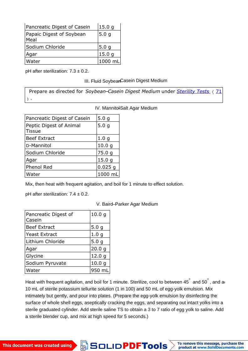
Pancreatic Digest of Casein 15.0 gPapaic Digest of SoybeanMeal
5.0 g
Sodium Chloride 5.0 gAgar 15.0 gWater 1000 mL
pH after sterilization: 7.3 ± 0.2.
III. Fluid Soybean–Casein Digest Medium
Prepare as directed for Soybean–Casein Digest Medium under Sterility Tests 71.
IV. Mannitol–Salt Agar Medium
Pancreatic Digest of Casein 5.0 gPeptic Digest of AnimalTissue
5.0 g
Beef Extract 1.0 gD-Mannitol 10.0 gSodium Chloride 75.0 gAgar 15.0 gPhenol Red 0.025 gWater 1000 mL
Mix, then heat with frequent agitation, and boil for 1 minute to effect solution.
pH after sterilization: 7.4 ± 0.2.
V. Baird–Parker Agar Medium
Pancreatic Digest ofCasein
10.0 g
Beef Extract 5.0 gYeast Extract 1.0 gLithium Chloride 5.0 gAgar 20.0 gGlycine 12.0 gSodium Pyruvate 10.0 gWater 950 mL
Heat with frequent agitation, and boil for 1 minute. Sterilize, cool to between 45 and 50 , and add10 mL of sterile potassium tellurite solution (1 in 100) and 50 mL of egg-yolk emulsion. Mixintimately but gently, and pour into plates. (Prepare the egg-yolk emulsion by disinfecting thesurface of whole shell eggs, aseptically cracking the eggs, and separating out intact yolks into asterile graduated cylinder. Add sterile saline TS to obtain a 3 to 7 ratio of egg yolk to saline. Add toa sterile blender cup, and mix at high speed for 5 seconds.)
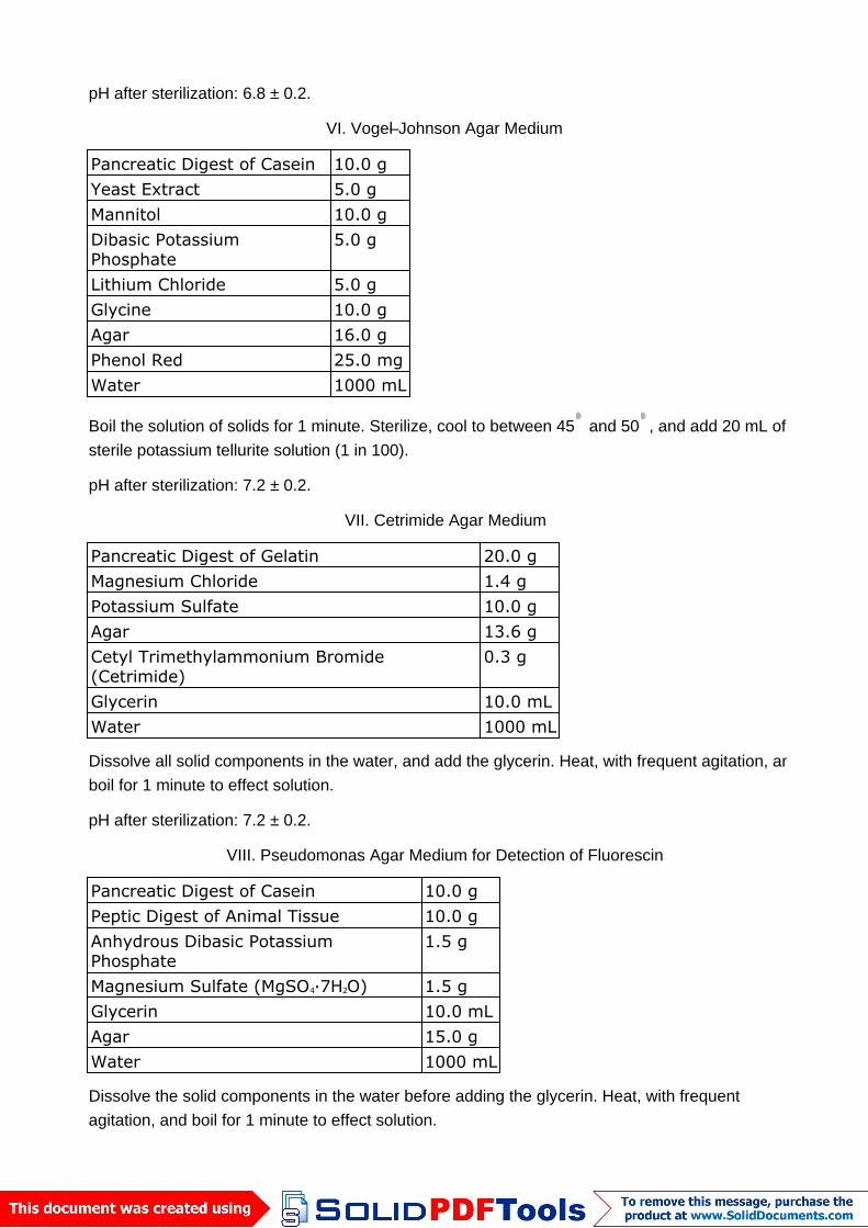
pH after sterilization: 6.8 ± 0.2.
VI. Vogel–Johnson Agar Medium
Pancreatic Digest of Casein 10.0 gYeast Extract 5.0 gMannitol 10.0 gDibasic PotassiumPhosphate
5.0 g
Lithium Chloride 5.0 gGlycine 10.0 gAgar 16.0 gPhenol Red 25.0 mgWater 1000 mL
Boil the solution of solids for 1 minute. Sterilize, cool to between 45 and 50 , and add 20 mL ofsterile potassium tellurite solution (1 in 100).
pH after sterilization: 7.2 ± 0.2.
VII. Cetrimide Agar Medium
Pancreatic Digest of Gelatin 20.0 gMagnesium Chloride 1.4 gPotassium Sulfate 10.0 gAgar 13.6 gCetyl Trimethylammonium Bromide(Cetrimide)
0.3 g
Glycerin 10.0 mLWater 1000 mL
Dissolve all solid components in the water, and add the glycerin. Heat, with frequent agitation, andboil for 1 minute to effect solution.
pH after sterilization: 7.2 ± 0.2.
VIII. Pseudomonas Agar Medium for Detection of Fluorescin
Pancreatic Digest of Casein 10.0 gPeptic Digest of Animal Tissue 10.0 gAnhydrous Dibasic PotassiumPhosphate
1.5 g
Magnesium Sulfate (MgSO4·7H2O) 1.5 gGlycerin 10.0 mLAgar 15.0 gWater 1000 mL
Dissolve the solid components in the water before adding the glycerin. Heat, with frequentagitation, and boil for 1 minute to effect solution.
pH after sterilization: 7.2 ± 0.2.
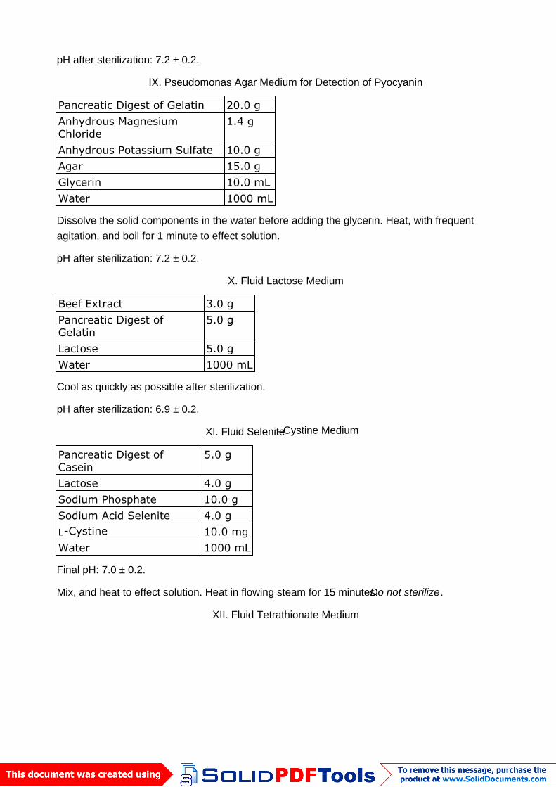
IX. Pseudomonas Agar Medium for Detection of Pyocyanin
Pancreatic Digest of Gelatin 20.0 gAnhydrous MagnesiumChloride
1.4 g
Anhydrous Potassium Sulfate 10.0 gAgar 15.0 gGlycerin 10.0 mLWater 1000 mL
Dissolve the solid components in the water before adding the glycerin. Heat, with frequentagitation, and boil for 1 minute to effect solution.
pH after sterilization: 7.2 ± 0.2.
X. Fluid Lactose Medium
Beef Extract 3.0 gPancreatic Digest ofGelatin
5.0 g
Lactose 5.0 gWater 1000 mL
Cool as quickly as possible after sterilization.
pH after sterilization: 6.9 ± 0.2.
XI. Fluid Selenite–Cystine Medium
Pancreatic Digest ofCasein
5.0 g
Lactose 4.0 gSodium Phosphate 10.0 gSodium Acid Selenite 4.0 gL-Cystine 10.0 mgWater 1000 mL
Final pH: 7.0 ± 0.2.
Mix, and heat to effect solution. Heat in flowing steam for 15 minutes.Do not sterilize.
XII. Fluid Tetrathionate Medium
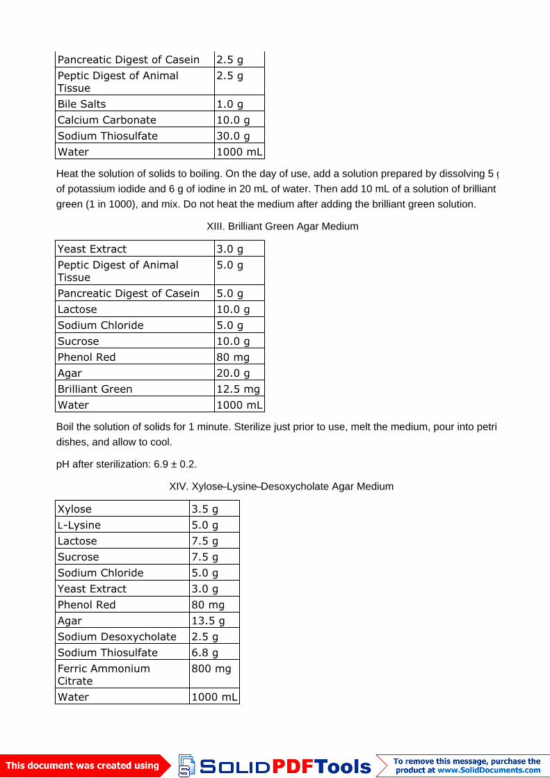
Pancreatic Digest of Casein 2.5 gPeptic Digest of AnimalTissue
2.5 g
Bile Salts 1.0 gCalcium Carbonate 10.0 gSodium Thiosulfate 30.0 gWater 1000 mL
Heat the solution of solids to boiling. On the day of use, add a solution prepared by dissolving 5 gof potassium iodide and 6 g of iodine in 20 mL of water. Then add 10 mL of a solution of brilliantgreen (1 in 1000), and mix. Do not heat the medium after adding the brilliant green solution.
XIII. Brilliant Green Agar Medium
Yeast Extract 3.0 gPeptic Digest of AnimalTissue
5.0 g
Pancreatic Digest of Casein 5.0 gLactose 10.0 gSodium Chloride 5.0 gSucrose 10.0 gPhenol Red 80 mgAgar 20.0 gBrilliant Green 12.5 mgWater 1000 mL
Boil the solution of solids for 1 minute. Sterilize just prior to use, melt the medium, pour into petridishes, and allow to cool.
pH after sterilization: 6.9 ± 0.2.
XIV. Xylose–Lysine–Desoxycholate Agar Medium
Xylose 3.5 gL-Lysine 5.0 gLactose 7.5 gSucrose 7.5 gSodium Chloride 5.0 gYeast Extract 3.0 gPhenol Red 80 mgAgar 13.5 gSodium Desoxycholate 2.5 gSodium Thiosulfate 6.8 gFerric AmmoniumCitrate
800 mg
Water 1000 mL
Final pH: 7.4 ± 0.2.
Heat the mixture of solids and water, with swirling, just to the boiling point.Do not overheat or
sterilize. Transfer at once to a water bath maintained at about 50 , and pour into plates as soon asthe medium has cooled.
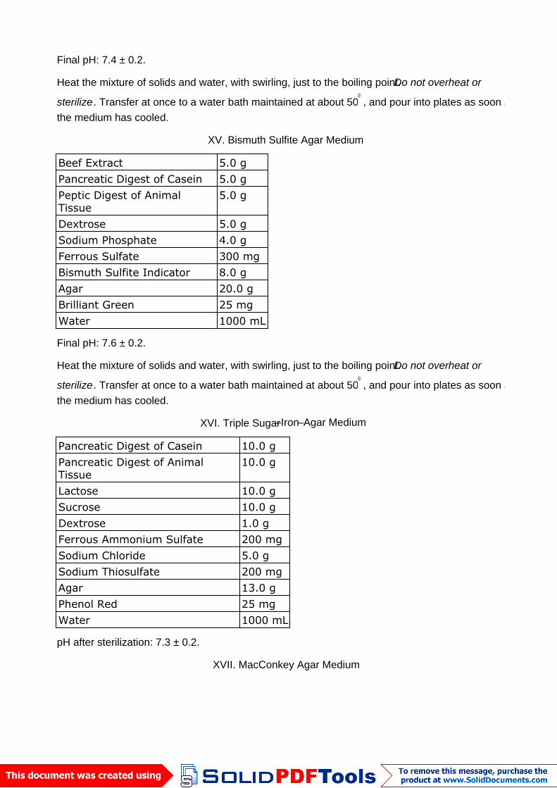
XV. Bismuth Sulfite Agar Medium
Beef Extract 5.0 gPancreatic Digest of Casein 5.0 gPeptic Digest of AnimalTissue
5.0 g
Dextrose 5.0 gSodium Phosphate 4.0 gFerrous Sulfate 300 mgBismuth Sulfite Indicator 8.0 gAgar 20.0 gBrilliant Green 25 mgWater 1000 mL
Final pH: 7.6 ± 0.2.
Heat the mixture of solids and water, with swirling, just to the boiling point.Do not overheat or
sterilize. Transfer at once to a water bath maintained at about 50 , and pour into plates as soon asthe medium has cooled.
XVI. Triple Sugar–Iron–Agar Medium
Pancreatic Digest of Casein 10.0 gPancreatic Digest of AnimalTissue
10.0 g
Lactose 10.0 gSucrose 10.0 gDextrose 1.0 gFerrous Ammonium Sulfate 200 mgSodium Chloride 5.0 gSodium Thiosulfate 200 mgAgar 13.0 gPhenol Red 25 mgWater 1000 mL
pH after sterilization: 7.3 ± 0.2.
XVII. MacConkey Agar Medium
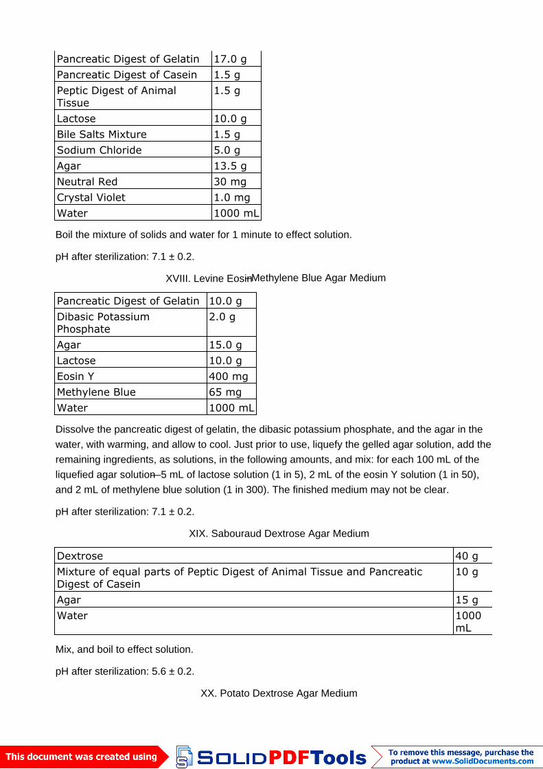
Pancreatic Digest of Gelatin 17.0 gPancreatic Digest of Casein 1.5 gPeptic Digest of AnimalTissue
1.5 g
Lactose 10.0 gBile Salts Mixture 1.5 gSodium Chloride 5.0 gAgar 13.5 gNeutral Red 30 mgCrystal Violet 1.0 mgWater 1000 mL
Boil the mixture of solids and water for 1 minute to effect solution.
pH after sterilization: 7.1 ± 0.2.
XVIII. Levine Eosin–Methylene Blue Agar Medium
Pancreatic Digest of Gelatin 10.0 gDibasic PotassiumPhosphate
2.0 g
Agar 15.0 gLactose 10.0 gEosin Y 400 mgMethylene Blue 65 mgWater 1000 mL
Dissolve the pancreatic digest of gelatin, the dibasic potassium phosphate, and the agar in thewater, with warming, and allow to cool. Just prior to use, liquefy the gelled agar solution, add theremaining ingredients, as solutions, in the following amounts, and mix: for each 100 mL of theliquefied agar solution—5 mL of lactose solution (1 in 5), 2 mL of the eosin Y solution (1 in 50),and 2 mL of methylene blue solution (1 in 300). The finished medium may not be clear.
pH after sterilization: 7.1 ± 0.2.
XIX. Sabouraud Dextrose Agar Medium
Dextrose 40 gMixture of equal parts of Peptic Digest of Animal Tissue and PancreaticDigest of Casein
10 g
Agar 15 gWater 1000
mL
Mix, and boil to effect solution.
pH after sterilization: 5.6 ± 0.2.
XX. Potato Dextrose Agar Medium
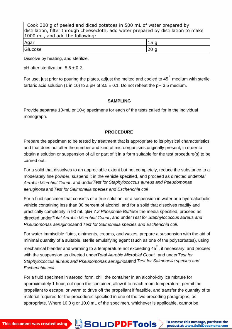
Cook 300 g of peeled and diced potatoes in 500 mL of water prepared bydistillation, filter through cheesecloth, add water prepared by distillation to make1000 mL, and add the following:Agar 15 gGlucose 20 g
Dissolve by heating, and sterilize.
pH after sterilization: 5.6 ± 0.2.
For use, just prior to pouring the plates, adjust the melted and cooled to 45 medium with steriletartaric acid solution (1 in 10) to a pH of 3.5 ± 0.1. Do not reheat the pH 3.5 medium.
SAMPLING
Provide separate 10-mL or 10-g specimens for each of the tests called for in the individualmonograph.
PROCEDURE
Prepare the specimen to be tested by treatment that is appropriate to its physical characteristicsand that does not alter the number and kind of microorganisms originally present, in order toobtain a solution or suspension of all or part of it in a form suitable for the test procedure(s) to becarried out.
For a solid that dissolves to an appreciable extent but not completely, reduce the substance to amoderately fine powder, suspend it in the vehicle specified, and proceed as directed underTotalAerobic Microbial Count, and underTest for Staphylococcus aureus and Pseudomonasaeruginosa andTest for Salmonella species and Escherichia coli .
For a fluid specimen that consists of a true solution, or a suspension in water or a hydroalcoholicvehicle containing less than 30 percent of alcohol, and for a solid that dissolves readily andpractically completely in 90 mL ofpH 7.2 Phosphate Buffer or the media specified, proceed asdirected underTotal Aerobic Microbial Count , and underTest for Staphylococcus aureus andPseudomonas aeruginosa and Test for Salmonella species and Escherichia coli.
For water-immiscible fluids, ointments, creams, and waxes, prepare a suspension with the aid of aminimal quantity of a suitable, sterile emulsifying agent (such as one of the polysorbates), using a
mechanical blender and warming to a temperature not exceeding 45 , if necessary, and proceedwith the suspension as directed underTotal Aerobic Microbial Count , and underTest forStaphylococcus aureus and Pseudomonas aeruginosa and Test for Salmonella species andEscherichia coli .
For a fluid specimen in aerosol form, chill the container in an alcohol-dry ice mixture forapproximately 1 hour, cut open the container, allow it to reach room temperature, permit thepropellant to escape, or warm to drive off the propellant if feasible, and transfer the quantity of testmaterial required for the procedures specified in one of the two preceding paragraphs, asappropriate. Where 10.0 g or 10.0 mL of the specimen, whichever is applicable, cannot be
obtained from 10 containers in aerosol form, transfer the entire contents from 10 chilled containersto the culture medium, permit the propellant to escape, and proceed with the test on the residues.If the results of the test are inconclusive or doubtful, repeat the test with a specimen from 20 morecontainers.
Total Aerobic Microbial Count
For specimens that are sufficiently soluble or translucent to permit use of thePlate Method, usethat method; otherwise, use theMultiple-Tube Method. With either method, first dissolve orsuspend 10.0 g of the specimen if it is a solid, or 10 mL, accurately measured, if the specimen is aliquid, inpH 7.2 Phosphate Buffer, Fluid Soybean–Casein Digest Medium, or Fluid CaseinDigest–Soy Lecithin-Polysorbate 20 Medium to make 100 mL. For viscous specimens that cannotbe pipeted at this initial 1:10 dilution, dilute the specimen until a suspension is obtained, i.e., 1:50or 1:100, etc., that can be pipeted. Perform the test for absence of inhibitory (antimicrobial)properties as described underPreparatory Testing before the determination ofTotal AerobicMicrobial Count. Add the specimen to the medium not more than 1 hour after preparing theappropriate dilutions for inoculation.
PLATE METHOD
Dilute further, if necessary, the fluid so that 1 mL will be expected to yield between 30 and 300colonies. Pipet 1 mL of the final dilution onto each of two sterile petri dishes. Promptly add to eachdish 15 to 20 mL ofSoybean–Casein Digest Agar Medium that previously has been melted and
cooled to approximately 45 . Cover the petri dishes, mix the sample with the agar by tilting orrotating the dishes, and allow the contents to solidify at room temperature. Invert the petri dishes,and incubate for 48 to 72 hours. Following incubation, examine the plates for growth, count thenumber of colonies, and express the average for the two plates in terms of the number ofmicroorganisms per g or per mL of specimen. If no microbial colonies are recovered from thedishes representing the initial 1:10 dilution of the specimen, express the results as “less than 10microorganisms per g or per mL of specimen.”
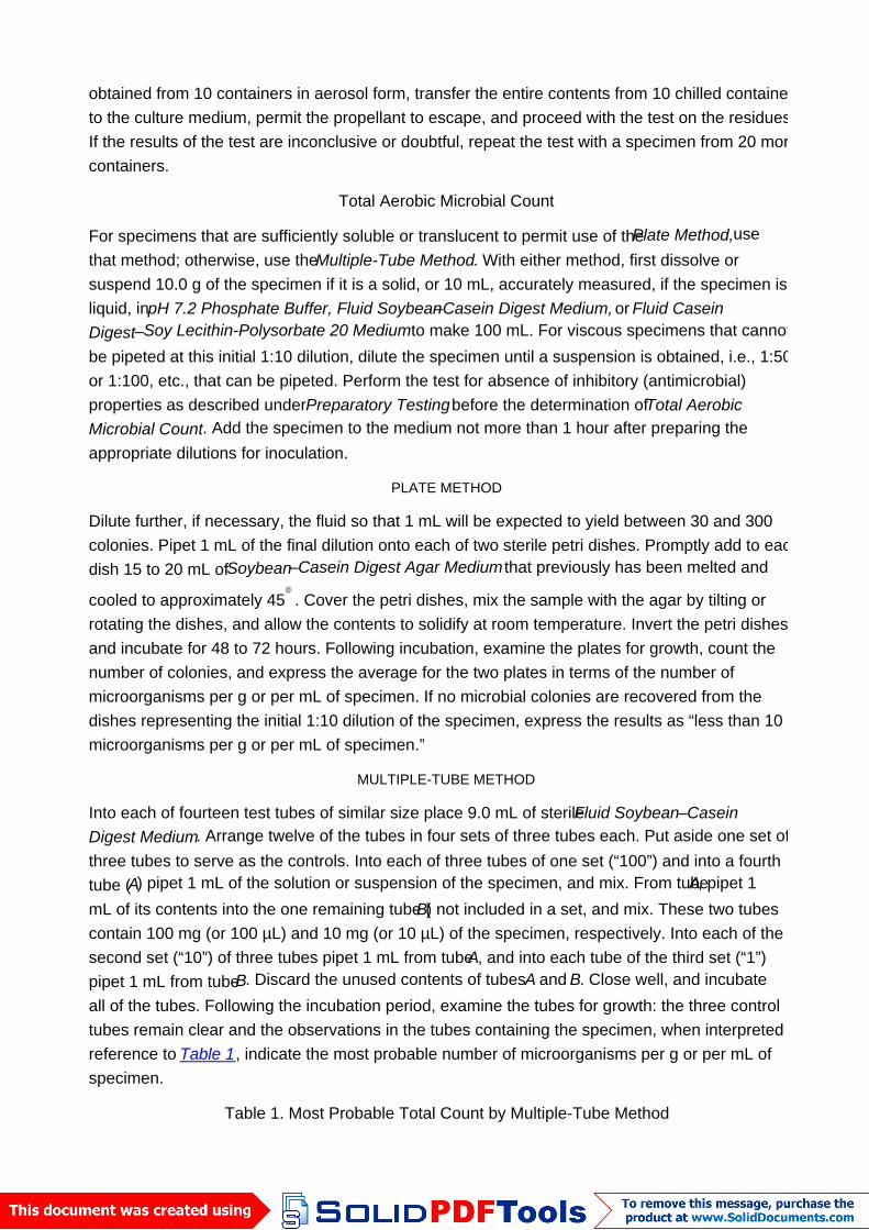
MULTIPLE-TUBE METHOD
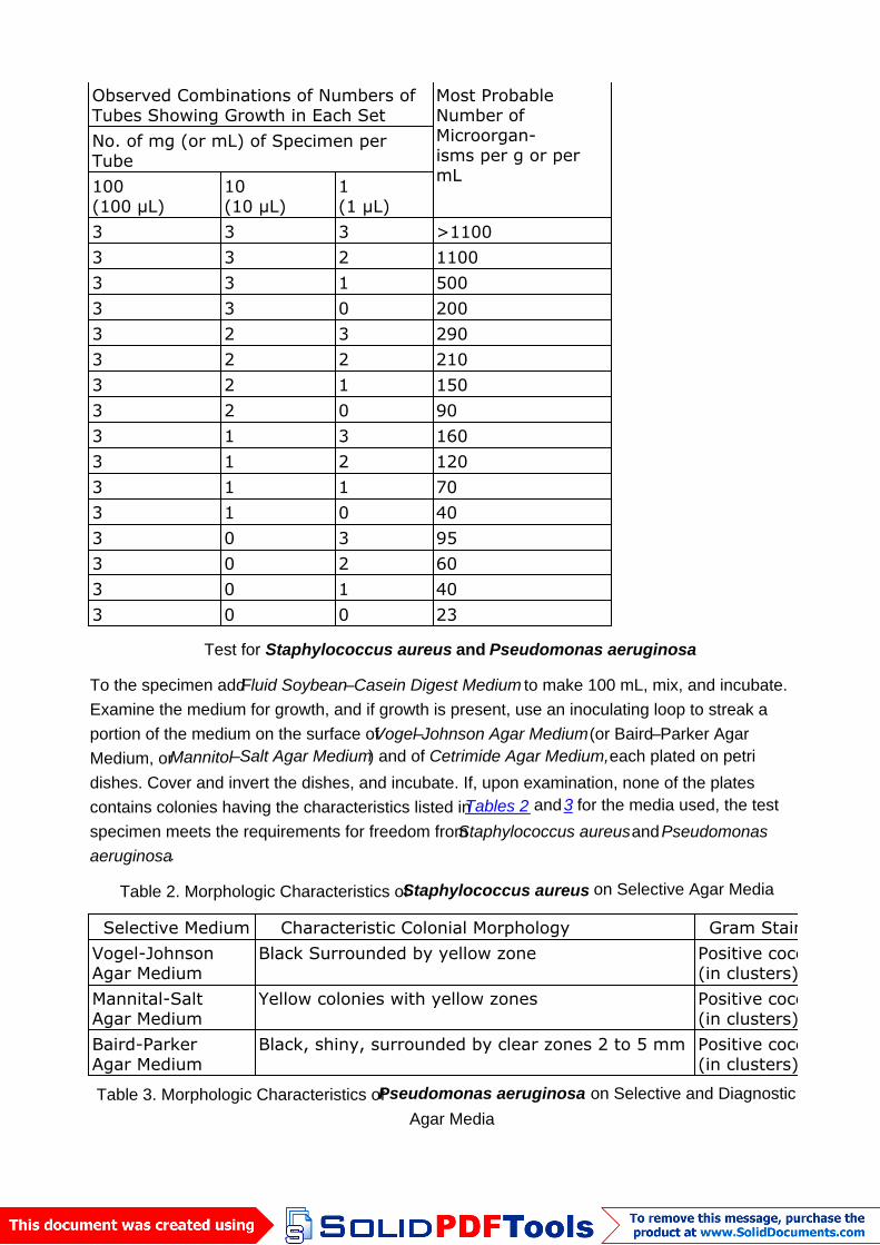
Into each of fourteen test tubes of similar size place 9.0 mL of sterileFluid Soybean–CaseinDigest Medium. Arrange twelve of the tubes in four sets of three tubes each. Put aside one set ofthree tubes to serve as the controls. Into each of three tubes of one set (“100”) and into a fourthtube (A) pipet 1 mL of the solution or suspension of the specimen, and mix. From tubeA, pipet 1mL of its contents into the one remaining tube (B) not included in a set, and mix. These two tubescontain 100 mg (or 100 µL) and 10 mg (or 10 µL) of the specimen, respectively. Into each of thesecond set (“10”) of three tubes pipet 1 mL from tubeA, and into each tube of the third set (“1”)pipet 1 mL from tubeB. Discard the unused contents of tubesA and B. Close well, and incubateall of the tubes. Following the incubation period, examine the tubes for growth: the three controltubes remain clear and the observations in the tubes containing the specimen, when interpreted byreference to Table 1, indicate the most probable number of microorganisms per g or per mL ofspecimen.
Table 1. Most Probable Total Count by Multiple-Tube Method
Observed Combinations of Numbers ofTubes Showing Growth in Each Set
Most ProbableNumber ofMicroorgan-isms per g or permL
No. of mg (or mL) of Specimen perTube100(100 µL)
10(10 µL)
1(1 µL)
3 3 3 >11003 3 2 11003 3 1 5003 3 0 2003 2 3 2903 2 2 2103 2 1 1503 2 0 903 1 3 1603 1 2 1203 1 1 703 1 0 403 0 3 953 0 2 603 0 1 403 0 0 23
Test for Staphylococcus aureus and Pseudomonas aeruginosa
To the specimen addFluid Soybean–Casein Digest Medium to make 100 mL, mix, and incubate.Examine the medium for growth, and if growth is present, use an inoculating loop to streak aportion of the medium on the surface ofVogel–Johnson Agar Medium (or Baird–Parker AgarMedium, orMannitol–Salt Agar Medium) and of Cetrimide Agar Medium, each plated on petridishes. Cover and invert the dishes, and incubate. If, upon examination, none of the platescontains colonies having the characteristics listed inTables 2 and3 for the media used, the testspecimen meets the requirements for freedom fromStaphylococcus aureus andPseudomonasaeruginosa.
Table 2. Morphologic Characteristics ofStaphylococcus aureus on Selective Agar Media
Selective Medium Characteristic Colonial Morphology Gram StainVogel-JohnsonAgar Medium
Black Surrounded by yellow zone Positive cocci(in clusters)
Mannital-SaltAgar Medium
Yellow colonies with yellow zones Positive cocci(in clusters)
Baird-ParkerAgar Medium
Black, shiny, surrounded by clear zones 2 to 5 mm Positive cocci(in clusters)
Table 3. Morphologic Characteristics ofPseudomonas aeruginosa on Selective and DiagnosticAgar Media
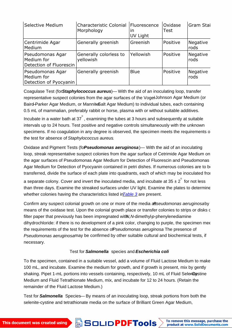
Selective Medium Characteristic ColonialMorphology
FluorescenceinUV Light
OxidaseTest
Gram Stain
Centrimide AgarMedium
Generally greenish Greenish Positive Negativerods
Pseudomonas AgarMedium forDetection of Fluorescin
Generally colorless toyellowish
Yellowish Positive Negativerods
Pseudomonas AgarMedium forDetection of Pyocyanin
Generally greenish Blue Positive Negativerods
Coagulase Test (forStaphylococcus aureus)— With the aid of an inoculating loop, transferrepresentative suspect colonies from the agar surfaces of the Vogel–Johnson Agar Medium (orBaird–Parker Agar Medium, or Mannitol–Salt Agar Medium) to individual tubes, each containing0.5 mL of mammalian, preferably rabbit or horse, plasma with or without suitable additives.
Incubate in a water bath at 37 , examining the tubes at 3 hours and subsequently at suitableintervals up to 24 hours. Test positive and negative controls simultaneously with the unknownspecimens. If no coagulation in any degree is observed, the specimen meets the requirements ofthe test for absence of Staphylococcus aureus.
Oxidase and Pigment Tests (forPseudomonas aeruginosa)— With the aid of an inoculatingloop, streak representative suspect colonies from the agar surface of Cetrimide Agar Medium onthe agar surfaces of Pseudomonas Agar Medium for Detection of Fluorescin and PseudomonasAgar Medium for Detection of Pyocyanin contained in petri dishes. If numerous colonies are to betransferred, divide the surface of each plate into quadrants, each of which may be inoculated from
a separate colony. Cover and invert the inoculated media, and incubate at 35 ± 2 for not lessthan three days. Examine the streaked surfaces under UV light. Examine the plates to determinewhether colonies having the characteristics listed inTable 3 are present.
Confirm any suspect colonial growth on one or more of the media asPseudomonas aeruginosa bymeans of the oxidase test. Upon the colonial growth place or transfer colonies to strips or disks offilter paper that previously has been impregnated withN,N-dimethyl-p-phenylenediaminedihydrochloride: if there is no development of a pink color, changing to purple, the specimen meetsthe requirements of the test for the absence ofPseudomonas aeruginosa. The presence ofPseudomonas aeruginosa may be confirmed by other suitable cultural and biochemical tests, ifnecessary.
Test for Salmonella species and Escherichia coli
To the specimen, contained in a suitable vessel, add a volume of Fluid Lactose Medium to make100 mL, and incubate. Examine the medium for growth, and if growth is present, mix by gentlyshaking. Pipet 1-mL portions into vessels containing, respectively, 10 mL of Fluid Selenite–CystineMedium and Fluid Tetrathionate Medium, mix, and incubate for 12 to 24 hours. (Retain theremainder of the Fluid Lactose Medium.)
Test for Salmonella Species— By means of an inoculating loop, streak portions from both theselenite-cystine and tetrathionate media on the surface of Brilliant Green Agar Medium,
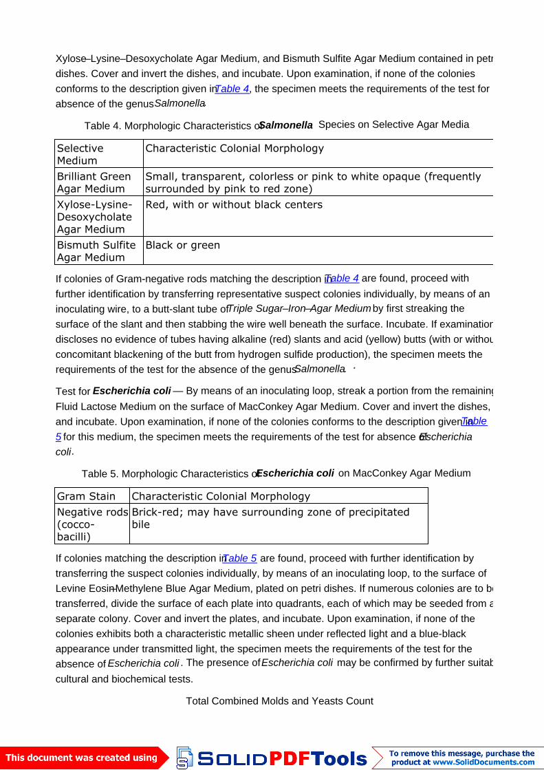
Xylose–Lysine–Desoxycholate Agar Medium, and Bismuth Sulfite Agar Medium contained in petridishes. Cover and invert the dishes, and incubate. Upon examination, if none of the coloniesconforms to the description given inTable 4, the specimen meets the requirements of the test forabsence of the genusSalmonella.
Table 4. Morphologic Characteristics ofSalmonella Species on Selective Agar Media
SelectiveMedium
Characteristic Colonial Morphology
Brilliant GreenAgar Medium
Small, transparent, colorless or pink to white opaque (frequentlysurrounded by pink to red zone)
Xylose-Lysine-DesoxycholateAgar Medium
Red, with or without black centers
Bismuth SulfiteAgar Medium
Black or green
If colonies of Gram-negative rods matching the description inTable 4 are found, proceed withfurther identification by transferring representative suspect colonies individually, by means of aninoculating wire, to a butt-slant tube ofTriple Sugar–Iron–Agar Medium by first streaking thesurface of the slant and then stabbing the wire well beneath the surface. Incubate. If examinationdiscloses no evidence of tubes having alkaline (red) slants and acid (yellow) butts (with or withoutconcomitant blackening of the butt from hydrogen sulfide production), the specimen meets therequirements of the test for the absence of the genusSalmonella. *
Test for Escherichia coli — By means of an inoculating loop, streak a portion from the remainingFluid Lactose Medium on the surface of MacConkey Agar Medium. Cover and invert the dishes,and incubate. Upon examination, if none of the colonies conforms to the description given inTable5 for this medium, the specimen meets the requirements of the test for absence ofEscherichiacoli.
Table 5. Morphologic Characteristics ofEscherichia coli on MacConkey Agar Medium
Gram Stain Characteristic Colonial MorphologyNegative rods(cocco-bacilli)
Brick-red; may have surrounding zone of precipitatedbile
If colonies matching the description inTable 5 are found, proceed with further identification bytransferring the suspect colonies individually, by means of an inoculating loop, to the surface ofLevine Eosin–Methylene Blue Agar Medium, plated on petri dishes. If numerous colonies are to betransferred, divide the surface of each plate into quadrants, each of which may be seeded from aseparate colony. Cover and invert the plates, and incubate. Upon examination, if none of thecolonies exhibits both a characteristic metallic sheen under reflected light and a blue-blackappearance under transmitted light, the specimen meets the requirements of the test for theabsence of Escherichia coli . The presence ofEscherichia coli may be confirmed by further suitablecultural and biochemical tests.
Total Combined Molds and Yeasts Count
Proceed as for thePlate Method underTotal Aerobic Microbial Count , except for using the sameamount of Sabouraud Dextrose Agar Medium or Potato Dextrose Agar Medium, instead ofSoybean Casein Digest Medium, and except for incubating the inverted petri dishes for 5 to 7 days
at 20 to 25 .
Retest
For the purpose of confirming a doubtful result by any of the procedures outlined in the foregoingtests following their application to a 10.0-g specimen, a retest on a 25-g specimen of the productmay be conducted. Proceed as directed forProcedure, but make allowance for the largerspecimen size.
* Additional, confirmatory evidence may be obtained by use of procedures set forth in Official Methods of Analysisof the AOAC , 12th ed. (1975), sections 46.013-46.026.
Auxiliary Information—Staff Liaison : Radhakrishna S Tirumalai, Scientist
Expert Committee : (MSA05) Microbiology and Sterility Assurance
USP30–NF25 Page 83
Phone Number : 1-301-816-8339