Embed Size (px)
Citation preview

SKILLS CENTER
STANDARD OPERATING PROCEDURE
A BIOFIZZ
PRODUCTON
Macromolecule Structure Analysis
Using Chimera Module Hours: 3
Effective Date: 03/01/2021 PRQs
Introduction to Chimera
Revision # 1.0 A. Siclair
Checked: M. Guzie
- 1 -
Analyzing the structure and function of macromolecules is imperative to draw connections between different proteins. By considering structural similarity or conserved regions of proteins between species, it becomes easier to describe the functionality and structure of those that have been studied less or have yet to be fully described. Applications like UCSF Chimera allow for this sort of comparison with the ability to analyze sequences and specified components of the structures like distances, bonding formation and hydrophobicity.
The Chimera application, established by the
Resource for Biocomputing, Visualization and Informatics (RBVI), allows for detailed 3D molecular visualization and interactive assessment of macromolecules (RBVI, 2018). When specifically looking at proteins, this program analyzes characteristics such as chemical makeup, bond formation, and can even be utilized for detailed structure comparison.
The information about specific proteins and their corresponding structures is kept in separate databases and can be converted for use within Chimera. Although the Worldwide Protein Data Bank currently sits at the top of the protein database food-chain, with many smaller databases contributing to it, other reliable sources like the RCSB Protein Data Bank have been around for nearly 50 years and have been accumulating protein information since their creation (Berman et al., 2012). The RCSB Protein Data Bank originally consisted of a mere 7 structures, but now contains documentation about thousands of proteins from numerous different organisms (RCSB PDB).
The most important benefits of protein analysis technology lie in the ability to build upon existing knowledge of structures. Through comparison of various domains, residues, etc., more headway can be made in uncovering homologous regions, functions, and entire proteins between organisms. It is also beneficial for analyzing the role of mutation and protein misfolding and misfunction in disease. Possessing the capacity to magnify such tiny structures and analyze them virtually with a fine-tooth comb is something that could never be feasible without the computing applications of today such as Chimera.
PDB dopamine receptors 3PBL and 6CM4 with overlapping structural chains.

SKILLS CENTER
STANDARD OPERATING PROCEDURE
A BIOFIZZ
PRODUCTON
Macromolecule Structure Analysis
Using Chimera Module Hours: 3
Effective Date: 03/01/2021 PRQs
Introduction to Chimera
Revision # 1.0 A. Siclair
Checked: M. Guzie
- 2 -
1. PURPOSE
The purpose of this procedure is to become comfortable with analyzing and comparing various components of the structures of macromolecules, specifically emphasizing proteins.
2. SCOPE
This procedure applies to qualified skills center users. 3. RESPONSIBILITY
3.1. It is the responsibility of the user to understand and perform the procedure described in this document.
3.2. It is the responsibility of the user performing the procedure to fully document any deviations from the written procedure.
3.3. It is the responsibility of the user to become trained on the procedure. 4. DEFINITIONS
4.1. Protein Data Bank – A database that houses information about 3D macromolecule structures, such as proteins and nucleic acids.
4.2. RCSB Protein Data Bank – The first open access digital data resource in all of biology that has been around since 1971.
4.3. UCSF Chimera – An application that allows for detailed molecular visualization and interactive assessment of macromolecules.
4.4. Chimera X – A condensed version of the UCSF Chimera application that can be used on slower computing devices or with larger molecular structures.
4.5. Biological assembly – the macromolecular assembly that has been presumed or shown to be the functional form of the macromolecule.
5. MATERIALS/EQUIPMENT
5.1. UCSF Chimera Application 5.2. RCSB PDB 5.3. Worldwide PDB 5.4. 7zip software
6. PROCEDURE

SKILLS CENTER
STANDARD OPERATING PROCEDURE
A BIOFIZZ
PRODUCTON
Macromolecule Structure Analysis
Using Chimera Module Hours: 3
Effective Date: 03/01/2021 PRQs
Introduction to Chimera
Revision # 1.0 A. Siclair
Checked: M. Guzie
- 3 -
Before beginning this procedure, make sure to become familiar with all of the tools and techniques used in the Chimera operating procedure.
6.1. Choosing and Importing the Proteins of Interest
6.1.1. Visit the RCSB protein databank website: https://www.rcsb.org/ - The Worldwide Protein Data Bank is also an optimal source:
http://www.wwpdb.org/ (But not used in this Procedure). 6.1.2. Choose two proteins that are related in some way and download the
biological assembly file. - For this example, 2 dopamine receptors are being used: PDB 3PBL and
6CM4. 6.1.3. Unzip the biological assembly downloaded files. 6.1.4. Open the chimera application and import the molecules. Color each chain
of each molecule different colors.
6.2. Structure Alignment Comparison Between Proteins 6.2.1. Click Tools, Structure Comparison, and MatchMaker. A new window
should pop up. (Figure 1)
6.2.2. In the pop-up window, select one of the molecules as the reference
structure and the other as the structure to match. Click Apply and then OK. (Figure 2) - This should align a portion of the structures if there is similarity present.
Figure 1 Figure 2

SKILLS CENTER
STANDARD OPERATING PROCEDURE
A BIOFIZZ
PRODUCTON
Macromolecule Structure Analysis
Using Chimera Module Hours: 3
Effective Date: 03/01/2021 PRQs
Introduction to Chimera
Revision # 1.0 A. Siclair
Checked: M. Guzie
- 4 -
- It is possible to only align specific chains as well, if the third option for chain pairing is selected. Choose a reference chain and a chain to match, then Apply. (Figure 3)
6.2.3. Once the structures are aligned, the chains that differ between the 2
structures can be hidden. Select the chains to be hidden and click Actions, Atoms/Bonds and then hide. (Figure 4)
6.2.4. Click Actions and then Focus to fit the superimposed molecules to the frame. (Figure 4)
6.2.5. To compare the sequences of the superimposed region, click Tools, Sequence and Align Chain Sequences. (Figure 5)
Figure 4
Figure 3
Figure 5
Figure 6
SKILLS CENTER
STANDARD OPERATING PROCEDURE
A BIOFIZZ
PRODUCTON
Macromolecule Structure Analysis
Using Chimera Module Hours: 3
Effective Date: 03/01/2021 PRQs
Introduction to Chimera
Revision # 1.0 A. Siclair
Checked: M. Guzie
- 5 -
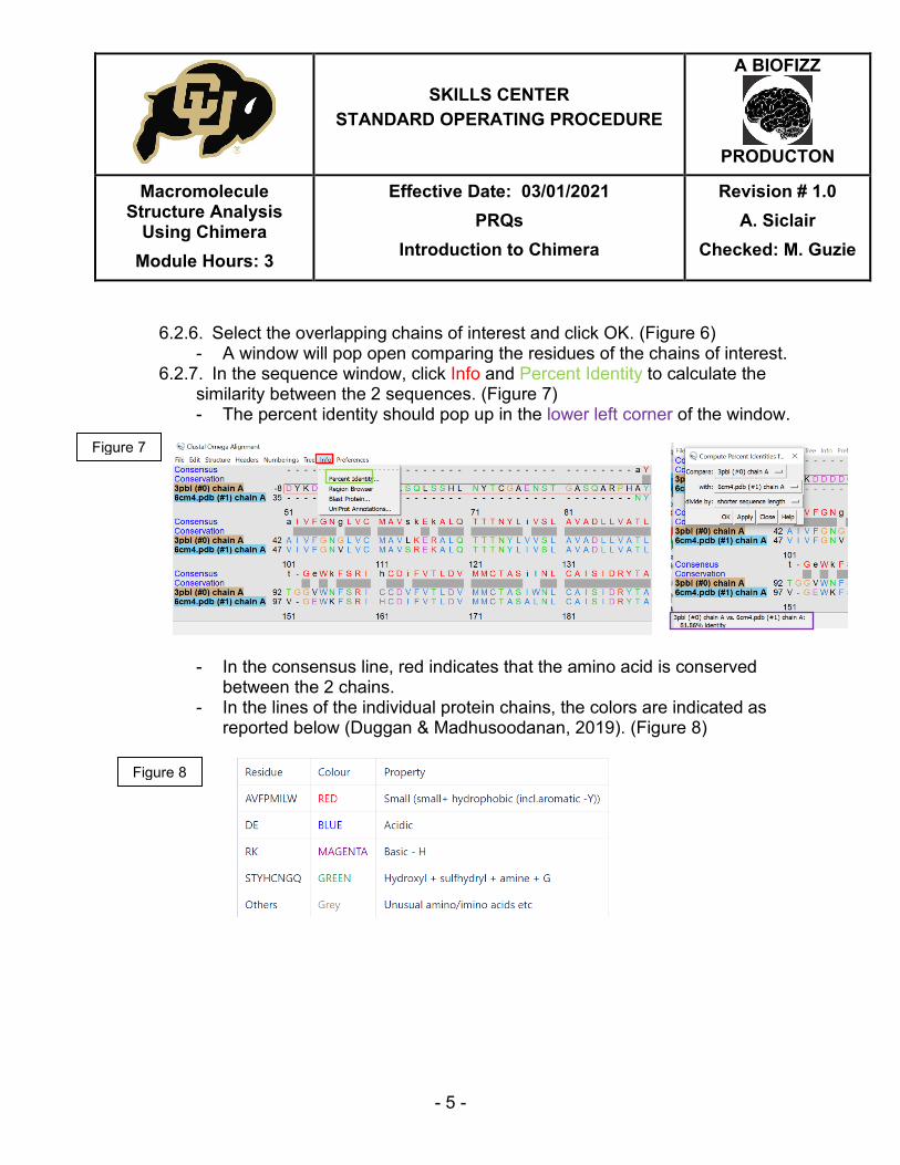
6.2.6. Select the overlapping chains of interest and click OK. (Figure 6)
- A window will pop open comparing the residues of the chains of interest. 6.2.7. In the sequence window, click Info and Percent Identity to calculate the
similarity between the 2 sequences. (Figure 7) - The percent identity should pop up in the lower left corner of the window.
- In the consensus line, red indicates that the amino acid is conserved
between the 2 chains. - In the lines of the individual protein chains, the colors are indicated as
reported below (Duggan & Madhusoodanan, 2019). (Figure 8)
Figure 7
Figure 8

SKILLS CENTER
STANDARD OPERATING PROCEDURE
A BIOFIZZ
PRODUCTON
Macromolecule Structure Analysis
Using Chimera Module Hours: 3
Effective Date: 03/01/2021 PRQs
Introduction to Chimera
Revision # 1.0 A. Siclair
Checked: M. Guzie
- 6 -
6.3. Analysis of Chain Structure Within a Single Protein
6.3.1. To view the superimposed section of each of the 2 molecules separately, click Favorites and then Model Panel. (Figure 9)
6.3.2. Deselect Shown for one of the molecules to hide its chains. (Figure 10) 6.3.3. Establish the hydrogen bonding in the structure by clicking Tools,
Structure Analysis and FindHBond. (Figure 11) - In the pop-up window, use all of the default controls and click OK. The
hydrogen bonds should appear in the selected chain. (Figure 12)
Figure 9
Figure 10
Figure 11
Figure 12
SKILLS CENTER
STANDARD OPERATING PROCEDURE
A BIOFIZZ
PRODUCTON
Macromolecule Structure Analysis
Using Chimera Module Hours: 3
Effective Date: 03/01/2021 PRQs
Introduction to Chimera
Revision # 1.0 A. Siclair
Checked: M. Guzie
- 7 -
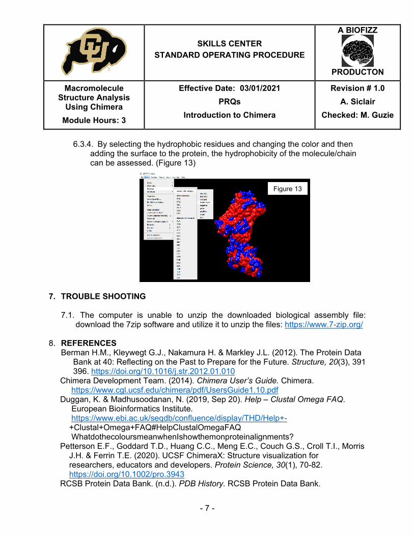
6.3.4. By selecting the hydrophobic residues and changing the color and then adding the surface to the protein, the hydrophobicity of the molecule/chain can be assessed. (Figure 13)
7. TROUBLE SHOOTING
7.1. The computer is unable to unzip the downloaded biological assembly file:
download the 7zip software and utilize it to unzip the files: https://www.7-zip.org/
8. REFERENCES Berman H.M., Kleywegt G.J., Nakamura H. & Markley J.L. (2012). The Protein Data
Bank at 40: Reflecting on the Past to Prepare for the Future. Structure, 20(3), 391 396. https://doi.org/10.1016/j.str.2012.01.010
Chimera Development Team. (2014). Chimera User’s Guide. Chimera. https://www.cgl.ucsf.edu/chimera/pdf/UsersGuide1.10.pdf Duggan, K. & Madhusoodanan, N. (2019, Sep 20). Help – Clustal Omega FAQ. European Bioinformatics Institute. https://www.ebi.ac.uk/seqdb/confluence/display/THD/Help+- +Clustal+Omega+FAQ#HelpClustalOmegaFAQ WhatdothecoloursmeanwhenIshowthemonproteinalignments? Petterson E.F., Goddard T.D., Huang C.C., Meng E.C., Couch G.S., Croll T.I., Morris J.H. & Ferrin T.E. (2020). UCSF ChimeraX: Structure visualization for researchers, educators and developers. Protein Science, 30(1), 70-82. https://doi.org/10.1002/pro.3943 RCSB Protein Data Bank. (n.d.). PDB History. RCSB Protein Data Bank.
Figure 13

SKILLS CENTER
STANDARD OPERATING PROCEDURE
A BIOFIZZ
PRODUCTON
Macromolecule Structure Analysis
Using Chimera Module Hours: 3
Effective Date: 03/01/2021 PRQs
Introduction to Chimera
Revision # 1.0 A. Siclair
Checked: M. Guzie
- 8 -
https://www.rcsb.org/pages/about-us/history RCSB Protein Data Bank. (2010). 3PBL: Structure of the human dopamine D3 receptor in complex with eticlopride. RCSB Protein Data Bank. https://www.rcsb.org/structure/3PBL RCSB Protein Data Bank. (2018). 6CM4: Structure of the D2 Dopamine Receptor Bound to the Atypical Antipsychotic Drug Risperidone. RCSB Protein Data Bank. https://www.rcsb.org/structure/6CM4 Resource for Biocomputing, Visualization and Informatics. (2018). About Us. UCSF Resource for Biocomputing, Visualization and Informatics. https://www.cgl.ucsf.edu/Overview/index.html Resource for Biocomputing, Visualization and Informatics. (2018). UCSF Chimera: An Extensible Molecular Modeling System. UCSF Resource for Biocomputing, Visualization and Informatics. https://www.cgl.ucsf.edu/chimera/ Shuchismita, D. [RCSBProteinDataBank]. (2014, Dec 23). UCSF Chimera: Structure Analysis [Video]. Youtube. https://www.youtube.com/watch?v=eLxhKc7Ljjk Shuchismita, D. [RCSBProteinDataBank]. (2014, Dec 23). UCSF Chimera: Structure Comparisons [Video]. Youtube. https://www.youtube.com/watch?v=oThN3LG8LQU Zabriskie, C. [BioPandit]. (2018, Mar 1). Basic Guide to Protein Data Bank and PDB Files. Youtube. https://www.youtube.com/watch?v=Fz5t5QPUyi8 9. MODULE MASTERY TASK This quiz will test your knowledge of the basic visualization and structure comparison techniques that can be implemented in the UCSF Chimera application. Choose 2 related proteins from the RCSB Protein Data Bank to complete the following questions.
1. What are the PDB codes for your chosen proteins? 2. Import the proteins into Chimera and use the MatchMaker function to align the
proteins. Submit an image of the result. 3. Align 2 specific chains in the chosen proteins and then hide the remaining chains.
Submit an image of the result. 4. Compare the sequences of the overlapping chains of the proteins. What is the
percent identity match of the regions you chose? 5. Alter the visualization so that the superimposed chain of only one of the proteins
is in the window. Add hydrogen bonds to that region and submit and image.

SKILLS CENTER
STANDARD OPERATING PROCEDURE
A BIOFIZZ
PRODUCTON
Macromolecule Structure Analysis
Using Chimera Module Hours: 3
Effective Date: 03/01/2021 PRQs
Introduction to Chimera
Revision # 1.0 A. Siclair
Checked: M. Guzie
- 9 -
6. Color the hydrophobic residues a different color and implement the surface function. Submit the resulting image.
7. Repeat steps 4 and 5 with the other protein’s superimposed region. 8. Does the hydrophobicity of the 2 superimposed regions look similar?