Embed Size (px)
Citation preview
WWW.KJOG.ORG 361
A CASE OF ENDOMETRIAL OSSEOUS METAPLASIA TREATED BY HYSTEROSCOPIC OPERATIONJi Yeon Lee, MD, Hyang Ah Lee, MD, PhD, Hyuk Min Kwon, MD, Sung Hoon Na, MD, Jong Yun Hwang, MD, PhD, Dong Heon Lee, MD, PhD Department of Obstetrics and Gynecology, Kangwon National University School of Medicine, Chuncheon, Korea
Endometrial osseous metaplasia is a very rare disease related to secondary infertility. A 42-year-old woman, a North Korean defector, visited for gynecological exam. She presented with a history of secondary infertility. She had two previous pregnancies and both were voluntarily terminated at 14 and 16 years ago. Vaginal delivery at about 20 weeks in the fi rst gestation 16 years ago; and dilatation and curettage at about 14 weeks 14 years ago were done in a medical facility of North Korea. Vaginal ultrasonography showed an intrauterine structure described as a hyperechogenic image suggesting calcifi cation. Hysteroscopy revealed multiple coral-like white spicules about 1 cm in length in the uterine cavity. The lesion was treated by hysteroscopic removal without complications. Histology established the diagnosis of endometrial osseous. In our case, hysteroscopy was effective in the diagnosis and treatment of endometrial osseous metaplasia.
Keywords: Osseous metaplasia; Hysteroscopy; Secondary infertility
Received: 2012.2.9. Revised: 2012.3.29. Accepted: 2012.4.23.Corresponding author: Hyang Ah Lee, MD, PhDDepartment of Obstetrics and Gynecology, Kangwon National University Hospital, Kangwon National University School of Medicine, 156 Baengnyeong-ro, Chuncheon 200-722, KoreaTel: +82-33-258-2307 Fax: +82-33-256-1376E-mail: [email protected]
Th is is an Open Access article distributed under the terms of the Creative Commons Attribution Non-Commercial License (http://creativecommons.org/licenses/by-nc/3.0/) which permits unrestricted non-commercial use, distribution, and reproduction in any medium, provided the original work is properly cited.
Copyright © 2012. Korean Society of Obstetrics and Gynecology
Osseous metaplasia is a very rare disorder of endometrium that usually leads to secondary infertility. Endometrial ossification is frequently associated with a history of recurrent abortions. Its clinical presentation may include vaginal bleeding or discharge, menometrorrhagia, dysmenorrhea, and pelvic pain [1].Although the etiology of this rare condition is unknown, the most widely accepted hypothesis is that ossification is related to re-tained fetal bones, following abortion suggesting endochondral ossifi cation. It also may be related to transformation of mesenchy-mal tissue to osseous tissue in response to infl ammation and the reparative process induced by abortion. Some cases of endome-trial ossifi cation arise after abortion at an early age of gestation or even without abortion, suggesting a phenomenon of true hetero-topias with metaplasia of mature endometrial stromal cells [2].In this report, we present a patient with endometrial osseous metaplasia after previous termination of pregnancy. To our knowl-edge, this is the second case that the patient was successfully treated by hysteroscopic operation in Korea.
Case Report
A 42-year-old woman, a North Korean defector, visited our hos-
pital to take health gynecological screening. She fled from the North to South Korea a year ago. She had menarche at the age of 13 years followed by regular menstrual cycles. She had no history of endocrine abnormalities. Her obstetric history was notable for two pregnancies. Both of which were electively terminated in a medical facility of North Korea. Her fi rst pregnancy was a vaginal delivery at about 20 weeks of gestation 16 years ago. Her second pregnancy showed dilatation and curettage at about 14 weeks of gestation 14 years ago. She has been infertile and failed to conceive since then. She had a regular menstrual cycle and had no
CASE REPORTKorean J Obstet Gynecol 2012;55(5):361-365http://dx.doi.org/10.5468/KJOG.2012.55.5.361pISSN 2233-5188 · eISSN 2233-5196
WWW.KJOG.ORG362
KJOG Vol. 55, No. 5, 2012
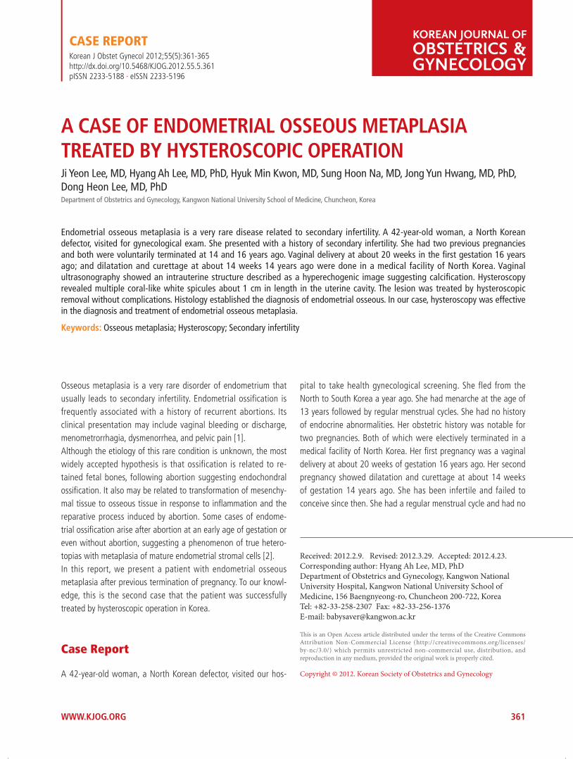
menstrual disorders. She, however, presented with symptoms of intermittent vaginal discharge and chronic pelvic pain.The pelvic examination revealed a normal size, anteverted uterus and normal adnexa. There was white colored leucorrhea, but no cervical motion tenderness. Laboratory studies included blood count, chemistry including serum calcium, and urine analysis, all of which were within the normal limits. Vaginal culture revealed infection of Mycoplasma hominis and Chlamydia trachomatis (Later she was treated with doxycycline and 3rd generation cephalospo-rin. The repeated culture demonstrated that there was no more infection).On transvaginal ultrasound examination, about 2 cm sized highly echogenic and linear endometrial cavity was noted (Fig. 1). It ap-peared as an intrauterine device, but she denied any experience of having a procedure of intrauterine device insertion. We performed
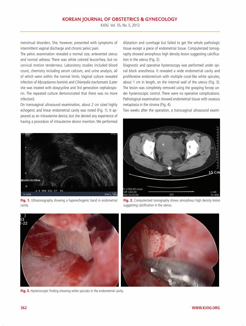
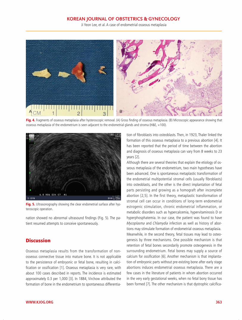
dilatation and curettage but failed to get the whole pathologic tissue except a piece of endometrial tissue. Computerized tomog-raphy showed amorphous high density lesion suggesting calcifi ca-tion in the uterus (Fig. 2).Diagnostic and operative hysteroscopy was performed under spi-nal block anesthesia. It revealed a wide endometrial cavity and proliferative endometrium with multiple coral-like white spicules, about 1 cm in length, on the internal wall of the uterus (Fig. 3). The lesion was completely removed using the grasping forcep un-der hysteroscopic control. There were no operative complications. Pathological examination showed endometrial tissue with osseous metaplasia in the stroma (Fig. 4).Two weeks after the operation, a transvaginal ultrasound exami-
Fig. 1. Ultrasonography showing a hyperechogenic band in endometrial cavity.
Fig. 2. Computerized tomography shows amorphous high density lesion suggesting calcifi cation in the uterus.
Fig. 3. Hysteroscopic fi nding showing white spicules in the endometrial cavity.
WWW.KJOG.ORG 363
Ji Yeon Lee, et al. A case of endometrial osseous metaplasia
nation showed no abnormal ultrasound fi ndings (Fig. 5). The pa-tient resumed attempts to conceive spontaneously.
Discussion
Osseous metaplasia results from the transformation of non-osseous connective tissue into mature bone. It is not applicable to the persistence of embryonic or fetal bone, resulting in calci-fi cation or ossifi cation [1]. Osseous metaplasia is very rare, with about 100 cases described in reports. The incidence is estimated approximately 0.3 per 1,000 [3]. In 1884, Virchow attributed the formation of bone in the endometrium to spontaneous differentia-
tion of fi broblasts into osteoblasts. Then, in 1923, Thaler linked the formation of this osseous metaplasia to a previous abortion [4]. It has been reported that the period of time between the abortion and diagnosis of osseous metaplasia can vary from 8 weeks to 23 years [2].Although there are several theories that explain the etiology of os-seous metaplasia of the endometrium, two main hypotheses have been advanced. One is spontaneous metaplastic transformation of the endometrial multipotential stromal cells (usually fibroblasts) into osteoblasts, and the other is the direct implantation of fetal parts persisting and growing as a homograft after incomplete abortion [2,5]. In the first theory, metaplastic transformation of stromal cell can occur in conditions of long-term endometrial estrogenic stimulation, chronic endometrial inflammation, or metabolic disorders such as hypercalcemia, hypervitaminosis D or hyperphosphatemia. In our case, the patient was found to have Mycoplasma and Chlamydia infection as well as history of abor-tions may stimulate formation of endometrial osseous metaplasia.Meanwhile, in the second theory, fetal tissues may lead to osteo-genesis by three mechanisms. One possible mechanism is that retention of fetal bones secondarily promote osteogenesis in the surrounding endometrium. Fetal bones may supply a source of calcium for ossifi cation [6]. Another mechanism is that implanta-tion of embryonic parts without pre-existing bone after early stage abortions induces endometrial osseous metaplasia. There are a few cases in the literature of patients in whom abortion occurred in the very early gestational weeks, when no fetal bony tissue has been formed [7]. The other mechanism is that dystrophic calcifi ca-
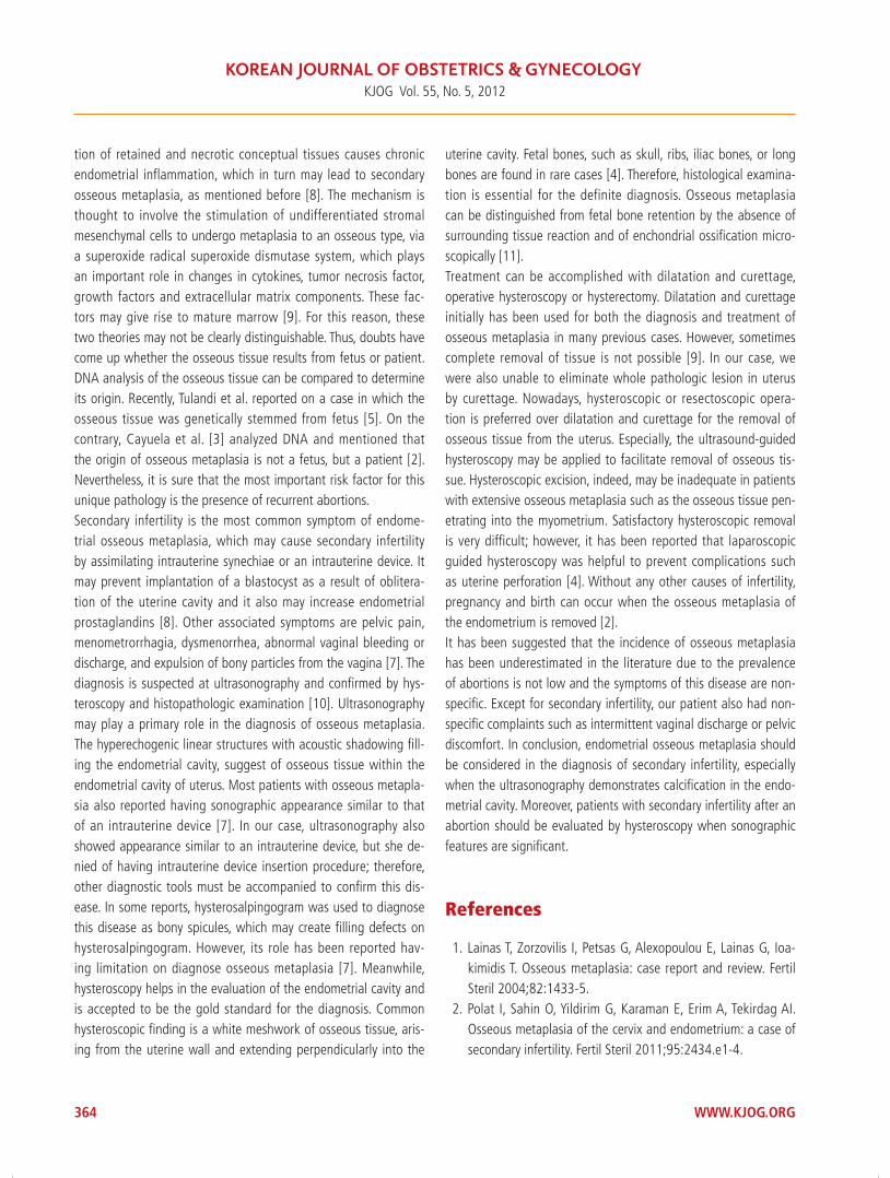
Fig. 4. Fragments of osseous metaplasia after hysteroscopic removal. (A) Gross fi nding of osseous metaplasia. (B) Microscopic appearance showing that osseous metaplasia of the endometrium is seen adjacent to the endometrial glands and stroma (H&E, ×100).
A B
Fig. 5. Ultrasonography showing the clear endometrial surface after hys-teroscopic operation.
WWW.KJOG.ORG364
KJOG Vol. 55, No. 5, 2012
tion of retained and necrotic conceptual tissues causes chronic endometrial inflammation, which in turn may lead to secondary osseous metaplasia, as mentioned before [8]. The mechanism is thought to involve the stimulation of undifferentiated stromal mesenchymal cells to undergo metaplasia to an osseous type, via a superoxide radical superoxide dismutase system, which plays an important role in changes in cytokines, tumor necrosis factor, growth factors and extracellular matrix components. These fac-tors may give rise to mature marrow [9]. For this reason, these two theories may not be clearly distinguishable. Thus, doubts have come up whether the osseous tissue results from fetus or patient. DNA analysis of the osseous tissue can be compared to determine its origin. Recently, Tulandi et al. reported on a case in which the osseous tissue was genetically stemmed from fetus [5]. On the contrary, Cayuela et al. [3] analyzed DNA and mentioned that the origin of osseous metaplasia is not a fetus, but a patient [2]. Nevertheless, it is sure that the most important risk factor for this unique pathology is the presence of recurrent abortions. Secondary infertility is the most common symptom of endome-trial osseous metaplasia, which may cause secondary infertility by assimilating intrauterine synechiae or an intrauterine device. It may prevent implantation of a blastocyst as a result of oblitera-tion of the uterine cavity and it also may increase endometrial prostaglandins [8]. Other associated symptoms are pelvic pain, menometrorrhagia, dysmenorrhea, abnormal vaginal bleeding or discharge, and expulsion of bony particles from the vagina [7]. The diagnosis is suspected at ultrasonography and confi rmed by hys-teroscopy and histopathologic examination [10]. Ultrasonography may play a primary role in the diagnosis of osseous metaplasia. The hyperechogenic linear structures with acoustic shadowing fi ll-ing the endometrial cavity, suggest of osseous tissue within the endometrial cavity of uterus. Most patients with osseous metapla-sia also reported having sonographic appearance similar to that of an intrauterine device [7]. In our case, ultrasonography also showed appearance similar to an intrauterine device, but she de-nied of having intrauterine device insertion procedure; therefore, other diagnostic tools must be accompanied to confi rm this dis-ease. In some reports, hysterosalpingogram was used to diagnose this disease as bony spicules, which may create fi lling defects on hysterosalpingogram. However, its role has been reported hav-ing limitation on diagnose osseous metaplasia [7]. Meanwhile, hysteroscopy helps in the evaluation of the endometrial cavity and is accepted to be the gold standard for the diagnosis. Common hysteroscopic fi nding is a white meshwork of osseous tissue, aris-ing from the uterine wall and extending perpendicularly into the
uterine cavity. Fetal bones, such as skull, ribs, iliac bones, or long bones are found in rare cases [4]. Therefore, histological examina-tion is essential for the definite diagnosis. Osseous metaplasia can be distinguished from fetal bone retention by the absence of surrounding tissue reaction and of enchondrial ossifi cation micro-scopically [11]. Treatment can be accomplished with dilatation and curettage, operative hysteroscopy or hysterectomy. Dilatation and curettage initially has been used for both the diagnosis and treatment of osseous metaplasia in many previous cases. However, sometimes complete removal of tissue is not possible [9]. In our case, we were also unable to eliminate whole pathologic lesion in uterus by curettage. Nowadays, hysteroscopic or resectoscopic opera-tion is preferred over dilatation and curettage for the removal of osseous tissue from the uterus. Especially, the ultrasound-guided hysteroscopy may be applied to facilitate removal of osseous tis-sue. Hysteroscopic excision, indeed, may be inadequate in patients with extensive osseous metaplasia such as the osseous tissue pen-etrating into the myometrium. Satisfactory hysteroscopic removal is very diffi cult; however, it has been reported that laparoscopic guided hysteroscopy was helpful to prevent complications such as uterine perforation [4]. Without any other causes of infertility, pregnancy and birth can occur when the osseous metaplasia of the endometrium is removed [2].It has been suggested that the incidence of osseous metaplasia has been underestimated in the literature due to the prevalence of abortions is not low and the symptoms of this disease are non-specifi c. Except for secondary infertility, our patient also had non-specifi c complaints such as intermittent vaginal discharge or pelvic discomfort. In conclusion, endometrial osseous metaplasia should be considered in the diagnosis of secondary infertility, especially when the ultrasonography demonstrates calcifi cation in the endo-metrial cavity. Moreover, patients with secondary infertility after an abortion should be evaluated by hysteroscopy when sonographic features are signifi cant.
References
1. Lainas T, Zorzovilis I, Petsas G, Alexopoulou E, Lainas G, Ioa-kimidis T. Osseous metaplasia: case report and review. Fertil Steril 2004;82:1433-5.
2. Polat I, Sahin O, Yildirim G, Karaman E, Erim A, Tekirdag AI. Osseous metaplasia of the cervix and endometrium: a case of secondary infertility. Fertil Steril 2011;95:2434.e1-4.
WWW.KJOG.ORG 365
Ji Yeon Lee, et al. A case of endometrial osseous metaplasia
3. Cayuela E, Perez-Medina T, Vilanova J, Alejo M, Cañadas P. True osseous metaplasia of the endometrium: the bone is not from a fetus. Fertil Steril 2009;91:1293.e1-4.
4. Rosa-E-Silva JC, Barcelos ID, Navarro PA, Rosa-E-Silva AC, Nogueira AA, Ferriani RA. Osseous metaplasia of the endome-trium associated with infertility: a case report and review of the literature. J Med Case Reports 2009;3:7427.
5. Tulandi T, Al-Sunaidi M, Arseneau J, Tonin PN, Arcand SL. Calci-fi ed tissue of fetal origin in utero. Fertil Steril 2008;89:217-8.
6. Torné A, Jou P, Pagano R, Sanchez I, Ordi J, Vanrell JA. En-dometrial ossification successfully treated by hysteroscopic resection. Eur J Obstet Gynecol Reprod Biol 1996;66:75-7.
7. Onderoglu LS, Yarali H, Gultekin M, Katlan D. Endometrial os-seous metaplasia: an evolving cause of secondary infertility.
Fertil Steril 2008;90:2013.e9-11. 8. Kumar S, Gupta P, Subbaiah M. Left behind: the patient’s
secondary infertility was traced to a previous pregnancy. Am J Obstet Gynecol 2010;202:319.e1-2.
9. Basu M, Mammen C, Owen E. Bony fragments in the uterus: an association with secondary subfertility. Ultrasound Obstet Gynecol 2003;22:402-6.
10. Coccia ME, Becattini C, Bracco GL, Scarselli G. Ultrasound-guided hysteroscopic management of endometrial osseous metaplasia. Ultrasound Obstet Gynecol 1996;8:134-6.
11. Bolaji, II, Saridogan E, Hasan N, Baithun S, Djahanbakhch O. Prolonged retention of fetal bones with osseous metaplasia of the endometrium. Int J Gynaecol Obstet 1995;50:65-6.
자궁경수술로 치료된 자궁내막 골화생 1예
강원대학교 의학전문대학원 산부인과학교실
이지연, 이향아, 권혁민, 나성훈, 황종윤, 이동헌
자궁내막 골화생은 이차성 불임과 관련 있는 매우 드문 질환이다. 이전에 유산시술을 받은 42세 여성이 새터민을 대상으로 하는 건강검
진을 받기 위해 내원하였다. 질식초음파 결과 자궁내강에 고에코의 음영을 보이는 병변이 보였으며 마치 자궁내 피임기구처럼 보였다. 자
궁내막 소파술을 시행하였으나 병변을 성공적으로 제거하지 못하여, 자궁경수술을 시행하였다. 자궁경을 통해 자궁후벽에 있는 여러 개
의 약 1 cm 길이 내외의 산호초 같은 하얀 침골을 관찰하였으며, 자궁경을 이용하여 병변을 모두 제거하였다. 조직 검사를 통하여 자궁내
막 골화생을 진단하였다. 저자들은 이와 같이 세계적으로 드물게 보고되고 있는 자궁내막 골화생을 진단하고 자궁경을 이용하여 합병증
없이 성공적으로 치료하였기에 이를 간단한 문헌고찰과 함께 보고하는 바이다.
중심단어: 자궁내막 골화생, 자궁경, 이차성 불임