Embed Size (px)
Citation preview

316
Received:November 19, 2015, Revised:December 23, 2015, Accepted:December 24, 2015
Corresponding to:Hee Jin Park, Department of Rheumatology, Catholic Kwandong University International St. Mary’s Hospital, 25 Simgok-ro 100beon-gil, Seo-gu, Incheon 22711, Korea. E-mail:[email protected]
pISSN: 2093-940X, eISSN: 2233-4718Copyright ⓒ 2016 by The Korean College of Rheumatology. All rights reserved.This is a Free Access article, which permits unrestricted non-commerical use, distribution, and reproduction in any medium, provided the original work is properly cited.
Case ReportJournal of Rheumatic Diseases Vol. 23, No. 5, October, 2016https://doi.org/10.4078/jrd.2016.23.5.316
A Case of Idiopathic Massive Rice Bodies in the Knee Joint without Rheumatoid Arthritis or Tuberculosis and a Literature Review
Whan Yong Chung1, Ji-Sun Song2, Hwa Eun Oh3, Hee Jin Park4
Departments of 1Orthopedics, 2Pathology, and 4Rheumatology, Catholic Kwandong University International St. Mary’s Hospital, Incheon, 3Department of Pathology, Korea University Ansan Hospital, Ansan, Korea
Rice bodies are materials with an amorphous nucleus and a fibrin layer found floating in the synovial space and bursa. These bodies have often been detected in patients with rheumatoid arthritis, tuberculous arthritis, and bursitis. Although the etiology and pathogenesis of rice bodies are not yet fully understood, it has been hypothesized that they might be caused by chronic in-flammation originating from the synovium. However, we report on a case of idiopathic massive rice bodies in the knee joint without evidence of inflammatory articular disease or infection including rheumatoid arthritis, seronegative spondyloarthri-tides, tuberculosis, or bacterial or fungal infection. (J Rheum Dis 2016;23:316-320)
Key Words. Rice body, Knee, Rheumatoid arthritis, Tuberculosis
INTRODUCTION
Floating rice-like particles in the synovial space could be found in the inflammatory joint diseases. These particles are known as rice bodies and were first reported by Reise in 1895 in tuberculous arthritis [1]. Rice bodies have been found in the synovial space in patients with rheuma-toid arthritis or seronegative spondyloarthritis [2] and in the bursa [3] and around tendon sheaths [4] in associa-tion with inflammation. However, we experienced a case with massive rice bodies in the knee joint without any in-flammatory or infective evidence and report these idio-pathic rice bodies with an associated literature review.
CASE REPORT
A 46-year-old male had developed discomfort in the right knee one year ago, and aggravated swelling and pain two month ago. For these symptoms, he visited to the out-patient clinic of orthopedics department. He did not have
any medical history and previous joint problems. He worked in an office and had not experienced any traumatic events involving the right knee joint. He felt discomfort when climbing stairs but no definite tenderness and warmth in the right knee. He did not have any symptoms in the other joints and had not experienced inflammatory back pain, uveitis, psoriasis, or inflammatory bowel disease. At admission, his white blood cell count was 3,870/μL, hemoglobin was 14.9 g/dL, and platelet count was 224,000/μL. Erythrocyte sediment rate was 6 mm/hr, and C-reactive protein was 3.18 mg/L (normal range, 0∼5 mg/L). Antinuclear antibody, rheumatoid fac-tor, anti-cyclic citrullinated protein antibody, and human histocompatibility leukocyte antigen (HLA) B27 were all negative. Magnetic resonance imaging (MRI) of the knee showed a large amount of joint effusion with numerous low-signal foci in the suprapatellar bursa against a back-ground of fluid signal intensity on T2-weighted image and intact anterior and posterior cruciate ligaments and collat-eral ligaments, as shown in Figure 1. Arthroscopic syno-

Idiopathic Massive Rice Bodies in the Knee Joint
www.jrd.or.kr 317
Figure 2. Gross morphology of rice bodies in the washing synovial fluid and arthroscopic findings. (A) Many white amorphous ma-terials were found in the washing synovial fluid collected through arthroscopic irrigation. Arthroscopic findings were consistent with rice bodies in the suprapatellar space (B) and revealed a normal synovium (C).
Figure 1. Magnetic resonance images of the right knee. (A) T1-weighted image (T1WI) and (B∼D) T2-weighted images (T2WI) showed a large amount of joint effusion with numerous low-signal foci against a back-ground of fluid signal intensity on T2WI. Anterior and posteri-or cruciate ligaments and collat-eral ligaments were intact.
vectomy of the right knee joint was conducted for diag-nosis and revealed numerous rice bodies in the synovial space, but no definite synovial proliferation, as shown in Figure 2. Microscopic pathology of these materials re-vealed multiple nodular fibrocartilaginous tissues con-sistent with rice bodies and clean synovium without any synovial hyperplasia and inflammatory cells as shown in Figure 3. The results of bacterial, fungal, and acid-fast ba-
cilli (AFB) culture studies from the synovium and washing synovial fluid were all negative, and real-time polymerase chain reaction (PCR) for non-tuberculous mycobacterium (NTM) of synovium was also negative. His symptoms im-proved after synovectomy without any medications and continue to be stable 6 months later.

Whan Yong Chung et al.
318 J Rheum Dis Vol. 23, No. 5, October, 2016
Figure 3. Pathology of rice body and synovium. (A) Microscopic examination (H&E, x40) revealed multiple nodular fi-brocartilaginous tissues consistent with rice bodies. (B) The synovium of the knee joint (H&E, x100) showed a histologically cleansynovial surface.
Table 1. Clinical characteristics and prognosis after treatment of idiopathic rice bodies
Age (yr)/
gender
Site ofrice body
Duration of symptom
Laboratory data
Culture Pathology TreatmentFollow-up duration
Prog-nosis
Refe-rence
38/F Right ankle 2 yr RF/ANA negative
PPD skin test (−)
Tenosynovitis Tenosynovectomy 5 yr No recurr
4
51/M Left wrist 2 yr RF/ANA/HLA B27 negative
Tenosynovitis Radicalsynovectomy
1 yr No recurr
11
31/M Left knee 3 mo ESR/CRP normal
Bacterial culture (−)
Synovitis Synovectomy 2 yr No recurr
12
RF/ANA negative
AFB (−)
4/F Both shoulder and knee
Unknown ESR (18 mm/hr)/CRP normalRF/ANA negative
Surgery without synovectomy
1 yr No recurr
13
11/M Left knee 4 mo ESR/CRP normal
Chronic synovitis
Subtotal synovectomy
2.5 yr No recurr
14
RF negative54/F Both shoulder 14 mo ESR/CRP
normalInflammatory
bursitisSynovectomy 3 mo No
recurr 9
RF/ANA negative
F: female, M: male, ANA: anti-nuclear antibodies, AFB: acid-fast bacilli, CRP: C-reactive protein, ESR: erythrocyte sedimentation rate, HLA: human histocompatibility leukocyte antigen, PPD: purified protein derivative, RF: rheumatoid factor, recurr: recurrence.
DISCUSSION
Rice bodies were most commonly detected in rheuma-toid arthritis [2], tuberculous arthritis, and bursitis [5,6]. Rice bodies had been found in 72% of joints affected by rheumatoid arthritis after aspiration and lavage of syno-
vial fluid [2]. These materials could occur adjunctive to inflamed synovium, in the bursa [3], and around tendon sheaths [4], as well as in the pleural fluid of patients with rheumatoid arthritis [7]. They have also been reported in patients with juvenile arthritis [8], seronegative spondy-loarthritis [2], and osteoarthritis [2]. In the tuberculous
Idiopathic Massive Rice Bodies in the Knee Joint
www.jrd.or.kr 319
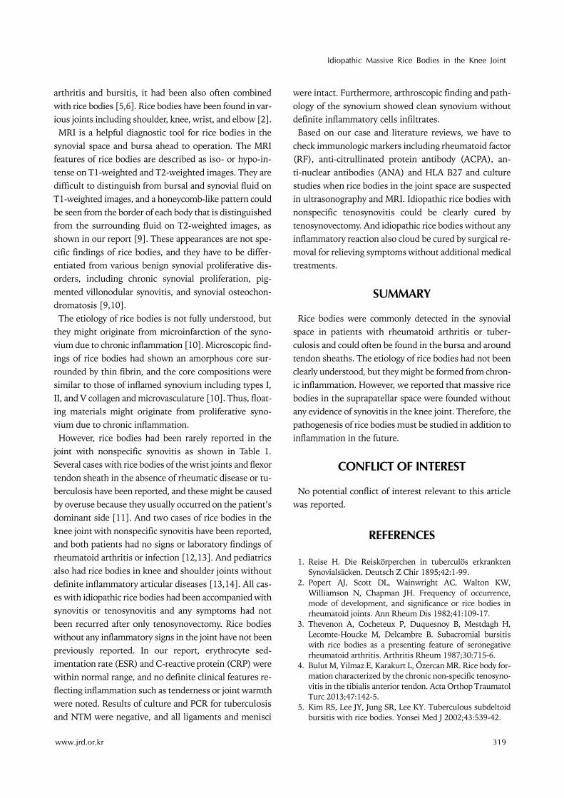
arthritis and bursitis, it had been also often combined with rice bodies [5,6]. Rice bodies have been found in var-ious joints including shoulder, knee, wrist, and elbow [2]. MRI is a helpful diagnostic tool for rice bodies in the
synovial space and bursa ahead to operation. The MRI features of rice bodies are described as iso- or hypo-in-tense on T1-weighted and T2-weighted images. They are difficult to distinguish from bursal and synovial fluid on T1-weighted images, and a honeycomb-like pattern could be seen from the border of each body that is distinguished from the surrounding fluid on T2-weighted images, as shown in our report [9]. These appearances are not spe-cific findings of rice bodies, and they have to be differ-entiated from various benign synovial proliferative dis-orders, including chronic synovial proliferation, pig-mented villonodular synovitis, and synovial osteochon-dromatosis [9,10]. The etiology of rice bodies is not fully understood, but
they might originate from microinfarction of the syno-vium due to chronic inflammation [10]. Microscopic find-ings of rice bodies had shown an amorphous core sur-rounded by thin fibrin, and the core compositions were similar to those of inflamed synovium including types I, II, and V collagen and microvasculature [10]. Thus, float-ing materials might originate from proliferative syno-vium due to chronic inflammation. However, rice bodies had been rarely reported in the
joint with nonspecific synovitis as shown in Table 1. Several cases with rice bodies of the wrist joints and flexor tendon sheath in the absence of rheumatic disease or tu-berculosis have been reported, and these might be caused by overuse because they usually occurred on the patient’s dominant side [11]. And two cases of rice bodies in the knee joint with nonspecific synovitis have been reported, and both patients had no signs or laboratory findings of rheumatoid arthritis or infection [12,13]. And pediatrics also had rice bodies in knee and shoulder joints without definite inflammatory articular diseases [13,14]. All cas-es with idiopathic rice bodies had been accompanied with synovitis or tenosynovitis and any symptoms had not been recurred after only tenosynovectomy. Rice bodies without any inflammatory signs in the joint have not been previously reported. In our report, erythrocyte sed-imentation rate (ESR) and C-reactive protein (CRP) were within normal range, and no definite clinical features re-flecting inflammation such as tenderness or joint warmth were noted. Results of culture and PCR for tuberculosis and NTM were negative, and all ligaments and menisci
were intact. Furthermore, arthroscopic finding and path-ology of the synovium showed clean synovium without definite inflammatory cells infiltrates. Based on our case and literature reviews, we have to
check immunologic markers including rheumatoid factor (RF), anti-citrullinated protein antibody (ACPA), an-ti-nuclear antibodies (ANA) and HLA B27 and culture studies when rice bodies in the joint space are suspected in ultrasonography and MRI. Idiopathic rice bodies with nonspecific tenosynovitis could be clearly cured by tenosynovectomy. And idiopathic rice bodies without any inflammatory reaction also cloud be cured by surgical re-moval for relieving symptoms without additional medical treatments.
SUMMARY
Rice bodies were commonly detected in the synovial space in patients with rheumatoid arthritis or tuber-culosis and could often be found in the bursa and around tendon sheaths. The etiology of rice bodies had not been clearly understood, but they might be formed from chron-ic inflammation. However, we reported that massive rice bodies in the suprapatellar space were founded without any evidence of synovitis in the knee joint. Therefore, the pathogenesis of rice bodies must be studied in addition to inflammation in the future.
CONFLICT OF INTEREST
No potential conflict of interest relevant to this article was reported.
REFERENCES
1. Reise H. Die Reiskörperchen in tuberculös erkrankten Synovialsäcken. Deutsch Z Chir 1895;42:1-99.
2. Popert AJ, Scott DL, Wainwright AC, Walton KW, Williamson N, Chapman JH. Frequency of occurrence, mode of development, and significance or rice bodies in rheumatoid joints. Ann Rheum Dis 1982;41:109-17.
3. Thevenon A, Cocheteux P, Duquesnoy B, Mestdagh H, Lecomte-Houcke M, Delcambre B. Subacromial bursitis with rice bodies as a presenting feature of seronegative rheumatoid arthritis. Arthritis Rheum 1987;30:715-6.
4. Bulut M, Yilmaz E, Karakurt L, Özercan MR. Rice body for-mation characterized by the chronic non-specific tenosyno-vitis in the tibialis anterior tendon. Acta Orthop Traumatol Turc 2013;47:142-5.
5. Kim RS, Lee JY, Jung SR, Lee KY. Tuberculous subdeltoid bursitis with rice bodies. Yonsei Med J 2002;43:539-42.

Whan Yong Chung et al.
320 J Rheum Dis Vol. 23, No. 5, October, 2016
6. Chau CL, Griffith JF, Chan PT, Lui TH, Yu KS, Ngai WK. Rice-body formation in atypical mycobacterial tenosynovi-tis and bursitis: findings on sonography and MR imaging. AJR Am J Roentgenol 2003;180:1455-9.
7. Kassimos D, George E, Kirwan JR. Rice bodies in the pleural aspirate of a patient with rheumatoid arthritis. Ann Rheum Dis 1994;53:427-8.
8. Chung C, Coley BD, Martin LC. Rice bodies in juvenile rheu-matoid arthritis. AJR Am J Roentgenol 1998;170:698-700.
9. Griffith JF, Peh WC, Evans NS, Smallman LA, Wong RW, Thomas AM. Multiple rice body formation in chronic sub-acromial/subdeltoid bursitis: MR appearances. Clin Radiol 1996;51:511-4.
10. Cheung HS, Ryan LM, Kozin F, McCarty DJ. Synovial origins
of Rice bodies in joint fluid. Arthritis Rheum 1980;23:72-6.11. Forse CL, Mucha BL, Santos ML, Ongcapin EH. Rice body
formation without rheumatic disease or tuberculosis in-fection: a case report and literature review. Clin Rheumatol 2012;31:1753-6.
12. Kang DJ, Ahn JM, Rhee SJ. A case of multiple rice bodies by the nonspecific synovitis in the knee joint. J Korean Knee Soc 2010;22:306-9.
13. Mutlu H, Silit E, Pekkafali Z, Karaman B, Omeroglu A, Basekim CC, et al. Multiple rice body formation in the sub-acromial-subdeltoid bursa and knee joint. Skeletal Radiol 2004;33:531-3.
14. Aşik M, Eralp L, Cetik O, Altinel L. Rice bodies of synovial origin in the knee joint. Arthroscopy 2001;17:E19.