Embed Size (px)
Citation preview

Vol. 22, No. 2, 2010 219
Fig. 1. Pruritic, solitary, well-demarcated, brown to black plaque,3.8×1.2 cm in size, on the abdomen.
Ann Dermatol Vol. 22, No. 2, 2010 DOI: 10.5021/ad.2010.22.2.219
CASE REPORT
Received February 2, 2009, Revised June 10, 2009, Accepted for publication September 1, 2009*This study was supported by a grant of the Korea Healthcare technology R&D Project. Ministry for Health, Welfare & Family Affairs, Republic of Korea (A090084).
Corresponding author: Young Min Park, M.D., Department of Dermatology, Seoul St. Mary’s Hospital, College of Medicine, The Catholic University of Korea, 505 Banpo-dong, Seocho-gu, Seoul 137-701, Korea. Tel: 82-2-2258-6223, Fax: 82-2-594-3255, E-mail: yymmpark6301@ hotmail.com
A Case of Lymphomatoid Keratosis
Min Jee Choi, M.D., Hei Sung Kim, M.D., Hyung Ok Kim, M.D., Kye Yong Song, M.D.1, Young Min Park, M.D.
Department of Dermatology, Seoul St. Mary's Hospital, College of Medicine, The Catholic University of Korea, 1Department of Pathology, College of Medicine, Chung-Ang University, Seoul, Korea
Lymphomatoid keratosis (LK) is considered to be a rare variant of cutaneous lymphoid hyperplasia, with epidermo-tropism. We herein report a case of LK which developed on the abdomen of an elderly Korean woman. A 60-year-old woman presented with a 10-year history of a pruritic, solitary, brown to black plaque on the abdomen. Histopathologically, the specimen showed hyperkeratosis, parakeratosis, acanthosis and Pautrier’s micro-abscess in the epidermis, and a lichenoid infiltration of lymphocytes in the dermis, which expressed both B cell and T cell lineage on the immune-histochemical staining. Based on these clinical and histopathological findings, our case was diagnosed as LK. To our knowledge, this is the first case report of LK in the Korean dermatologic literature. (Ann Dermatol 22(2) 219∼222, 2010)
-Keywords-Cutaneous lymphoid hyperplasia, Epidermotropism, Lymp-homatoid keratosis
INTRODUCTION
Lymphomatoid keratosis (LK) has been considered to be a variant of uni-lesional mycosis fungoides (MF), as it shows epidermotropism that is characterstic of MF1. With its li-chenoid keratotic features, it also has been recognized as
a subtype of benign lichenoid keratosis2. Recently, Arai et al.3 proposed that LK is a epidermotropic type of cuta-neous lymphoid hyperplasia, where differentiation from MF and benign lichenoid keratosis is possible, by compar-ing the clinocopathological, immunohistochemical and molecular biological findings. To date, there have been 26 case reports of LK in the English literature, but none in the Korean dermatologic literature. Herein, we report the first case of LK in Korea and review the previous cases.
CASE REPORT
A 60-year-old woman presented with a 10-year history of a pruritic, solitary, well-demarcated, scaly, brown to black plaque on the abdomen, measuring 3.8×1.2 cm in size, which had started as a tiny papule (Fig. 1). Dermoscopic findings revealed numerous brown dots and globules at the periphery (Fig. 2). She had no previous personal or familial history of skin cancer and no other significant cu-taneous or medical history was found. There was no his-tory of trauma. A skin biopsy from a local clinic 4 years before showed hyperkeratosis, parakeratosis, acanthosis, papillomatosis and hypergranulosis in the epidermis, which was consistent with seborrheic keratosis. Additio-
MJ Choi, et al
220 Ann Dermatol
Fig. 4. (A, B) Epidermal hyperplasia and epidermotropism in the epidermis and formation of lymphoid follicle and lichenoid infiltrationof lymphocytes in the reticular dermis (H&E stain, ×40, ×100). (C) Epidermotropism with Pautrier’s microabscess in the epidermis(H&E stain, ×400). (D) Lichenoid infiltration of lymphocytes in the reticular dermis (H&E stain, ×400).
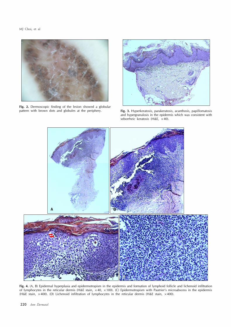
Fig. 2. Dermoscopic finding of the lesion showed a globular pattern with brown dots and globules at the periphery. Fig. 3. Hyperkeratosis, parakeratosis, acanthosis, papillomatosis
and hypergranulosis in the epidermis which was consistent withseborrheic keratosis (H&E, ×40).

A Case of Lymphomatoid Keratosis
Vol. 22, No. 2, 2010 221
Fig. 5. Immunohistochemical study for CD3 (A), CD4 (B), CD8 (C), CD20 (D), showed positive staining with lymphocytes (H&E, ×400).
nally, a dense infiltration of lymphocytes in the superficial dermis was observed (Fig. 3).To confirm the previous diagnosis and rule out malig-nancy, we performed a second skin biopsy from the same lesion. Histopathological findings showed hyperkeratosis, parakeratosis, acanthosis and epidermotropism with Pau-trier’s micro-abscess in the epidermis, a lichenoid infiltra-tion of lymphocytes, the formation of lymphoid follicle and hemorrhage in the reticular dermis (Fig. 4). Solar elas-tosis was not seen. Immuno-histopathologically, the exo-cytic lymphocytes in the epidermis and the lymphocytes of the lymphoid follicle in the dermis were positive for CD3, CD4, CD8, CD20, CD30 and CD79a (Fig. 5). Genotypically, rearrangement of TCRγ was partially demonstrated. Based on these clinical and histopatho-logical findings, we made a final diagnosis of LK. Complete excision of the lesion was performed. No local
recurrence occurred during the ensuing 3 months.
DISCUSSION
Evans et al.1 first reported a case of uni-lesional MF on the right flank area of a 44-year-old male, where the lesion demonstrated histopathological features of epidermotrop-ism with lichenoid activity. Later on, Kossard2 suggested that the case reported by Evans et al.1 should have been categorized as LK, thereby introducing the term LK for the first time in 1997. Thereafter, there have been 23 cases of LK reported in the English literature.LK clinically presents as an asymptomatic, scaly, eryth-ematous plaque on the face or upper trunk of the middle aged personnel, simulating basal cell carcinoma, actinic keratosis, or seborrheic keratosis2,4,5. Most cases of LK are idiopathic, however several have been associated with

MJ Choi, et al
222 Ann Dermatol
newly encountered antigens from anthropod bites, stings, tattoo, vaccinations, trauma, injection of foreign sub-stances, pierced ear jewelry, and drugs6. Al-Hoqail and Crawford4 reported cases of LK adjacent to seborrheic ker-atosis and solar lentigo. Coexistence with these diseases make us suspect that LK might be related to solar damage. In our case, though, solar elastosis was not observed in the dermis. Further evaluation is needed to explain the clear relationship between LK and solar damage. The most pathognomic pathological finding of LK is epidermotro-pism. Lymphocytes with tropism for the epidermis are composed of both B cells and T cells3.Special attention is needed to differentiate LK from uni-le-sional MF because these 2 diseases largely resemble one another. Uni-lesional MF is a rare variant of MF which usually appears as a single, isolated lesion comprising of less than 5% of the body’s surface area. The disease is usually benign and has an excellent response to locally ablative treatments7. Uni-lesional MF is different from LK in that the epidermotropic lymphocytes in uni-lesional MF mainly consist of helper T cells possessing T cell clonality, which conveys the malignant transformation3.Other major differentials to consider are lichenoid actinic keratosis and benign lichenoid keratosis. Both of them commonly show lichenoid infiltrates of lymphocytes with epidermal hyperplasia. However, lichenoid actinic kera-tosis differs from LK in that atypical basal cells and kerati-nocytes can be found whereas epidermotropism of the lymphocytes are not seen. Benign lichenoid keratosis shows necrotic keratinocytes, spongiosis and Max-Joseph spaces without epidermotropism8. Also, irritated sebor-rheic keratosis and inflamed seborrheic keratosis should be included in the differential diagnosis of LK in that the biopsy specimen from the same lesions 4 years earlier was consistent with seborrheic keratosis. Irritated seborrheic keratosis differs from LK in that in the former, squamous cell eddies or pearls can be found, and epidermotropism of the lymphocytes are not seen (even though in-flammation is severe). Inflamed seborrheic keratosis differs from LK in that spongiosis and exocytosis of lymphocytes in the epidermis can be found in the former, and epi-dermotropism of the lymphocytes are not seen9,10.Based on the histopathological and immunohistochemical findings, our case was diagnosed as LK. The unusual fea-ture in our case is the history of a transformation from se-borrheic keratosis to LK. Either the transformation of se-
borrheic keratosis to LK is incidental or else these two dis-eases may be truly related. Chronic rubbing may induce hyperkeratotic changes in the epidermis and exocytosis and spongiosis of lymphocytes in the dermis, reflecting the unidentified culprit factor of LK like other spongiotic dermatitis. For a clear explanation, further investigation is needed.In conclusion, we experienced a typical case of LK that developed on the abdomen and was transformed from se-borrheic keratosis. LK is an epidermotropic kind of cuta-neous lymphoid hyperplasia, with hyperkeratotic changes in the epidermis, therefore it should be included in the dif-ferential diagnosis of benign keratotic dermatosis.
REFERENCES
1. Evans LT, Mackey SL, Vidmar DA. An asymptomatic scaly plaque. Unilesional mycosis fungoides (MF). Arch Dermatol 1997;133:231, 234.
2. Kossard S. Unilesional mycosis fungoides or lymphomatoid keratosis? Arch Dermatol 1997;133:1312-1313.
3. Arai E, Shimizu M, Tsuchida T, Izaki S, Ogawa F, Hirose T. Lymphomatoid keratosis: an epidermotropic type of cuta-neous lymphoid hyperplasia: clinicopathological, immuno-histochemical, and molecular biological study of 6 cases. Arch Dermatol 2007;143:53-59.
4. Al-Hoqail IA, Crawford RI. Benign lichenoid keratoses with histologic features of mycosis fungoides: clinicopathologic description of a clinically significant histologic pattern. J Cutan Pathol 2002;29:291-294.
5. Lever WF, Schaumburg-Lever G. Histopathology of the skin. 6th ed. London: Lippincott, 1983:762-768.
6. Bergman R, Khamaysi Z, Sahar D, Ben-Arieh Y. Cutaneous lymphoid hyperplasia presenting as a solitary facial nodule: clinical, histopathological, immunophenotypical, and mole-cular studies. Arch Dermatol 2006;142:1561-1566.
7. Heald PW, Glusac EJ. Unilesional cutaneous T-cell lympho-ma: clinical features, therapy, and follow-up of 10 patients with a treatment-responsive mycosis fungoides variant. J Am Acad Dermatol 2000;42:283-285.
8. Murphy GF, Schwarting R. Cutaneous lymphomas and leukemias. In: Elder DE, Elenitsas R, Johnson BL Jr, Murphy GF, editors. Lever's histopathology of the skin. 9th ed. Philadelphia: Lippincott Williams & Wilkins, 2005:927-978.
9. Bazza MA, Ryatt KS, Dharmagunawardena PV. Mycosis fungoides masquerading as seborrhoeic keratosis. Br J Dermatol 2002;147:1264-1265.
10. Yoo SS, Viglione M, Moresi M, Vonderheid E. Unilesional mycosis fungoides mimicking Bowen’s disease. J Dermatol 2003;30:417-419.


![l c a l Derma Journal of Clinical & Experimental t i n o i ... · dermatologic conditions, including tinea pedis, seborrheic dermatitis, and axillary granular parakeratosis [14]](https://img.pdfslide.net/doc/110x75/5ce96bfc88c9932e468d82af/l-c-a-l-derma-journal-of-clinical-experimental-t-i-n-o-i-dermatologic.jpg)


![Evaluation of the Trivedi Effect - Energy of … symptoms include parakeratosis, hypogeusia, anorexia, dysosmia, geophagia, hypogonadism, growth retardation, etc. [5-7]. Recently,](https://img.pdfslide.net/doc/110x75/5d209b2b88c993a5378d16d5/evaluation-of-the-trivedi-effect-energy-of-symptoms-include-parakeratosis-hypogeusia.jpg)