Embed Size (px)
Citation preview
Narita et al. BMC Nephrology (2015) 16:151 DOI 10.1186/s12882-015-0147-9
CASE REPORT Open Access
A case of membranoproliferativeglomerulonephritis associated withcurved fibril deposition
Ikuyo Narita1*, Michiko Shimada1, Takeshi Fujita1, Reiichi Murakami1, Masayuki Nakamura1, Norio Nakamura1,2,Hideaki Yamabe1 and Ken Okumura1Abstract
Background: It is sometimes challenging to diagnose unsusual cases of fibrillary glomerulonephritis (FGN) andimmunotactoid glomerulopathy (ITG), the rare causes of nephrotic syndrome.
Case presentation: A 75-year-old Japanese woman presented with nephrotic syndrome, microhematuria andrenal insufficiency. Renal biopsy revealed membranoproliferative glomerulonephritis (MPGN) with IgM and weak C3deposition. Congo red stain was negative. Electron microscopy demonstrated massive fibrils in the subendothelium,mesangium and subepithelium. The fibrils were partially parallel, partially curved and 17 nm in diameter. Cryoglobulin,hepatitis B virus (HBV) antigen, hepatitis C virus (HCV) antibody or antinuclear antibody were negative.
Conclusion: We report a case of MPGN associated with peculiar non-amyloid fibril deposition corresponding toneither FGN nor ITG.
Keywords: Membranoproliferative glomerulonephritis, Fibrillary glomerulonephritis, Immunotactoid glomerulopathy
BackgroundFGN and ITG are glomerular deposition diseases withimmunoglobulin-derived non-amyloid fibrils. The dis-tinction between ITG and FGN is generally based onthe appearance, thickness and the arrangement of thedeposited fibrils. Typically, in FGN, fibrils are solid,12–24 nm in diameter and the arrangement is random.Whereas, in ITG, fibrils are microtubule, diameteris >30 nm and parallel arrangement is observed [1].In this report, we describe a case of MPGN associated
with peculiar fibril deposits corresponding to neitherFGN nor ITG.
Case presentationA 75-year-old Japanese woman presented with nephroticsyndrome, microhematuria and renal insufficiency. Shehad been treated as hypertension for 15 years. She wasdeaf since she was 6 years old for unknown reason.
* Correspondence: [email protected] of Cardiology and Nephrology, Hirosaki University GraduateSchool of Medicine, 5 Zaifu-cho, Hirosaki 036-8562, JapanFull list of author information is available at the end of the article
© 2015 Narita et al. Open Access This articleInternational License (http://creativecommonsreproduction in any medium, provided you gthe Creative Commons license, and indicate if(http://creativecommons.org/publicdomain/ze
6 months prior to admission, she developed erythemanodosum but Crohn’s disease, Sarcoidosis, Mycobacter-ium tuberculosis and malignancy were denied, and hadbeen treated with prednisolone 10 mg per day. Then shenoted pretibial edema and was referred to our hospital asnephrotic syndrome.At the time of admission, blood pressure was 177/
99 mmHg, the height was 148 cm, and the weight was58.4 kg. She exhibited edema in her face and both lowerextremities. Pigmentation was seen on her legs buterythema was not present. The results of laboratory teston admission were as follows: hemoglobin 9.4 g/dl; redblood count 336 × 104/μl; white cell count 11500/μl; plate-let count 569 × 103/μl; blood urea nitrogen 18 mg/dl;serum creatinine (sCr) 1.55 mg/dl; serum total cholesterol286 mg/dl; triglycerides, 249 mg/dl; total protein 4.9 g/dl;albumin 2.3 g/dl; serum IgG 301 mg/dl; IgA 42 mg/dl;IgM 28 mg/dl. The levels of serum complement, freeκ and λ light chains were within normal limits. Elec-trophoresis of serum and urine revealed no monoclo-nal spikes. Cryoglobulin, HBV antigen, HCV antibodyand antinuclear antibody were not detected. Urinalysis
is distributed under the terms of the Creative Commons Attribution 4.0.org/licenses/by/4.0/), which permits unrestricted use, distribution, andive appropriate credit to the original author(s) and the source, provide a link tochanges were made. The Creative Commons Public Domain Dedication waiverro/1.0/) applies to the data made available in this article, unless otherwise stated.
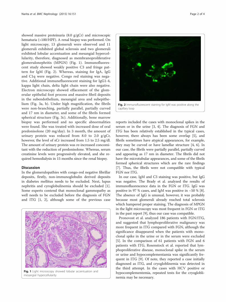
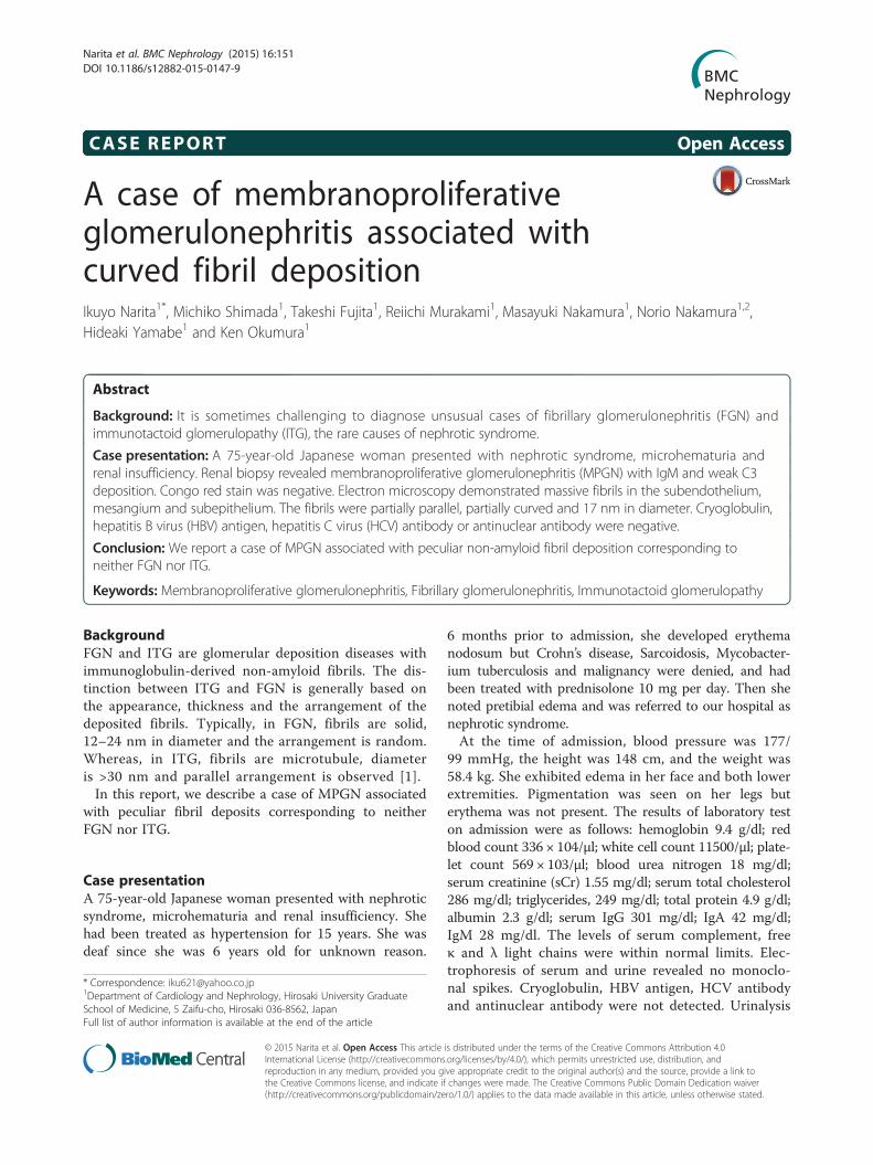
Fig. 2 Immunofluorescent staining for IgM was positive along thecapillary loop
Narita et al. BMC Nephrology (2015) 16:151 Page 2 of 4
showed massive proteinuria (8.0 g/gCr) and microscopichematuria (>100/HPF). A renal biopsy was performed. Onlight microscopy, 13 glomeruli were observed and 11glomeruli exhibited global sclerosis and two glomeruliexhibited lobular accentuation and mesangial hypercel-lularity, therefore, diagnosed as membranoproliferativeglomerulonephritis (MPGN) (Fig. 1). Immunofluores-cent study showed weakly positive C3 and fringe pat-tern for IgM (Fig. 2). Whereas, staining for IgA, IgGand C1q were negative. Congo red staining was nega-tive. Additional immunefluorescent staining for IgG1-4,kappa light chain, delta light chain were also negative.Electron microscopy showed effacement of the glom-erular epithelial foot process and massive fibril depositsin the subendothelium, mesangial area and subepithe-lium (Fig. 3a, b). Under high magnification, the fibrilswere non-branching, partially parallel, partially curvedand 17 nm in diameter, and some of the fibrils formedspherical structure (Fig. 3c). Additionally, bone marrowbiopsy was performed and no specific abnormalitieswere found. She was treated with increased dose of oralpredonisolone (20 mg/day). In 3 month, the amount ofurinary protein was reduced from 8.0 to 2.0 g/gCr,however, the level of sCr increased from 1.5 to 2.5 mg/dl.The amount of urinary protein was re-increased concomi-tant with the reduction of predonisolone. Whereas, serumcreatinine levels were progressively elevated, and she re-quired hemodialysis in 15 months since the renal biopsy.
DiscussionIn the glomerulopathies with congo-red negative fibrillardeposits, firstly, non-immunoglobulin derived depositsin diabetes mellitus needs to be excluded. Next, lupusnephritis and cyroglobulinemia should be excluded [1].Some experts contend that monoclonal gammopathy aswell needs to be excluded before the diagnosis of FGNand ITG [1, 2], although some of the previous case
Fig. 1 Light microscopy showed lobular accentuation andmesangial hypercellularity
reports included the cases with monoclonal spikes in theserum or in the urine [3, 4]. The diagnosis of FGN andITG has been relatively established in the typical cases,however, there always has been some overlap [5], andfibrils sometimes have atypical appearances, for example,they may be curved or have lamellar structure [4, 6]. Inour case, the fibrils were partially parallel, partially curvedand appearing as 17 nm in diameter. The fibrils did nothave the microtubular appearances, and some of the fibrilsformed spherical structures which are the rare findings[7]. Thus, the fibrils were not compatible with typicalFGN nor ITG.In our case, IgM and C3 staining was positive, but IgG
was negative. The Brady et al. analyzed the results ofimmunofluorescence data in the FGN or ITG. IgG waspositive in 97 % cases, and IgM was positive in ~50 % [8].The absence of IgG is unusual, however, it was probablybecause most glomeruli already reached total sclerosiswhich hampered proper staining. The diagnosis of MPGNin the light microscopy was most frequent in FGN or ITGin the past report [9], thus our case was compatible.Pronovost et al. analyzed 186 patients with FGN/ITG,
and suggested that lymphoproliferative malignancy wasmore frequent in ITG compared with FGN, although thesignificance disappeared when the patients with mono-clonal spike in the urine or in the serum were excluded[5]. In the comparison of 61 patients with FGN and 6patients with ITG, Rosenstock et al. reported that lym-phoproliferative disease, monoclonal spike in the serumor urine and hypocomplementamia was significantly fre-quent in ITG [9]. Of note, they reported a case initiallydiagnosed as ITG, and cryogloblinemia was detected inthe third attempt. In the cases with HCV positive orhypocomplementemia, repeated tests for the cryoglobli-nemia may be necessary.
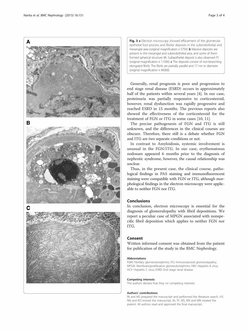
Fig. 3 a Electron microscopy showed effacement of the glomerularepithelial foot process and fibrilar deposits in the subendothelial andmesangial area (original magnification × 5750). b Massive deposits arepresent in the mesangial and subendothelial area, and some of themformed spherical structure (#). Subepithelial deposit is also observed (*)(original magnification × 11500). c The deposits consist of non-branchingelongated fibrils. The fibrils are partially parallel and 17 nm in diameter(original magnification × 46000)
Narita et al. BMC Nephrology (2015) 16:151 Page 3 of 4
Generally, renal prognosis is poor and progression toend stage renal disease (ESRD) occurs in approximatelyhalf of the patients within several years [4]. In our case,proteinuria was partially responsive to corticosteroid,however, renal dysfunction was rapidly progressive andreached ESRD in 15 months. The previous reports alsoshowed the effectiveness of the corticosteroid for thetreatment of FGN or ITG in some cases [10, 11].The precise pathogenesis of FGN and ITG is still
unknown, and the differences in the clinical courses areobscure. Therefore, there still is a debate whether FGNand ITG are two separate conditions or not.In contrast to Amyloidosis, systemic involvement is
unusual in the FGN/ITG. In our case, erythematosusnodosum appeared 6 months prior to the diagnosis ofnephrotic syndrome, however, the causal relationship wasunclear.Thus, in the present case, the clinical course, patho-
logical findings in PAS staining and immunofluorescentstaining were compatible with FGN or ITG, although mor-phological findings in the electron microscopy were applic-able to neither FGN nor ITG.
ConclusionsIn conclusion, electron microscopy is essential for thediagnosis of glomerulopathy with fibril depositions. Wereport a peculiar case of MPGN associated with nonspe-cific fibril deposition which applies to neither FGN norITG.
ConsentWritten informed consent was obtained from the patientfor publication of the study in the BMC Nephrology.
AbbreviationsFGN: Fibrillary glomerulonephritis; ITG: Immunotactoid glomerulopathy;MPGN: Membranoproliferative glomerulonephritis; HBV: Hepatitis B virus;HCV: Hepatitis C virus; ESRD: End stage renal disease.
Competing interestsThe authors declare that they no competing interests.
Authors’ contributionsIN and MS prepared the manuscript and performed the literature search. HY,NN and KO revised the manuscript. IN, TF, MS, RM and MN treated thepatient. All authors read and approved the final manuscript.
Narita et al. BMC Nephrology (2015) 16:151 Page 4 of 4
AcknowledgementWe thank Prof. Charles E. Alpers for his critical advice in the assessment ofrenal histological findings.
Author details1Department of Cardiology and Nephrology, Hirosaki University GraduateSchool of Medicine, 5 Zaifu-cho, Hirosaki 036-8562, Japan. 2CommunityMedicine, Hirosaki University Graduate School of Medicine, 5 Zaifu-cho,Hirosaki 036-8562, Japan.
Received: 28 April 2015 Accepted: 30 June 2015
References1. Schwartz MM. Immunotactoid glomerulopathy. J Am Soc Nephrol.
2002;13(5):1390–7.2. Korbet SM, Schwartz MM, Lewis EJ. Immuotactoid glomerulopathy (fibrillary
glomerulonephritis). Clin J Am Soc Nephrol. 2006;1(6):1351–6.3. Ivanyi B, Degrell P. Fibrillary glomerulonephritis and immunotactoid
glomerulopathy. Nephrol Dial Transplant. 2004;19(9):2166–70.4. Alpers CE, Kowalewska J. Fibrillary glomerulonephritis and immunotactoid
glomerulopathy. J Am Soc Nephrol. 2008;19(1):34–7.5. Pronovost PH, Brady HR, ME G, Espinoza O, Rennke HG. Clinical
features, predictors of disease progression and results of renal. Nephrol DialTransplant. 1996;11:837–42.
6. Joh K, Aizawa S, Takahashi T, Hatakeyama M, Muto S, Asano Y, et al.Microlamellar structures in lobular glomerulonephritis associated withmonoclonal IgG lambda paraproteinemia. A case report and review of theliterature. Acta Pathol Jpn. 1990;40(12):913–21.
7. Jain S, Chhabra D. A case of immunotactoid glomerulopathy with rapidprogression to end-stage renal disease. TheScientificWorldJOURNAL.2009;9:1348–54.
8. Hugh RB. Fibrillary glomerulopathy. Kidney Int. 1998;53(1998):1421–9.9. Rosenstock JL, Markowitz GS, Valeri AM, Sacchi G, Appel GB, D’Agati VD.
Fibrillary and immunotactoid glomerulonephritis Distinct. Kidney Int.2003;63(4):1450–61.
10. Dickenmann M, Schaub S, Nickeleit V, Mihatsch M, Steiger J, Brunner F.Fibrillary glomerulonephritis: early diagnosis associated with steroidresponsiveness. Am J Kidney Dis. 2002;40(3):E9.
11. Kinomura M, Maeshima Y, Kodera R, Morinaga H, Saito D, Nakao K, et al.A case of immunotactoid glomerulopathy exhibiting nephrotic syndromesuccessfully treated with corticosteroids and antihypertensive therapy. ClinExp Nephrol. 2009;13(4):378–84.
Submit your next manuscript to BioMed Centraland take full advantage of:
• Convenient online submission
• Thorough peer review
• No space constraints or color figure charges
• Immediate publication on acceptance
• Inclusion in PubMed, CAS, Scopus and Google Scholar
• Research which is freely available for redistribution
Submit your manuscript at www.biomedcentral.com/submit