Embed Size (px)
Citation preview

Case ReportMembranoproliferative Glomerulonephritis inPatients with Chronic Venous Catheters: A CaseReport and Literature Review
John Sy,1 Cynthia C. Nast,2 Phuong-Thu T. Pham,3 and Phuong-Chi T. Pham1
1 Division of Nephrology and Hypertension, Department of Internal Medicine, UCLA-Olive View Medical Center,14445 Olive View Drive, 2B-182, Sylmar, CA 91342, USA
2Cedars Sinai Medical Center, Department of Pathology, Los Angeles, CA 90048, USA3David Geffen School of Medicine at UCLA, Kidney and Pancreas Transplant Program, Los Angeles, CA 90095, USA
Correspondence should be addressed to Phuong-Chi T. Pham; [email protected]
Received 31 August 2013; Accepted 22 December 2013; Published 30 January 2014
Academic Editors: Y. Fujigaki, K. Hirayama, A. K. Saxena, and W. Sułowicz
Copyright © 2014 John Sy et al. This is an open access article distributed under the Creative Commons Attribution License, whichpermits unrestricted use, distribution, and reproduction in any medium, provided the original work is properly cited.
Chronic indwelling catheters have been reported to be associated withmembranoproliferative glomerulonephritis (MPGN) via theactivation of the classical complement pathway in association with bacterial infections such as coagulase negative staphylococcus.We herein provide supporting evidence for the direct causal relationship between chronic catheter infections andMPGN via a caseof recurrent MPGN associated with recurrent catheter infections used for total parenteral nutrition (TPN) in a man with short gutsyndrome. We also present a literature review of similar cases and identify common clinical manifestations that may serve to aidclinicians in the early identification of MPGN associated with infected central venous catheterization or vice versa. The importanceof routine monitoring of kidney function and urinalysis among patients with chronic central venous catheterization is highlightedas kidney injury may herald or coincide with overtly infected chronic indwelling central venous catheters.
1. Introduction
Membranoproliferative glomerulonephritis (MPGN) is a pat-tern of disease characterized by the deposition of immun-oglobulins, complement factors, or both along capillary wallsand within the glomerular mesangium.The classic finding oflobular accentuation of glomerular tufts on light microscopyis attributed tomesangial hypercellularity, endocapillary pro-liferation, and capillary wall remodeling resulting in the for-mation of “double contours.” Depositions of the third compo-nent of complement (C3) with or without immunoglobulinsmay be observed on immunofluorescent studies [1]. Theunderlying etiologies of MPGN comprise a spectrum ofconditions including infection, monoclonal gammopathy,autoimmune or rheumatologic disease, and dysregulation ofthe alternative complement pathway. It is well known thatchronic infection from indwelling ventriculosystemic shuntscan cause “shunt nephritis”, an entity first reported in 1965by Black et al. after the placement of a ventriculoatrial shunt
for the relief of hydrocephalus in two pediatric patients[2, 3]. Further experiments in animal studies have similarlyshown a relation between chronic infections associated withindwelling catheters andMPGN[4, 5]. Althoughuncommon,there have been few reports of MPGN associated with centralvenous catheters placed for total parenteral nutrition (TPN)[6].Weherein report a case of recurrentMPGN in associationwith recurrent coagulase negative Staphylococcus epidermidisHickman catheter infection, and review the literature forcommon clinical presentations of MPGN in patients requir-ing chronic central venous catheter placement.
2. Case Report
2.1. Clinical History and Initial Laboratory Data. A 23-year-oldmale with priormultiple gunshot wounds to the abdomenrequiring complete small bowel resection and chronic TPNsupport via a Hickman catheter since the age of 17 presentedwith anasarca and low grade fevers in June 1996. Basic urine
Hindawi Publishing CorporationCase Reports in NephrologyVolume 2014, Article ID 159370, 5 pageshttp://dx.doi.org/10.1155/2014/159370
2 Case Reports in Nephrology
(a) (b)
(c)
Figure 1: Glomerular renal biopsy findings. (a) Mesangial and endocapillary hypercellularity with a lobular pattern and segmental capillarydouble contours (periodic acid methenamine silver ×400). (b) Peripheral granular staining for C3 (×400). (c) Capillary wall subendothelialelectron dense deposits with peripheralmesangial migration and new subendothelial basementmembranematerial forming a double contour(×19,000).
evaluations revealed 2+ blood without evidence of castsand 2.0 g proteinuria from a 24-hour collection. A serumchemistry panel revealed creatinine of 1.9mg/dL (estimatedglomerular filtration rate of 50mL/min/1.73m2), blood ureanitrogen (BUN) of 37mg/dL, and albumin of 2 gm/dL. Hisbaseline creatinine levels were unknown. Routine serologyevaluation including human immunodeficiency virus (HIV),rapid plasma reagin (RPR), antinuclear antibody (ANA), andhepatitis B and C screen were all negative. Complement stud-ies revealed C3 of 68mg/dL (reference range, 90–180mg/dL),fourth component of complement (C4) of 19mg/dL (ref-erence range 16–47mg/dL), and total complement lev-els (CH50) of <28mg/dL (reference range 60–90mg/dL).Echocardiogram showed no vegetations. Blood cultures werepositive for coagulase negative staphylococcus. Kidney ultra-sound revealed right kidney measuring 10.6 cm and leftkidney measuring 10.8 cm without structural abnormalitiesor evidence of obstruction. A kidney biopsy was performed.
2.2. Kidney Biopsy (June 1996). Light microscopy revealed 15glomeruli showing a lobular patternwithmesangial hypercel-lularity, amoderate number of capillary wall double contours,
and leukocytes within capillary lumina. Three glomeruli hadsegmental crescents.Therewere interstitial inflammation andedema, associated with acute tubular cell injury. Immunoflu-orescence microscopy disclosed five glomeruli staining forIgM (3+), C3 (3+), and kappa (trace to 1+) and lambda (trace)light chains along capillary walls in a granular pattern andperipheral distribution. Mesangial regions were stained forIgM (1 to 2+), C1q (trace), C3 (1+), and kappa (trace) andlambda (trace) light chains in a granular pattern. Electronmicroscopy of three glomeruli revealed small subendothelialand few mesangial electron dense deposits. There wereno tubuloreticular structures in the cytoplasm of any cells(Figure 1).
Diagnoses of membranoproliferative glomerulonephritistype I and acute tubulointerstitial nephritis were rendered.
2.3. Clinical Follow-Up. Thepatient received vancomycin andunderwent Hickman catheter replacement with subsequentrapid and complete resolution of his acute kidney injury(serum creatinine improved to 1.3mg/dL), proteinuria, andanasarca. Due to the temporal association of treatment and

Case Reports in Nephrology 3
(a) (b)
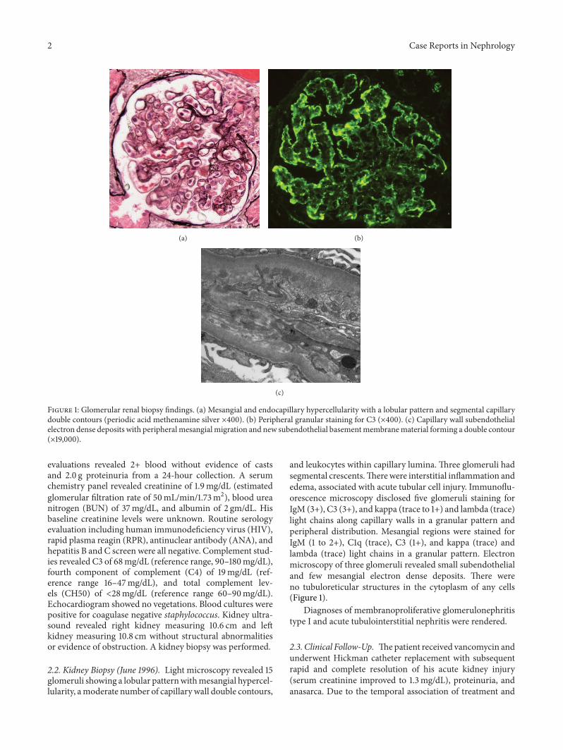
Figure 2: Glomerular features of second renal biopsy. (a) Lobular hypercellular glomerulus with capillary wall double contours (periodic acidmethenamine silver ×600). (b) Capillary wall with subendothelial deposits and peripheral mesangial migration and interposition producinga double contour (×7200).
renal disease resolution, the MPGN was presumed to besecondary to staphylococcal bacteremia.
He was lost to follow-up for several years until Febru-ary 2010 when he presented with upper extremity edemaand chills. On admission he had anemia, reduced kidneyfunction, and hypoalbuminemia. Again, he was found tobe infected with coagulase negative staphylococcus bac-teremia (S. epidermidis). His spot urine protein to creatinineratio at presentation was 2.4 g/g creatinine but increased to5.8 g/g over several days without associated blood pressurechanges. Routine laboratory investigations revealed creati-nine of 2.2mg/dL, BUN 19mg/dL, WBC 4,400/mm3, andhemoglobin 5.9 g/dL. Urinalysis revealed 300mg/dL protein,large blood, large leukocyte esterase, 196 WBC/high powerfield (HPF), 224 RBC/HPF, 33 hyaline casts, few WBCclumps, 14 granular casts, and 24 cellular casts. Hewas treatedwith vancomycin pending repeat evaluation of the underlyingnephrotic syndrome. Of interest, the patient commentedthat “every time I swell up, they give me antibiotics andthe swelling goes away.” Further evaluation of his renaldisease was again pursued as all his previous medical recordswere lost in a hospital fire. Serum protein electrophoresis(SPEP), urine protein electrophoresis (UPEP), serum pro-tein immunofixation (SPIF), urine protein immunofixation(UPIF), antineutrophil cytoplasmic antibody (ANCA), RPR,ANA, and HIV were all negative. C3 was low at 71mg/dLwith normal C4 of 23mg/dL. A kidney ultrasound revealednormal sized kidneys (right 11.0 cm and left 11.7 cm) with-out structural abnormalities. Evaluation for subacute bac-terial endocarditis was negative. His Hickman catheter wasreplaced and subsequent blood cultures confirmed resolu-tion of his bacteremia. His proteinuria improved markedly(greater than 50% reduction) within a few days of antibiotictherapy initiation. Serial creatinine measurements docu-mented improvement in his creatinine to 1.75mg/dL within20 days of presentation. A repeat kidney biopsy performedin June 2010 confirmed MPGN type I (Figure 2), with acute
tubulointerstitial nephritis and mild-to-moderate chronicrenal parenchymal injury.
In July 2010, he presented for the third timewith anasarca,fevers, and acute kidney injury (creatinine of 3.29mg/dL,elevated from baseline level 1.7–2.0mg/dL). Initial urinalysisshowed 100mg/dL protein, large blood, 215 WBC/HPF, 252RBC/HPF, and 42 hyaline casts. Complement levels revealedC3 of 45mg/dL, C4 of 18mg/dL, and CH50 < 13mg/dL.Blood cultures revealed coagulase negative staphylococcusbacteremia (S. epidermidis) and spot urine protein-creatinineratios rapidly rose from 5.1 g/g to a peak of 9.5 g/g within 2days without accompanying blood pressure changes. Treat-ment with vancomycin and Hickman line replacement led torapid reduction in proteinuria and anasarca with creatinineimproving to his recent baseline of 1.7mg/dL by October2010. As the two previous kidney biopsies demonstratedMPGN type I, this third episode of acute kidney injuryaccompanied by hematuria and nephrotic range proteinuriawas attributed to a recurrence of MPGN secondary torecurrent Hickman catheter infection.
3. Discussion
The pathophysiology of MPGN caused by chronic indwellingcentral catheters has been previously described [4, 6, 8].The production of immunoglobulins against an infectiousagent and the subsequent binding of two or more of theseimmunoglobulins result in activation of C1, which thencleaves C4 and C2 to generate C4b and C2a to form C4b2a,the classical pathway convertase, leading to the activation ofC3 convertase and generation of the terminal complementcomplex. Glomerular involvement is instigated by depositionof immune-complexes and complement factors of the clas-sical and terminal pathway in the subendothelial region ofcapillary walls [6]. The injury phase of MPGN is character-ized by the influx of leukocytes with associated cytokine andprotease release, inducing capillary wall damage and ensuing

4 Case Reports in Nephrology
Table1:Clinicalmanifestations
ofrepo
rted
casesa
ndcurrentcase.
References
Medicalhisto
ryInitialpresentatio
nBa
selin
ecreatin
ine
(mg/dL
)
Presentin
gcreatin
ine
(mg/dL
)Urin
alysis
Com
plem
ents
(mg/dL
)Re
nalB
iopsy
Bloo
dcultu
res
Num
bero
fcatheter
changes
Yaredetal.[7]
66-year-oldmalew
ithmesenteric
ischemia
andbo
welresection
with
parenteral
nutrition
for
hyperalim
entatio
n
Worsening
kidn
eyfunctio
nandnew
skin
rash
1.53.2
>25
RBC/HPF,
proteinu
ria,4–10
granular
casts
“Normal”
complem
ents
(valuesn
otrepo
rted)
MPG
NS.epidermidis
and
C.jeikeium
6
Yaredetal.[7]
45-year-oldfemale
TAH/BSO
complicated
byisc
hemicbo
wel
requ
iring
resection,
requ
iredparenteral
nutrition
for
hyperalim
entatio
n
Worsening
kidn
eyfunctio
n,newskin
rash,and
severe
anem
ia
1.87.7
Proteinu
riaand
hematuriawith
RBCand
mixed-cellcasts
Initiallyno
rmal
complem
ents,
then
C3andC4
levels
slightly
depressed
MPG
NUnk
nown
5
Ohara
etal.[6]
13-year-oldmale
midgutvolvulusa
ndresectionof
necrotic
ileum
,required
parenteralnu
trition
for
hyperalim
entatio
n
Hem
aturiaand
proteinu
riaon
routineu
rinary
screening
Unk
nown
0.6
ManyRB
Cs,10–
15WBC
,1-2
granular
casts
/HPF
C330
(low),C4
8(lo
w),CH
50<10
(low)
MPG
NS.epidermidis
7
Currentcase
repo
rt
23-year-oldmale
multip
legunsho
twou
ndstoabdo
men
atage17,requ
ired
parenteralnu
trition
for
hyperalim
entatio
n
Firstepisode
July1996:
proteinu
ria,
hematuria,and
renalinsuffi
ciency
onroutinetestin
g
Unk
nown
1.92+
bloo
d,>100R
BC,no
cellu
larc
asts
C369
(low),C4
19(lo
w-normal),
CH50<28
(low)
MPG
NS.epidermidis
Unk
nown
Second
episo
deFebruary
2010
atage
37:fevers,anasarca,
andrenal
insufficiency
1.3–1.5
2.2
Protein300m
g/dL
,largeb
lood
,WBC
196,RB
C224,
+hyalin
e,granular,
andcellu
lar
casts
/HPF
C371
(low),C4
23(lo
w-normal),
CH50<13
(low)
MPG
N(biopsydo
neJune
2010)
S.epidermidis
>2
Third
episo
deJuly2010:anasarca
andfatig
ue1.7–2.0
3.3
Protein100m
g/dL
,largeb
lood
,WBC
215,RB
C252,42
hyalinec
asts/
HPF
C345
(low),C4
18(lo
w-normal),
CH50<13
(low)
Nobiop
sy∗
S.epidermidis
>2
Abbreviatio
ns:TAH/BSO
:totalabdo
minalhyste
rectom
yand
bilateralsalpingooo
pherectomies;S.epidermidis:
Staphylococcus
epidermidis;
C.jeikeium:C
lostridiumjeikeium;R
BC:red
bloo
dcells;W
BC:w
hitebloo
dcells;H
PF:highpo
wer
field.
∗Presum
ptived
iagn
osisof
recurrentM
PGNbasedon
previous
biop
syfin
ding
s,clinicalcou
rse,andrespon
seto
approp
riatetherapy.

Case Reports in Nephrology 5
hematuria and proteinuria. In addition to Staphylococcus,other bacteria reported in association with MPGN includeMycobacterium tuberculosis, streptococci, Propionibacteriumacnes, Mycoplasma pneumoniae, brucella, Coxiella burnetii,nocardia, andMeningococcus [8].
A literature search for biopsy proven MPGN associatedwith chronic central venous catheterization revealed onlythree cases [6, 7]. In these three reported cases, the centralvenous catheter was used for home parenteral nutrition forshort bowel syndrome (Table 1). Of note, all patients hadmultiple (five to seven) episodes of infectious catheter com-plications prior to overt renal manifestations. Specific renalpresentations ranged from incidental finding of microscopichematuria,mild proteinuria (0.3 g/g creatinine), and granularcasts in one patient to an insidious or relatively rapid rise inserum creatinine over 18 days to 2 months in the two otherpatients. Patients with increasing serum creatinine hadconcurrent significant proteinuria and an active urinarysediment including microscopic hematuria with or withoutcellular casts. Associated extrarenal clinical manifestationsreported include edema/anasarca, fevers, and/or palpablepurpura due to biopsy proven leukocytoclastic vasculitis.Complement levels varied from normal to significantlydepressed. Blood and catheter tip cultures obtained inthree out of four cases revealed Staphylococcus epidermidis.Following catheter replacement and appropriate antibioticadministration, all patients promptly andmarkedly improvedin renal function, proteinuria, and/or hematuria. Of interest,signs of recovery such as reduction in proteinuria may benoted within a few days and fall in serum creatinine within1-2 weeks of antibiotic therapy and catheter removal (8,present case). Complete recovery of renal functionmay occurwithin three to 10 months. Unfortunately, in our current case,only partial renal functional recovery and reduction inproteinuria were observed following the third documentedepisode of infection with glomerulonephritis. This likely wasdue to significant renal parenchymal scarring consequent toinadequate treatment of prior infectious insults in associationwith poor patient compliance and follow-up.
Recurrent biopsy proven MPGN in parallel with recur-rent line infection/bacteremia, as observed in the currentcase, leaves little doubt regarding a direct causal relationshipbetween chronic central line infections and MPGN. Subtlerenal manifestations such as microscopic hematuria and slowrise in serum creatininemay herald overtly apparentmanifes-tations of catheter infections; therefore, routine urinalysis andclosemonitoring of serum creatinine in patients with chroniccentral venous catheterization are indicated to allow earlydetection of catheter infection and prevention of progressivekidney injury. It should be noted that animal studies involvingsheep and baboons have shown that renal parenchymal injurymay occur even prior to overt renal manifestations [4, 5].Nevertheless, some extent of disease reversal is expectedwith prompt intervention, including appropriate antimicro-bial therapy and removal or replacement of the indwellingcatheter, as reported [6, 7].
In summary, we have presented a case of recurrentMPGN associated with recurrent bacteremia from a chron-ically indwelling Hickman catheter in an adult. Given the
repeated strong temporal correlations of MPGN with recur-rent catheter infections, the latter is likely the key factor in thedevelopment of MPGN in this setting. In a patient with aninfected catheter and concurrent evidence of kidney injury,with or without a prior biopsy-proven MPGN, short timeallowance for antibiotic treatment and catheter replacementmay be indicated prior to renal biopsy performance, partic-ularly when the only abnormal serologic finding is a low C3level. However, it should be emphasized that a kidney biopsyshould be considered if renal function and/or proteinuria donot resolve within 2–4 weeks, or if serologic testing suggeststhe possibility of another disease entity.
In conclusion, patients with chronic indwelling centralvenous catheters should be given routine surveillance forboth infections and markers of kidney injury includingserum creatinine and urinalysis. Similarly, patients should beeducated to recognize early signs and symptoms of infectionsas well as development of unusual urinary foaming and/orchange in urine output and color.
Conflict of Interests
The authors declare that there is no conflict of interestsregarding the publication of this paper.
References
[1] H. G. Rennke, “Secondary membranoproliferative glomeru-lonephritis,” Kidney International, vol. 47, no. 2, pp. 643–656,1995.
[2] E. Noiri, S. Kuwata, K. Nosaka et al., “Shunt nephritis: efficacy ofan antibiotic trial for clinical diagnosis,” Internal Medicine, vol.32, no. 4, pp. 291–294, 1993.
[3] J. A. Black, D. N. Challacombe, and B. G. Ockenden, “Nephroticsyndrome associated with bacteraemia after shunt operationsfor hydrocephalus,” The Lancet, vol. 286, no. 7419, pp. 921–924,1965.
[4] V. P. Rao, T. Poutahidis, R. P. Marini, H. Holcombe, A. B.Rogers, and J. G. Fox, “Renal infarction and immune-mediatedglomerulonephritis in sheep (Ovis aries) chronically implantedwith indwelling catheters,” Journal of the American Associationfor Laboratory Animal Science, vol. 45, no. 4, pp. 14–19, 2006.
[5] S. L. Leary, W. D. Sheffield, and J. D. Strandberg, “Immunecomplex glomerulonephritis in baboons (Papio cynocephalus)with indwelling intravascular catheters,” Laboratory AnimalScience, vol. 31, no. 4, pp. 416–420, 1981.
[6] S. Ohara, Y. Kawasaki, K. Takano et al., “Glomerulonephri-tis associated with chronic infection from long-term centralvenous catheterization,” Pediatric Nephrology, vol. 21, no. 3, pp.427–429, 2006.
[7] G. Yared, D. L. Seidner, E. Steiger, P. M. Hall, and J. V. Nally,“Tunneled right atrial catheter infection presenting as renalfailure,” Journal of Parenteral and Enteral Nutrition, vol. 23, no.6, pp. 363–365, 1999.
[8] S. Sethi and F. C. Fervenza, “Membranoproliferative glom-erulonephritis—a new look at an old entity,” The New EnglandJournal of Medicine, vol. 366, no. 12, pp. 1119–1131, 2012.

Submit your manuscripts athttp://www.hindawi.com
Stem CellsInternational
Hindawi Publishing Corporationhttp://www.hindawi.com Volume 2014
Hindawi Publishing Corporationhttp://www.hindawi.com Volume 2014
MEDIATORSINFLAMMATION
of
Hindawi Publishing Corporationhttp://www.hindawi.com Volume 2014
Behavioural Neurology
EndocrinologyInternational Journal of
Hindawi Publishing Corporationhttp://www.hindawi.com Volume 2014
Hindawi Publishing Corporationhttp://www.hindawi.com Volume 2014
Disease Markers
Hindawi Publishing Corporationhttp://www.hindawi.com Volume 2014
BioMed Research International
OncologyJournal of
Hindawi Publishing Corporationhttp://www.hindawi.com Volume 2014
Hindawi Publishing Corporationhttp://www.hindawi.com Volume 2014
Oxidative Medicine and Cellular Longevity
Hindawi Publishing Corporationhttp://www.hindawi.com Volume 2014
PPAR Research
The Scientific World JournalHindawi Publishing Corporation http://www.hindawi.com Volume 2014
Immunology ResearchHindawi Publishing Corporationhttp://www.hindawi.com Volume 2014
Journal of
ObesityJournal of
Hindawi Publishing Corporationhttp://www.hindawi.com Volume 2014
Hindawi Publishing Corporationhttp://www.hindawi.com Volume 2014
Computational and Mathematical Methods in Medicine
OphthalmologyJournal of
Hindawi Publishing Corporationhttp://www.hindawi.com Volume 2014
Diabetes ResearchJournal of
Hindawi Publishing Corporationhttp://www.hindawi.com Volume 2014
Hindawi Publishing Corporationhttp://www.hindawi.com Volume 2014
Research and TreatmentAIDS
Hindawi Publishing Corporationhttp://www.hindawi.com Volume 2014
Gastroenterology Research and Practice
Hindawi Publishing Corporationhttp://www.hindawi.com Volume 2014
Parkinson’s Disease
Evidence-Based Complementary and Alternative Medicine
Volume 2014Hindawi Publishing Corporationhttp://www.hindawi.com