Embed Size (px)
Citation preview
219
A CASE OF NOCARDIA MYCETOMA DEVELOPING AT THE SITE OF SKIN GRAFTING PRZYPADEK ROZWOJU NOCARDIA MYCETOMA W MIEJSCU SZCZEPIENIA Hosahalli Rajaiah Yogeesh1, Sujatha Chankramath1, Seema Srinivasa1, Raja Parthiban Sravana Rajendran2, Poornima Kamalaksha Shenoy3 1Department of Dermatology, M.V.J.Medical College & Research Hospital, Hoskote, Bangalore, India 2Department of Pathology, M.V.J.Medical College & Research Hospital, Hoskote, Bangalore, India 3Department of Microbiology, M.V.J.Medical College & Research Hospital, Hoskote, Bangalore, India Corresponding author: Dr. Hosahalli Rajaiah Yogeesh [email protected]
Our Dermatol Online. 2011; 2(4): 219-223 Date of submission: 12.07.2011 / acceptance: 30.08.2011 Conflicts of interest: None
Abstract Mycetomas are chronic infections of the skin, subcutaneous tissue and deeper tissues caused by fungi or filamentous bacteria and are characterized by tumefaction, nodules and sinuses. Eumycetomas are caused by fungi and actinomycetomas are caused by filamentous bacteria. Bacteria causing actinomycetomas are saprophytes found in soil, on plants, and on dead and decaying matter. They are aerobic, gram positive and weakly acid fast and form aggregates of micro colonies that appear as grains in the sinuses. Clinically, mycetomas present as areas of tumefaction with nodules and discharging sinuses. Bacterial mycetomas are sensitive to many antibiotics like penicillin, tetracyclines, sulphonamides, rifampcin, aminoglycocides etc, and long term combination therapies with variable success have been reported from different parts of the world. We have presented here a 23 year old male patient who presented with one year history of developing nodules and sinuses in the region of left flank. Patient had undergone skin grafting at the same site 3 years back for a wound he developed in the area following a road accident. He was unsuccessfully treated with antituberculous therapy elsewhere. He was admitted in our institute; the diagnosis of nocardia mycetoma was established and patient was successfully treated with complete clearance of lesions.
Streszczenie Mycetoma są przewlekłymi, skórnymi chorobami zakaźnymi, obejmujące tkankę podskórną i głębiej połoŜone tkanki, spowodowane przez grzyby lub/i nitkowate bakterie, charakteryzujące się nabrzmieniem, guzkami i zatokami. Eumycetoma są spowodowane przez grzyby, a actinomycetoma są spowodowane przez nitkowate bakterie. Bakterie wywołujące actinomycetoma są saprofitami, które moŜna znaleźć w glebie, na roślinach oraz na martwej i gnijącej materii. Są one tlenowe, gram-dodatnie i słabo kwaśne; występują w formie agregatów w mikro koloniach, które pojawiają się jako ziarna w zatokach. Klinicznie, mycetoma obecne są jako obszary nabrzmienia z guzkami i wydalającymi zatokami. Bakteryjne mycetoma są wraŜliwe na wiele antybiotyków jak penicyliny, tetracykliny, sulfonamidy, ryfampicyna, aminoglikozydy itp., a po długim okresie kombinowanej terapii wyzdrowienia ze zmiennym sukcesem zostały zgłaszane z róŜnych stron świata. Przedstawiamy 23-letniego pacjenta, który prezentował jednoroczną historię rozwoju guzków i zatok w regionie lewego boku. Przed 3-ma laty pacjent przeszedł przeszczep skóry w tym samym miejscu z powodu rany w wyniku wypadku drogowego. Był bezskutecznie leczony antytuberkulinową terapią w innej jednostce. Pacjent był przyjęty do naszego instytutu; postawiono diagnozę nocardia mycetoma i wdroŜono skuteczną terapię z całkowitym wyleczeniem zmian skórnych. Key words: Nocardia; mycetoma; penicillin Słowa klucze: Nocardia; mycetoma; penicylina
Introduction
Mycetomas are infections caused by fungi or filamentous bacteria and are characterized by tumefaction with nodules and sinuses. Organisms are
saprophytes found in soil or on plants [1]. Mostly infections follow penetrating injuries, especially amongst those people who walk barefoot; the disease is more common in tropical and sub-tropical regions [2]. After
Case Report / Opis przypadku
© Our Dermatol Online 4.2011
220
gaining access into the subcutaneous space, a swelling develops in the region which is followed by numerous nodules and sinuses over a span of months and sometimes years [3]. Grains that are found in the discharge from sinuses are aggregates of microcolonies of the organisms. Failure to recognize the condition early may lead to deeper involvement of infection causing damage to bones, muscles and other deeper tissues [4]. Early institution of antifungals/antibiotics can lead to complete cure of this condition. Late and neglected case require debridement of tissues and sometimes even amputation of limbs along with pharmacotherapy. In this article we have discussed a case of actinomycetoma caused by nocardia species. This patient was diagnosed as cutaneous tuberculosis in some other institute and was treated with anti-tuberculosis therapy. He was admitted in our institute MVJ Medical College and Research Hospital in a rural area of outskirts of Bangalore where he was instigated, and treated initially with amikacin and dapsone; later showed excellent response and complete clearance of lesions with injection crystalline penicillin followed by oral phenoxymethyl penicillin. Case report
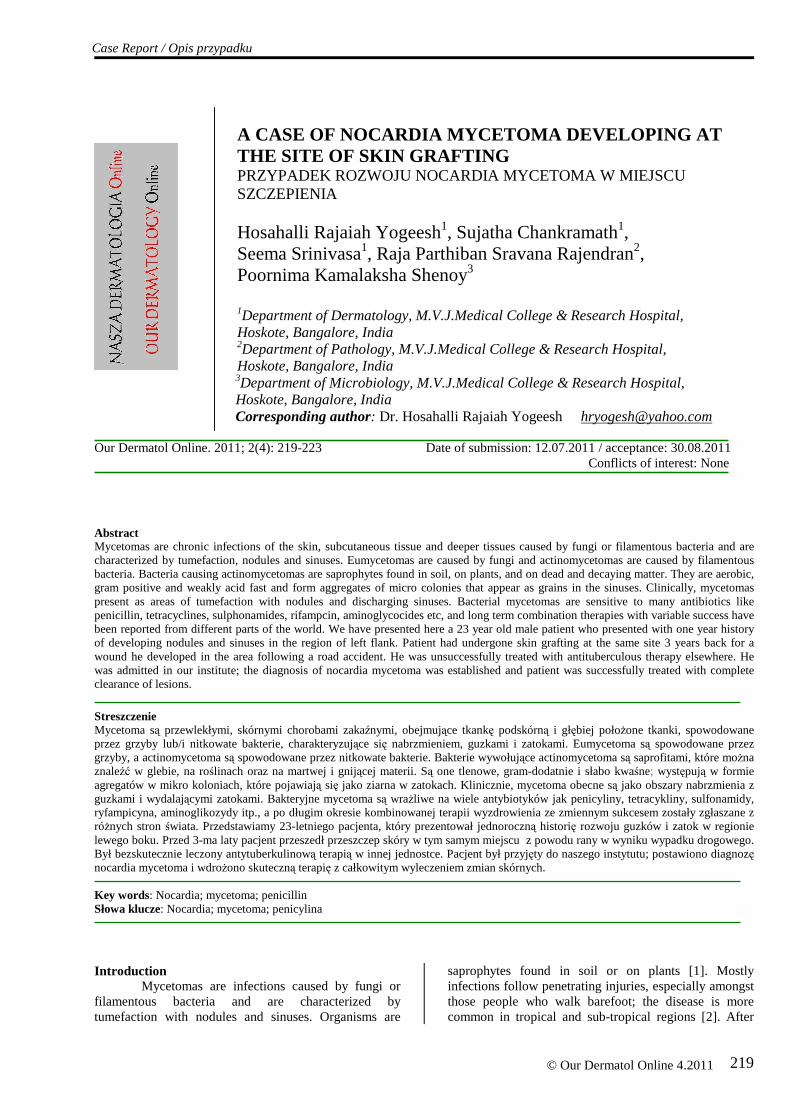
A 23 years old male patient presented with a swelling and multiple discharging sinuses over the left flank one year duration. He had undergone a skin graft 3 years back at the same site after he met with a road traffic accident and sustained a wound in the region. About six months after the skin grafting patient developed swelling and discharging sinuses in the region. His condition was diagnosed as cutaneous tuberculosis in another institute and he was placed on anti-tuberculous therapy for 6 months with no improvement of his condition. Details of the investigations prior to initiation of anti-tuberculous therapy were not available with the patient. On examination, an area of atrophic dyspigmented puckered scarring with nodules and multiple discharging sinuses measuring 15cms x 7cms was present on the left flank. Some sinuses showed seropurulent or bloody discharge while others were covered with hemorrhagic crusts (Fig.1,2). However, no granules were available on expressing the sinuses. Some areas showed moderate tenderness. Systemic examination was normal.
On investigating, his routine blood counts, urine routine examination, biochemical parameters and chest x-ray did not reveal any abnormalities. X-ray of flank was normal. Ultrasound examination of the region showed multiple subcutaneous and intramuscular abscesses in the left paraspinal area. KOH mounting of the discharge obtained from the lesion did not reveal any fungal elements. Gram staining and modified Ziel-Neelsen staining showed gram positive and weakly acid-fast bacilli. Discharge was sent for culture for fungi, bacteria and mycobacterium species.
Figure 1. Clinical photograph showing nodules and sinuses
Figure 2. Clinical photograph showing close-up view of lesions
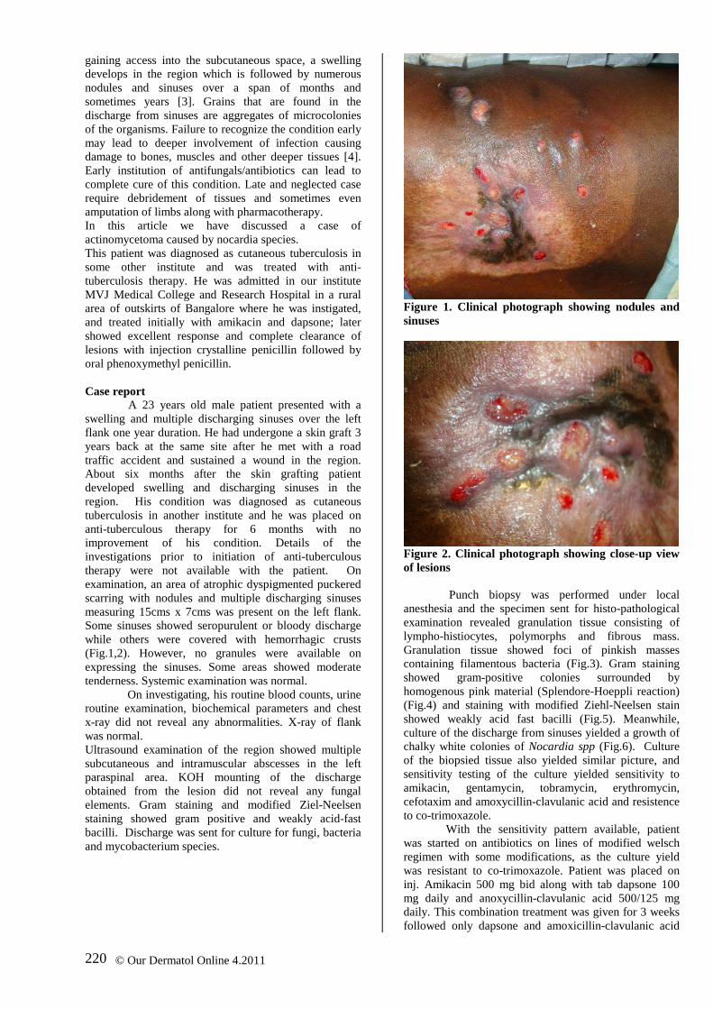
Punch biopsy was performed under local anesthesia and the specimen sent for histo-pathological examination revealed granulation tissue consisting of lympho-histiocytes, polymorphs and fibrous mass. Granulation tissue showed foci of pinkish masses containing filamentous bacteria (Fig.3). Gram staining showed gram-positive colonies surrounded by homogenous pink material (Splendore-Hoeppli reaction) (Fig.4) and staining with modified Ziehl-Neelsen stain showed weakly acid fast bacilli (Fig.5). Meanwhile, culture of the discharge from sinuses yielded a growth of chalky white colonies of Nocardia spp (Fig.6). Culture of the biopsied tissue also yielded similar picture, and sensitivity testing of the culture yielded sensitivity to amikacin, gentamycin, tobramycin, erythromycin, cefotaxim and amoxycillin-clavulanic acid and resistence to co-trimoxazole.
With the sensitivity pattern available, patient was started on antibiotics on lines of modified welsch regimen with some modifications, as the culture yield was resistant to co-trimoxazole. Patient was placed on inj. Amikacin 500 mg bid along with tab dapsone 100 mg daily and anoxycillin-clavulanic acid 500/125 mg daily. This combination treatment was given for 3 weeks followed only dapsone and amoxicillin-clavulanic acid
© Our Dermatol Online 4.2011
221
for 2 weeks. Three such cycles were given followed by daily continuous doses of oral dapsone and amoxycillin-clavulanic acid. Patient showed partial healing of the sinuses and decrease in size of nodules as long as he was on cycles of injection amikacin; however the improvement was not sustained while he was switched to oral medication. Fresh sinuses and nodules started appearing and he patient developed tenderness in the region. Attempt at fresh culture for sensitivity testing failed.
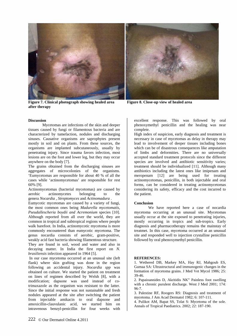
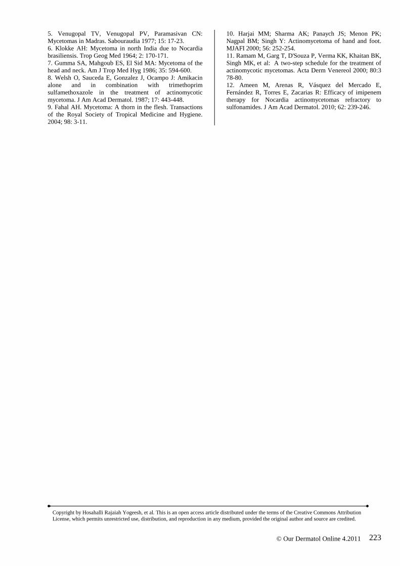
We decided to place the patient on benzyl penicillin and started him on 1.2 mega units of benzyl penicillin intravenously 6th hourly, while continuing the dapsone and stopping amoxicillin-clavulanic acid. In four weeks of this therapy patient showed excellent response with near healing of the sinuses and nodules. We decided to continue therapy with oral phenoxymethyl penicillin (pentids) at the dose of 800 mg four times daily while continuing dapsone. Patient has completed 4 months of oral phenoxymethyl pencillin and dapsone. The lesions showed complete resolution with healing of all nodules and sinuses (Fig.7,8).
Figure 3. HPE showing grains surrounded by Figure 4. HPE showing grains surrounded by inflammatory cells (H&E Staining, 400 X) homogenous pinkish material (Splendore-Hoeppli reaction) (Gram staining, 400X)
Figure 5. Modified Z-N staining showing weakly acid fast grains (400X)
Figure 6. Nocardia colonies in culture tube
© Our Dermatol Online 4.2011
222
Figure 7. Clinical photograph showing healed area Figure 8. Close-up view of healed area after therapy Discussion
Mycetomas are infections of the skin and deeper tissues caused by fungi or filamentous bacteria and are characterized by tumefaction, nodules and discharging sinuses. Causative organisms are saprophytes present mostly in soil and on plants. From these sources, the organisms are implanted subcutaneously, usually by penetrating injury. Since trauma favors infection, most lesions are on the foot and lower leg, but they may occur anywhere on the body [7]. The grains obtained from the discharging sinuses are aggregates of microcolonies of the organisms. ‘Eumycetomas are responsible for about 40 % of all the cases while ‘actinomycetomas’ are responsible for rest 60% [9]. Actinomycetomas (bacterial mycetomas) are caused by aerobic actinomycetes belonging to the genera Nocardia , Streptomyces and Actinomadura . Eumycotic mycetomas are caused by a variety of fungi, the most common ones being Madurella mycetomatis, Pseudallescheria boydii and Acremonium species [10]. Although reported from all over the world, they are common in tropical and subtropical regions where people walk barefoot. In India, actinomycotic mycetoma is more commonly encountered than eumycotic mycetoma. The genus nocardia consists of aerobic, gram-positive, weakly acid fast bacteria showing filamentous structure. They are found in soil, wood and water and also in decaying matter. In India the first report on N. brasiliensis infection appeared in 1964 [5]. In our case mycetoma occurred at an unusual site (left flank) where skin grafting was done in the region following an accidental injury. Nocardia spp was obtained on culture. We started the patient on treatment on lines of regimen described by Welsh [8], with a modification; dapsone was used instead of co-trimaxazole as the organism was resistant to the latter. Since the initial response was not sustainable and fresh nodules appeared at the site after switching the patient from injectable amikacin to oral dapsone and amoxicillin-clauvulanic acid, we started him on intravenous benzyl-penicillin for four weeks with
excellent response. This was followed by oral phenoxymethyl penicillin and the healing was near complete. High index of suspicion, early diagnosis and treatment is necessary in case of mycetomas as delay in therapy may lead to involvement of deeper tissues including bones which can be of disastrous consequences like amputation of limbs and deformities. There are no universally accepted standard treatment protocols since the different species are involved and antibiotic sensitivity varies; treatment should be individualized [11]. Although many antibiotics including the latest ones like imipenam and meropenam [12] are being used for treating actinomycetomas, penicillin, in both injectable and oral forms, can be considered in treating actinomycetomas considering its safety, efficacy and the cost incurred to the patient. Conclusion
We have reported here a case of nocardia mycetoma occurring at an unusual site. Mycetomas usually occur at the site exposed to penetrating injuries, mostly occurring in tropics and sub-tropics. Early diagnosis and pharmacotherapy remains the mainstay of treatmet. In this case, mycetoma occurred at an unusual site and responded well to injection crystalline penicillin followed by oral phenoxymethyl penicillin. REFERENCES: 1. Wethered DB, Markey MA, Hay RJ, Mahgoub ES, Gumaa SA: Ultrastructural and immunogenic changes in the formation of mycetoma grains. J Med Vet Mycol 1986; 25: 39-46. 2. Papaioannides D, Akritidis NK” Painless foot swelling with a chronic purulent discharge. West J Med 2001; 174: 96-7. 3. Palestine RF, Roogers RS: Diagnosis and treatment of mycetoma. J Am Acad Dermatol 1982; 6: 107-111. 4. Pulikot AM, Bapat SS, Tolat S: Mycetoma of the sole. Annals of Tropical Paediatrics. 2002; 22: 187-190.
© Our Dermatol Online 4.2011
223
5. Venugopal TV, Venugopal PV, Paramasivan CN: Mycetomas in Madras. Sabouraudia 1977; 15: 17-23. 6. Klokke AH: Mycetoma in north India due to Nocardia brasiliensis. Trop Geog Med 1964; 2: 170-171. 7. Gumma SA, Mahgoub ES, El Sid MA: Mycetoma of the head and neck. Am J Trop Med Hyg 1986; 35: 594-600. 8. Welsh O, Sauceda E, Gonzalez J, Ocampo J: Amikacin alone and in combination with trimethoprim sulfamethoxazole in the treatment of actinomycotic mycetoma. J Am Acad Dermatol. 1987; 17: 443-448. 9. Fahal AH. Mycetoma: A thorn in the flesh. Transactions of the Royal Society of Tropical Medicine and Hygiene. 2004; 98: 3-11.
10. Harjai MM; Sharma AK; Panaych JS; Menon PK; Nagpal BM; Singh Y: Actinomycetoma of hand and foot. MJAFI 2000; 56: 252-254. 11. Ramam M, Garg T, D'Souza P, Verma KK, Khaitan BK, Singh MK, et al: A two-step schedule for the treatment of actinomycotic mycetomas. Acta Derm Venereol 2000; 80:3 78-80. 12. Ameen M, Arenas R, Vásquez del Mercado E, Fernández R, Torres E, Zacarias R: Efficacy of imipenem therapy for Nocardia actinomycetomas refractory to sulfonamides. J Am Acad Dermatol. 2010; 62: 239-246.
© Our Dermatol Online 4.2011
Copyright by Hosahalli Rajaiah Yogeesh, et al. This is an open access article distributed under the terms of the Creative Commons Attribution License, which permits unrestricted use, distribution, and reproduction in any medium, provided the original author and source are credited.