Embed Size (px)
Citation preview
557
INTRODUCTION
Although locally-transmitted malaria has been under con-trol, imported malaria, including Plasmodium knowlesi and Plasmodium ovale infections, are still being monitored in China [1], the Republic of Korea [2], and Japan [3]. In 2010, the Min-istry of Health in China launched an action plan for malaria elimination, with the goals of eradicating local malaria cases in regions outside of the Yunnan border area by the end of 2015 and eliminating malaria in the entire country by the end of 2020 [4]. However, the proportion of imported malaria cas-es caused by 4 traditional species including falciparum, vivax, malariae, and ovale rapidly increased to 66.4% in 2011, since monitoring of imported malaria cases first started in 2008 [1,5]. Effective surveillance of every malaria case is important, and malaria strategies should range from the control stage to elimi-nation of malaria.
In 2011, the national malaria genetic diagnosis protocol was released by Chinese Center for Disease Control and Prevention. In this protocol, the gene encoding the small subunit (SSU)
rRNA was identified as the target gene for differentiation, be-cause this gene has highly conserved and variable regions that not only allow discrimination of different Plasmodium species but can also be used for obtaining information about the phy-logenetic characterization of different malaria parasites. Fifty malaria cases in Hainan Province have been confirmed, and 18 cases were excluded by this protocol since the Hainan ma-laria reference laboratory was established in 2011.
In this article, we present a case of Plasmodium ovale wallikeri in China imported from West Africa. The case was diagnosed by microscopic examinations, rapid diagnosis test (RDT), nest-ed PCR, and sequencing analysis of the SSU rRNA gene. Inter-estingly, this was a case of relapse and was not a new infection.
CASE DESCRIPTION
On 28 February 2013, a 39-year-old Chinese patient pre-sented to Baoxian Township Hospital in Hainan Province with a 2-day history of daily fever, mild headache, chills, and tem-perature up to 38˚C. He had worked in Sierra Leone from July 2012 to January 2013, after which he returned to Hainan. He had cold and fever symptoms on one occasion in July 2012 in Ghana, and was diagnosed with malaria by a local hospital and treated with artemether injection daily for 3 days.
After he was admitted to Baoxian Township Hospital, thin and thick blood smears were taken and stained with Giemsa
ISSN (Print) 0023-4001ISSN (Online) 1738-0006
Korean J Parasitol Vol. 51, No. 5: 557-562, October 2013 http://dx.doi.org/10.3347/kjp.2013.51.5.557▣ CASE REPORT
A Case of Plasmodium ovale wallikeri Infection in a Chinese Worker Returning from West Africa
Yuchun Li, Guangze Wang, Dingwei Sun, Feng Meng, Shigan Lin, Ximin Hu* and Shanqing Wang*Parasitic Diseases Department, Hainan Provincial Center for Disease Control and Prevention, Haikou, Hainan 570203, P. R. China
Abstract: In contrast to the gradual reduction in the number of locally transmitted malaria cases in China, the number of imported malaria cases has been increasing since 2008. Here, we report a case of a 39-year-old Chinese man who ac-quired Plasmodium ovale wallikeri infection while staying in Ghana, West Africa for 6 months in 2012. Microscopic exami-nations of Giemsa-stained thin and thick blood smears indicated Plasmodium vivax infection. However, the results of rap-id diagnostic tests, which were conducted 3 times, were not in agreement with P. vivax. To further check the diagnosis, standard PCR analysis of the small-subunit rRNA gene was conducted, based on which a phylogeny tree was construct-ed. The results of gene sequencing indicated that this malaria is a variant of P. ovale (P. ovale wallikeri). The infection in this patient was not a new infection, but a relapse of the infection from the one that he had contracted in West Africa.
Key words: Plasmodium ovale wallikeri, imported malaria, case report, China
•Received 11 July 2013, revised 11 August 2013, accepted 4 September 2013.*Corresponding authors ([email protected]; [email protected])
© 2013, Korean Society for Parasitology and Tropical MedicineThis is an Open Access article distributed under the terms of the Creative Commons Attribution Non-Commercial License (http://creativecommons.org/licenses/by-nc/3.0) which permits unrestricted non-commercial use, distribution, and reproduction in any medium, provided the original work is properly cited.
558 Korean J Parasitol Vol. 51, No. 5: 557-562, October 2013
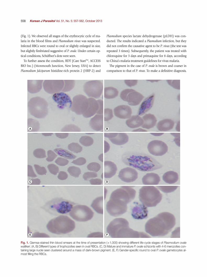
(Fig. 1). We observed all stages of the erythrocytic cycle of ma-laria in the blood films and Plasmodium vivax was suspected. Infected RBCs were round to oval or slightly enlarged in size, but slightly fimbriated suggestive of P. ovale. Under certain op-tical conditions, Schüffner’s dots were seen.
To further assess the condition, RDT (Care StartTM, ACCESS BIO Inc.) (Monmouth Junction, New Jersey, USA) to detect Plasmodium falciparum histidine-rich protein 2 (HRP-2) and
Plasmodium species lactate dehydrogenase (pLDH) was con-ducted. The results indicated a Plasmodium infection, but they did not confirm the causative agent to be P. vivax (the test was repeated 3 times). Subsequently, the patient was treated with chloroquine for 3 days and primaquine for 8 days, according to China’s malaria treatment guidelines for vivax malaria.
The pigment in the case of P. ovale is brown and coarser in comparison to that of P. vivax. To make a definitive diagnosis,
A B
C D
E F
Fig. 1. Giemsa-stained thin blood smears at the time of presentation (×1,000) showing different life-cycle stages of Plasmodium ovale wallikeri. (A, B) Different types of trophozoites seen in oval RBCs. (C, D) Mature and immature P. ovale schizonts with 4-6 merozoites con-taining large nuclei seen clustered around a mass of dark-brown pigment. (E, F) Gender-specific round to oval P. ovale gametocytes al-most filling the RBCs.
Li et al.: An imported case of oval malaria wallikeri type in China 559
the blood samples were transferred to the Hainan Provincial Center for Disease Control and Prevention for further PCR am-plification and genetic analyses. Genomic DNA was extracted from 80 µl of the whole blood sample by using a QIAamp Blood Mini kit (Qiagen, Valencia, California, USA) according to the national malaria diagnosis protocol. The partial gene of the SSU rRNA gene was amplified by PCR with Plasmodium-specific primers (rPLU1 and rPLU5) in Nest1 PCR, and spe-cies-specific primers (rFAL 1 and 2, rMAL 1 and 2, rVIV 1 and 2, and rOVA 1 and 2) and genus-specific primers (rPLU3 and rPLU4) in Nest 2 PCR. The Nest 1 amplification conditions were as follows: step 1, 94˚C for 5 min; step 2, denaturation at 94˚C for 30 sec; step 3, annealing at 55˚C for 1 min; step 4, ex-tension at 72 C̊ for 2 min; steps 2-4 were then repeated 35 times, followed by step 4 for 5 min. Two microliters of the Nest 1 am-plification product served as the DNA template for each of the 20-ml Nest 2 amplifications. The concentration of the Nest 2 primers and other constituents were identical to those for the
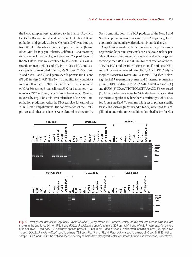
Nest 1 amplifications. The PCR products of the Nest 1 and Nest 2 amplifications were analyzed by 2.5% agarose gel elec-trophoresis and staining with ethidium bromide (Fig. 2).
Amplification results with the species-specific primers were negative for faciparum, vivax, malariae, and ovale malaria par-asites. However, positive results were obtained with the genus-specific primers rPLU3 and rPLU4. For confirmation of the re-sults, the PCR products from the genus-specific primers rPLU1 and rPLU5 were sequenced using the 3,730× l DNA Analyzer (Applied Biosystems, Foster City, California, USA) after TA clon-ing; the M13 sequencing primer and 2 internal sequencing primers, KR1 (5́ -TAA CCAGACAAATCATATTCACGAAC-3́ ) and rPLU6 (5́ -TTAAAATTGTTGCAGTTAAAACG-3́ ), were used [6]. Analysis of sequences in the NCBI database indicated that the causative species may have been a variant type of P. ovale, i.e., P. ovale wallikeri. To confirm this, a set of primers specific for P. ovale wallikeri (rOVA1v and rOVA2v) were used for am-plification under the same conditions described before for Nest
Fig. 2. Detection of Plasmodium spp. and P. ovale wallikeri DNA by nested PCR assays. Molecular size markers in base pairs (bp) are shown in the end lanes (M). A: rFAL 1 and rFAL 2, P. falciparum-specific primers (205 bp); rVIV 1 and rVIV 2, P. vivax-specific primers (144 bp); rMAL 1 and rMAL 2, P. malariae-specific primer (112 bp); rOVA 1 and rOVA 2, P. ovale curtisi-specific primers (800 bp); rOVA 1v and rOVA 2v, P. ovale wallikeri-specific primers (782 bp); rPLU 3 and rPLU 4, Plasmodium-specific primers (240 bp). B: HNS: Hainan sample; SHS1 and SHS2: the first and second delivery samples from Shanghai Center for Disease Control and Prevention, respectively.
560 Korean J Parasitol Vol. 51, No. 5: 557-562, October 2013
2, and a 782 bp positive fragment was obtained [7,8]. The se-quence of our specimen was deposited in GenBank, NCBI un-der the accesion no. KF048920.
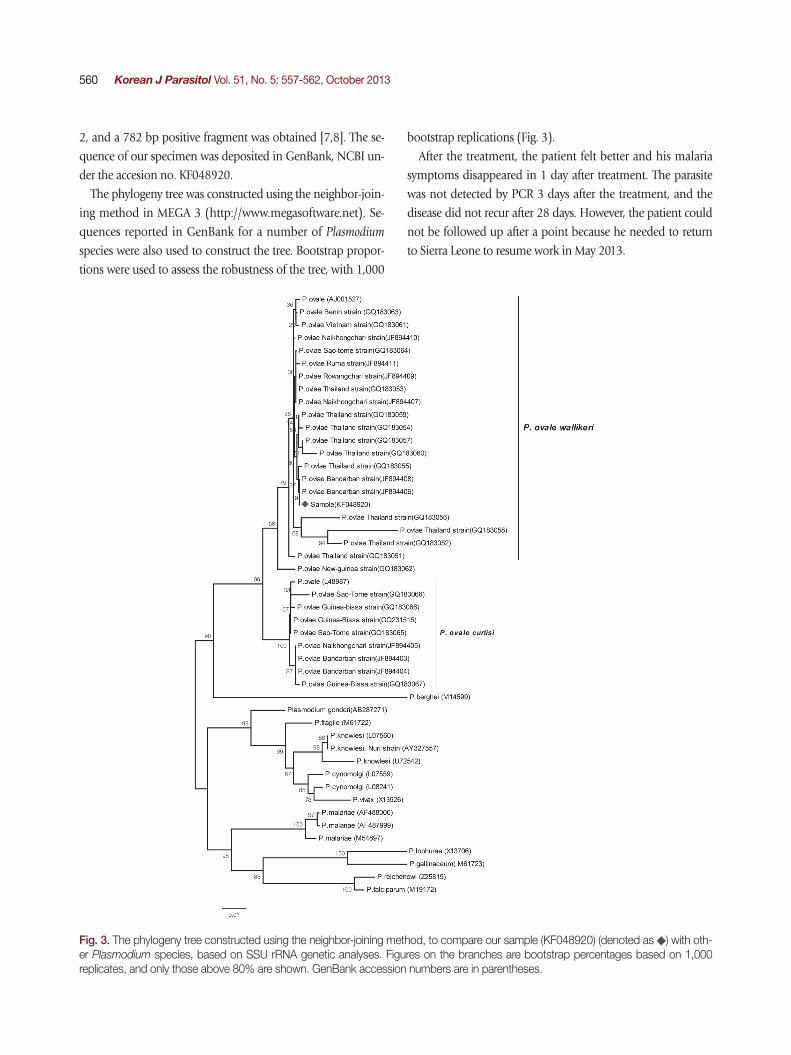
The phylogeny tree was constructed using the neighbor-join-ing method in MEGA 3 (http://www.megasoftware.net). Se-quences reported in GenBank for a number of Plasmodium species were also used to construct the tree. Bootstrap propor-tions were used to assess the robustness of the tree, with 1,000
bootstrap replications (Fig. 3). After the treatment, the patient felt better and his malaria
symptoms disappeared in 1 day after treatment. The parasite was not detected by PCR 3 days after the treatment, and the disease did not recur after 28 days. However, the patient could not be followed up after a point because he needed to return to Sierra Leone to resume work in May 2013.
Fig. 3. The phylogeny tree constructed using the neighbor-joining method, to compare our sample (KF048920) (denoted as ) with oth-er Plasmodium species, based on SSU rRNA genetic analyses. Figures on the branches are bootstrap percentages based on 1,000 replicates, and only those above 80% are shown. GenBank accession numbers are in parentheses.
Li et al.: An imported case of oval malaria wallikeri type in China 561
DISCUSSION
P. ovale is the last reported malaria parasite in humans (be-fore P. knowlesi), and it is mainly found in West Africa [9]. In 2010, P. ovale was reported to be divided into 2 subspecies, P. ovale curtisi (classic type) and P. ovale wallikeri (variant type), with the estimated time of divergence between the 2 subspe-cies being 1 and 3 million years ago [10]. In China, faciparum and vivax malaria were frequently detected in most endemic areas, but malariae and ovale were only detected in the border of Yunnan Province with Myanmar in recent 30 years.
Microscopy-based diagnosis is a traditional tool for diagnos-ing malaria, especially in cases located in remote areas, as it is more convenient and time-saving. However, this method is not very efficient in accurately distinguishing between certain malarial species that have morphological similarities, for ex-ample, between P. malariae and P. knowlesi, and between P. ova-le with P. vivax [3]. In our case, P. ovale wallikeri was mistakenly identified as P. vivax because our microscopist was unfamiliar with morphological stages of the former species. Therefore, there are chances of the causative species being wrongly identi-fied in malaria cases. RDTs are believed to overcome these lim-itations; these tests have been commercially available, and in recent years, they have allowed quick diagnosis in endemic malaria areas with high or low incidence. In this method, the diagnosis is based on identification of some very specific pro-teins expressed by the malaria parasite, and it has been shown to have high sensitivity and specificity in the field. However, there are still some reports of failure and misdiagnosis [11]. The failures were believed to be caused by low levels of parasit-emia, the absence of specific monoclonal antibodies, and ge-netic variations. Further research on this method is therefore essential.
Diagnosis of malaria by PCR has quickly emerged as a stan-dard methodology in the molecular identification of Plasmodi-
um spp., ever since Snounou et al. [12] introduced a species-specific nested PCR technique targeting conserved regions of the SSU rRNA gene. However, even species-specific primers, such as rOVA1 and rOVA2 for P. ovale, were found to be inef-fective in the case of variant forms of this species, until specific primers for the variant forms (rOVA1v and rOVA2v) were in-troduced later in 2007 [7]. This article is the first to use primers of the variants of P. ovale and to confirm their usefulness by constructing the phylogeny tree for the SSU rRNA gene. From the phylogenetic tree, it seems that this strain of P. ovale be-
longs to the P. ovale wallikeri cluster, as it shows a close rela-tionship with members of this cluster. These methods were also applied to 2 mailed samples from the Shanghai Center for Disease Control and Prevention, 1 of which was also iden-tified as P. ovale wallikeri. These findings indicate that these vari-ants of P. ovale may be found in other localities in China, espe-cially among imported malaria cases originating from West Af-rica and South Asia.
When we tried to trace the origin of the infection in our pa-tient, we found that the infection was initiated from hypnozo-ites in his liver, which indicates that it was a relapse and not a new infection. However, cases of relapse of P. ovale infections are rare. At the time of admission, it had been 56 days since his return from Sierra Leone; this period exceeded the incuba-tion period (12-18 days) of P. ovale [13]. Therefore, based on the date of the illness and incubation period of this species, we inferred that this patient had a relapse and not a new infection.
From the findings of this study, we think that it is important to develop standardized protocols for detection of Plasmodium parasites and that the protocols need to be more comprehen-sive so as to allow detection of possibly unknown variants of malarial parasites.
ACKNOWLEDGMENTS
This study received a financial support from the Hainan Provincial Scientific Research grant (Grant no. 310174 and 813251). We would like to thank the patients who participated in this report and the staff from Ledong County’s Center for Disease Control and Prevention and from Baoxian Township Hospital. We would also like to thank Dr. Zaixing Zhang from WHO Country Liaison Office for support with grammar and writing style of this manuscript. None of the authors have com-peting interests to declare.
ConsentThe objectives of this report were explained to the patient,
after which his oral informed consent for publication of this case report and any accompanying images was obtained.
REFERRENCES
1. Zhou SS, WangY, Fang W, Tang LH. Malaria situation in the Peo-ple’s Republic of China in 2008. Chinese J Parasitol Parasit Dis 2009; 27: 455-457 (in Chinese).
2. Kang Y, Yang J. A case of Plasmodium ovale malaria imported from
562 Korean J Parasitol Vol. 51, No. 5: 557-562, October 2013
West Africa. Korean J Parasitol 2013; 51: 213-218.3. Tanizaki R, Ujiie M, Kato Y, Iwagami M, Hashimoto A, Kutsuna S,
Takeshita N, Hayakawa K, Kanagawa S, Kano S, Ohmagari N. First case of Plasmodium knowlesi infection in a Japanese traveler re-turning from Malaysia. Malar J 2013; 12: 128.
4. China Action Plan for Malaria Elimination (http://www.moh.gov. cn/ publicfiles/business/htmlfiles/mohjbyfkzj/s3593/201005/ 47529.htm).
5. Xia ZG, Yang MN, Zhou SS. Malaria situation in the People’s Re-public of China in 2011. Chinese J Parasitol Parasit Dis 2012; 30: 419-422 (in Chinese).
6. Singh B, Sung KL, Matusop A, Radhakrishnan A, Shamsul SSG, Cox-Singh J, Thomas A, Conway DJ. A large focus of naturally acquired Plasmodium knowlesi infections in human beings. Lan-cet 2004; 363: 1017-1024.
7. Calderaro A, Piccolo G, Perandin F, Gorrini C, Peruzzi S, Zuelli C, Ricci L, Manca N, Dettori G, Chezzi C, Snounou G. Genetic poly-morphisms influence Plasmodium ovale PCR detection accuracy. J Clin Microbiol 2007; 45: 1624-1627.
8. Fuehrer HP, Stadler MT, Buczolich K, Bloeschl I, Noedla H. Two techniques for simultaneous identification of Plasmodium ovale curtisi and Plasmodium ovale wallikeri by use of the small-subunit
rRNA gene. J Clin Microbiol 2012; 50: 4100-4102.9. Collins WE, Jeffery GM. Plasmodium ovale: parasite and disease.
Clin Microbiol Rev 2005; 18, 570-581.10. Sutherland CJ, Tanomsing N, Nolder D, Oguike M, Jennison C,
Pukrittayakamee S, Dolecek C, Hien TT, do Rosário VE, Arez AP, Pinto J, Michon P, Escalante AA, Nosten F, Burke M, Lee R, Blaze M, Otto TD, Barnwell JW, Pain A, Williams J, White NJ, Day NPJ, Snounou G, Lockhart PJ, Chiodini PL, Imwong M, Polley SD. Two non-recombining sympatric forms of the human malaria parasite Plasmodium ovale occur globally. J Infect Dis 2010; 201: 1544-1550.
11. Bauffe F, Desplans J, Fraisier C, Parzy D. Real-time PCR assay for discrimination of Plasmodium ovale curtisi and Plasmodium ovale wallikeri in the Ivory Coast and in the Comoros Islands. Malar J 2012; 11: 307.
12. Snounou G, Viriyakosol S, Zhu XP, Jarra W, Pinheiro L, do Rosa-rio VE, Thaithong S, Brown KN. High sensitivity of detection of human malaria parasites by the use of nested polymerase chain reaction. Mol Biochem Parasitol 1993; 61: 315-320.
13. Heymanm DL. Control of Communicable Disease Manual. 18th ed. Washington DC, USA. American Public Health Association. 2004, p 335.


![Exotic infection presentation of Plasmodium ovale malaria ... · of plasmodium transmitted, the severity of the disease, and the region where the infection was acquired [9]. According](https://img.pdfslide.net/doc/110x75/5ced06a188c993627a8cdf84/exotic-infection-presentation-of-plasmodium-ovale-malaria-of-plasmodium.jpg)


![Life Sciences...76 3 Contribution of Natural Products to Drug Discovery in Tropical Diseases mosquito [2]. Plasmodium falciparum, Plasmodium vivax, Plasmodium ovale, Plasmodium malariae,andPlasmodium](https://img.pdfslide.net/doc/110x75/6049cbda4f3447749747f712/life-sciences-76-3-contribution-of-natural-products-to-drug-discovery-in-tropical.jpg)