Embed Size (px)
Citation preview
http://jsms.sch.ac.kr 99
A Case of Systemic Amyloidosis Found by Goiter in Crohn’s DiseaseHyo Chul Kang1, Seong Ran Jeon1, Hyun Gun Kim1, Ho Eun Jung1, Jin Nyoung Kim1, Dong-Jae Han1, So Young Jin2, Jin-Oh Kim1
1Department of Internal Medicine, Institute of Digestive Research, Digestive Disease Center; 2Department of Pathology, Soonchunhyang University Seoul Hospital, Soonchunhyang University College of Medicine, Seoul, Korea
Secondary amyloidosis is characterized by accumulation of insoluble, fibrous amyloid proteins in various tissues and organs, ac-companied by infectious or inflammatory diseases. Amyloidosis may involve the thyroid, gastrointestinal tract, kidneys, liver, or bone marrow. Amyloidosis as a complication of Crohn’s disease is rare but serious, and may worsen the prognosis. We have experi-enced a case of amyloid goiter and gastrointestinal amyloidosis secondary to Crohn’s disease. A 74-year-old female patient with Crohn’s disease was admitted to Soonchunhyang University Hospital with general weakness and poor oral intake. Anterior-neck diffuse goiter and tenderness around the navel were found. Amyloid goiter and gastrointestinal amyloidosis diagnosed by sono-guided needle biopsy of the thyroid and endoscopic biopsies of the stomach and duodenum.
Keywords: Amyloidosis; Crohn’s disease; Amyloid; Goiter; Congo red
INTRODUCTION
Amyloidosis is a disorder characterized by abnormal folding of proteins; the deposition of these proteins which compose insoluble fibrils disrupts tissue structure and function. Amyloidosis can be occurred as primary or secondary, and the deposition of amyloid fibrils can be localized or systemic [1]. Amyloidosis can be occurred secondary to chronic inflammation or chronic infection such as inflammatory bowel disease [2], rheumatoid arthritis, tuberculo-sis, myeloma, or bronchiectasis [3]. Amyloidosis is not common; however, it can cause serious complication in inflammatory dis-eases, but it is a serious complication [4,5]. It is known from clini-cal experience that amyloidosis only rarely appears as amyloid goiter accompanied by enlargement of the thyroid. After the first reported case of renal amyloidosis accompanying Crohn’s disease was reported in 2006 [6], a case of gastrointestinal amyloidosis ac-companying Crohn’s disease was reported in 2011. In Korea, how-ever, any case of systemic amyloidosis in Crohn’s disease diag-nosed based on amyloid goiter has not been reported to-date [7]. We report here a case of secondary amyloidosis in which amyloid goiter and gastrointestinal amyloidosis occurred simultaneously in a patient with Crohn’s disease.
CASE REPORT
A 74-year-old female patient was admitted to Soonchunhyang University Hospital because of general weakness and poor oral in-take. She had been diagnosed with Crohn’s disease 3 years earlier, and was being treated with 5 mg prednisolone and 3,000 mg me-salazine at the time of admission. Her abdominal pain worsened during the last 30 days. 14 days before hospitalization, we conduct-ed abdominal computed tomography (CT), an enteroenteric fistu-la was observed in addition to deterioration of her Crohn’s disease. 10 days before hospitalization, we began to administer 287 mg of infliximab to the patient. Because she had been diagnosed with hypertension 6 years earlier and coronary artery disease 4 years earlier, she was taking 100 mg of aspirin, 120 mg of isosorbide di-nitrate, and 90 mg of diltiazem. When hospitalized, she had a blood pressure of 120/80 mmHg, pulse of 88 beats per minute, and body temperature of 36°C. She was alert but appeared chronically ill. Unlike the follow-up observations during the previous outpatient treatment, anterior-neck diffuse goiter was found, but there was no tenderness. In the abdominal examination, it was found that the bowel sound was decreased, and there was tenderness around the navel. Laboratory examination demonstrated leukocytes 12,000/
Soonchunhyang Medical Science 19(2):99-103, December 2013 pISSN: 2233-4289 I eISSN: 2233-4297
CASE REPORT
Correspondence to: Jin-Oh KimInstitute for Digestive Research, Digestive Disease Research Center, Soonchunhyang University Seoul Hospital, 59 Daesagwan-ro, Yongsan-gu, Seoul 140-743, KoreaTel: +82-2-709-9202, Fax: +82-2-709-9696, Email: [email protected]: Jan. 7, 2013 / Accepted after revision: Oct. 21, 2013
© 2013 Soonchunhyang Medical Research InstituteThis is an Open Access article distributed under the terms of the
Creative Commons Attribution Non-Commercial License (http://creativecommons.org/licenses/by-nc/3.0/).
Kang HC, et al. • Systemic Amyloidosis Found by Goiter in Crohn’s Disease
Soonchunhyang Medical Science 19(2):99-103100 http://jsms.sch.ac.kr
A B
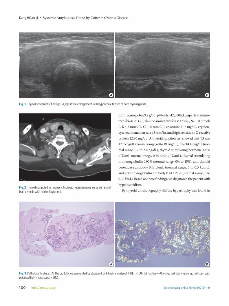
Fig. 1. Thyroid sonographic findings. (A, B) Diffuse enlargement with hypoechoic texture of both thyroid glands.
Fig. 2. Thyroid computed tomography findings. Heterogeneous enhancement of both thyroids with mild enlargement.
A B
Fig. 3. Pathologic findings. (A) Thyroid follicles surrounded by abundant pink hyaline material (H&E, × 100). (B) Positive with congo red staining (congo red stain with polarized light microscope, × 200).
mm3, hemoglobin 9.2 g/dL, platelets 142,000/µL, aspartate amino-transferase 23 U/L, alanine aminotransferase 12 U/L, Na 138 mmol/L, K 4.5 mmol/L, Cl 100 mmol/L, creatinine 1.16 mg/dL, erythro-cyte sedimentation rate 48 mm/hr, and high-sensitivity C-reactive protein 22.80 mg/dL. A thyroid function test showed that T3 was 12.55 ng/dL (normal range, 60 to 190 ng/dL), free T4 1.2 ng/dL (nor-mal range, 0.7 to 2.0 ng/dL), thyroid stimulating hormone 12.88 μIU/mL (normal range, 0.25 to 0.4 μIU/mL), thyroid stimulating immunoglobulin 0.90% (normal range, 0% to 15%), anti-thyroid peroxidase antibody 0.14 U/mL (normal range, 0 to 0.3 U/mL), and anti- thyroglobulin antibody 0.44 U/mL (normal range, 0 to 0.3 U/mL). Based on these findings, we diagnosed the patient with hypothyroidism.
By thyroid ultrasonography, diffuse hypertrophy was found in
Systemic Amyloidosis Found by Goiter in Crohn’s Disease • Kang HC, et al.
Soonchunhyang Medical Science 19(2):99-103 http://jsms.sch.ac.kr 101
A B
C D
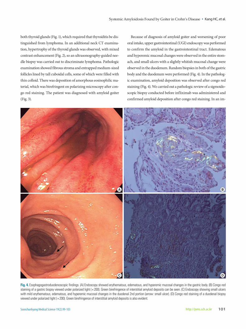
Fig. 4. Esophagogastroduodenoscopic findings. (A) Endoscopy showed erythematous, edematous, and hyperemic mucosal changes in the gastric body. (B) Congo red staining of a gastric biopsy viewed under polarized light (× 200). Green birefringence of interstitial amyloid deposits can be seen. (C) Endoscopy showing small ulcers with mild erythematous, edematous, and hyperemic mucosal changes in the duodenal 2nd portion (arrow: small ulcer). (D) Congo red staining of a duodenal biopsy viewed under polarized light (× 200). Green birefringence of interstitial amyloid deposits is also evident.
both thyroid glands (Fig. 1), which required that thyroiditis be dis-tinguished from lymphoma. In an additional neck CT examina-tion, hypertrophy of the thyroid glands was observed, with mixed contrast enhancement (Fig. 2), so an ultrasonography-guided nee-dle biopsy was carried out to discriminate lymphoma. Pathologic examination showed fibrous stroma and entrapped medium-sized follicles lined by tall cuboidal cells, some of which were filled with thin colloid. There was deposition of amorphous eosinophilic ma-terial, which was birefringent on polarizing microscopy after con-go red staining. The patient was diagnosed with amyloid goiter (Fig. 3).
Because of diagnosis of amyloid goiter and worsening of poor oral intake, upper gastrointestinal (UGI) endoscopy was performed to confirm the amyloid in the gastrointestinal tract. Edematous and hyperemic mucosal changes were observed in the entire stom-ach, and small ulcers with a slightly whitish mucosal change were observed in the duodenum. Random biopsies in both of the gastric body and the duodenum were performed (Fig. 4). In the patholog-ic examination, amyloid deposition was observed after congo red staining (Fig. 4). We carried out a pathologic review of a sigmoido-scopic biopsy conducted before infliximab was administered and confirmed amyloid deposition after congo red staining. In an im-
Kang HC, et al. • Systemic Amyloidosis Found by Goiter in Crohn’s Disease
Soonchunhyang Medical Science 19(2):99-103102 http://jsms.sch.ac.kr
munoelectrophoretic examination of blood and urine conducted to discriminate primary amyloidosis, there were no specific find-ings. But since polyclonal gammopathy was observed by immuno-fixation, the patient was diagnosed with secondary amyloidosis involving both the thyroid and the gastrointestinal tract accompa-nying Crohn’s disease. It was planned that infliximab would be constantly administered to the patient for the treatment of system-ic secondary amyloidosis. But she had a blood pressure of 90/60 mmHg, body temperature of 38.2°C, rapidly decreased renal func-tion, and identified methicillin resistant staphylococcus epidermis from blood culture on 32th day of hospitalization. We treated sep-tic shock with hydration, vancomycin and meropenem antibiotics. But due to sepsis, she expired on the 35th day of hospitalization while receiving treatment.
DISCUSSION
Amyloidosis occurs when an insoluble fibrous protein, amyloid, is deposited beside cells, and may induce failure of the involved or-gans. As a disease related to chronic inflammation or an infectious disease, secondary amyloidosis most commonly involves the kid-neys, and it also may occur in the gastrointestinal tract, liver, auto-nomic nerve tissue, and even the heart, albeit rarely [8]. Amyloid infiltration in the thyroid was first reported by von Rokitansky in 1855 [9]. Although amyloid was found in the thyroid of 50% to 80% of primary and secondary amyloidosis patients [10], it rarely appears clinically as diffuse goiter, which von Eisenberg named
‘amyloid goiter.’ One characteristic of amyloid goiter is its rapid growth, which makes to need to discriminate from a malignant tumor [8]. Patients with amyloid goiter rarely have thyroid dys-function, but some studies have reported cases accompanied by hyperthyroidism or hypothyroidism [11]. In this case, as the pa-tient had thyroid goiter that occurred suddenly, and had a suspi-cious of a malignant tumor, we performed thyroid ultrasonogra-phy and neck CT and observed diffuse thyroid goiter on both sides. Afterward, by ultrasonography-guided needle biopsy conducted for confirmation, amyloid deposition was observed in the thyroid. Accordingly, when the thyroid is rapidly enlarged in patients with chronic inflammatory diseases, such as inflammatory bowel dis-ease, rheumatoid arthritis, and sarcoidosis, it is necessary to deter-mine the presence of amyloid goiter.
In this case, the patient was found to have abdominal pain and decreased ingestion, and when she was confirmed to have amyloid
goiter, UGI endoscopy was carried out to find out whether amy-loid involved the gastrointestinal tract. In random biopsies of both the gastric body and the duodenum, amyloid deposition was ob-served after congo red staining. Based on these results, the patient was diagnosed with gastrointestinal amyloidosis.
In a large-scale study of secondary amyloidosis occurring in in-flammatory bowel disease, its prevalence was 0.9% in Crohn’s dis-ease and 0.07% in ulcerative colitis. In gastrointestinal tract-in-volved amyloidosis, symptoms may include macroglossia, vesicu-lar lesions, ulcers, and xerostomia in the oral cavity; motor distur-bance, hematemesis, achalasia, aperistalsis, portal hypertension, and esophageal varices in the esophagus; and early satiety, nausea, vomiting, abdominal pain, and hematemesis if the stomach is in-volved [1,12]. The small intestine is the most common area of amy-loidosis, and due to intestinal hypoperistalsis and intestinal amy-loid deposition, symptoms such as diarrhea, intestinal obstruc-tion, intestinal perforation, and intestinal bleeding can also occur [1,13]. When the large intestine is involved, amyloidosis may also cause intestinal obstruction, intestinal perforation, and intestinal bleeding [1]. As mentioned earlier, gastrointestinal amyloidosis has such varied symptoms and nonspecific findings that it is diffi-cult to diagnose; indeed, radiologic test results and endoscopic findings are also non-specific [14]. Using a polarization micro-scope, congo red staining after biopsy of organs suspected of being involved will reveal an apple green color, which indicates deposi-tion of amyloid fibroid material.
One of the principles of treatment of secondary amyloidosis is to reduce the creation of serum amyloid A protein [15]. No effec-tive treatment has yet been developed, but it has been reported that inflammation-suppressive or immunosuppressive therapies, main-ly using tumor necrosis factors such as cytokines, are efficacious [16]. In this case, the patient with Crohn’s disease had deteriorated, and an enteroenteric fistula was discovered, so infliximab was ad-ministered. After this, we were diagnosed with amyloidosis. After the patient had been diagnosed with amyloidosis, it was planned that infliximab would be administered again, but because of the general deterioration due to sepsis, this did not occur.
Amyloidosis accompanying Crohn’s disease is rare, and its symp-toms and endoscopic findings are non-specific, which may delay diagnosis. Amyloidosis can involve several organs such as the kid-ney, gastrointestinal tract, heart, liver, and thyroid, and when a pa-tient receives treatment lately the prognosis may be poor. There-fore, it is necessary to confirm symptoms attentively during follow-
Systemic Amyloidosis Found by Goiter in Crohn’s Disease • Kang HC, et al.
Soonchunhyang Medical Science 19(2):99-103 http://jsms.sch.ac.kr 103
up observations, and when abnormal findings are found in a physi-cal examination such as cervical goiter, specific tests should be car-ried out to determine whether systemic amyloidosis is present.
REFERENCES
1. Sattianayagam PT, Hawkins PN, Gillmore JD. Systemic amyloidosis and the gastrointestinal tract. Nat Rev Gastroenterol Hepatol 2009;6:608-17.
2. Schmidt H, Riemann JF. Amyloidosis in Crohn disease. Dtsch Med Wo-chenschr 1983;108:795-7.
3. Rashid H, Blake D, Gokal R, Gooptu D, Kerr DN. The association of re-nal amyloidosis with regional enteritis (Crohn’s disease): report of two cases and review of the literature. Clin Nephrol 1980;14:154-7.
4. Greenstein AJ, Sachar DB, Panday AK, Dikman SH, Meyers S, Heimann T, et al. Amyloidosis and inflammatory bowel disease: a 50-year experi-ence with 25 patients. Medicine (Baltimore) 1992;71:261-70.
5. Wester AL, Vatn MH, Fausa O. Secondary amyloidosis in inflammatory bowel disease: a study of 18 patients admitted to Rikshospitalet University Hospital, Oslo, from 1962 to 1998. Inflamm Bowel Dis 2001;7:295-300.
6. Jin GB, Yoon JS, Lee KT, Hwang EA, Han SY, Park SB, et al. A case of sec-ondary renal amyloidosis complicating Crohn’s disease. Korean J Med 2006;70:309-12.
7. Park KT, Kang DH, Choi CW, Park SB, Lee JH, Kim BG, et al. A case of
gastrointestinal amyloidosis as a complication of Crohn’s disease. Korean J Gastrointest Endosc 2011;42:401-5.
8. Gertz MA, Kyle RA. Secondary systemic amyloidosis: response and sur-vival in 64 patients. Medicine (Baltimore) 1991;70:246-56.
9. Von Rokitansky C. Allgemeine pathologische Anatomie und anomalien des Blotes. In: von Rokitansky C, editor. Lehrbuch der pathologischen Anatomie. Wien: Braumuller; 1855. p. 326-8.
10. Bell GO, Mena BA. Amyloid goiter associated with the nephrotic syn-drome. Med Clin North Am 1963;47:385-9.
11. Kimura H, Yamashita S, Ashizawa K, Yokoyama N, Nagataki S. Thyroid dysfunction in patients with amyloid goitre. Clin Endocrinol (Oxf) 1997; 46:769-74.
12. Menke DM, Kyle RA, Fleming CR, Wolfe JT 3rd, Kurtin PJ, Oldenburg WA. Symptomatic gastric amyloidosis in patients with primary systemic amyloidosis. Mayo Clin Proc 1993;68:763-7.
13. Hayman SR, Lacy MQ, Kyle RA, Gertz MA. Primary systemic amyloido-sis: a cause of malabsorption syndrome. Am J Med 2001;111:535-40.
14. Moon W, Lee OY, Cho YJ, Yang SY, Park HY, Han SH, et al. The endo-scopic findings and clinical characteristics of gastrointestinal amyloido-sis. Korean J Gastrointest Endosc 2005;31:216-20.
15. Gillmore JD, Lovat LB, Persey MR, Pepys MB, Hawkins PN. Amyloid load and clinical outcome in AA amyloidosis in relation to circulating concentration of serum amyloid A protein. Lancet 2001;358:24-9.
16. Park YK, Han DS, Eun CS. Systemic amyloidosis with Crohn’s disease treated with infliximab. Inflamm Bowel Dis 2008;14:431-2.