Embed Size (px)
Citation preview
See discussions, stats, and author profiles for this publication at: https://www.researchgate.net/publication/316683710
A Case Report of Mucocele
Article · January 2014
CITATIONS
2READS
146
2 authors, including:
Some of the authors of this publication are also working on these related projects:
Maxillary growth after primary palate repair View project
Mandibular reconstruction View project
Dwi Ariawan
University of Indonesia
5 PUBLICATIONS 2 CITATIONS
SEE PROFILE
All content following this page was uploaded by Dwi Ariawan on 05 May 2017.
The user has requested enhancement of the downloaded file.
253
International Journal of Clinical Preventive Dentistry Volume 9, Number 4, December 2013
A Case Report of Mucocele
Ike Siti Indiarti1, Dwi Ariawan2
Departments of 1Pediatric Dentistry, 2Oral Surgery, Faculty of Dentistry, Indonesia University, Jakarta, Indonesia
Mucocele is a common benign lesion in the oral cavity. A mucocele is a swelling in mouth caused by a blocked salivary gland. Mucoceles are painless but can be bothersome for patients to eating and speaking. Most mucoceles are visually identified. Only a few of mucoceles goes away without any special treatment, but most of mucoceles are removed by surgi-cal process. The most common location of the extravasation mucocele is the lower lip, while retention mucoceles can be found at any other site. Mucoceles Can affect the general population, but most commonly young patients. Trauma is the main etiologic factor involved in the development of mucoceles in children. The proper treatment can remove mucoceles without side effects. It is essential for a dentist to visually recognize oral lesions such as mucocele and for the proper treatment. This paper reports a case of mucocele in child.has been threatment by surgical removal and the result is no recurrence.
Keywords: mucocele, surgical, child
Corresponding author Ike Siti IndiartiDepartment of Pediatric Dentistry, Faculty of Dentistry, Indonesia University, Salemba Raya 4, Central Jakarta, [10430], Indonesia. Tel: +62-21-727-0006, Fax: +62-21- 7867-7222, E-mail: [email protected] June, 11, 2013, Revised June, 24, 2013, Accepted September, 16, 2013
Introduction
A mucocele is a swelling in mouth, also known as a “mucous cyst of the oral mucosa”. Although mucocele is benign, it can affect the oral functions such as chewing and speaking and oral hygiene. Mucoceles can have very similar features with other oral lesions. Thus, a dentist must inform patients on their oral lesions advise them to seek further diagnosis and treatment in order to prevent any potential medical threats (1). Etiology of Mucocele can happen when a salivary gland is in-jured or blocked. Many salivary glands exist in mouth, secreting saliva. Saliva is made of water, mucus and enzymes. Saliva moves from a gland into mouth through tiny tubes called ducts. Sometimes, one of these ducts can be cut, the saliva pools at the cut spot, and causes a swelling, or a mucocele. Mucocele com-monly occur inside the lower lip because of the biting habit, but
it also can be found in other places inside the mouth, including the roof of the mouth and the floor of the mouth. A mucocele can form around piercings that have been inserted into the lips or tongue. Some medicines can thicken saliva, and it can plug up a salivary gland and cause mucocele. Mucoceles also can oc-cur if one of ducts is blocked and saliva is congested up in the duct. If swelling occurs because the submandibular duct is blocked, the mucocele is called a ranula. A ranula is quite large and appears under the tongue or on the floor of the mouth. A mucocele developed on the gums is called epulis (1,2). Mucoceles occur on the tongue, inside the cheeks, the roof of the mouth, the floor of the mouth, or around tongue or lip piercings. Most mucoceles are painless but can be bother some because patients are sensitive to the bumps in their mouth. Mucoceles can get in the way of eating or speaking and are prone to biting. Shallow mucoceles may burst and release straw-col-ored fluid. Deeper ones can last longer and are more likely to bother patients. Mucoceles are benign. However, if they are left untreated, they can organize and form a permanent bump on the oral surface. Mucoceles occur where salivary glands exist. The dentist should look for a rubbery, bubble-like swelling. Mucoceles are usually found inside the lower lip, but they can also be found on the inner side of the cheek, the anterior ventral tongue, the roof of the mouth, the floor of the mouth, and rarely the upper lip (3,4).
International Journal of Clinical Preventive Dentistry
254 Vol. 9, No. 4, December 2013
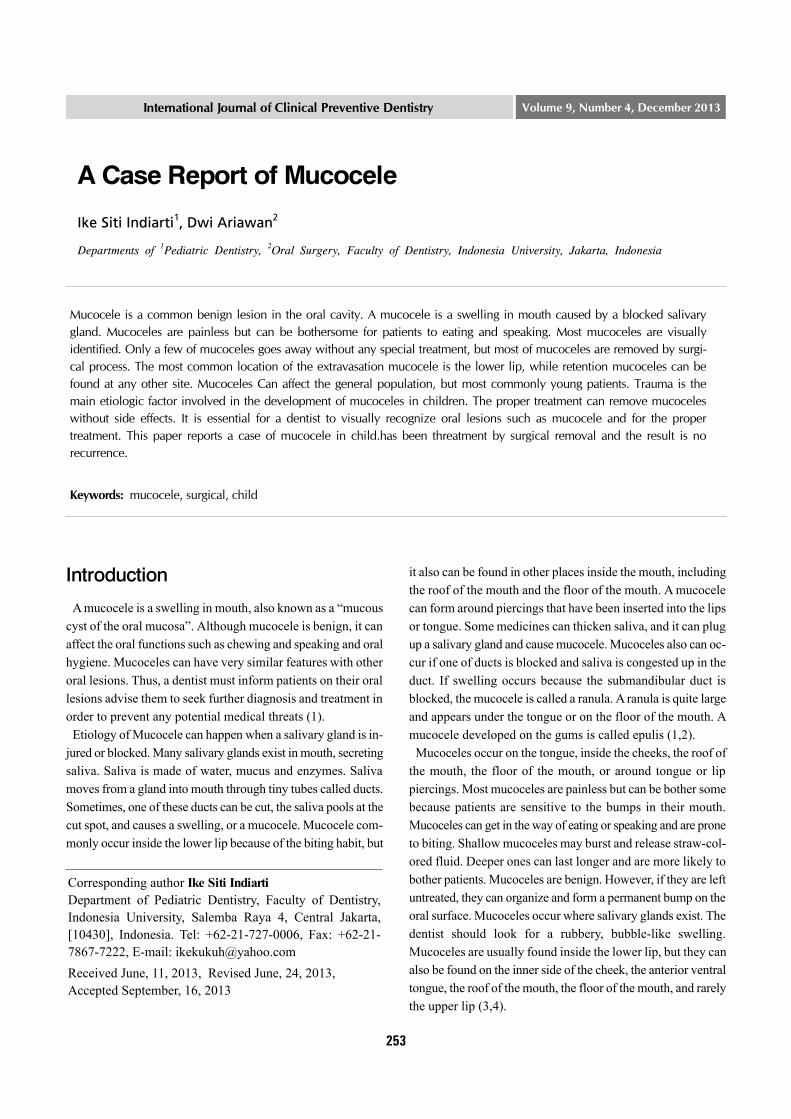
Figure 1. Clinical features of patient before surgery.
Mucoceles semitransparent and tough and their size range from a few millimeters to centimeters. Superficial mucoceles have bluish color while mucoceles in a deeper lesion have the mucosa color. A dentist should ask if a patient has experienced trauma in that area. The patient might have bitten the lip or was hit in the face with something. Also, some medicines can thick-en saliva, and it can plug up a salivary gland and cause a mucocele. Mucoceles do not change color when pressure is ap-plied on them. If a patient has a blue swelling that looks like a mucocele, the dentist may put pressure on it to see if it changes color. If it does, it may be a harmless growth made of blood vessels. This is known as a hemangioma. The dentist may take out the swollen tissue and send it to a laboratory. The laboratory can tell if the tissue is a mucocele, or if it is some other form of oral lesion. The patient may get an X-ray of the area, and the X-ray will show if the patient has a salivary gland stone. X-rays are often done for patients who have ranulas (3,5). With proper diagnosis and treatment, mucoceles can be cured with an excellent outcome. Trauma is a leading cause mucocele. A patient should avoid piercing in mouth piercing and try not to bite his/her lip (3). Infrequently, a mucocele goes away without treatment. But if some mucoceles remain untreated, they can scar over. The den-tist should examine any swelling in patients’ mouth. Mucoceles are usually removed by surgery. The dentist may use a scalpel or a laser to remove a mucocele. The removed mucocele tissue will be sent to a laboratory for further evaluation. There is a chance that another mucocele develops after the one is removed. Doctors also can use corticosteroid injections before attempting a surgical treatment. These injections sometimes bring down the swelling, and then the patient may not need surgery. A few cases of mucoceles and ranulas resolve naturally. Without treatment, especially in infants and young children. As an alternative to surgery, lesion aspiration and follow-up for about 6 months have been suggested if symptoms are minimal
in infants and young children (1,4,6). The main dental implication of mucoceles would be tissue trauma to the buccal mucosa or the lip region because of the swollen tissue and tendency for the patient to bite the lesion while chewing. The dentist should inform the prevention of mu-coceles to patients who has tendency of biting mouth tissues.
Case Report
A girl 5 years old reported to go to “The Dental Spesiality Clinic” with the chief complaint of a big swelling on the right side of the lower lip since two months. The swelling was diag-nosed as a extravasation mucocele after history and clinical examination. The treatment involved surgical resection of the tissue. The mucocele was removed under infiltrating local anes-thesia (2% lidocaine with epinephrine 1:100.000; one cartridge). The child was sit in the dental chair. Its histopathological exami-nation & regular follow up to check for uneventful healing. After one week regular recall and checkup for the reoccurrence of the lesion was done. No recurrence was observed.
Discussion
The incidence of mucoceles in the general population is 0.4-0.8%, with scant differences between males and females (7). Our own series coincides with this, since 55% of the lesions were found in males (Figure 1). As regards patient age, different authors report the peak incidence to be in the second or third dec-ades of life (8). An interesting and controversial aspect of muco-celes is their origin. Bhaskar et al. suggested obstruction of the salivary gland ducts as the cause of mucoceles, though this hy-pothesis has lost support in favor of a traumatic origin of the le-sions (9,10). The literature contains a number of studies that confirm the traumatic etiology of these lesions. In our series we identified an antecedent of trauma or of nibbling in 34% of the patients - this figure being low in comparison to the percentages
Ike Siti Indiarti and Dwi Ariawan:A Case Report of Mucocele
IJCPD 255
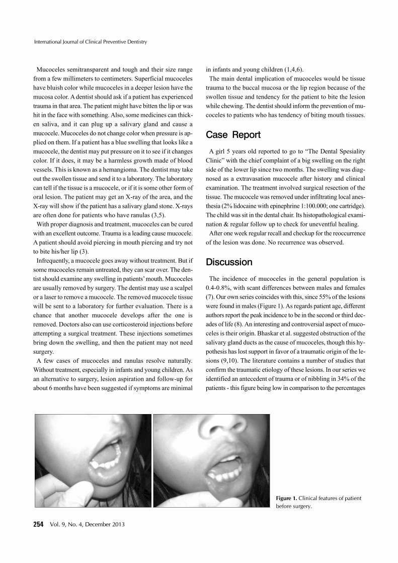
Figure 2. The conditions while being treated and after treated.
reported in other studies. In any case, the typical location of these lesions in the lower lip (more susceptible to accidental traumatism or nibbling and suction habits), their presence in young patients, and the exceptional presence of calculi in the minor salivary glands, all support this etiopathogenic theory (Figure 2). As regards mucocele location in the oral cavity, most investigators consider the lower lip to be the most frequently affected location (40-80% of all cases), followe by the cheek mucosa and floor of the mouth. The tongue, palate and upper lip are infrequent locations. The present study coincides with these observations, since 73.5% of the lesions were in the lower lip, with very little involvement of other locations. In addition, the lower lip mucoceles were predominantly located on one side-with very few medial lower lip locations. These data would be directly related to the greater capacity of certain teeth to exert trauma upon the lip, as a result of their spatial distribution. In this sense, there also have been reports of mucoceles produced as a result of the action of dental braces. In the present study mu-cocele growth was generally seen to be slow. According to Harrison, the lesions develop over a period of one week to five years, though the most common duration is three weeks to three months (8). Mucoceles may be located either as a fluid filled vesicle or blis-ter in the superficial mucosa or as a fluctuant nodule deep within the connective tissue. Spontaneous drainage of the inspisatted mucin especially in superficial lesions followed by subsequent recurrence may occur. The surface of long standing lesions may show fibrosis (2). The development of Mucoceles usually depends on the dis-ruption of the flow of saliva from the secretory apparatus of the salivary glands. The lesions are most often associated with mu-cus extravasation into the adjacent soft tissues caused by a trau-matic ductal insult, which may include a crush-type injury and severance of the excretory duct of the minor salivary gland.
Conventional treatment is commonly surgical extirpation of the surrounding mucosa and glandular tissue down to the muscle layer. With a simple incision of the mucocele the content would drain out but the lesion would reappear as soon as the wound heals (11). There is no need for treatment if superficial extravasation mu-coceles resolve spontaneously. Small mucoceles can be re-moved completely with the marginal glandular tissue before suture. In the case of larger mucoceles, marsupialization would avoid damage to vital structures. Clinically there is no differ-ence between both types of mucocele, and are therefore treated in the same manner (12).
Conclusion
Mucoceles can develop from any population. Although muco-celes are painless and intransmissible, they can affect regular eating and speaking routine of patients. Thus they should be treated properly and prevented if possible. The dentist should help patients prevent mucoceles by diagnosis and education and advise them to receive the proper treatment for their oral health and comfort. Mucoceles of the oral cavity are more common in young males. Traumatisms are the usual cause and the most frequent location is the mucosa of the lower lip. The treatment of lesions such as mucocele must be planned taking into consideration the various clinical parameters and any oral habits that are probable as these lesions have a propen-sity of recurrence. Prevention for mucocele, a patient should try not to bite his/her lip and preventive from traumatic injury.
References
1. Ata-Ali J, Carrillo C , Bonet C, et al. Oral mucocele: review of
International Journal of Clinical Preventive Dentistry
256 Vol. 9, No. 4, December 2013
the literature. J Clin Exp Dent 2010;2(1):10-3.2. Mcdonald RE, Avery DR. Dentistry for the child and adolescent.
9th ed. London: Mosby; 2011:131-2, 681-2.3. Pinkham JR. Pediatric dentistry: infancy through adolescence.
3rd ed. Philadelphia: WB Saunders Co,; 1999:3.4. Mathewson RJ, Primosch RE. Fundamentals of pediatric
dentistry. 3rd ed. New Zealand: Quintessence Pub. Co. Inc.; 1995:72-3.
5. Yamasoba T, Tayama N, Syoji M, Fukuta M. Clinicostatistical study of lower lip mucoceles. Head Neck 1990;12(4):316-20.
6. Wilcox JW, History JE. Nonsurgical resolution of mucoceles. J Oral Surg 1978;36:478.
7. Knapp MJ. Oral disease in 181,338 consecutive oral examina-
tions. J Am Dent Assoc 1971;83:1288-93.8. Jose YG, Antonio JET, Leonardo BA, Cosme GE. Treatment of
oral mucocele - scalpel versus C02 laser. Med Oral Patol Oral Cir Bucal 2009;14(9):469-74.
9. Bhaskar SN, Bolden TE, Weinmann JP. Pathogenesis of muco-celes. J Dent Res 1956;35:863-74.
10. Arendorf TM, Van Wyk CW. The association between perioral injury and mucoceles. Int J Oral Surg 1981;10:328-32.
11. Huang IY, Chen CM, Kao YH, Worthington P. Treatment of mu-cocele of the lower lip with carbon dioxide laser. J Oral Maxillofac Surg 2007;65:855-8.
12. Baurmash HD. Mucoceles and ranulas. J Oral Maxillofac Surg 2003;61:369-78.
View publication statsView publication stats


![Case Report Recurrent Oral Mucocele Management with …Mucocele is a common oral lesion affecting minor salivary glands. It develops by extravasation or retention of mucous [1–4]](https://img.pdfslide.net/doc/110x75/609f5e3ba9bf391a1f34e3c3/case-report-recurrent-oral-mucocele-management-with-mucocele-is-a-common-oral-lesion.jpg)


![Mucocele Expo[1]](https://img.pdfslide.net/doc/110x75/577cdb5c1a28ab9e78a805d7/mucocele-expo1.jpg)