Embed Size (px)
Citation preview
1The Journal of Contemporary Dental Practice, Volume 11, No. 1, January 1, 2010©2010 Seer Publishing LLC
A Comparison of Panoramic Image Quality between a Digital Radiography Storage Phosphor System and a Film-Based SystemNikos Parissis, DDS, PhD; Christos Angelopoulos, DDS, MS; Stephen Mantegari, DDS, MD; Stelios Karamanis, DDS; Farah Masood, DDS, MS; Anastasios Tsirlis, DDS, PhD
Abstract
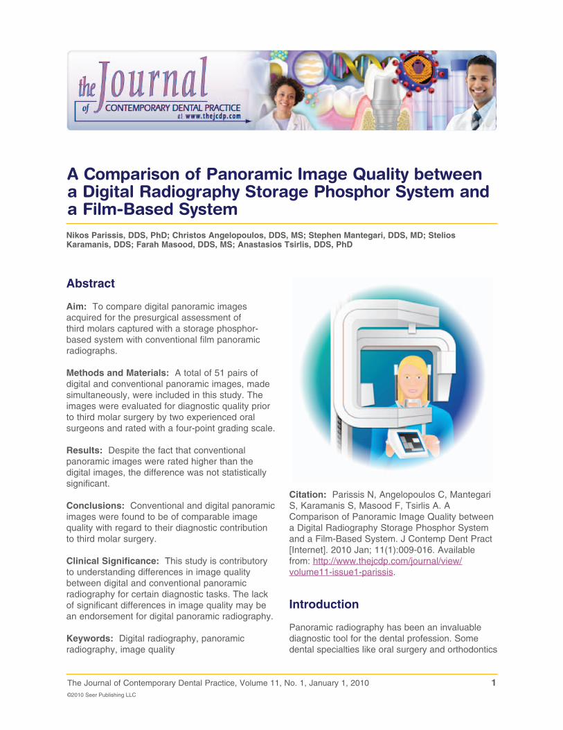
Aim: To compare digital panoramic images acquired for the presurgical assessment of third molars captured with a storage phosphor-based system with conventional film panoramic radiographs.
Methods and Materials: A total of 51 pairs of digital and conventional panoramic images, made simultaneously, were included in this study. The images were evaluated for diagnostic quality prior to third molar surgery by two experienced oral surgeons and rated with a four-point grading scale.
Results: Despite the fact that conventional panoramic images were rated higher than the digital images, the difference was not statistically significant.
Conclusions: Conventional and digital panoramic images were found to be of comparable image quality with regard to their diagnostic contribution to third molar surgery.
Clinical Significance: This study is contributory to understanding differences in image quality between digital and conventional panoramic radiography for certain diagnostic tasks. The lack of significant differences in image quality may be an endorsement for digital panoramic radiography.
Keywords: Digital radiography, panoramic radiography, image quality
Citation: Parissis N, Angelopoulos C, Mantegari S, Karamanis S, Masood F, Tsirlis A. A Comparison of Panoramic Image Quality between a Digital Radiography Storage Phosphor System and a Film-Based System. J Contemp Dent Pract [Internet]. 2010 Jan; 11(1):009-016. Available from: http://www.thejcdp.com/journal/view/volume11-issue1-parissis.
Introduction
Panoramic radiography has been an invaluable diagnostic tool for the dental profession. Some dental specialties like oral surgery and orthodontics
2The Journal of Contemporary Dental Practice, Volume 11, No. 1, January 1, 2010©2010 Seer Publishing LLC
that generate an electrical charge in proportion to the amount of light or X-rays striking them.1–3 A scintillator (material that produces light energy when hit by X-rays) is fiberoptically coupled with the sensor. As a result, the X-ray energy is converted to light energy just before striking the sensor so the light will excite the sensitive pixels of the sensor. This process actually reduces the patient exposure because the presence of the scintillator intensifies the X-ray energy when converting it to light (each X-ray photon striking the scintillator produces several light photons).3
The electrical charges generated in each of the pixels of the CCD are transferred to the computer component of the system, where they will be identified and stored. An analog to digital converter (ADC) will convert all these charges to digital data by assigning a number to each one of them, in proportion to the electrical energy. This number will eventually represent the pixel intensity value (shade of gray) of the specific location of the digital image.
Storage phosphor-based digital panoramic systems, or simply storage phosphor plates (SPPs), capture images in a way similar to that of film-based panoramic systems. Radiographic film is replaced by a reusable plate in an ordinary film holder (cassette) without any intensifying screen. SPPs use a phosphor layer to capture X-ray energy. These phosphors (most commonly, Europium-doped barium fluorohalide) are coated on a plastic base very similar to conventional film. In fact, when X-rays reach the plates, they cause a series of electron changes in the crystal lattice of the phosphors and, in a way, they form a latent image similar to conventional film.4 The difference is that with SPPs the latent image will be detected by special laser scanners instead of regular film processors.5 In these scanners, after proper light stimulation, the SPPs will release the X-ray energy they have stored, which in turn will be converted to an electrical charge. The electrical signal is then assigned a number in proportion to its intensity by an ADC. This number will eventually represent the pixel intensity value (shade of gray) of the specific location of the digital image and will be based on the X-ray energy that has been initially stored on that area of the plate. After the completion of the scanning process, the SPP is flooded with light. This will erase any remainder of the latent image and will render the plate ready for additional exposures. The additional time needed
rely almost exclusively on panoramic radiographs to obtain the majority of the radiographic information needed for diagnostic procedures. One common diagnostic use in oral surgery is the evaluation of third molars prior to surgical removal. The frequent incomplete eruption, or impaction, and the location of third molars makes assessment with intraoral radiographs difficult and necessitates the utilization of panoramic radiographs most of the time.
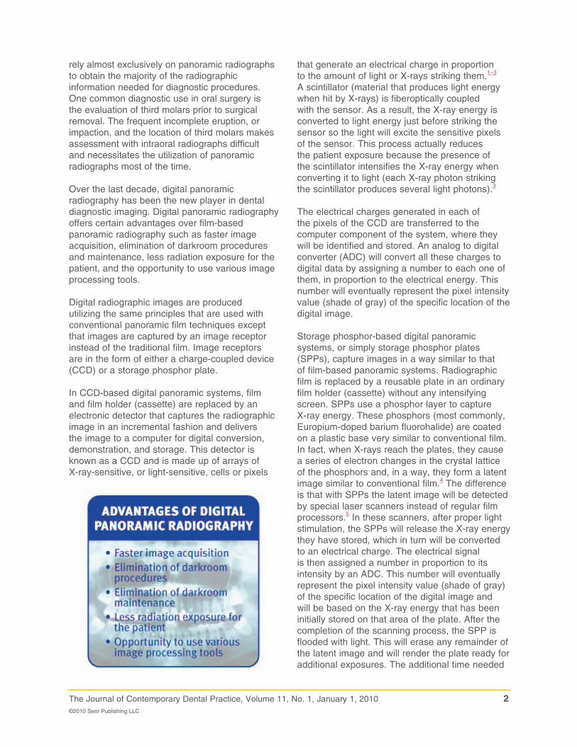
Over the last decade, digital panoramic radiography has been the new player in dental diagnostic imaging. Digital panoramic radiography offers certain advantages over film-based panoramic radiography such as faster image acquisition, elimination of darkroom procedures and maintenance, less radiation exposure for the patient, and the opportunity to use various image processing tools.
Digital radiographic images are produced utilizing the same principles that are used with conventional panoramic film techniques except that images are captured by an image receptor instead of the traditional film. Image receptors are in the form of either a charge-coupled device (CCD) or a storage phosphor plate.
In CCD-based digital panoramic systems, film and film holder (cassette) are replaced by an electronic detector that captures the radiographic image in an incremental fashion and delivers the image to a computer for digital conversion, demonstration, and storage. This detector is known as a CCD and is made up of arrays of X-ray-sensitive, or light-sensitive, cells or pixels
3The Journal of Contemporary Dental Practice, Volume 11, No. 1, January 1, 2010©2010 Seer Publishing LLC
prior to third molar surgery. A dental radiologist reviewed all panoramic images produced for general image quality and technique errors. Only patients that had all four third molars present (erupted or impacted) were included in this study. Images that demonstrated positioning errors or exposure errors were excluded from the study along with their matching counterpart in the pair of images. At the end of the selection process, 51 pairs of conventional and digital panoramic radiographs were included in the study.
The digital images were scanned immediately after exposure in a high resolution mode (at 300 dpi), identified, and stored in a separate directory as uncompressed (TIFF) files. Two experienced oral surgeons evaluated all panoramic images during two different sessions, selected in random order, with an interval of one week between rating sessions. The order of the images in each viewing session was random as well. All sessions were carried out in the same room under standardized conditions (dimmed light, same masked light box for the conventional panoramic radiographs and the same 17” LCD monitor for the digital panoramic images). The digital images were viewed using EMAGO® software (Oral Diagnostic Systems, Amsterdam, Netherlands).
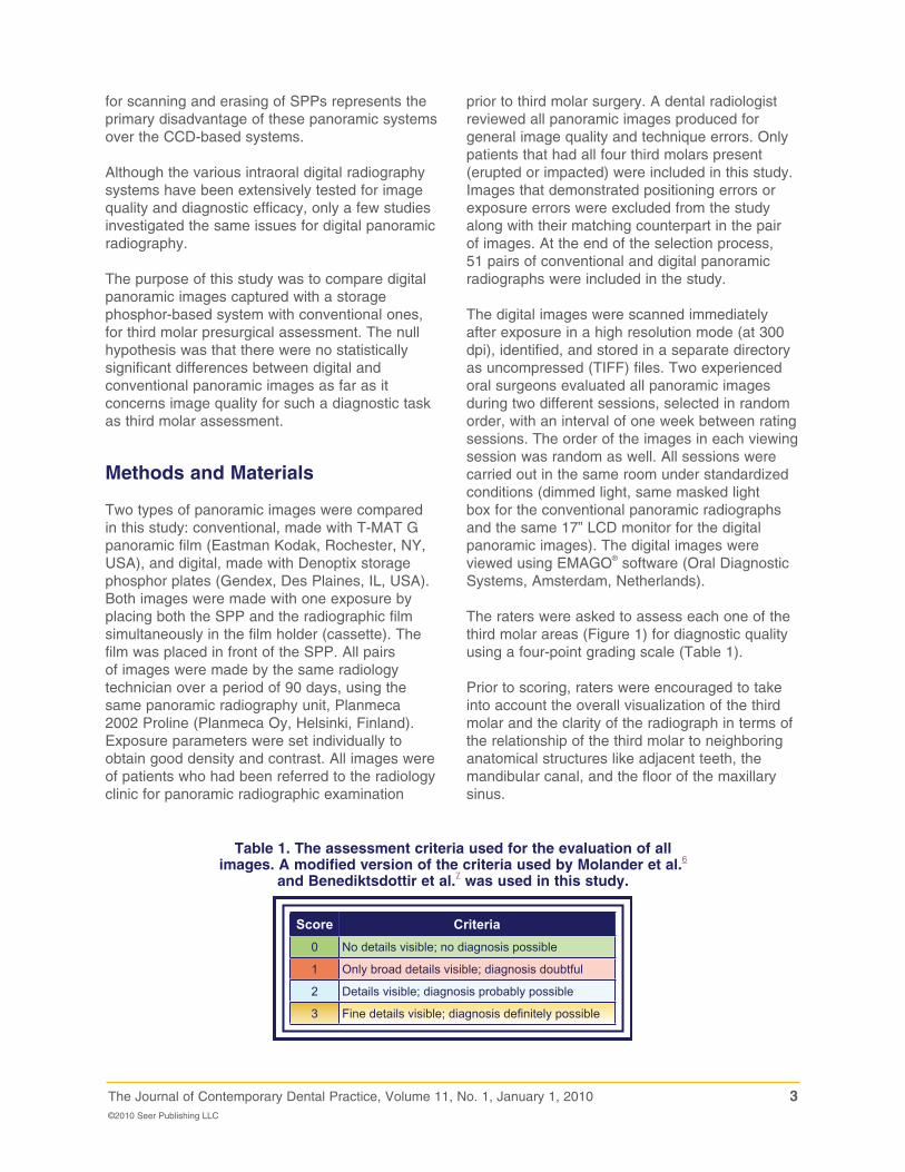
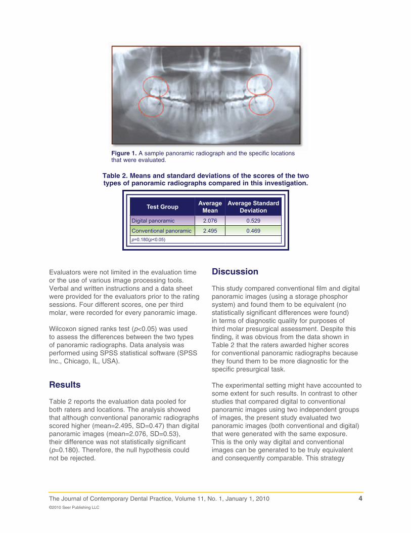
The raters were asked to assess each one of the third molar areas (Figure 1) for diagnostic quality using a four-point grading scale (Table 1).
Prior to scoring, raters were encouraged to take into account the overall visualization of the third molar and the clarity of the radiograph in terms of the relationship of the third molar to neighboring anatomical structures like adjacent teeth, the mandibular canal, and the floor of the maxillary sinus.
for scanning and erasing of SPPs represents the primary disadvantage of these panoramic systems over the CCD-based systems.
Although the various intraoral digital radiography systems have been extensively tested for image quality and diagnostic efficacy, only a few studies investigated the same issues for digital panoramic radiography.
The purpose of this study was to compare digital panoramic images captured with a storage phosphor-based system with conventional ones, for third molar presurgical assessment. The null hypothesis was that there were no statistically significant differences between digital and conventional panoramic images as far as it concerns image quality for such a diagnostic task as third molar assessment.
Methods and Materials
Two types of panoramic images were compared in this study: conventional, made with T-MAT G panoramic film (Eastman Kodak, Rochester, NY, USA), and digital, made with Denoptix storage phosphor plates (Gendex, Des Plaines, IL, USA). Both images were made with one exposure by placing both the SPP and the radiographic film simultaneously in the film holder (cassette). The film was placed in front of the SPP. All pairs of images were made by the same radiology technician over a period of 90 days, using the same panoramic radiography unit, Planmeca 2002 Proline (Planmeca Oy, Helsinki, Finland). Exposure parameters were set individually to obtain good density and contrast. All images were of patients who had been referred to the radiology clinic for panoramic radiographic examination
Table 1. The assessment criteria used for the evaluation of all images. A modified version of the criteria used by Molander et al.6
and Benediktsdottir et al.7 was used in this study.
Score Criteria0 No details visible; no diagnosis possible
1 Only broad details visible; diagnosis doubtful
2 Details visible; diagnosis probably possible
3
4The Journal of Contemporary Dental Practice, Volume 11, No. 1, January 1, 2010©2010 Seer Publishing LLC
Discussion
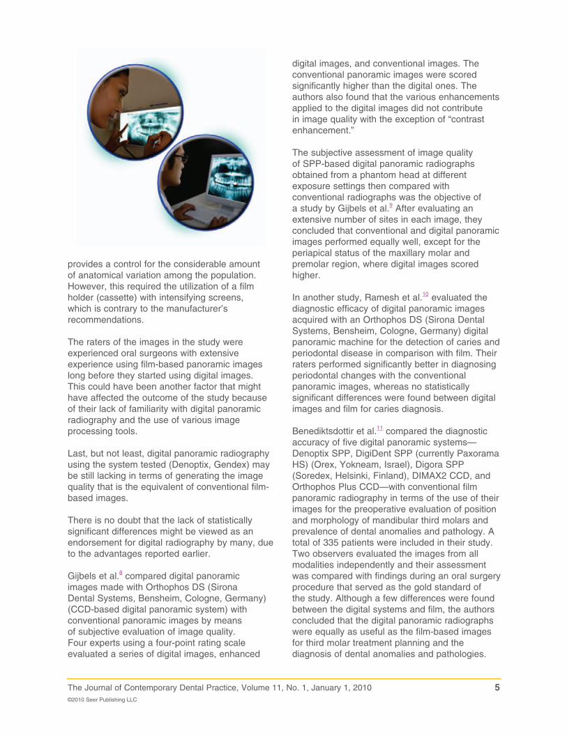
This study compared conventional film and digital panoramic images (using a storage phosphor system) and found them to be equivalent (no statistically significant differences were found) in terms of diagnostic quality for purposes of third molar presurgical assessment. Despite this finding, it was obvious from the data shown in Table 2 that the raters awarded higher scores for conventional panoramic radiographs because they found them to be more diagnostic for the specific presurgical task.
The experimental setting might have accounted to some extent for such results. In contrast to other studies that compared digital to conventional panoramic images using two independent groups of images, the present study evaluated two panoramic images (both conventional and digital) that were generated with the same exposure. This is the only way digital and conventional images can be generated to be truly equivalent and consequently comparable. This strategy
Evaluators were not limited in the evaluation time or the use of various image processing tools. Verbal and written instructions and a data sheet were provided for the evaluators prior to the rating sessions. Four different scores, one per third molar, were recorded for every panoramic image.
Wilcoxon signed ranks test (p<0.05) was used to assess the differences between the two types of panoramic radiographs. Data analysis was performed using SPSS statistical software (SPSS Inc., Chicago, IL, USA).
Results
Table 2 reports the evaluation data pooled for both raters and locations. The analysis showed that although conventional panoramic radiographs scored higher (mean=2.495, SD=0.47) than digital panoramic images (mean=2.076, SD=0.53), their difference was not statistically significant (p=0.180). Therefore, the null hypothesis could not be rejected.
Figure 1. A sample panoramic radiograph and the specific locations that were evaluated.
Table 2. Means and standard deviations of the scores of the two types of panoramic radiographs compared in this investigation.
Test Group Average Mean
Average Standard Deviation
Digital panoramic 2.076 0.529
Conventional panoramic 2.495 0.469p=0.180(p<0.05)
5The Journal of Contemporary Dental Practice, Volume 11, No. 1, January 1, 2010©2010 Seer Publishing LLC
digital images, and conventional images. The conventional panoramic images were scored significantly higher than the digital ones. The authors also found that the various enhancements applied to the digital images did not contribute in image quality with the exception of “contrast enhancement.”
The subjective assessment of image quality of SPP-based digital panoramic radiographs obtained from a phantom head at different exposure settings then compared with conventional radiographs was the objective of a study by Gijbels et al.9 After evaluating an extensive number of sites in each image, they concluded that conventional and digital panoramic images performed equally well, except for the periapical status of the maxillary molar and premolar region, where digital images scored higher.
In another study, Ramesh et al.10 evaluated the diagnostic efficacy of digital panoramic images acquired with an Orthophos DS (Sirona Dental Systems, Bensheim, Cologne, Germany) digital panoramic machine for the detection of caries and periodontal disease in comparison with film. Their raters performed significantly better in diagnosing periodontal changes with the conventional panoramic images, whereas no statistically significant differences were found between digital images and film for caries diagnosis.
Benediktsdottir et al.11 compared the diagnostic accuracy of five digital panoramic systems—Denoptix SPP, DigiDent SPP (currently Paxorama HS) (Orex, Yokneam, Israel), Digora SPP (Soredex, Helsinki, Finland), DIMAX2 CCD, and Orthophos Plus CCD—with conventional film panoramic radiography in terms of the use of their images for the preoperative evaluation of position and morphology of mandibular third molars and prevalence of dental anomalies and pathology. A total of 335 patients were included in their study. Two observers evaluated the images from all modalities independently and their assessment was compared with findings during an oral surgery procedure that served as the gold standard of the study. Although a few differences were found between the digital systems and film, the authors concluded that the digital panoramic radiographs were equally as useful as the film-based images for third molar treatment planning and the diagnosis of dental anomalies and pathologies.
provides a control for the considerable amount of anatomical variation among the population. However, this required the utilization of a film holder (cassette) with intensifying screens, which is contrary to the manufacturer’s recommendations.
The raters of the images in the study were experienced oral surgeons with extensive experience using film-based panoramic images long before they started using digital images. This could have been another factor that might have affected the outcome of the study because of their lack of familiarity with digital panoramic radiography and the use of various image processing tools.
Last, but not least, digital panoramic radiography using the system tested (Denoptix, Gendex) may be still lacking in terms of generating the image quality that is the equivalent of conventional film-based images.
There is no doubt that the lack of statistically significant differences might be viewed as an endorsement for digital radiography by many, due to the advantages reported earlier.
Gijbels et al.8 compared digital panoramic images made with Orthophos DS (Sirona Dental Systems, Bensheim, Cologne, Germany) (CCD-based digital panoramic system) with conventional panoramic images by means of subjective evaluation of image quality. Four experts using a four-point rating scale evaluated a series of digital images, enhanced
6The Journal of Contemporary Dental Practice, Volume 11, No. 1, January 1, 2010©2010 Seer Publishing LLC
Clinical Significance
The utilization of digital imaging (intraoral and panoramic) is steadily growing in the dental practice. Digital radiography offers a number of advantages in comparison to film-based radiography. These include reduced radiation exposure to the patient, fast image acquisition, elimination of the darkroom, and others. However, evidence needs to be provided that it does offer diagnostic images of comparable quality to that of conventional images. This project intended to shed some light into this issue by comparing panoramic images (digital and conventional) that were acquired on the same patient, at the same time, and with the same exposure settings. Its results indicated that the modalities under investigation generated images of comparable quality. This may be considered an endorsement of digital panoramic imaging by many.
References
1. van der Stelt PF. Principles of digital imaging. Dent Clin North Am. 2000; 44(2):237-48.
2. Farman AG, Farman TT. Extraoral and panoramic systems. Dent Clin North Am. 2000; 44(2):257-72, v-vi.
3. Angelopoulos C, Bedard A, Katz JO, Karamanis S, Parissis N. Digital panoramic radiography: An overview. Semin Orthod. 2004; 10(3):194-203.
4. Hildebolt CF, Couture RA, Whiting BR. Dental photostimulable phosphor radiography. Dent Clin North Am. 2000; 44(2):273-97, vi.
5. Parks ET, Williamson GF. Digital radiography: an overview. J Contemp Dent Pract. 2002; 3(4):23-39.
6. Molander B, Ahlqwist M, Gröndahl HG. Image quality in panoramic radiography. Dentomaxillofac Radiol. 1995; 24(1):17-22.
7. Benediktsdottir IS, Hintze H, Petersen JK, Wenzel A. Image quality of two solid-state and three photostimulable phosphor plate digital panoramic systems, and treatment planning of mandibular third molar removal. Dentomaxillofac Radiol. 2003; 32(1):39-44.
8. Gijbels F, De Meyer AM, Bou Serhal C, Van den Bossche C, Declerck J, Persoons M, Jacobs R. The subjective image quality of
In a different study, the same authors7 using a larger sample (n=433 patients), compared the diagnostic quality of digital panoramic radiographs made with the same five digital systems (Denoptix SPP, DigiDent SPP [currently Paxorama HS], Digora SPP, DIMAX2 CCD, and Orthophos Plus CCD). Image quality was assessed in six regions on all radiographs by three raters using a four-point scale. The raters were allowed to use brightness, contrast, and/or gamma enhancements. The highest quality scores were obtained with Digora (SPP), DIMAX2 (CCD), and Orthophos Plus (CCD) digital panoramic systems, whereas images from Denoptix (SPP) and DigiDent (SPP) were ranked significantly lower.The lower default scanning resolution of these systems in comparison to the Digora system might account for their lower performance.
The results of studies investigating image quality and diagnostic efficacy of digital imaging modalities in comparison to film are mixed. Some of the studies have shown that film-based images are still ahead of digital images in terms of image quality, whereas others report comparable results. Some factors that may have contributed to differences in the results include different viewing conditions such as
• Viewingofdigitalimagesonacomputermonitor instead of a traditional viewbox.
• Limitationsofthemonitorscurrentlyavailablein terms of resolution, gray scale, contrast ratios, and refresh rates.
• Thelackofraterfamiliaritywiththenumerousimage enhancement software tools.
These factors may be controlled with time as physicians become better acquainted with contemporary technology and as technology itself overcomes the current limitations. Since digital panoramic radiography is a fairly new imaging modality, the number of studies done to evaluate it remains limited. Further investigation is required in order to assess its value in maxillofacial diagnosis.
Conclusions
Digital panoramic images were found to be of comparable quality to conventional ones for such a diagnostic task as third molar assessment.

7The Journal of Contemporary Dental Practice, Volume 11, No. 1, January 1, 2010©2010 Seer Publishing LLC
College of Dental Medicine in New York, NY, USA. He is a diplomate of the American Board of Oral and Maxillofacial Radiology.
e-mail: [email protected]
Stephen Mantegari, DDS, MD
Dr. Mantegari is an associate professor of Oral and Maxillofacial Surgery at the University of Missouri–Kansas City, School of Dentistry. He is a diplomate of the American Board of Oral and Maxillofacial Surgery.
Stelios Karamanis, DDS
Dr. Karamanis is a teaching assistant and a doctoral student in the Department of Oral Surgery, Implantology and Roentgenology of the Aristotle University, School of Dentistry in Thessaloniki, Greece.
Farah Masood, DDS, MS
Dr. Masood is an associate professor and the Oral and Maxillofacial Radiology Clinic director at the Oklahoma University, College of Dentistry. She is a diplomate of the American Board of Oral and Maxillofacial Radiology.
Anastasios Tsirlis, DDS, PhD
Dr. Tsirlis is an associate professor in the Department of Oral Surgery, Implantology and Roentgenology of the Aristotle University, School of Dentistry in Thessaloniki, Greece.
direct digital and conventional panoramic radiography. Clin Oral Investig. 2000; 4(3):162-7.
9. Gijbels F, Sanderink G, Bou Serhal C, Pauwels H, Jacobs R. Organ doses and subjective image quality of indirect digital panoramic radiography. Dentomaxillofac Radiol. 2001; 30(6):308-13.
10. Ramesh A, Tyndall DA, Ludlow JB. Evaluation of a new digital panoramic system: a comparison with film. Dentomaxillofac Radiol. 2001; 30(2):98-100.
11. Benediktsdottir IS, Hintze H, Petersen JK, Wenzel A. Accuracy of digital and film panoramic radiographs for assessment of position and morphology of mandibular third molars and prevalence of dental anomalies and pathologies. Dentomaxillofac Radiol. 2003; 32(2):109-15.
About the Authors
Nikos Parissis, DDS, PhD
Dr. Parissis is an associate professor and the chairman of the Department of Oral Surgery, Implantology and Roentgenology of the Aristotle University, School of Dentistry in Thessaloniki, Greece.
Christos Angelopoulos, DDS, MS (Corresponding Author)
Dr. Angelopoulos is an associate professor and the director of the Division of Oral and Maxillofacial Radiology at Columbia University,