Embed Size (px)
Citation preview
Antiviral Research 41 (1999) 35–43
A high throughput colorimetric cell proliferation assay for theidentification of human cytomegalovirus inhibitors
Jean Bedard *, Suzanne May, Dominique Barbeau, Leonard Yuen, Robert F. Rando,Terry L. Bowlin
Department of Virology, BioChem Pharma Inc., 275 Boule6ard Armand-Frappier, La6al, Que. H7V 4A7, Canada
Received 1 June 1998; accepted 26 October 1998
Abstract
A colorimetric assay based on the cleavage of the tetrazolium salt WST-1 has been developed for humancytomegalovirus (HCMV) antiviral susceptibility testing and adapted to a microtiter plate format. Optimal conditionswere determined and the standard routine assay was calibrated with a viral input of 0.05–0.10 plaque forming unit(p.f.u.)/cell with a density of 2000 cells/well in a 96-well microtiter plate for an incubation period of 7 days.Ganciclovir (9-(2-hydroxy-1(hydroxymethyl) ethyoxymethyl) guanine; DHPG), and cidofovir ((S)-1-(3-hydroxy-2-phosphonylmethoxypropyl) cytosine; HPMPC) were used as positive control test compounds to validate the assay.The effective EC50 concentration values obtained with the two antiviral compounds in the present assay were in goodagreement with plaque reduction assay results performed in parallel experiments. This method presents the advantageof being easy and rapid to perform, reliable, reproducible, and convenient for use in a high throughput screeningcapacity. © 1999 Elsevier Science B.V. All rights reserved.
Keywords: Cytomegalovirus; Cell viability; Colorimetric assay; HPMPC; DHPG
1. Introduction
Human cytomegalovirus is a serious life threat-ening opportunistic pathogen in immunocompro-mized individuals such as HIV-1 infected
individuals, patients with cancer, and organ trans-plant recipients (Alford and Britt, 1993). It is alsoone of the principal causes of congenital malfor-mation and neurological defects in infected new-borns (Alford and Britt, 1993).
Ganciclovir (9-(2-hydroxy-1(hydroxymethyl)ethyoxymethyl) guanine; DHPG), foscarnet(phosphonic acid), and cidofovir ((S)-1-(3-hy-droxy-2-phosphonylmethoxypropyl) cytosine;
* Corresponding author. Tel.: +1-514-9787864; fax: +1-514-9787946.
E-mail address: [email protected] (J. Bedard)
0166-3542/99/$ - see front matter © 1999 Elsevier Science B.V. All rights reserved.
PII: S 0166 -3542 (98 )00061 -8
J. Bedard et al. / Anti6iral Research 41 (1999) 35–4336
HPMPC) are the three anti-HCMV compoundscurrently approved for the treatment of cy-tomegalovirus retinitis, which is one of the mostcommon complications encountered in patientswith AIDS. Serious side-effects are associatedwith prolonged use of these antiviral agents suchas nephrotoxicity and granulocytopenia (Collabo-rative DHPG Treatment Study Group, 1986;Causey, 1991; Jacobson et al., 1991; Palestine etal., 1991; Jacobson, 1997). The emergence of clin-ical resistant isolates is also an additional compli-cation (Jacobson et al., 1991; Palestine et al.,1991; Stanat et al., 1991; Chou et al., 1995; Han-son et al., 1995; Jacobson, 1997). There is apossibility of cross-resistance with cidofoviramong highly DHPG resistant clinical isolateswhich are characterized by a mutation in both theviral UL97 and viral DNA polymerase genes(Smith et al., 1997). Thus, there is still a greatneed for identifying new compounds for the treat-ment of HCMV infections.
Several in vitro assay systems are available forevaluating molecules associated with anticy-tomegaloviral activity. These methods can be te-dious and time consuming to perform. The plaquereduction assay is the most commonly used assayand is the gold standard for in vitro susceptibilitytesting of anti-HCMV drug candidates. In thepast few years, numerous methodologies havebeen developed to increase the rapidity of viralquantification and some of them have beenadapted for use in large-scale screening programsfor anti-herpes agents. The technologies for directmonitoring of viral production involve the use ofrecombinant HSV and HCMV viruses (Dicker etal., 1995; Hippenmeyer and Dilworth, 1996), nu-cleic acid hybridisation (Gadler, 1983; Dankner etal., 1990), immunocytochemical staining (Musianiet al., 1988), and in situ enzyme-linked immuno-sorbent assay (ELISA) (Tatarowicz et al., 1991).In addition, an HSV-1 cytopathic effect assaybased on a vital dye uptake has also been re-ported in the literature (McLaren et al., 1983).
We present here a quantitative colorimetric as-say adapted to a microtiter plate format for thedetermination of HCMV susceptibility to antiviralcompounds. In the system described in this re-port, virus-induced cytopathic effects (CPE) with
respect to cell viability and proliferation, are as-sessed by reduction of the tetrazolium salt WST-1into a water soluble formazan product which canbe directly quantified using a microtiter ELISAreader.
2. Material and methods
2.1. Cells and 6irus
Primary newborn human fibroblast (Hs68) cellsand HCMV strain AD169 were obtained from theAmerican Type Culture Collection (ATCC)(Rockville, MD, USA). Cells were passaged inDulbecco’s modified Eagle’s medium (DMEM)(Life Technologies, Gaithersburg, MD, USA)supplemented with 10% fetal bovine serum (FBS)(Hyclone Laboratories, Logan, UT, USA), and 2mM glutamine (Life Technologies). Penicillin andstreptomycin (Life Technologies) were added at500 U/ml and 50 mg/ml final concentrations, re-spectively. Cells were used for the plaque reduc-tion and CPE assays between passages 13 and 24.
2.2. Reagents
Cell proliferation reagent WST-1 was pur-chased from Boehringer Mannheim (Laval, Que-bec, Canada). DHPG was a gift from RocheBioscience (Palo Alto, CA, USA) and (S)-1-(3-hy-droxy-2-phosphonylmethoxypropyl) cytosine(HPMPC) was synthesized at BioChem Pharma.Trypsin-EDTA solution was obtained from LifeTechnologies and crystal violet from Sigma (St.Louis, MO, USA).
2.3. Colorimetric cell proliferation assay
Hs68 cells were seeded at a density of 8×10⁵
cells in DMEM/10% FBS in a polystyrene flaskculture vessel of 75-cm2 surface area (Nunc,No.147589, Roskilde, Denmark) and incubatedfor 18 h at 37°C in 5% CO2 in order to obtain, inthese conditions, 80% confluence. The cell mono-layer was then inoculated with HCMV at a multi-plicity of infection (MOI) of 0.05–0.1 plaqueforming unit (p.f.u.) /cell in a final volume of 3 ml
J. Bedard et al. / Anti6iral Research 41 (1999) 35–43 37
in DMEM/2% FBS. Viral adsorption was allowedto proceed for 2 h at 37°C with rocking of theplates every 30 min. Infected monolayers weretrypsinized, cells were resuspended in DMEM/2%FBS, counted, and cell suspensions adjusted to adensity of 2×10⁴ cells/ml. An aliquot of 100 mlwas added to wells of a 96-well microtiter plate(Nunc, No.167008) containing 100 ml of test com-pound at the appropriate concentrations. After 7days of incubation at 37°C in 5% CO2, themedium was then removed and cell viability, as anindication of the CPE reached, was assessed bythe addition of 100 ml of pre-warmed DMEM/2%FBS containing WST-1 cell proliferating reagentdiluted 1/40 to each well. After a period of incu-bation of 2 h at 37°C, the absorbance was mea-sured on a microplate Dynatech MR5000Microelisa Autoreader (Dynatech, Alexandria,VA, USA) set at a wavelength of 410 nm. DHPGand HPMPC were used as positive controls forthe inhibition of HCMV CPE. Stock solutionswere prepared in sterile water at 2 and 10 mg/ml,respectively.
The concentration of drug which reduces theviral CPE by 50% (EC50) was calculated using asigmoidal dose response (variable slope) equationto perform a non-linear regression analysis usingthe GraphPad Prism software: version 2.0(GraphPad Software, San Diego, CA, USA):� A: Optical density obtained with infected cells
cultured in the presence of the minimal drugconcentration required to obtained the maxi-mal inhibition of CPE (top plateau)
� B: Optical density obtained with infected cellscultured in the absence of drug (bottomplateau)
� Y: Optical density obtained with infected cellscultured in the presence of a specific drugconcentration X
� Hill’s coefficient: Variable that controls theslope of the curve. Values of 1.15 and 2.58 wereobtained for HPMPC and DHPG, respectively.
Y=B+ (A−B)/1+10(logEC50−X)Hill’s coefficient
As a negative control, mock-infected cells weretreated with DHPG and HPMPC at the sameconcentrations used for the experiments with in-fected cells in order to determine a potential drug-
induced CPE. The drugs were tested at aconcentration range of 6.1×10⁻⁴ to 2.5 mg/mlfor DHPG and of 9.8×10⁻⁵ to 0.4 mg/ml forHPMPC. All experiments were performed threetimes each in triplicate.
2.4. Plaque reduction assay
In a 12-well tissue culture dish (Corning Costar,No.25815, Orneonta, NY, USA), Hs68 cells wereplated at a density of 1.5×10⁵ cells/well in 2 mlof culture medium and incubated overnight in 5%CO2 at 37°C. Medium was removed and cellswere infected with 0.5 ml of DMEM/2% FBScontaining approximately 125 p.f.u./ml ofHCMV. After an adsorption at 37°C for 2 h, theinoculate was removed and the monolayer wasoverlaid with 1 ml of DMEM/2% FBS containingthe test compounds at concentrations rangingfrom 0.1 to 2.0 mg/ml for DHPG and 0.005 to 0.2mg/ml for HPMPC. After 7 days of incubation,cells were fixed with 1 vol. of a solution of 8%formaldehyde in water for 30 min, then the solu-tion was removed and cell monolayers stainedwith 2% crystal violet in 20% ethanol for a fewseconds. Cells were rinsed with tap water, dried,and monolayer examined for the presence ofplaques using an inverted microscope at 40×magnification.
The percentage of plaque reduction was deter-mined for DHPG and HPMPC by comparison ofthe mean number of plaques (drug-treated cells)with the infected cells and the 50% effective con-centration (EC50) was calculated by non-linearregression analysis.
3. Results
3.1. Optimization of the colorimetric cellproliferation assay in a microtiter plate format:effect of cell density, 6iral input, and incubationperiod
The degree of HCMV susceptibility to ganci-clovir in the colorimetric cell proliferation assaywas highly dependent upon several conditions,including the number of Hs68 cells seeded, the
J. Bedard et al. / Anti6iral Research 41 (1999) 35–4338
Table 1Effect of cell density on DHPG-related CPE on mock-infected Hs68 fibroblasts
DHPG (mg/ml) Cell densitya
3000 cells/well2000 cells/well1000 cells/well
% cell viabilityO.D.O.D. % cell viabilityO.D. % cell viabilityb
0.605 100 0.667 1000 0.569 1001050.7131080.156 0.6500.600 105
104 0.6910.313 0.575 101 0.631 102107 0.7070.625 0.561 99 0.649 104
0.715110 1051.250 0.6650.550 970.645 106 0.6902.500 0.463 102810.710 118 0.6765.000 0.300 10053
a Hs68 cells were cultured at various densities/well in a 96-well microtiter plate in the presence of DHPG at concentrations up to5 mg/ml. Cell viability was recorded at day 7 post-inoculation as described in Section 2. Experiments were performed in triplicates.
b Cell viability of uninfected Hs68 cells obtained in the absence of DHPG was recorded as 100%.
viral input used, and the period of incubation.The effects of these factors on the dynamic rangeof the assay was investigated.
The assay was calibrated initially for the opti-mum cell density. Cell viability was investigatedover a period of incubation up to 10 days usingseeding concentrations of 1000–4000 uninfectedcells/well in a 96-well microtiter plate. Results onHs68 cell viability when cultured at initial densi-ties of 1000, 2000, and 3000 cells/well in thepresence of various concentrations of DHPG upto 5 mg/ml are presented in Table 1. The dataindicate a drug-related CPE at a density of 1000cells/well using a concentration of DHPG of 2.5and 5.0 mg/ml. A cell viability of 53% wasrecorded with 5 mg/ml of DHPG when a densityof 1000 cells/well was used whereas no such effectwas seen at higher cell densities (Table 1). There-fore, if conditions having less than 2000 cells/wellwere used, Hs68 cells were found to be sensitive toDHPG-induced cytotoxicity. At this sub-conflu-ent plating, cells would enter log-phase growthand hence become sensitive to DHPG-inducedtoxicity. When more than 3000 fibroblasts wereadded to the well, cells were less susceptible toDHPG-induced CPE, however cells soon becameconfluent during the period of incubation. Theoptimum amount of cells to be added was deter-mined to be within the range of 2000–3000 cells/well.
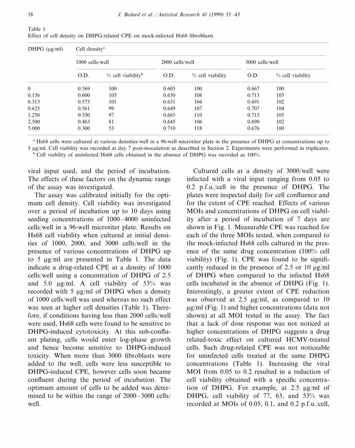
Cultured cells at a density of 3000/well wereinfected with a viral input ranging from 0.05 to0.2 p.f.u./cell in the presence of DHPG. Theplates were inspected daily for cell confluence andfor the extent of CPE reached. Effects of variousMOIs and concentrations of DHPG on cell viabil-ity after a period of incubation of 7 days areshown in Fig. 1. Measurable CPE was reached foreach of the three MOIs tested, when compared tothe mock-infected Hs68 cells cultured in the pres-ence of the same drug concentration (100% cellviability) (Fig. 1). CPE was found to be signifi-cantly reduced in the presence of 2.5 or 10 mg/mlof DHPG when compared to the infected Hs68cells incubated in the absence of DHPG (Fig. 1).Interestingly, a greater extent of CPE reductionwas observed at 2.5 mg/ml, as compared to 10mg/ml (Fig. 1) and higher concentrations (data notshown) at all MOI tested in the assay. The factthat a lack of dose response was not noticed athigher concentrations of DHPG suggests a drugrelated-toxic effect on cultured HCMV-treatedcells. Such drug-related CPE was not noticeablefor uninfected cells treated at the same DHPGconcentrations (Table 1). Increasing the viralMOI from 0.05 to 0.2 resulted in a reduction ofcell viability obtained with a specific concentra-tion of DHPG. For example, at 2.5 mg/ml ofDHPG, cell viability of 77, 63, and 53% wasrecorded at MOIs of 0.05, 0.1, and 0.2 p.f.u./cell,
J. Bedard et al. / Anti6iral Research 41 (1999) 35–43 39
Fig. 1. Effect of variation in viral multiplicity of infection and DHPG concentrations on cell viability after a period of incubationof 7 days. Hs68 cells were infected at an MOI of 0.05, 0.10, or 0.20 p.f.u./cell and seeded at a density of 3000 cells/well in a 96-wellmicrotiter plate containing DHPG at 0 ("), 2.5 (), and 10 mg/ml (�). Cell viability of mock-infected Hs68 cells obtained in thepresence of a specific concentration of DHPG was recorded as 100%. Experiments were performed in triplicate, the standardvariation of the mean not exceeding 3.7, 12.1, and 7.8% for the experimental points obtained using MOIs of 0.05, 0.1, and 0.2,respectively.
respectively (Fig. 1). It has been reported that theefficacy of DHPG is not sensitive to the MOI ofHCMV used, at least up to an MOI of 2.0 (Lewiset al., 1994). Therefore, it is unlikely that thereduction in cell viability was due to virus-inducedCPE, but more is likely due to a drug-relatedtoxicity.
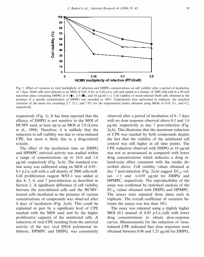
The effect of the incubation time on DHPGand HPMPC antiviral activity was studied withina range of concentrations up to 16.0 and 1.6mg/ml, respectively (Fig. 2a,b). The standard rou-tine assay was calibrated using an MOI of 0.05–0.1 p.f.u./cell with a cell density of 2000 cells/well.Cell proliferation reagent WST-1 was added atday 4, 5, 6, and 7 post-infection as described inSection 2. A significant difference of cell viabilitybetween the non-infected cells and the HCMV-treated cells incubated in the presence of variousconcentrations of compounds was observed after4 days of incubation (Fig. 2a,b). This could beexplained in part by a significant level of CPEreached with the MOI used and by the higherproliferative capacity of the uninfected cells. Areduction of viral CPE resulting from the antiviralactivity of the two viral DNA polymerase in-hibitors, HPMPC and DHPG, was consistently
observed after a period of incubation of 6–7 dayswith no dose response observed above 0.1 and 1.0mg/ml, respectively at day 7 post-infection (Fig.2a,b). This illustrates that the maximum reductionof CPE was reached by both compounds despitethe fact that the viability of the uninfected cellcontrol was still higher at all time points. TheCPE reduction observed with DHPG at 16 mg/mlwas not as pronounced as compared with lowerdrug concentrations which indicates a drug re-lated-toxic effect consistent with the results de-scribed above. Cell viability values obtained atday 7 post-infection (Fig. 2a,b) suggest EC50 val-ues B1 and :0.05 mg/ml for DHPG andHPMPC, respectively. The reproducibility of theassay was confirmed by statistical analysis of theEC50 values obtained with DHPG and HPMPC.The assays were repeated three times each intriplicate. The overall coefficient of variation be-tween the assays was less than 10%.
The assay was repeated using a slightly higherMOI (0.1 instead of 0.05 p.f.u./cell) with lowerdrug concentrations to obtain dose-responsecurves. Measurements for the reduction of virus-induced CPE indicated that dose responses wereobtained between 0.08 and 1.25 mg/ml for DHPG,
J. Bedard et al. / Anti6iral Research 41 (1999) 35–4340
Fig. 2. (a). Effect of DHPG on cell viability over a 7 days incubation period. Hs68 cells were infected at an MOI of 0.05 p.f.u./celland seeded at a density of 2000 cells/well in a 96-well microtiter plate and cultured in the presence of serial dilutions of DHPG at0 (�), 1.0 (�), 2.0 (!), 4.0 ( ), 8.0 (�), and 16 mg/ml (�). Non-infected cells () were included as a control for the assessment ofthe cytopathic effect reached during the course of the infection. Cell viability was measured at day 4, 5, 6, and 7 post-infection.Experiments were performed in triplicate, and the standard variation of the mean was less than 15% for each time point. (b) Effectof HPMPC on cell viability over a 7-day incubation period. Hs68 cells were infected at an MOI of 0.05 p.f.u./cell and seeded at adensity of 2000 cells/well in a 96-well microtiter plate and cultured in the presence of serial dilution of HPMPC at 0 (�), 0.05 (�),0.1 (!), 0.2 ( ), 0.4 (�), 0.8 (�), and 1.6 mg/ml ("). Non-infected cells () were included as a control for the assessment of thecytopathic effect reached during the course of the infection. Cell viability was measured at day 4, 5, 6, and 7 post-infection.Experiments were performed in triplicate, the standard variation of the mean was less than 15% for each time point.
J. Bedard et al. / Anti6iral Research 41 (1999) 35–43 41
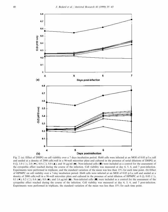
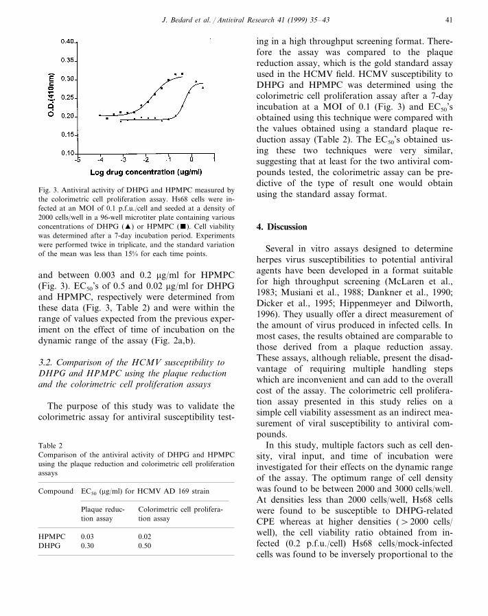
Fig. 3. Antiviral activity of DHPG and HPMPC measured bythe colorimetric cell proliferation assay. Hs68 cells were in-fected at an MOI of 0.1 p.f.u./cell and seeded at a density of2000 cells/well in a 96-well microtiter plate containing variousconcentrations of DHPG (�) or HPMPC (). Cell viabilitywas determined after a 7-day incubation period. Experimentswere performed twice in triplicate, and the standard variationof the mean was less than 15% for each time points.
ing in a high throughput screening format. There-fore the assay was compared to the plaquereduction assay, which is the gold standard assayused in the HCMV field. HCMV susceptibility toDHPG and HPMPC was determined using thecolorimetric cell proliferation assay after a 7-dayincubation at a MOI of 0.1 (Fig. 3) and EC50’sobtained using this technique were compared withthe values obtained using a standard plaque re-duction assay (Table 2). The EC50’s obtained us-ing these two techniques were very similar,suggesting that at least for the two antiviral com-pounds tested, the colorimetric assay can be pre-dictive of the type of result one would obtainusing the standard assay format.
4. Discussion
Several in vitro assays designed to determineherpes virus susceptibilities to potential antiviralagents have been developed in a format suitablefor high throughput screening (McLaren et al.,1983; Musiani et al., 1988; Dankner et al., 1990;Dicker et al., 1995; Hippenmeyer and Dilworth,1996). They usually offer a direct measurement ofthe amount of virus produced in infected cells. Inmost cases, the results obtained are comparable tothose derived from a plaque reduction assay.These assays, although reliable, present the disad-vantage of requiring multiple handling stepswhich are inconvenient and can add to the overallcost of the assay. The colorimetric cell prolifera-tion assay presented in this study relies on asimple cell viability assessment as an indirect mea-surement of viral susceptibility to antiviral com-pounds.
In this study, multiple factors such as cell den-sity, viral input, and time of incubation wereinvestigated for their effects on the dynamic rangeof the assay. The optimum range of cell densitywas found to be between 2000 and 3000 cells/well.At densities less than 2000 cells/well, Hs68 cellswere found to be susceptible to DHPG-relatedCPE whereas at higher densities (\2000 cells/well), the cell viability ratio obtained from in-fected (0.2 p.f.u./cell) Hs68 cells/mock-infectedcells was found to be inversely proportional to the
and between 0.003 and 0.2 mg/ml for HPMPC(Fig. 3). EC50’s of 0.5 and 0.02 mg/ml for DHPGand HPMPC, respectively were determined fromthese data (Fig. 3, Table 2) and were within therange of values expected from the previous exper-iment on the effect of time of incubation on thedynamic range of the assay (Fig. 2a,b).
3.2. Comparison of the HCMV susceptibility toDHPG and HPMPC using the plaque reductionand the colorimetric cell proliferation assays
The purpose of this study was to validate thecolorimetric assay for antiviral susceptibility test-
Table 2Comparison of the antiviral activity of DHPG and HPMPCusing the plaque reduction and colorimetric cell proliferationassays
Compound EC50 (mg/ml) for HCMV AD 169 strain
Colorimetric cell prolifera-Plaque reduc-tion assay tion assay
HPMPC 0.03 0.020.30DHPG 0.50
J. Bedard et al. / Anti6iral Research 41 (1999) 35–4342
cell density (data not shown). This could be ex-plained by the fact that the rate of cellular replica-tion of uninfected cells in log-phase growth ismuch higher than the rate of viral replication inthe infected cells. Basically, the number of viablecells after 7 days of incubation represents theend-point result of a competition between the cellcapacity to proliferate (uninfected cells) and theviral propagation resulting in a cell killing or/andreduction of cell proliferation (infected cells).
Cell viability studies of HCMV-infected Hs68cells, cultured in the absence of drug, havedemonstrated that 25% of the cells, when initiallyinfected at an MOI of 0.2, remained viable after 7days of incubation (Fig. 1). In order to increasethe CPE (cell viability closer to a zero value),higher MOIs or a longer period of incubationcould have been used. However, at MOIs higherthan 0.2, cells became more susceptible to drug-related CPE (data not shown), as illustrated forMOIs between 0.05 and 0.2 (Fig. 1). Increasingthe incubation period resulted in the same phe-nomenon as described above for experiments us-ing relatively high cell densities.
The overall cell viability of infected cells cul-tured in the presence of HPMPC and DHPGnever reached the level obtained for the unin-fected cell control during the 7-day period ofincubation. This could be explained in part by thefact that initially infected cells, even those cul-tured in the presence of DHPG, are condemnedto die. A higher degree of toxicity was observedwith increasing concentrations of DHPG onHs68-infected cells (Fig. 1) when compared tonon-infected cells at the same density (Table 1).This is independent of the overwhelming CPEobserved in a lytic infection. Mock-infected cells(control) have an initial growth advantage whencompared to the infected cells incubated in thepresence of compounds (Fig. 2a,b, day 4). Thisadvantage is amplified after 7 days of incubationsince several cell division cycles are allowed toproceed. The positive effect of HPMPC andDHPG on cell viability was observed 6–7 dayspost-infection (Fig. 2a,b). This is most likely re-lated to the antiviral mechanism of action of thetwo nucleotide analogs, which is exhibited duringthe major peak of replication cycle of HCMV
DNA, which does not occur until 72–92 h post-infection (Stinski, 1990). This would result in areduced amount of infectious virions available forthe second round of infection. In addition, thiscould be related to the toxicity of the compoundon the log phase growing infected cells whichsubsides as the cells reach confluence.
The EC50 values obtained with both DHPGand HPMPC were in good agreement with theplaque reduction assay results. A higher level ofinhibitory effect was achieved with HPMPC inboth antiviral assays. Several 1,6-naphthyridinecompounds with various degrees of potencyagainst HCMV (Jin et al., 1997) have been testedat least twice in both CPE and plaque reductionassays and a similar trend of inhibitory potencywas observed in the two test systems (data notshown). The results obtained using this chemicallydistinct class of compound further support thereliability of the presented methodology. It will beinteresting to perform antiviral susceptibility test-ing of some representative drug resistant HCMVstrains as an additional means to validate theassay as well as to compare the present assay withthe MTT (3-[4,5-dimethylthiazol-2-yl]-2,5-diph-enyl-tetrazolium bromide) or neutral red uptakeassays.
References
Alford, C.A., Britt, W.J., 1993. Cytomegalovirus. In: Roiz-man, B., Whitley, R.J., Lopez, C. (Eds.), The HumanHerpesviruses. Raven Press, New York, pp. 227–255.
Causey, D., 1991. Concomitant ganciclovir and zidovudinetreatment for cytomegalovirus retinitis in patients withHIV infection: an approach to treatment. J. AcquiredImmune Defic. Syndr. 4, S16–S21.
Chou, S., Erice, A., Jordan, M.C., Vercellotti, G.M., Michels,K.R., Talarico, C.L., Stanat, S.C., Biron, K.K., 1995.Analysis of the UL97 phosphotransferase coding sequencein clinical cytomegalovirus isolates and identification ofmutations conferring ganciclovir resistance. Infect. Dis.171, 576–583.
Collaborative DHPG Treatment Study Group, 1986. Treat-ment of serious cytomegalovirus infections with 9-(1,3-di-hydroxy-2-propoxymethyl)guanine in patients with AIDSand other immunodeficiencies. N. Engl. J. Med. 27, 801–805.
Dankner, W.M., Scholl, D., Stanat, S.C., Martin, M., Sonke,R.L., Spector, S.A., 1990. Rapid antiviral DNA-DNA
J. Bedard et al. / Anti6iral Research 41 (1999) 35–43 43
hybridization assay for human cytomegalovirus. J. Virol.Methods 28, 293–298.
Dicker, I.B., Blasecki, J.W., Seetharam, S., 1995. Herpes simplextype 1: lacZ recombinant viruses. II Microtiter plate-basedcolorimetric assays for the discovery of new antiherpesagents and the points at which such agents disrupt the viralreplication cycle. Antiviral Res. 28, 213–224.
Gadler, H., 1983. Nucleic acid hybridization for measurementof effects of antiviral compounds on human cytomegalovirusDNA replication. Antimicrob. Agents Chemother. 24, 370–374.
Hanson, M.N., Preheim, L.C., Chou, S., Talarico, C.L., Biron,K.K., Erice, A., 1995. Novel mutation in the UL97 gene ofa clinical cytomegalovirus strain conferring resistance toganciclovir. Antimicrob. Agents Chemother. 39, 1204–1205.
Hippenmeyer, P.J., Dilworth, V.M., 1996. A rapid assay fordetermination of antiviral activity against human cy-tomegalovirus. Antiviral Res. 32, 35–42.
Jacobson, M.A., Drew, W.L., Feinberg, J., O‘Donnell, J.J.,Whitmore, P.V., Miner, R.D., Parenti, D., 1991. Foscarnettherapy for ganciclovir resistant cytomegalovirus retinitis inpatients with AIDS. J. Infect. Dis. 163, 1348–1351.
Jacobson, M.A., 1997. Ganciclovir therapy for severe cy-tomegalovirus infection in immunocompetent patients. Clin.Infect. Dis. 25, 1487–1488.
Jin, H., Chan, L., Stefanac, T., Wang, W., Lavallee, J.F.,Falardeau, G., Mansour, T., Bedard, J., May, S., Yuen, L.,1997. Discovery of a novel class of potent heterocyclichuman cytomegalovirus inhibitors and their structure-activ-ity relationship studies. In: Program and Abstracts of the37th Interscience Conference on Antimicrobial Agents andChemotherapy. American Society for Microbiology, Wash-ington, DC Abstract H65.
Lewis, A.F., Drach, J.C., Fennewald, S.M., Huffman, J.H.,
Ptak, R.G., Sommadossi, J-P., Revankar, G.R., Rando,R.F., 1994. Inhibition of human cytomegalovirus in cultureby alkenyl guanine analogs of the thiazolo [4,5-d ] pyrimidinering system. Antimicrob. Agents Chemother. 38, 2889–2895.
McLaren, C., Ellis, M.N., Hunter, G.A., 1983. A colorimetricassay for the measurement of the sensitivity of herpessimplex viruses to antiviral agents. Antiviral Res. 3, 223–234.
Musiani, M., Zerbini, M., Gentilomi, G., La Placa, M., 1988.Rapid quantitative assay of cytomegalovirus infectivity. J.Virol. Methods 20, 333–340.
Palestine, A.G., Polis, M.A., DeSmet, M.D., Baird, B.F.,Falloon, J., Kovacs, J.A., Davey, R.T., Zurlo, J.J., Zunich,K.M., Davis, M., et al., 1991. A randomized, controlled trialof foscarnet in the treatment of cytomegalovirus retinitis inpatients with AIDS. Ann. Intern. Med. 115, 665–673.
Smith, I.L., Cherrington, J.M., Jiles, R.E., Fuller, M.D., Free-man, W.R., Spector, S.A., 1997. High-level resistance ofcytomegalovirus to ganciclovir is associated with alterationsin both the UL97 and DNA polymerase genes. J. Infect. Dis.176, 69–77.
Stanat, S.C., Reardon, J.E., Erice, A., Jordan, M.C., Drew,W.L., Biron, K.K., 1991. Ganciclovir-resistant cy-tomegalovirus clinical isolates: mode of resistance to ganci-clovir. Antimicrob. Agents Chemother. 35, 2191–2197.
Stinski, M.F., 1990. Cytomegalovirus and its replication. In:Fields, B.N., Knipe, D.M., et al. (Eds.), Virology, 2nd ed.Raven Press, New York, pp. 1959–1980.
Tatarowicz, W.A., Lurain, N.S., Thompson, K.D., 1991. In situELISA for the evaluation of antiviral compounds effectiveagainst human cytomegalovirus. J. Virol. Methods 35,207–215.
.